The Journal of Heredity 72: 353-354. 1981.
Gametophytic transcription for
acid phosphatases in pollen of
Cucurbita species hybrids
David L. Mulcahy, Richard W. Robinson, Masaaki lhara, and Richard Kesseli
ABSTRACT: A microslab isoelectric focusing gel is demonstrated, using single pollen grains from F, hybrids of Cucurbita species. When zymograms of these single pollen grains were stained to indicate the presence of acid phosphatase zones, it was seen that at least 11 out of 37, or 29.7 percent of such zones are controlled by loci that are transcribed in the microgametophyte. These results suggest that gametophytically transcribed loci may be fairly common and also that their investigation is now technically unchallenging.
IT HAS LONG BEEN suggested that pollen, with
its haploid genotype and very large population sizes, might undergo selective pressures that greatly exceed those found in other parts of the angiosperm life cycle. This possibility is a double-edged one, representing, on one hand, a vulnerability in which "a gene which greatly accelerates pollen tube growth will spread through a species even if it causes moderately disadvantageous changes in the adult plant."1 It is perhaps this that prompted some investi-gators2 to suggest the number of genes actually transcribed in the pollen is probably quite small. Conversely, a gene that benefits the mature plant, if it benefits also the gameto-phyte, could be increased rapidly by pollen tube selection. Finally it must be considered that if genes are not transcribed in the pollen, then both deleterious and beneficial effects of pollen tube selection are precluded. Evidence that genes are transcribed in pollen comes from several sources; but many of these rely on specialized phenomena such as selfin-compatibility or uniquely convenient bio-chemical differences such as the presence or absence of alcohol dehydrogenases or waxy versus nonwaxy starch. These phenomena have provided valuable insights into the biology of pollen but, at the same time, they raise a critical question. Does the rarity of such pollen makers indicate that much of the pollen genome is indeed inactive, or is it that we have not yet learned to detect other gametophyti-cally transcribed loci? Mascarenhas3 has
The authors are affiliated, respectively, with the Department of Botany, University of Massachusetts, Amherst, MA 01003; the Department of Seed and Vegetable Research, Cornell University, Geneva, NY 14456; the Department of Biochemistry, National Institute of Genetics, Mishima, 444 |apan, and the Department of Agronomy, University of California, Davis, CA 95616. This research was supported in part by NSF grant DEB 79-03685 and USDA/SEA grant 5901-0410-9-0365-0.
© 1981, American Genetic Association.
demonstrated that there is extensive protein synthesis in pollen but he points out the pos-sibility that some of this could be the transla-tion of longlived messenger transcribed by sporophytic tissues.
A resolution of this issue may be provided by electrophoretic studies of pollen, using ei-ther of two approaches. The first of these, re-cently employed by Weeden and Gottlieb8 is to examine di- or multi-meric enzymes. If pollen from segregating sporophytes exhibits homomultimers and, unlike the sporophytes, lacks heteromultimers, then the locus in question must be transcribed post-meiotically. Products of segregation would then be capable of responding to selection.
An alternative electrophoretic test for postmeiotic expression involves the analysis of individual pollen grains. In this case, post-meiotic transcription is indicated by segrega-tion of isozymes in pollen from heterozygous individuals. It is the latter method that is de-scribed in the present paper. Specifically, the method employs a microslab gel and isoelec-tric focusing.
Materials and Methods
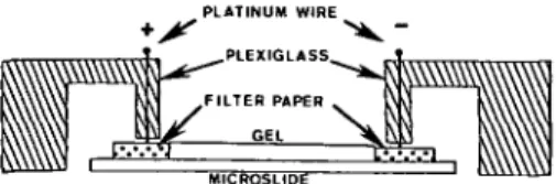
To form a gel, a sheet of GelBond film (Bioproducts, Rockland, Maine 04841) was placed, hydrophobic side up, on a sheet of glass or other smooth surface. Two strips of plastic, each approximately 0.5 X 4.0 X 80.0 mm, were then placed on the GelBond film, parallel to each other and about 20.0 mm apart. On top of these plastic strips was placed a microscope slide, treated with Silane A174 (Pharmacia) to promote gel adhesion. The space between the silanized slide and the gelbond film was then filled with the polyac-rylamide solution: 180 jtl of acpolyac-rylamide and bisacrylamide stock (4.75 g acryl. + 0.25 g bis. in 10 ml H2O), 30 fi\ TEMED (0.20 ml TEMED in 10 ml H2O), 180 fi\ ammonium persulfate solution (0.025 g/10 ml H2O) 90 fi\ Serva am-pholytes (pH 4-9), and 0.750 ml H2O. (This mixture is slightly modified from Ruchel7.)
Once polymerization has taken place, the gels may be used at once. This is done by lift-ing and turnlift-ing over the gel form and either sliding or peeling the GelBond film off from the gel. Free liquid on the gel surface should be removed by touching a piece of absorbent paper to the lower edge of the upturned gel. Single pollen grains are placed within 2-3 mm of one edge of the gel, moistened with an aqueous solution containing 0.01 percent of ascorbic acid, 0.01 percent of cysteine, 0.2 M urea and 1.0 percent Serva ampholyte (pH 2-4). Within a few seconds, the grains increase diameter by approximately 30 percent and then are crushed with fine forceps. (This op-eration is facilitated by bringing forceps to a very sharp point with a fine grained abrasive stone.) The gel is then placed on a rubber or other nonslip surface, while strips of filter paper (8.0 X 70.0 mm) are moistened with 0.025 M aspartic acid and 0.025 M glutamic acid in water or 0.025 M arginine and 0.025 M lysine in 2.0 M ethylenediamine (see Radola6). The aspartic-glutamic strip is placed at one edge of the gel and the arginine-lysine strip to
• > <
PLATINUM WIRE
AlCROSLIDt
FIGURE 1 Electrophoresis apparatus. The mi-croscope slide is 2.5 cm wide. See text for details.
the other. (Strips should be touching but not atop the gel.) The anode is connected to the first strip and the cathode to the other (see Figure 1). Electrophoresis is started at 100 volts and after 2-3 minutes, changed to constant power (approximately 0.5 watts). All runs are conducted at room temperature. Within 20-40 minutes, voltage increases as the conductivity of the focusing gel declines. The end of the run is signaled by a rapid increase of voltage to the maximum capability of the power supply (500 or 1000 v). No advantage is gained by using the 1000 v setting. After focusing, the gel is placed in a standard stain for acid phosphatases (fast garnet GBC and alpha-naphthol phosphate in 0.2 M acetate buffer, pH 5.0) for approxi-mately 1 hour at room temperature, less at 37 C. At the end of this time (45-60 minutes) the staining solution is exhausted and thus re-placed with another freshly prepared fast garnet and alpha naphthol phosphate solution. At the end of a second 45 to 60-minute stain, gels can then be cleaned with cotton swabs, photographed and, if desired, stained for general proteins using the simplified silver method of Oakley et al.5 (It is necessary to use the pre-wash that Oakley et al. suggested for 10 percent acrylamide gels in order to remove ampholytes from the slab before staining.)
The above method is technically less de-manding than the microcolumn system pre-viously described4 and the slab configuration permits precise comparison and interpretation of samples. Also, the isoelectric focusing seems to provide a greater degree of repro-ducibility between separate gels than do other systems.
Results and Discussion
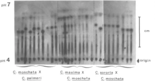
When single pollen grains from the Ft hy-brid, Cucurbita moschata X C. palmeri, are subjected to isoelectric focusing and then stained for acid phosphatase activity, eight zones of staining are seen (Figure 2). Of these eight, six are found in all of the samples. The remaining two zones are apparently deter-mined by a single segregating locus since each is found in approximately one of the samples. It may thus be concluded with confidence that at least two of the eight acid phosphatase zones in the pollen of this hybrid are under the control of the gametophytic genotype. As is shown, also in Figure 2, there is no evidence of segregation in any of the eight acid phos-phatase zones produced from pollen of the Fi hybrid, C. maxima X C. moschata. Corre-sponding data for the F1 between C. sororia X C. moschata (Figure 2) are four segregating outPH P H ' cm
4 origin
moschata X C. paimeri C_. maxima X C_. sororia X C. moschata C moschataFIGURE 2 Zymograms of single pollen grains from three interspecific hybrids of Cucurbita. Both the C. moschata X C. paimeri and C. sororia X C. moschata exhibit segregation in the pH range of 5.5. The latter hybrid is segregating also at pH 6.5. The pollen from C. maxima X C. moschata hybrid shows no segregation for acid phosphatases in this gel.
of a total of seven. For C. texana X C. palmata (not illustrated), three out of seven, and for C. pepo X C. texana (not illustrated), two out of seven. In all, 11 out of the 37, i.e., 29.7 percent of the acid phosphatase zones observed in these gels were definitely the products of gametophytic transcription. Furthermore, it must be considered that this method indicates
gametophytic transcription for heterozygous loci only. Without further information, it is not possible to determine the origin of the 26 other acid phosphatase zones. Some or all may be the products of sporophytic transcription, or they may be products of gametophytically transcribed loci that are not segregating in this material. These results thus provide a
con-servative estimate of gametophytic tran-scription. Nonetheless, they suggest that such transcription may be quite extensive. They also demonstrate that it is now possible to undertake a detailed investigation of game-tophytic genetic activity.
References
1. HALDANE, ). B. S. The Causes of Evolution. Longmans, Green, and Co., London. 1932. 2. HESLOP-HARRISON, J. Summary and
perspec-tives in higher plants as monitors of environ-mental mutagens. Environ. Health Perspect. 27:197-206. 1978.
3. MASCARENHAS, J. P. The biochemistry of an-giosperm pollen development. Bot. Rev. 41: 259-314.1975.
4. MULCAHY, D. L., G. B. MULCAHY, and R. W. ROBINSON. Evidence for postmeiotic genetic activity in pollen of Cucurbita species. ]. Hered. 70:365-368.1979.
5. OAKLEY, B. R., D. R. KIRSCH, and N. R. MORRIS. A simplified ultrasensitive silver stain for de-tecting proteins in polyacrylamide gels. Anal. Biochem. 105:361-363.1980.
6. RADOLA, B. J. Ultrathin-layer isoelectric focusing in polyacrylamide gels. Electrophoresis 1:43-56. 1980.
7. RUCHEL, R. Two-dimensional micro-separation technique for proteins and peptides, combining isoelectric focusing and gradient gel electro-phoresis. J. Chromatog. 132:451-468.1977. 8. WEEDEN, N. F. and L. D. GOTTLIEB.
Distin-guishing allozymes and isozymes of phospho-glucoisomerases by electrophoretic comparisons of pollen and somatic tissues. Biochem. Genet. 17:287-296. 1979.
The Journal of Heredity 72: 354-355. 1981.
A compensating trisomic in
pearl millet
R. S. Saini and J. L. Mlnocha
ABSTRACT: A compensating trisomic was isolated from the progeny of a multiple interchange trisomic in pearl millet, Pennisetum typhoides. It showed the character-istic morphological features of trisomy and formed a chain-of-seven chromosomes with the absence of a ring-of-four-or-six chromosomes.
ANEUPLOIDS, particularly trisomics, have been extensively used in genetic analyses and mapping of chromosomes of diploid plant species1'3. In pearl millet, Pennisetum
ty-The authors are, respectively, scientist (S-l), In-dian Council of Agricultural Research, New Delhi; and senior geneticist, Punjab Agricultural Univer-sity, Ludhiana, India. This study is based on research conducted by the senior author in partial fulfillment of the requirements for the Ph.D. degree. Please address reprint requests to Dr. Minocha. © 1981, American Genetic Association.
phoides (2n = 14), an important cereal of semiarid regions, a complete series of primary trisomics, a number of interchange stocks and tertiary trisomics are available267. Compen-sating trisomics in which one missing chro-mosome is compensated for by two modified chromosomes, have not been reported in pearl millet. However, such trisomics have been successfully used for locating genes on par-ticular arms of chromosomes in the tomato4. The present communication is the first report on the origin, isolation, and cytological be-havior of a compensating trisomic in pearl millet.
A compensating trisomic was isolated from the progeny of a multiple interchange tri-somic. The origin of multiple interchange trisomics was described by Minocha and Brar5. The expected types of aneuploids arising from the progeny of multiple inter-change trisomics are shown in Figure 2, where 'n' designates the haploid chromosome com-plement with standard or interchanged chromosomes. Chromosome composition of the extra dose in the gametes and the zygotes is also given. For simplicity, the male gametes are shown with only the standard chromo-some complement. One of the five combina-tions (square 3) involving the union of a nor-mal nor-male gamete (n chromosome number)
with a female gamete (n + 1 chromosome number) possessing two modified chromo-somes in place of normal chromosome (1S.1L) results in the compensating trisomic. This was confirmed cytologically by the presence of a chain configuration of seven chromosomes (lvn + 4n) and the absence of a ring-of-four-or-six chromosomes (Figure 1A). The meiotic analysis of the compensating trisomic showed a chain of seven chromosomes in 14.4 percent of the cells. The other chromosome
associa-B
FIGURE 1 A—diakinesis in a compensating tri-somic showing a chain of 7 chromosomes. B—ana-phase I with 8-7 chromosome distribution in the compensating trisomic.