Development of transition state analogues targeting chitinases and
oligosaccharyl transferase
by Seungjib Choi M.S. B.S.Chemistry, Seoul National Chemistry, Seoul National
University, 1999 University, 1997
Submitted to the Department of Chemistry in Partial Fulfillment of the Requirements for
the Degree of Doctor of Philosophy at the
Massachusetts Institute of Technology July 2006
©2006, Massachusetts Institute of Technology All Rights Reserved
MASSACHLUS.S INS E OF TECHNOLOGY
OCT 13 2006
LIBRARIES
ARCHIVE.
Signature of Author: Department of Chemistry July 11, 2006 Certified by: Accepted by: Barbara Imperiali Class of 1922 Professor of Chemistry and Professor of Biology/
Thesis SupervisorRobert W. Field Chairman, Department Committee on Graduate Students
This doctoral thesis has been examined by a committee of the Department of Chemistry as follows:
Professor Rick Danheiser.. .. ....- .-. ... .w... Chair
A.C. Cope Professor of Chemistry
Professor Barbara Imperiali ... Thesis Supervisor
Class of 1922 Professor of Chemistry and Prof. ssor of Biology
Professor Timothy M. Swager . ... ... ... , .
John D. MacArthur Professor of Chemistry U 7...
Development of transition state analogues targeting chitinases and
oligosaccharyl transferase
By
Seungjib Choi
Submitted to the Department of Chemistry On July 11, 2006 in partial fulfillment of the requirements for the Degree of Doctor of Philosophy
ABSTRACT
Oligosaccharyl transferase (OT) plays a central role in the biosynthesis of asparagine-linked glycoproteins in eukaryotic systems. The glycosylation step catalyzed by OT involves the co-translational transfer of a tetradecasaccharide from a dolichyl-pyrophosphate carrier to an asparagine side-chain within the Asn-Xaa-Ser/Thr sequence of a nascent polypeptide. Chitinases, which was emerged as a therapeutic target in combating asthma, are
13-1,4-N-acetylglucosaminidases that hydrolyze chitin to generate the disaccharide chitobiose
Although the reactions catalyzed by the two enzymes follow different pathways, they are believed to share similar transition states involving an oxocarbenium ion. To understand the mechanism of OT and discover potent and selective inhibitors against different chitinases, our intent was to utilize the common transition state analogue for both enzymes and systematically introduce additional binding determinants. The pseudo-disaccharides containing an imino sugar
were designed to target the oxocarbenium ion like transition state.
The pseudo-disaccharides containing imino sugar were synthesized and evaluated at inhibitors for OT and chitinases. Highlights and supporting studies from this work include : (1) the use of the Amadori rearrangement to generate the acyclic substrate; (2) the glycosylation of 3-hydroxy ketone; (3) the intramolecular reductive amination between the in-situ generated amine from azido and ketone moieties; (4) the determination of the stereo-chemical outcome by NOE difference experiments.
The pseudo-disaccharides containing imino sugars exhibited IC0s in the low micromolar range versus chitinase, yet significant inhibitory activity against OT was not observed.
Thesis Advisor: Barbara Imperiali
Acknowledgments
First of all, I would thank my advisor, Professor Barbara Imperiali, for the opportunity to work in the laboratory and for her guidance and support during the past years. I have enjoyed these challenging projects and I believe I have become a better chemist under her guidance. Especially, the pig liver preparation with Barbara was one of the most exciting events in my life. I eagerly await the day I can apply the knowledge gained from that experience. Thanks to my thesis committee members Professor Rick Danheiser and Timothy M. Swager for help and advice through years.
I also would like to thank my former advisor, Professor Eun Lee at Seoul National University. In the end it was his encouragement and support that brought me here.
I cannot express enough thanks to Eranthie Weerapara. She did the all OT assay and helped me to get through my graduate study. As a bench mate, I have occupied her hood and bench space most of time and she has never complained about it. Not a word. I really appreciate that and wish your great success.
Since almost everyone in the lab helped me to write this thesis, it is not fair to say thanks to them in order. But, first, I would thank the LBT (Langdon Martin, Bianca Sculimbrene and Anne 'Terrific' Reynolds) people for organizing, rewriting and proofreading every chapter. Nelson Oliver helped me a lot with biological aspects. Andreas Aemissegger and Elvedin Lukovic proofread chapter 2 and 4. Beth Vogel helped me to finish introduction. Dora Carrico-Moniz and Wendy Iskendrian kindly corrected the experimental section. (I knew it was boring.) Also thanks to Mettieu Sainlos for computer tips. Last but not least, Galen Loving read and corrected almost every word in my whole thesis, even the acknowledgments "in spite of his busy schedule". I really appreciate it and blame them for remaining typos.
So many of my former and current labmates have been much more than that to me. I am grateful to Mary O'Reilly for introducing me to this lab and for her advice and compassion. She will be my favorite 'O'Reilly' forever. Loving Mister Galen Loving, my best cubby mate. I have really enjoyed time with you and all the conversations and coffee breaks. I am looking forward to transplanting your kidney. As a reward, you are allowed to blame me for your lost items in the mean time. Many thanks to Mark Nitz, for teaching me the carbohydrate chemistry and answering my continuing questions. To Guofeng Zhang for critical thinking and sharing Asian jokes. To Jebrell Glover, for the nice conversation at the lunch time. To Major Nelson, for his wisdom and strength about the research as well as life. To Bianca, a consulting guru, of course for her advise through the years and knowledge of chemistry and baseball. To Elvedin, my first gay friend (that I know of), for his humor in any situation and spirit. Langdon, from protector to extraordinaire. I have been impressed by his unselfishness and congratulate on him on his wedding in advance.
Also I would like to acknowledge everyone during this journey. To all current lab members as well as past members Brenda, Carlos, Christian, Christina, Debbie, Eugenio,
Kevin, Maria, Mayssam, Mellisa, Rob, it has been a great pleasure working with you. Especially, Mark 'tough guy' Chan, Angelyn 'mercury' Larkin and Meredith 'hardly' Hartley, I firmly believe your great success on your project. You are and will remain the true 'glyco' members. You will find your own strength in your journey.
There have been many people outside the lab, who were supportive of me and have not forgotten me during past years. Thanks to Sungho Yoon, Yongwon Jung and Yoona Choi for their friendship and many many coffee breaks.
I would also like to thank my ever supportive parents and my sisters Seung-gu and Seung-lin. My Dad, my biggest fan all the time, I would never been able to finish this journey without his love and help. I am greatly thankful for to my Mom's unconditional love and sacrifice. I thank my sisters for taking care of Mom and Dad and for being proud of their little brother all the time.
Finally, I would like to give special thanks to my wife, Junhee and daughter- my desired product-Emily. Meeting them is the best thing that happened in my life. Their presence has always been a really great consolation and support to me. I apologize that we could not spend more time together over theses last few years. I know it is not enough to write down a few words in acknowledgment. However, don't forget that my heart has
Dedication
Table of Contents List of Figures ... 9 List of Schemes ... 10 List of Tables ... 10 List of Spectra... 11 List of Abbreviations ... 12
Chapter 1 Overview of glycosidases and glycosyltransferases 1-1 Introduction ... 14
1-2 Glycosidases ... 14
1-3 Inhibition studies of glycosidases ... ... ... 18
1-4 Chitinases ... 21
1-5 Inhibition studies of chitinases ... 23
1-6 Glycosyltransferases... 26
1-7 Inhibitions studies of glycosyltransferases ... 28
1-8 Oligosaccharyl transferase (OT) ... 32
1-9 Peptide substrate specificity studies of OT ... ... 34
1-10 Carbohydrate substrate specificity studies of OT... 36
1-11 Design of an inhibitor candidate for chitinase and OT ... 39
References ... 43
Chapter 2 The synthetic efforts toward a pseudo-disaccharide containing imino sugar by the convergent approach 2-1 Introduction ... 52
2-2 Synthesis of the imino sugar and biological evaluations ... 53
2-3 Synthesis of imino sugar building block ... 59
2-4 Glycosylation studies of imino sugar building block 2-19 ... 63
Experimental procedures ... 69
References ... 75
Chapter 3 The synthesis of pseudo-disaccharides containing an imino sugar by the linear approach 3-1 Introduction ... 79
3-2 Synthesis of the substrate for glycosylation studies ... 80
3-3 Glycosylation of the P-hydroxyl ketone with a glucose derivative ... 82
3-4 Construction of the imino sugar by intramolecular reductive amination...88
3-5 Synthesis of the imino sugar linked to N-acetylglucosamine ... 90
3-6 Determination of the stereo-chemical outcome of the reductive amination.100 3-7 The inhibition study of 3-1 and 3-2 against chitinase ... 04
3-8 Conclusion ... 106
Experimental procedures ... 107
References ... 127
Chapter 4 The synthesis of pseudo-disaccharide derivatives 4-1 Introduction ... 130
4-2 Synthesis of the pseudo-disaccharide precursor ... 132
4-3 Synthesis of pseudo-disaccharide derivatives and biological evaluations .. 140
4-4 Conclusion ... 141
4-5 Discussion and Future directions ... 142
Experimental procedures ... 145
References ... 157
List of Figures:
Figure 1-1. The proposed mechanism of the inverting glycosidases
in the active site of enzym e ... ... 16
Figure 1-2. The proposed mechanism of the retaining glycosidases in the active site of enzym e ... ... 17
Figure 1-3. The proposed ring distortion in the active site of enzyme ... 18
Figure 1-4. The examples of glycosidase inhibitors ... ... 19
Figure 1-5. The selected examples of the transition state analogues of glycosidases ... 21
Figure 1-6. The scheme of the reaction catalyzed chitinase...21
Figure 1-7. The proposed mechanism of family 18 chitinases... .... 23
Figure 1-8. Natural product chitinase inhibitors ... ... 25
Figure 1-9. The biological activity of allosamidin and its analogues ... 26
Figure 1-10. The proposed mechanism of GnT I based on the protein crystal structure ... 27
Figure 1-11. Selected inhibitors of glycosyltransferases based on the nucleotide analogue ... ... 29
Figure 1-12. Selected inhibitors of glycosyltransferases based on the transition state analogue ... ... 30
Figure 1-13. The scheme of reaction catalyzed by ac-1,3-Fuc-T... 30
Figure 1-14. The inhibition study of a- 1,3-Fuc-T VI with GDP analogues...31
Figure 1-15. The inhibition study of a-1,3-Fuc-T with transition state analogues ... 32
Figure 1-16. N-Linked glycosylation catalyzed oligosaccharyl transferase ... 33
Figure 1-17. The proposed mechanism of OT using Asx turn... 35
Figure 1-19. The sugar substrate specificity study for OT ... 36
Figure 1-20. The possible transition state of sugar substrate ... 37
Figure 1-21. The compounds tested in an OT inhibition study ... 38
Figure 1-22. Selected examples of five-membered imino sugars and their biological activities... ... 38
Figure 1-23. The design of inhibitors for chitinase and OT ... 39
Figure 2-1. Strategy toward the synthesis of pseudo-disaccharides containing an im ino sugar... ... 53
Figure 2-2. The general scheme of the chitinase assay ... .... 56
Figure 2-3. The inhibitory activity of 2-10 and 2-11 toward chitinase ... 56
Figure 2-4. The general scheme of OT assay... ... 58
Figure 2-5. The activity assay of 2-11 toward OT ... ... 59
Figure 3-1. The revised route toward the synthesis of the pseudo-disaccharide containing an imino sugar...80
Figure 3-2. The proposed stereo-outcome of reductive amination ... 100
Figure 3-3. The expected structure of 3-17 and possible diastereomers at C2 ... 103
Figure 3-4. The NOE difference spectrum of 3-17... 103
Figure 3-6. Compound 3-1 concentration dependent inhibition toward chitinase ... 105
Figure 3-7. Inhibition (%) of 3-1 toward chitinase ... 105
Figure 3-8. The inhibitory activity of 3-1, 3-2 and other natural products against chitinase from Streptomyces grises...106
Figure 3-9. The expected structure of 3-34 and possible diastereomers at C2 ... 125
Figure 3-10. The NOE difference spectrum of 3-34... 125
Figure 3-11. Inhibition (%) of 3-2 toward chitinase ... 126
Figure 4-1. The synthetic strategy toward derivatives of the pseudo-disaccharide... 131
Figure 4-2. The proposed inhibition mechanism of pseudo-disaccharide 3-2 against chitinase ... 143
List of Scheme:
Scheme 2-1. The synthesis of imino sugar developed by Sttitz ... 54
Scheme 2-2. The synthesis of imino sugar 2-10 and 2-11... .... 55
Schem e 2-3. The synthesis of 2-13... ... 60
Scheme 2-4. Attempted benzylation of 2-13...62
Scheme 2-5. The synthesis of building block 2-19... ... 63
Scheme 2-6. Attempted glycosylation of 2-19 with glycosyl imidates ... 65
Scheme 2-7. Attempted glycosylation of 2-19 with thioglycosides or glycosyl halides...66
Scheme 2-8. Attempted glycosylation of 2-19 with glucose derivative donors ... 67
Scheme 2-9. Attempted selective glycosylation of 2-32 ... ... 68
Scheme 3-1. The synthesis of 13-amino ketal 3-9... ... 81
Scheme 3-2. The synthesis of j3-hydroxyl ketone 3-10 ... .... 82
Scheme 3-3. The initial glycosylation studies of P-hydroxy ketone... 84
Scheme 3-4. The optimization of glycosylation of (S-hydroxyl ketone ... 86
Scheme 3-5. The attempted selective glycosylation of 3-9 ... ... 88
Scheme 3-6. Attempted intramolecular reductive amination of 3-13 ... 89
Scheme 3-7. Reductive amination and synthesis of pseudo-disaccharide 3-1...90
Scheme 3-8. Attempted conversion of N-Troc to N-Ac group of 3-20 ... 92
Scheme 3-9. Attempted intramolecular reductive amination of 3-21 ... 93
Scheme 3-10. Attempted intramolecular reductive amination of 3-24 ... 94
Scheme 3-11. Attempted intramolecular reductive amination of 3-27 ... 96
Scheme 3-12. The synthesis of the ketal 3-32 ... 98
Scheme 3-13. The synthesis of 3-33 ... 99
Scheme 3-14. The synthesis of the target pseudo-disaccharide 3-2... 100
Scheme 4-1. Comparison of the glycosylation results between 3-5 and 4-7 ... 133
Scheme 4-2. Optimization of the glycosylation of 4-7... 134
Scheme 4-3. A practical synthesis of 4-9... 135
Scheme 4-4. The synthesis of 3-hydroxy ketone 4-14 with Boc group... 136
Scheme 4-5. Attempted glycosylation of 4-14... 137
Scheme 4-6. The synthesis of 4-2... ... 139
Schem e 4-7. The synthesis of 4-19... 140
Scheme 4-8. The synthesis of pseudo-disaccharide derivatives ... 141
List of Tables: Table 3-1. The optimization of glycosylation... 86
List of Spectra:
Spectrum 1. 'H NMR spectrum of 2-10 ... A-1
Spectrum 2. 13C NMR spectrum of 2-10 ... A-2 Spectrum 3. 1H NMR spectrum of 2-11 ... A-3 Spectrum 4. 13C NMR spectrum of 2-11 ... A-4 Spectrum 5. 'H NMR spectrum of 2-16 ... ... ... A-5
Spectrum 6. '3C NM R spectrum of 2-16... ... A-6 Spectrum 7 'H NMR spectrum of 2-19 ... A-7 Spectrum 8 'H NMR spectrum of 3-7 ... A-8 Spectrum 9 'H NMR spectrum of 3-8 ... A-9 Spectrum 10 'H NMR spectrum of 3-10... A-10 Spectrum 11 '3C NMR spectrum 3-10 ... ... A-11
Spectrum 12 'H NMR spectrum 3-13 ... A-12 Spectrum 13 13C NMR spectrum 3-13 ... A-13 Spectrum 14 'H NMR spectrum 3-15 ... A-14 Spectrum 15 'H NMR spectrum 3-16 ... A-15 Spectrum 16 "'C NMR spectrum 3-16 ... A-16 Spectrum 17 'H NMR spectrum 3-1 ... A-17 Spectrum 18 13C NMR spectrum 3-1 ... A-18 Spectrum 19 'H NMR spectrum 3-31 ... A-19 Spectrum 20 'H NMR spectrum 3-33 ... A-20 Spectrum 21 13C NMR spectrum 3-33 ... A-21 Spectrum 22 'H NMR spectrum 3-34 ... A-22 Spectrum 23 'H NMR spectrum 3-34 ... A-23 Spectrum 24 'H NMR spectrum 3-35 ... A-24 Spectrum 25 'H NMR spectrum 3-2 ... A-25 Spectrum 26 'H NMR spectrum 4-10 ... A-26 Spectrum 27 'H NMR spectrum 4-11 ... A-27 Spectrum 28 '3C NMR spectrum 4-11 ... A-28 Spectrum 29 'H NMR spectrum 4-12 ... A-29 Spectrum 30 'H NMR spectrum 4-16 ... A-30 Spectrum 31 '3C NMR spectrum 4-16 ... A-31 Spectrum 32 'H NMR spectrum 4-3 ... A-32 Spectrum 33 'H NMR spectrum 4-2 ... A-33
List of Abberivations:
Standard three letter codes are used for naturally occurring amino acids, All dolichol-linked disaccharides are in P3-1,4 linkages.
Ac AgOTf Alloc Bn Bz Boc CAN Calcd Cbz DABCO DBU DIPEA DMF DMDP DMSO DPM Dol-PP EC ER ESI-MS Fuc-T GDP Glu GlcNAc GnT h HEPES HRMS LiHMDS min MeCN NaHMDS NIS NOE NMR N-TFA OT ppm PMB Pth acetyl silver trifluromethanesulfonate allyloxycarbonyl benzyl benzoyl tert-butyloxycarbonyl
ceric ammonium nitrate calculated benzyloxycarbonyl 1 ,4-diazabicyclo[2,2,2]octane 1,8-diazabicyclo[5.4.0]-undec-7-ene N,N-diisopropylethylamine N,N-dimethylformamide 2,5-dideoxy-2,5-imino-D-mannitol dimethylsulfoxide
disintegrations per minute dolichyl pyrophosphate enzyme category endoplasmic reticulum
electrospray ionization mass spectrometry fucosyltransferase guanosine 5'-diphospho glucose N-acetylglucosamine N-acetylglucosaminyltransferases hour(s) N-(2-hydroxyethyl)piperazine-N'-(2-ethane-sulfonic acid)
high resolution mass spectrometry lithium hexamethyldisilazide minute(s)
acetonitrile
sodium hexamethyldisilazide N-iodosuccinimide
nuclear Overhauser effect nuclear magnetic resonance N-trifluroacetimide
oligosaccharyl transferase parts per million
p-methoxybenzyl phthalimide
TBAF TBAI TBDPSCI TBSOTf TCA TFA TfOH THF THP TLC TMSOTf Troc p-TsOH TUP UDP-GlcNAc tetra-n-butylammonium fluoride tetra-n-butylammonium iodide tert-butyldiphenylsilyl chloride tert-butyldimethyl trifluromethanesulfonate trichloroacetyl trifluroacetic acid trifluoromethanesulfonic acid tetrahydrofuran tetrahydropyranyl
thin layer chromatography
trimethylsilyl trifluoromethanesulfonate trichloroethyloxycarbonyl
p-toluenesulfonic acid theoretic upper phase
uridine 5'-diphopho-N-acetyl-a glucosamine
used to denote any amino acid Xaa
Chapter 1- Overview of glycosidase and glycosyltransferases
1-1 Introduction
Carbohydrates are some of the most widely distributed biopolymers in nature, accounting for as much as two-thirds of the carbon found in the biosphere.' Traditionally, carbohydrates have been known as a unit for energy storage or as inert polymers that provide structural support in the cellular environment. However, in the past 30 years, with the advent of glycobiology and synthetic carbohydrate chemistry,2-9 carbohydrates
have been found to play important roles in many biological processes such as intercellular communication, cell growth, cell adhesion, immune defense, viral replication, and inflammation."" 2 Most of the carbohydrates in cells exist as oligosaccharides of complex homo- or hetero-polymers linked to proteins (glycoproteins) or lipid (glycolipids). The assembly of complex glycans is facilitated by carbohydrate processing enzymes including glycosidases (glycosyl hydrolases) and glycosyltransferases.
1-2 Glycosidases
Glycosidases catalyze the hydrolysis of a glycosidic bond by facilitating the attack of a water molecule at the anomeric carbon of a sugar. A glycosidic bond is very stable against hydrolysis, with an estimated half-life of spontaneous hydrolysis of approximately 5 million years." Glycosidases are one of the most efficient and sophisticated catalysts, increasing the reaction rate by a factor of 10"7. To achieve such a
rate-acceleration, glycosidases are expected to have very high transition state affinities, on the order of 10-22 M.10 To date more than 6,300 different glycosidase genes have been found and, on the basis of primary sequence homology, these have been classified into more than 100 families.'4 Based on the stereochemistry of the anomeric center in the sugar substrate, glycosidases are termed as a-glycosidases or P3-glycosidases. Additionally, the enzymes are classified as retaining or inverting glycosidases according to the stereo chemical outcome of hydrolysis at the anomeric center.
The mechanisms of hydrolysis by both retaining and inverting glycosidases have been well characterized on a structural and biochemical level.15' 6 The majority of glycosidases follow one of these two mechanisms. 17
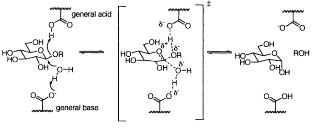
For inverting glycosidases, the glycosidic bonds are hydrolyzed via single direct displacement, facilitated by two carboxylic acids in the side chains of aspartic acid and/or glutamic acid located in the active site (Figure 1-1). One carboxylic acid acts as a general acid activating the leaving group by protonation of the exocyclic oxygen. The other acts as a general base, facilitating the attack of a water molecule on the anomeric carbon by the partial deprotonation.
o general acid 00 H HO R- OHgHO-O 0g
IL
general base 0 0 HOO H O \#- O H " 6R8O OH "O-H O I OH HO ROH O OH:!
Figure 1-1. The proposed mechanism of the inverting glycosidases in the active site of enzyme.
For retaining glycosidases, the reaction proceeds via a double displacement mechanism (Figure 1-2). In the first step, a general acid partially protonates the exocyclic oxygen, while the nucleophile carboxylate on the opposite side attacks the anomeric center to displace the leaving group, forming a covalently bonded glycosyl-enzyme intermediate. In the second step, the acidic residue near the active site acts as a general base, facilitating the hydrolysis of the glycosyl-enzyme intermediate, resulting in overall retention. The presence of the glyco-enzyme intermediate has been confirmed by mass spectrometry and protein crystallography.18' 19
o ogeneral acid 00 H HO83 O R OH1 OL nucleophile H
HOO_ HO- 0OH OH
0 O_~
O H HO OH6O 70
H
OHO -H OH'O
6
general base HHO_
H
ROH
ýO glycosyl-enzyme intermediate
Figure 1-2. The proposed mechanism of the retaining glycosidases in the active site of enzyme.
Reactions catalyzed by both inverting and retaining glycosidases are believed to proceed through an oxocarbenium-ion-like transition state.20 The degree of concertedness of bond breaking and bond making is not clear. Data from kinetic experiments suggest that the sp3-hybridized anomeric carbon is converted to an sp2 carbon, such that cleavage of the glycosidic bond precedes the formation of the new bond.21,22 In the transition state, the carbohydrate in the active site is believed to adopt a non-chair conformation.23 Especially for P(-glycosidases, the distortion of the carbohydrate substrate from the chair conformation to a boat,24 skew boat,25 or half-chair form has been observed in protein crystal structures. The distortion of the carbohydrate directs the departing glycosidic bond
to a pseudo-axial orientation, which is antiperiplanar to the lone pair of the ring oxygen or the nucleophile (Figure 1-3).26
RO
OH
OH OH
Figure 1-3. The proposed ring distortion in the active site of enzyme.
1-3 Inhibition studies of glycosidases
Since hydrolysis of the glycosidic bond is a ubiquitous biological process, glycosidase inhibitors have many potential applications, including use as agrochemicals and therapeutic agents (Figure 1-4).27,2 a-Glucosidases play an essential role in the
control of blood glucose levels in humans, and in the transport of glucose in insects and fungi.29 Several glucosidase inhibitors are being marketed to treat type II diabetes,30-32 and
others are being developed as powerful insecticides."'34 Glycosidases are also involved in the trimming of cell and viral surface complex glycans. Inhibition of these glycosidases can disrupt the biosynthesis of carbohydrates, and hence the cell-cell35 or the cell-virus recognition process.36 This principle is the basis for the anti-influenza neuraminidase inhibitors37 that have recently been marketed as well as the basis for potential HIV inhibitors.383 9 Genetic disorders,40'4 1 hepatitis C,42 and cancer43 are other promising targets for glycosidase inhibition.
OH OH D ON CO2Et
HO H O HO HO H2N
r
HN O DS HN - O Oseltamivir
OH HO OH neuraminidase inhibitor,
HO H FDA approved influenza drug
OH HO
oH
OHO OH OHOH
HO OH
Validamxylamine A Acarbose OH HO O
OH
trehalase inhibitor, insecticide candidate a amylase inhibitor, -deoxyglucuconojirimycin
FDA approved Type II diabetes drug
ceramide glucosyltransferase inhibitor, Type 1 Gaucher disease phase III clinical trial
Figure 1-4. The examples of glycosidase inhibitors.
In addition to the practical applications, understanding the mechanism of glycosidase catalyzed reactions has been another motive for the design and synthesis of transition state analogues. A substrate fits into the enzyme's active site, a pocket or groove on its surface. Upon binding, the shape and charge of the active site induce a conformational change of the substrate into its transition-state configuration. Thus, enzymes bind and stabilize the transition state, lowering the activation energy to allow the reaction to proceed at higher rates.44 In principle, a transition state analogue should bind more tightly than the substrate to the enzyme, resulting in the enzyme inhibition.45 When considering the design of an enzyme inhibitor, much focus has been placed on mimicking the geometry and charge of the assumed transition state. The relative importance of mimicking the shape and charge of the transition state has been the subject of much debate.46 It is also noteworthy that shape and charge are interdependent in most cases.47
The design of glycosidase inhibitors has focused on mimicking the charge and shape of the oxocarbenium-ion-like transition state (Figure 1-5).48 Natural and synthetic polyhydroxy alkaloids have shown potent and specific inhibitory activity against
glycosidases.495 ' The ring nitrogen would be protonated under physiological conditions to form a cation. Thus, these compounds are also referred to as imino sugars or aza sugars (Figure 1-5a, b, c, d).51 ~54 The five-membered ring is assumed to mimic the half-chair structure involved in the transition state, whereas the six-membered ring closely resembles the ground state.55 Incorporation of amidine (Figure 1-5e)56 and imidazole (Figure 1-5f) 5758 functionality into the sugar ring has been used to mimic the positive charge. To mimic the distorted structure involved in the transition state, a double bond or epoxide group59 has been introduced into the ring (Figure 1-5g). Alternatively, a bicyclic system6 has been utilized (Figure 1-5e, h).
Varying the position of the nitrogen often results in a change in inhibitory activity and selectivity.53"' These observations, in conjunction with analysis of a glycosidase crystal structure in the presence of these inhibitors, aided in understanding the mode of inhibition and mechanism for glycosidase catalytic activity.6
brewers' yeast a glycosidase almond B glycosidase OH HO- NH OH a ref. 52 IC50 12.6 pLM IC50 47 [iM OH
HO
-HO 3NH bref. 53 86 pLM 0.1 pM OH HO.- NH cref. 53 3.9 pgM 0.65 pM OH HO -NH HO OH dref. 54 almond 0 glycosidase IC 50= 17 piM OH OH H HO-HOO HO OHNH NH HOO OH
fref.56 eref. 58
almond 3 glycosidase almond 0 glycosidase Ki= 8.4 pM Ki = 0.1 VM OH ref. 59 OH
HO 0
HO NyS
ref 59 href.6
almond 0 glycosidase jack bean P-N-acetylglucosaminidase IC50 = 12.5 piM Ki = 0.28 pM
Figure 1-5. The selected examples of the transition state analogues of glycosidases.
1-4 Chitinases
Chitinases (EC 3.2.1.14) are P-1,4-N-acetylglucosaminidases that hydrolyze chitin, a homopolymer of 3-1,4-linked N-acetylglucosamine to generate the disaccharide chitobiose (Figure 1-6).
OH OH OH
HO
0
o
O OQ OHHO HO HO
AcHN AcHN AcHN
chitin
OH OH OH
HO HO -O ~H OH
AcHN AcHN AcHN
chitin +OH + OH chitinase HO- O OH AcHN AcHN chitoblose
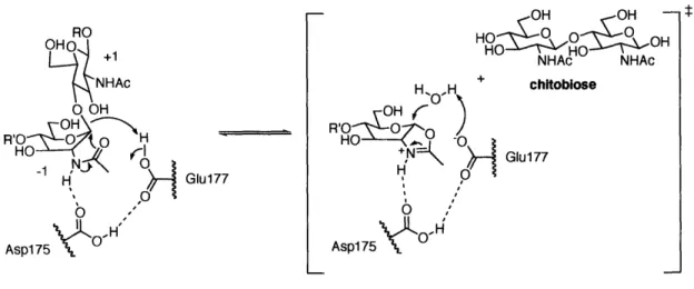
Through classification based on amino acid sequence, most chitinases belong to family 18. According to the stereo-chemical outcome of hydrolysis at the anomeric carbon, chitinases are classified as retaining glycosidases. However, chitinases and other glycosidases in family 18 adopt a pathway for enzyme catalysis that differs from the double-displacement mechanism through the glycosyl-enzyme intermediate adopted by other retaining glycosidases.
The mechanism of chitinase-mediated hydrolysis has been extensively studied and is well understood (Figure 1-7). All family 18 chitinases contain a conserved DXDXE sequence (Asp-Xaa-Asp-Xaa-Glu) in the active site, which is essential for the enzyme activity.5 5 By convention, the core sugar substrates at the binding site are termed as -2, -1, +1, and +2, and chitinases cleave between -1 and +1. Upon chitin binding to chitinase, the -1 sugar adopts a boat conformation to place the leaving group (chitobiose) in the pseudo-axial position, and Asp175 changes its orientation to interact with Glul77.6 -67 Aspl75 activates the amide proton of the C2 acetamido group in the -1 sugar. Anchimeric assistance by the neighboring C2 acetamido group produces an oxazoline ion intermediate, liberating chitobiose.6 Structural and theoretical investigations strongly support the presence of oxazoline ion intermediates.69' 70 The reorientation of Asp175 and Glu177 is believed to play an important role in the stabilization of the high-energy transition state. The hydrolysis of the oxazoline ion intermediate regenerates the N-acetyl group, resulting in overall retention of the anomeric configuration.
RO NHAc O OH
HO
N
I
O---
-1 H GI0u177 Asp 75 Aspl75 OH ' HOOR 00 OH NHAc NHAc H H chitobiose OHf
R'O • ýý O o 0 HI:N , Glu177 H ,O6
'"
Asp 75 Asp175 OFigure 1-7. The proposed mechanism of family 18 chitinases
1-5 Inhibition studies of chitinases
Family 18 chitinases include enzymes from mammals, insects, plants, nematodes, fungi, and bacteria. Chitinase is implicated in pathogenic fungal cell division,71 ecdysis of
insects7 and malaria transmission.73"74 Its involvement in those processes makes chitinases attractive targets for use in the development of fungicides, insecticides and antimalarials.2 4 However, chitinases may function in a beneficial role as well. Chitin is one of the main components in the cell walls of fungi, the exoskeletons of insects and other arthropods.75'" Since these chitin coats provide protection for pathogens inside the host and chitinases inhibit the growth of chitin containing organisms, a variety of life forms, including plants, insects, and fish, use chitinases as a weapon against chitin-expressing pathogens.7
Until a few years ago it was generally assumed that humans lack the ability to produce chitinase. However, a mammalian chitinase has recently been identified.7 8 Interestingly, the enzyme substrate, chitin, and chitin syntase have yet to be discovered in mammals. Though the role of chitinase remains to be defined, it was hypothesized that
mammalian chitinase contributes to the immune response on the basis of their functions in other species. The implication of mammalian chitinase in allergic airway responses was reported.55 The expression of mammalian chitinase by airway epithelia and pulmonary macrophages is dramatically increased with developments of asthma in mice and humans."79 Moreover, the enzyme activity is critical to disease manifestation in an experimental model of asthma. Inhibition of the chitinase with a natural product, allosamidin, decreases airway inflammation and airway hyperresponsiveness. The detailed mechanisms by which chitinase regulates allergic responses remain unclear. Considering that inhibition of chitinase ameliorates physiological maladies, its regulation is likely to be important. The development of therapeutics that targets mammalian chitinase has been suggested in combating allergic asthma.8 0
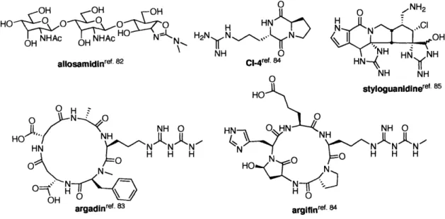
To date several natural chitinase inhibitors have been isolated,"8 including the pseudo-trisaccharide allosamidin and its derivatives,82 the cyclic pentapeptides argifine and argadin,83 amino acid-derived compounds," and the complex alkaloid
HO
0-0
o-
0
allNHA mOH NHAef 82 allosmidinref. 8 2 OH. z N NH O HOH NH 'kH H H
0 NO... N styloguanidinere NH 0 O H H H argadinref. 83 argifinref. 84Figure 1-8. Natural product chitinase inhibitors.
Allosamidin, first isolated from mycelial extracts of Streptomyces sp. 1713,82 has been extensively studied due to both its potent inhibitory activity and similarity of the corresponding molecular structure to the proposed transition state. 9"' As shown in Figure 1-8, allosamidin consists of a di N-acetylallosamine residue and an aminocyclitol aglycone, allosamizoline. Allosamidin strongly inhibits chitinases from human and the silkworm Bombyx mori, with IC5 values of 40 nM and 48 nM, while it inhibits the yeast chitinases from Saccharomyces cerevisiae 500-fold less potently.81 Interestingly, the pseudo-disaccharide derivative inhibits chitinase from Bombyx mori as efficiently as allosamidin. However, allosamizoline showed very little inhibitory activity against chitinases.
OH OOH OH OHOH OH
HO_ 0 0 0 &-7 HO 0HO HO37_O
O HAc -N HO H HA HO NcK
OH O H NKN OHNH N
allosamidin glucoallosamidin Aine
pseudo-disaccharide allosaizollne IC50 = 40 nM Bombys mori IC50= 60 nM Bombyx mori Very weak inhibition IC50 = 54 pM Saccharomyces cerevisiae Not active against Saccharomyces cerevisiaes
Figure 1-9. The biological activity of allosamine and its analogues.
1-6 Glycosyltransferases
Glycosyltransferases catalyze the transfer of a sugar moiety from an activated sugar donor onto saccharide or nonsaccharide acceptors such as proteins and lipids. Nucleotide pyrophosphate sugars, nucleotide monophosphate sugars, lipid pyrophosphate sugars, and lipid monophosphate sugars are all utilized as donors. To date 12,000 known and putative glycosyltransferases have been identified, and by amino acid sequence similarities divided into 78 families.92 Similar to glycosidases, glycosyltransferases are classified as either retaining or inverting, depending on the stereo chemical outcome of reaction at the anomeric center. Glycosyltransferases are believed to follow an analogous mechanistic pathway to that of glycosidases due to the similarity of their catalyzed reactions."" Unfortunately, a detailed structural and mechanistic understanding of glycosyltransferases is lacking due to difficulties with protein overexpression, purification and crystallization.
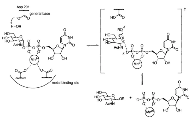
The reaction of inverting glycosyltransferases is thought to proceed through single direct displacement, similar to that proposed for the inverting glycosidase reaction. The protein crystal structure of rabbit N-acetylglucosaminyltransferase I (GnT 1) determined to 1.5 A resolution in the presence of uridine 5'-diphospho-N-acetyl-a-D-glucosamine
(UDP-GlcNAc) and Mn2+, provided insight into the reaction mechanism (Figure 1-10).96 GnT 1 catalyzes the transfer of GlcNAc from UDP-GlcNAc to a mannose residue on a complex N-linked glycoprotein. The general base, Asp291, which is positioned 4.7
A
from Cl of GIcNAc, deprotonates the incoming nucleophile (sugar acceptor), facilitating attack on the anomeric center of UDP-GlcNAc. The Mn2+ activates the leaving group
(pyrophosphate-nucleotide) by coordination to the pyrophosphate moiety. The structure also revealed the interaction between Mn2+ and the metal binding residues. The relative
positions of the catalytic base, the metal binding motif and the metal ion show that an in-line attack of the acceptor would lead to inversion of stereochemistry at the Cl position of the donor sugar. Additionally, other structural and kinetic evidence for inverting glycosyltransferases support a direct displacement involving an oxocarbenium ion-like transition state.""9 Asp 291
7O
general base
H-OR HOTO 6-RO HO 5+\ O HOO , N AcHN 11o-
P-o-
HP-O e Hd bH lingsite
11
OHO
O
O
N0
HO .S OR + -OR P- o• AcHN q PO HdO bHFigure 1-10. The proposed mechanism of GnT I based on the protein crystal structure.
T_
NH
The mechanism of retaining glycosyltransferases remains elusive. A double-displacement mechanism via a covalently bound glycosyl-enzyme intermediate has been postulated based on analogy with retaining glycosidases. However, numerous efforts to trap the glycosyl-enzyme intermediate in the active site have been unsuccessful.?'" To date, X-ray crystal structures of retaining glycosyltransferases in six families have been solved." In some of protein crystal structures, no nucleophile candidates, which are required to form glycosyl-enzyme intermediates, were observed in the active site.lo•0'02 Alternatively, a substitution nucleophilic internal (SNi)-like mechanism has been proposed, in which the acceptor attaches on the same side as leaving group.9 To understand the mechanism of retaining glycosyltransferase, more structural and kinetic experimental evidence is necessary.
1-7 Inhibition studies of glycosyltransferases
In contrast to the many potent inhibitors found for glycosidases, only limited success has been achieved in developing inhibitors of glycosyltransferases.'o3 The challenges in the design of glycosyltransferase inhibitors are 1) low binding affinity (Kn)
of the enzyme to the reaction substrate and 2) difficulties with rational design due to complex reaction partners, such as sugar donor, sugar acceptor, metal ion and nucleotide; and 3) limited structural information.'" However, a few natural and synthetic inhibitors have been found or synthesized."'5 They can be divided into one of two categories: nucleotide analogues and transition state analogues.
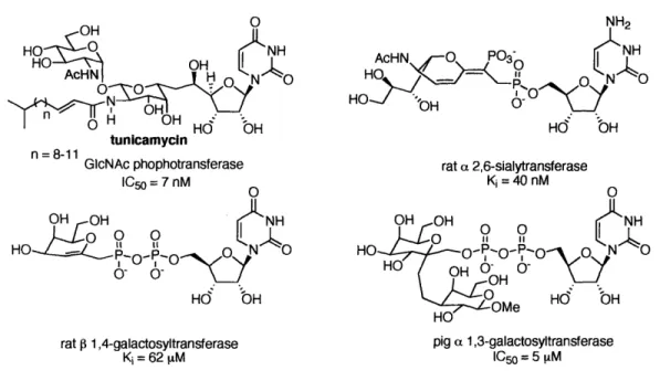
Construction of inhibitors starting with the nucleotide (or pyrophosphate-nucleotide portion) has become a widely adopted strategy (Figure 1-11). The natural product tunicamycin falls into this category of nucleotide analogues.i"', 7 Although no clear sequence homology has been found for the nucleotide-binding site, homology in conformation at the binding site has been observed. Since the nucleotide portion is identical to the native substrate, it has a basal binding affinity to the enzyme. Addition of sugar analogues or other functional groups to the nucleotide portion may therefore increase the inhibition and selectivity against other enzymes. 6,' 8-0' '1
OH 0 NH2
HO 0[NH AcHN O PO3 N H
H
HO
AcHN OH H HOAcHN
OOO3
O
n = 8-11
8-11 GcNAc phophotransferase rat a 2,6-sialytransferase
IC = 7 nM Ki = 40 nM
Ho NH
H- 0o-H O
Hd
bH
rat 0 1,4-galactosyltransferase pig a 1,3-galactosyltransferase Ki = 62 M C1050 = 5 1iM
Figure 1-11. Selected inhibitors of glycosyltransferses based on the nucleotide analogue.
As glycosyltransferases are believed to catalyze the reaction via similar mechanism, involving an oxocarbenium-ion-like transition state, it is rational to attempt the use of glycosidase inhibitors as glycosyltransferase inhibitors.'111 13 A few synthetic glycosidase inhibitors show micromolar inhibitory activity against glycosyltransferases
(Figure 1-12). Similar to glycosidases, the biological activity is very sensitive to changes in the structure of the inhibitor, and addition of functional groups to the core inhibitor has shown to increase the binding affinity. 14
OHOH P-1,4-galactosyltransferase IC 50 22 [iM cx-1,3-galatcosyltransferase IC so no inhibition OH OH HOO NH no inhibition 15 [tM OH OH OH no inhibition no inhibition
Figure 1-12. Selected inhibitors of glycosyltransferses based on the transition state analogue.
Studies of human a-1,3-fucosyltransferase (Fuc-T) inhibition represent both approaches. Fuc-T catalyzes the transfer of L-fucose from guanosine 5'-diphosphate 1-L-fucose (GDP-Fuc) to the C3 hydroxyl of GlcNAc in the glycoconjugate acceptor (Figure
1-13). The fucosylation is the elaboration step in the biosynthesis of sialyl Lewis x (sLex) and sialyl Lewis a (sLea), which play essential roles in inflammation and the immune response.' 15
R'OO
OOH
OOOH-
RNHA
o
0 NNH .0 - P O H 0 0Ný
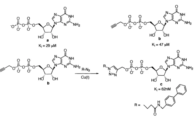
N ' NH2 OH, O O OOH ZH NHAc a-1,3-Fuc-T OHOH + O O 0N a 0 0 HNNHOne approach was to generate a GDP analogue library in a combinatorial fashion to increase inhibitory activity (Figure 1-14).116 The synthesis of 85 GDP analogues was achieved by copper-catalyzed cycloaddition between a GDP-alkyne and various azides having different functional groups. The GDP analogue having a hydrophobic biaryl turned out to be the most potent inhibitor (Figure 1-14c). Inhibitory activity was increased by 500-fold over GDP. It also showed high selectivity against other fucosyltransferases. 0 o 0 NH
.OoO- o
-0
O
N NrNH2
Hd bH a Ki= 29 pM 0 o o (7 S O O 0- 'O NNH2R-N3 Hd bH CU(1) b 0 o 1 0IfN /• -- •O 5 N N NH2 0- 0-OH Hd bH b o 0 R N:: NHR 'NNI - 'O• O"N4 N NH2
Nz O, 0- 0-Hdo OH
C
Ki= 62nM / H R= Nv/YN OFigure 1-14. the inhibition study of a-1,3-Fuc-T VI with GDP analogues.
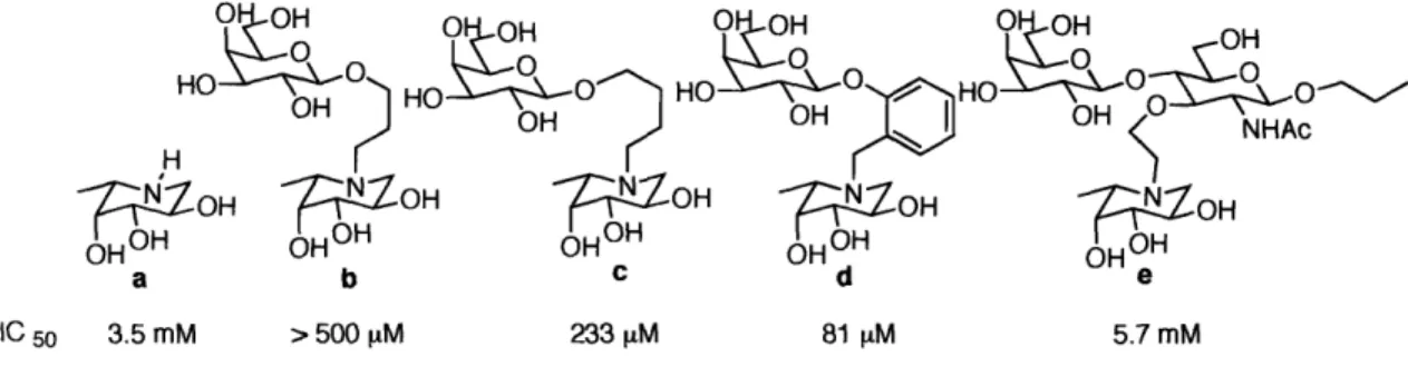
The other approach utilized a glycosidase inhibitor as the scaffold (Figure 1-15). The compound L-fuco-nojirimycin (Figure 1-15a) was known as a potent inhibitor against a-L-fucosidase (Ki = 1 nM),"7 but showed very little inhibitory activity against fucosyltransferase. Based on the structure of this transition state analogue, bisubstrate
analogue inhibitors, in which the sugar acceptor and sugar donor were connected by a linker, were synthesized. Addition of the sugar acceptor moiety showed an increase of inhibitory activity. By varying the length and geometry of the linker, the optimal orientation was found (Figure 1-15d)."8 Interestingly, the trisaccharide derivative showed poor inhibitory activity, probably due to an unfavorable orientation, despite its close resemblance to the proposed transition state (Figure 1-15e).
O 1- ,-| ... ,
HO
OH OH OH HOH OH
HOO 0 O HO O 0 OH OH NHAc_OH
OH
ZoH
OOH %,OH a b c d e IC 50 3.5 mM > 500 piM 233 [M 81 [M 5.7 mMFigure 1-15. The inhibition study of a-1,3-Fuc-T with transition state analogues.
1-9 Oligosaccharyl transferase (OT)
Oligosaccharyl transferase (OT, EC 2.4.1.119) plays a central role in the biosynthesis of asparagine-linked glycoproteins in eukaryotic systems. The glycosylation step catalyzed by OT involves the co-translational transfer of a tetradecasaccharide from a dolichyl-pyrophosphate carrier to an asparagine side-chain within the Asn-Xaa-Ser/Thr sequence of a nascent polypeptide, where Xaa can be any amino acid except proline (Figure 1-16.)."9 This is the first step in N-linked glycosylation and the resulting glycoprotein is further processed by various glycosidases and glycosyltransferases.'20
OH RO0N0 O 0 R' 0 S H O
OOH
HO
O-P-O-P-O-Dolichyl ROO NH Dolichyl-PP R=Glc3MangGlcNAc Dolichol =Figure 1-16. N-Linked glycosylation catalyzed by oligosaccharyl transferase.
OT is a membrane-associated multimeric protein localized in the lumen of the endoplasmic reticulum (ER). Eight subunits (Ostlp, Ost2p, Ost3p, Ost4p, Ostp5p, Wbplp, Swplp, Stt3) are assembled into a hetero-octameric OT complex in yeast
(Saccharomyces cerevisiae).12' Recent evidence indicates that Stt3 is the catalytic subunit
of the eukaryotic OT.'22'24 Stt3 family proteins are responsible for the catalytic activity of N-linked glycosylation in eukaryotes, archaebacteria and in selected eubacteria.12 5"126 In spite of its essential role in N-linked glycosylation, only limited structural or mechanistic information for Stt3 is available. The highly membrane-associated nature of Stt3, which is predicted to have 11 or 13 transmembrane domains, has hampered the investigation of this subunit.127
,128 Even the topology of the Stt3 protein is hard to predict. Therefore, studies that have focused on substrate specificity in vivo and in vitro have contributed to our understanding of catalysis.
1-10 Peptide substrate specificity studies of OT
The source of enhanced nucleophilicity of the amide nitrogen of asparagine is a prevailing mechanistic question in N-linked glycosylation, and several mechanisms have been proposed.2 9-131 The general agreement is that intramolecular hydrogen bonding
activates the carboximide of asparagine.'3 2
In addition, the Asx-turn motif is believed to be an important element to activate the peptide substrate (Figure 1-17). A peptide having this recognition sequence can adopt two conformations: an Asx-turn or a P3-turn. The major difference is the intramolecular hydrogen bonding site of the backbone amide. In the Asx-turn, the backbone amide interacts with the side-chain amide of the asparagine. In contrast, the p-turn is characterized by the hydrogen bonding between backbone amide groups.'33 The hydrogen-bonding network in the Asx-turn facilitates deprotonation of the amide to provide the neutral imidol species. The intermediate could react with the dolichyl-pyrophosphate-linked sugar donor to generate the glycopeptide. Using NMR studies, it was demonstrated that an unglycosylated peptide, based on a short sequence of hemagglutinin, adopts an Asx-turn conformation. Upon glycosylation, the structure changes to induce a type-I-P-turn conformation. 134,135 The fact that proline is not accepted at the Xaa site within Asn-Xaa-Ser/Thr sequence, and that 10-30% of recognition sites are not glycosylated, supports that local conformation plays an important role.' 136
H. Asx-turn L -P R=Glc3MangGIcNAc OH HO NH H R AcHN HN N O HN'
o
0o-
---
H-N.,.
HOyt<I P-turnFigure 1-17. The proposed mechanism of OT using Asx turn.
The mechanistic studies of peptide substrates guided the development of inhibitors against OT with low-nanomolar affinity. The establishment of backbone constraint and replacement of the carbonyl of asparagine with methylene unit, gave a tight binding inhibitor with a K, of 37 nM (Figure 1-18.).'37
~I
00 HN-Aý NO2 H2N HN H 0 OH N NN7COA NH2 0 O OH O Ki =37 nM in vitro
Figure 1-18. The potent OT peptide inhibtior with Asx turn.
1-11 Carbohydrate substrate specificity studies of OT
A tetradecasaccharide linked to dolichyl-pyrophosphate by an a-linkage is the preferred substrate for OT both in vivo and in vitro (Figure 1-19a). Due to the difficulty of obtaining the full substrate, most in vitro studies have been carried out using chitobiose linked to a dolichyl-pyrophosphate as the substrate (Figure 1-19b). The Km values for the full substrate (Figure 1-19a) and the truncated substrate (Figure 1-19b) were determined to be 33 [tM and 65 [tM respectively.'3 8 Interestingly, the synthetic substrate Dol-PP-GlcNAc-Glc (Figure 1-19c) showed 2.5 fold improvement in binding with a Km of 26 pM.'3 9 The replacement of the C2 acetamido group with trifluroacetamide (Figure 1-19d) or ethyl ether (Figure 1-19e) of the proximal GlcNAc residue leads to total loss of enzyme catalytic activity.'3 9 These analogues (Figure 1-19d,
e) were further analyzed as inhibitors of OT, which suggested that OT recognized these synthetic substrates without a C2 acetamide group, but OT could not transfer them to the peptide substrate. In contrast, Dol-PP-GlcNAc (Figure 1-19f) was found to be very poor substrate for OT.
D(distal) site P(proximal) site
OH OH
HO6 HHO O O0
AcHN AcHN
R=GIc3Man9 O-P-O-P-O-Dolichyl
a o-Km = 33 I&M HOH OH HO '-O HO OH HO O O OH AcHN •_ D
O-P-0-P-O-Dolichyl
c O O .- OH OH Km = 26 MCM HO HO 0 0 AcHN EtO I 11 O-P-O-P-O-DolichylHO O e no apparent Ki = 252 ILM OH OH HO - HO 0 0 AcHN AcHN 11 O-P-O-P-O-Dolichyl b 0 O Km = 65 IM OH OH HO"• 0 o HO •" HO-•'• 0 0 AcHN TFAHN ,1 O-P-O-P-O-Dolichyl d 0 0 no apparent activity OH Ki = 154 pMHO -O
O-P-O-P-O-Dolichylactivity f 6 very poor substrate0-r
Figure 1-19. The sugar substrate specificity study for OT.
Since OT is classified as an inverting glycosyltransferase, the reaction pathway of OT is believed to proceed via single direct displacement involving an oxocarbenium ion-like transition state. Activated asparagine attacks the anomeric center of the GlcNAc in the proximal (P) site, generating a 13-linked glycopeptide. Although no metal binding motif has been found in OT, it has been reported that divalent ions such as Mn2, or Mg2+ are essential for the catalytic activity.'" The results of a carbohydrate substrate study suggested that the C2 acetamide group plays an important role in enzyme catalysis, either directly or indirectly.139 A substrate specificity study of bacterial OT also supports the role of the C2 acetamide group in enzyme activity.'41 Based on these substrate study results, a reaction pathway via an oxazoline ion intermediate was proposed.14' A
high-energy oxocarbenium ion might be stabilized by the delocalization of positive charge through the participation of the C2 acetamido group. However, to establish the exact
function of the C2 acetamido related to the mechanism of OT, a detailed structural and kinetic study of OT is required.
Asn
OHRO -
OO NH
2 HO -' O O AcHN I i, , O-P-O-P-O-Dolichyl O-O-O .f
HN 6 HO 6+ ', HO 0 O0 H'N jO-P-O--O-Dolichyl0<
6-HN6 HO +' RO HO . 0 0
H
d
--P-O-P-O-Dolichyl
Asn OH O HO NH AcHNFigure 1-20. The possible transition state of sugar substrate.
Based on the similarity of the proposed transition states of chitinase and OT, the potent chitinase inhibitor glucoallosamdin A pseudo-disaccharide was considered as a potential inhibitor for OT (Figure 1-21a). Unfortunately, no inhibitory activity was observed up to 5 mM. Other chitobiose derivatives (Figure 1-21b, c) showed no inhibitory activity against OT up to 500 tM concentration.142
OH OH OH OH OH OH HO 00O• O HO 00O HO 0 SH O N HO O H'HOO O H ' O NHPh AcHN N S • AcHN C a H b N N AcH0N glucoallosamidin A pseudo-disaccharide
11 Design of an inhibitor candidate for chitinase and OT
The goal of this study was to design, synthesize, and evaluate potential inhibitors against OT from Saccharomyces cerevisiae and chitinase from Streptomyces grises. Although the reactions catalyzed by the two enzymes follow different pathways, they are believed to share similar transition states involving an oxocarbenium ion. Our intent was to utilize the common transition state analogue for both enzymes and systematically introduce additional binding determinants.
From the substrate and inhibitor studies and mechanistic considerations of chitinase and OT, common transition state analogues were designed. Polyhydroxypyrrolidine 1, also known as 2,5-dideoxy-2,5-imino-D-mannitol (DMDP),54" 43 is one of the most prevalent sugar mimics.49"'" Five-membered imino
sugars show potent inhibitory activity against the a broad range of retaining and inverting glycosidases and glycosyltransferases (Figure 1-22).145,146
HO
HO
OH
HO
OH
HOHAc
NHAc1 DMDP 2 3
almond B glycosidase coffee bean ca-galactosidase jack bean P-N-acetylglucosaminidase
IC 5o= 17 pM Ki= 50 nM Ki = 1.9 [tM
0-1,4-galactosyltransferase
IC 50 = 22 iM
Among imino sugar pyrrolidine analogues, we chose 3 as the target transition state analogue. In addition to its ability to mimic the charge and shape of the oxocarbenium ion, there were several attractive features of iminocyclitol 3. In chitinase and OT, the C2 N-Ac group of the proximal GIcNAc residue plays an important role in the enzyme catalysis either directly or indirectly. The Cl N-Ac group in the imino sugar could interact with the same key residues in the active site that bind to the C2 N-Ac group in the sugar substrate. The two pseudo-equatorial secondary alcohols have a similar configuration to that of N-acetylglucosamine. Moreover, functionalization of Cl amine or secondary ring amine by acylation or reductive amination have been reported, efficiently generating the corresponding derivatives.'47
The inhibition study of chitinase and the carbohydrate substrate study of OT suggest that the disaccharide unit is the minimal binding motif for OT and chitinase. To fulfill this requirement, we decided to synthesize a pseudo-disaccharide motif containing the transition state analogue (Figure 1-23). To mimic the glycosyl linkage in chitobiose, we planned to add glucose and N-acteylglucosamine to the C4 hydroxyl group of the imino sugar via a n-linkage.
Chitinase has emerged as a therapeutic target in combating asthma. To validate the exact role of mammalian chitinase in the allergic response, the effect of exogenous chitinases (from insect or fungi, for example) must be investigated. Although many natural product inhibitors have been discovered, the synthesis of derivatives based on a common motif has proven effective in the discovery of an inhibitor and in improvements to its biological activity. 47"48To discover potent and selective inhibitors against different
chitinases, the synthesis of pseudo-disaccharide derivatives will be useful. We were particularly interested in exploration of different groups such as aliphatic chains, aromatic and heterocyclic groups on the Cl amine of the imino sugar. These compounds will be valuable tools to elucidate the function of chitinases from various species in allergic response.
In spite of the importance of N-linked glycosylation, the structural and mechanistic study of OT has been limited due to the membrane-associated nature of this multimeric complex. Studies of the inhibition of OT are a more tractable approach to providing insight into the mechanism of transferring an oligosaccharide from the dolichyl lipid to protein. For the design of an OT inhibitor, the addition of a long aliphatic chain to the transition state analogue is expected to increase the binding affinity to the enzyme. The failure of glucoallosamdin A pseudo-disaccharide and chitobiose derivatives as OT inhibitors could be explained based on the possibility that these transition state analogues might not be accessible to the active site of OT. Considering the amphiphilic nature of the dolichyl-pyrophosphate-linked tetradecasaccharide and the membrane-associated nature OT, the active site of glycosylation may be at the interface between the membrane and soluble domains of OT.
pseudo-disaccharide
SI transition state analogue
H OOOH -OH
HO
o
Ho
X = OH, NHAc H R = aliphatic, aromatic
target molecules
A modular approach based on the pseudo-disaccharide core will reveal the critical binding determinants required for inhibition of chitinase and OT. A comparison of biological properties will give insight into the mechanistic differences between OT and chitinase in terms of inhibition.
References
1. Sinnott, M. L. Catalytic Mechanisms of Enzymatic Glycosyl Transfer. Chem.
Rev., 1990, 90, 1171-1202.
2. Bertozzi, C. R.; Kiessling, L. L. Chemical glycobiology. Science, 2001, 291, 2357-2364.
3. Alper, J. Glycobiology -Turning sweet on cancer. Science, 2003, 301, 159-160. 4. Lowe, J. B.; Marth, J. D. A genetic approach to mammalian glycan function.
Annu. Rev. Biochem., 2003, 72, 643-691.
5. Roseman, S. Reflections on glycobiology. J. Biol. Chem., 2001, 276, 41527-41542.
6. Feizi, T.; Mulloy, B. Carbohydrates and glycoconjugates: never a dull moment in glycoscience -Editorial overview. Curr. Opin. Struct. Biol., 2005, 15, 479-480.
7. Seeberger, P. H.; Haase, W. -C. Solid-phase oligosaccharide synthesis and combinatorial carbohydrate libraries. Chem. Rev., 2000, 100, 4349-4378.
8. Sears, P.; Wong, C. -H. Toward automated synthesis of oligosaccharides and glycoproteins. Science, 2001, 291, 2344-2350.
9. Nicolaou, K. C.; Mitchell, H. J. Adventures in carbohydrate chemistry: New synthetic technologies, chemical synthesis, molecular design, and chemical biology.
Angew. Chem. Int. Ed. Engl., 2001, 40, 1576-1624.
10. Helenius, A.; Aebi, M. Intracellular functions of N-linked glycans. Science, 2001,
291, 2364-2369.
11. Varki, A. Biological Roles of Oligosaccharides -All of the Theories Are Correct.
Glycobiology, 1993, 3, 97-130.
12. Upreti, R. K.; Kumar, M.; Shankar, V. Bacterial glycoproteins: Functions, biosynthesis and applications. Proteomics, 2003, 3, 363-379.
13. Wolfenden, R.; Lu, X. D.; Young, G. Spontaneous hydrolysis of glycosides. J.
Am. Chem. Soc., 1998, 120, 6814-6815.
14. Henrissat, B.; Davies, G. Structural and sequence-based classification of glycoside hydrolases. Curr. Opin. Struct. Biol., 1997, 7, 637-644.
15. Heightman, T. D.; Vasella, A. T. Recent insights into inhibition, structure, and mechanism of configuration-retaining glycosidases. Angew. Chem. Int. Ed. Engl, 1999,
38, 750-770.
16. Zechel, D. L.; Withers, S. G. Dissection of nucleophilic and acid-base catalysis in glycosidases. Curr. Opin. Chem. Biol., 2001, 5, 643-649.
17. Yip, V. L. Y.; Withers, S. G. Nature's many mechanisms for the degradation of oligosaccharides. Org. Biomol. Chem., 2004, 2, 2707-2713.
18. Vocadlo, D. J.; Davies, G. J.; Laine, R.; Withers, S. G. Catalysis by hen egg-white lysozyme proceeds via a covalent intermediate. Nature, 2001, 412, 835-838.
19. Sidhu, G.; Withers, S. G.; Nguyen, N. T.; McIntosh, L. P.; Ziser, L.; Brayer, G. D. Sugar ring distortion in the glycosyl-enzyme intermediate of a family G/11 xylanase.
Biochemistry, 1999, 38, 5346-5354.
20. Zechel, D. L.; Withers, S. G. Glycosidase mechanisms: Anatomy of a finely tuned catalyst. Acc. Chem. Res., 2000, 33, 11-18.
21. Vocadlo, D. J.; Wicki, J.; Rupitz, K.; Withers, S. G. Mechanism of
Thermoanaerobacterium saccharolyticum j-xylosidase: Kinetic studies. Biochemistry,
2002, 41, 9727-9735.
22. Huang, X. C.; Tanaka, K. S. E.; Bennet, A. J. Glucosidase-catalyzed hydrolysis of f-D-glucopyranosyl pyridinium salts: Kinetic evidence for nucleophilic involvement at the glucosidation transition state. J. Am. Chem. Soc., 1997, 119, 11147-11154.
23. Vasella, A.; Davies, G. J.; Bohm, M. Glycosidase mechanisms. Curr. Opin.
Chem. Biol., 2002, 6, 619-629.
24. Sabini, E.; Wilson, K. S.; Danielsen, S.; Schulein, M.; Davies, G. J. Oligosaccharide binding to family 11 xylanases: both covalent intermediate and mutant product complexes display B-2,B-5 conformations at the active centre. Acta Crystallogr.,
Sect. D, 2001, 57, 1344-1347.
25. Ducros, V. M. A.; Zechel, D. L.; Murshudov, G. N.; Gilbert, H. J.; Szabo, L.; Stoll, D.; Withers, S. G.; Davies, G. J. Substrate distortion by a P-mannosidase: Snapshots of the Michaelis and covalent-intermediate complexes suggest a B-2,B-5 conformation for the transition state. Angew. Chem. Int. Ed. Engl., 2002, 41, 2824-2827. 26. Sulzenbacher, G.; Driguez, H.; Henrissat, B.; Schulein, M.; Davies, G. J. Structure of the Fusarium oxysporum endoglucanase I with a nonhydrolyzable substrate analogue: Substrate distortion gives rise to the preferred axial orientation for the leaving group. Biochemistry, 1996, 35, 15280-15287.
27. Asano, N. Glycosidase inhibitors: update and perspectives on practical use.
Glycobiology, 2003, 13, 93R-104R.
28. El Ashry, E. S. H.; Rashed, N.; Shobier, A. H. S. Glycosidase inhibitors and their chemotherapeutic value, part 3. Pharmazie, 2000, 55, 403-415.
29. Elbein, A. D.; Pan, Y. T.; Pastuszak, I.; Carroll, D. New insights on trehalose: a multifunctional molecule. Glycobiology, 2003, 13, 17R-27R.
30. Joubert, P. H.; Foukaridis, G. N.; Bopape, M. L. Miglitol May Have a Blood-Glucose Lowering Effect Unrelated to Inhibition of a-Glucosidase. Eur. J. Clin.
Pharmacol., 1987, 31,723-724.
31. Joubert, P. H.; Venter, H. L.; Foukaridis, G. N. The Effect of Miglitol and Acarbose after an Oral Glucose-Load -a Novel Hypoglycemic Mechanism. Br. J. Clin.
Pharmacol., 1990, 30, 391-396.
32. Krentz, A. J.; Bailey, C. J. Oral antidiabetic agents - Current role in type 2 diabetes mellitus. Drugs, 2005, 65, 385-411.
33. Asano, N.; Takeuchi, M.; Kameda, Y.; Matsui, K.; Kono, Y. Trehalase Inhibitors, Validoxylamine-A and Related-Compounds as Insecticides. J. Antibiot., 1990, 43, 722-726.
34. Ando, O.; Kifune, M.; Nakajima, M. Effects of Trehazolin, a Potent Trehalase Inhibitor, on Bombyx-Mori and Plant-Pathogenic Fungi. Biosci. Biotechnol. Biochem.,
1995, 59, 711-712.
35. Bucior, I.; Burger, M. M. Carbohydrate-carbohydrate interactions in cell recognition. Curr. Opin. Struct. Biol., 2004, 14, 631-637.
36. Kim, C. U.; Lew, W.; Williams, M. A.; Liu, H. T.; Zhang, L. J.; Swaminathan, S.; Bischofberger, N.; Chen, M. S.; Mendel, D. B.; Tai, C. Y.; Laver, W. G.; Stevens, R. C. Influenza neuraminidase inhibitors possessing a novel hydrophobic interaction in the