R. Sutter C. Arber A. Tichelli A.J. Steck A. Czaplinski
Cranial and peripheral
neuropathy
due to leptomeningeal
infiltration in a patient
with Waldenstrom’s
macroglobulinemia
Received: 20 April 2006Received in revised form: 26 August 2006 Accepted: 7 September 2006
Published online: 3 August 2007
Sirs: Lymphoplasmacytic lym-phoma or Waldenstrom’s macro-globulinemia (WM) is a chronic mature B-cell lymphoproliferative disorder of plasmacytoid lympho-cytes usually involving bone mar-row, lymph nodes and spleen. A serum monoclonal immunoglob-ulin M (IgM) may be present and may be associated with hypervis-cosity and/ or cryoglobulinemia.
We present the case of a 70 year old male who was diagnosed with WM. Bone marrow infiltration at time of diagnosis was 40%, the monoclonal IgM kappa in periph-eral blood was 11.5g/l, and cryo-globulines were present. A therapy with four cycles of R-CHOP (rit-uximab, cyclophosphamid,
doxo-rubicin, vincristin, and prednison) was administered. The patient re-sponded well to therapy and achieved a good partial remission with only a residual bone marrow infiltration of 5–10% and the monoclonal IgM kappa in periph-eral blood was 0.7 g/l.
Six months later the patient gradually developed persistent headaches, nuchal rigidity, double vision, dysphagia and dysarthria. The clinical examination at admission showed meningism and severe bulbar paralysis along with symptoms of sensori-motor poly-neuropathy.
Examination of cerebrospinal fluid disclosed 106 cells/mm3, ele-vated protein level (3160 mg/l) and elevated beta-2-microglobulin (6.35 mg/l). Cytological analyses revealed atypical lymphocytes and flow cytometry analysis showed a kappa monoclonal CD19 positive B-cell population. Bone marrow examination showed a persistent multifocal infiltration with lym-phoplasmacytic lymphoma (15% of nucleated cells). Transformation in a diffuse large B cell lymphoma was excluded. The serum monoclonal IgM kappa was 0.4 g/l, cryoglobu-lines were low, and serum anti-MAG (myelin associated glycopro-tein) antibodies were negative. The brain MRI showed a mild thicken-ing of leptomenthicken-inges without tu-mor mass or meningeal
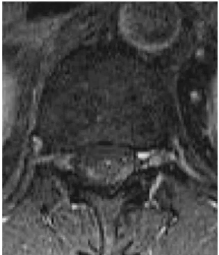
enhancement. In addition the MRI of the lumbar spinal cord after gadolinium injection showed an increased signal in perimedullary and cauda equina roots regions, as well as enlargement of the latter (Fig. 1 and 2).
We interpreted the symptoms as cranial and peripheral neurop-athy due to leptomeningeal and nerve root infiltration and the pa-tient was given a palliative radia-tion-therapy of the brain and the cauda with simultaneous steroid
therapy. The patient’s condition improved with a clear decrease of bulbar and polyneuropathic symptoms. Although these thera-peutic interventions were estab-lished, the patient died eleven weeks after diagnosis.
Sensori-motor neuropathy re-lated to the antibody activity of the monoclonal IgM to the MAG is a well-known neurological compli-cation of WM [2, 3]. However, involvement of the peripheral nervous system due to leptomen-ingeal infiltration by neoplastic cells is very rare and has been re-ported in few cases only [1, 4–6]. Remarkably, in contrast to other non-Hodgkin’s lymphomas where meningeal involvement develops in the setting of progressive dis-ease, in our patient meningeal infiltration occurred in a stable partial remission phase of the WM.
In conclusion, we hypothesize that WM may progress in the nervous system without transfor-mation into a more malignant B-cell-lymphoma (Richter syn-drome) even if the initial chemo-therapy leads to significant remission of the disease.
Dr. R. Sutter (&) Æ A.J. Steck Æ A. Czaplinski
Dept. of Neurology University of Basel Patersgraben 4 4031 Basel, Switzerland E-Mail: sutte r@uhbs.ch C. Arber Æ A. Tichelli
Dept. of Hematology, University of Basel, Switzerland
Fig. 1 Axial T1-weighted MRI of lumbar spinal cord after gadolinium showed an increased signal in perimedullary regions
LETTER TO THE EDITORS
J Neurol (2007) 254:1122–1123DOI 10.1007/s00415-006-0405-7
References
1. Abad S, Zagdanski A-M, Brechignac S, Thioliere B, Brouet JC, Mariette X (1999) Neurolymphomatosis in Waldenstro¨m’s macroglobulinaemie. Br J Haematol 106: 100–103
2. Braun PE, Frail DE, Latov N (1982) Myelin-associated glycoprotein ist the antigen for a monoclonal IgM in poly-neuropathy. J Neurochem 39: 1261–1265
3. Dellagi K, Dupouey P, Brouet JC, Bille-cocq A, Gomez D, Clauvel JP, Seligmann M (1983) M. Waldenstrom’s macro-globulinemia and peripheral neuropa-thy: a clinical study of 25 patients. Blood 62: 280–285
4. Hug A, Haas J, Storch-Hagenlocher B, Wildemann B (2004) Leptomeningeale Tumorzellinfiltration als Erstmanifesta-tion eines Immunozytoms (M. Wald-enstro¨m). Nervenarzt 75: 1012–1015
5. Massengo S, Riffaud L, Morandi X, Bernard M, Verin M (2003) Nervous system lymphoid infiltration in Wald-enstro¨m’s macroglobulinemia. A case report. J Neuro-Oncol 62: 353–358 6. Noel J, Gille M, Garbar Ch, Martens M,
Verhulst D, Vandeput Y (2002) Primary central nervous system lymphoma with Waldenstro¨m’s macroglobulinaemia. Eur Neurol 47: 184–185
Fig. 2 Sagittal T1-weighted MRI of lumbar spinal cord after gadolinium showed an increased signal in perimedullary and cauda equina roots regions