Diabetologia (2005) 48: 900–905 DOI 10.1007/s00125-005-1723-5
A RT I C L E
A. Biason-Lauber . B. Boehm . M. Lang-Muritano . B. R. Gauthier . T. Brun . C. B. Wollheim .
E. J. Schoenle
Association of childhood type 1 diabetes mellitus with a variant
of
PAX4: possible link to beta cell regenerative capacity
Received: 3 September 2004 / Accepted: 20 December 2004 / Published online: 15 April 2005
# Springer-Verlag 2005
Abstract Aims/hypothesis: Loss of pancreatic beta cells is the crucial event in the development of type 1 diabetes. It is the result of an imbalance between autoimmune de-struction and insufficient regeneration of islet cells. To study the role of islet cell regeneration in the pathogenesis of type 1 diabetes, we focused on PAX4, a paired homeodomain transcriptional repressor that is involved in islet cell growth. Methods: The study included 379 diabetic children and 1,070 controls from two distinct populations, and a cohort of children who had not developed type 1 diabetes, despite the presence of islet cell antibodies. Genomic DNA analysis of PAX4 was carried out via direct sequencing of PCR-amplified fragments and allelic discrimination. We com-pared the transrepression potential of the PAX4 variants in βTC3 cells and analysed their influence on beta cell growth. Results: The type 1 diabetic subjects are different from the normal individuals in terms of the genotype distribution of the A1168C single nucleotide polymorphism in PAX4. The C/C genotype is frequent among type 1 diabetic children (73%) and rare among the control population (32%). Con-versely, the A/C genotype is prevalent among control sub-jects (62%) and antibody-positive children without type 1 diabetes (73.6%), but uncommon among subjects with type 1 diabetes (17.5%). The combination of PAX4A and PAX4C is functionally more active than PAX4C alone (the‘diabetic’
variant). Beta cells expressing PAX4A and PAX4C efficient-ly proliferate when stimulated with glucose, whereas cells expressing the PAX4C variant alone do not. Conclusions/ interpretation: We have identified a link between beta cell regenerative capacity and susceptibility to type 1 diabetes. This finding could explain the fact that not all of the in-dividuals who develop autoimmunity against beta cells ac-tually contract the disease. The C/C genotype of the A1168C polymorphism in PAX4 can be viewed as a predisposition marker that can help to detect individuals prone to develop type 1 diabetes.
Keywords Diabetes mellitus, type 1 . Pancreas . Regeneration
Abbreviations BrdU: 5-bromo-2′-deoxyuridine . SNP: single nucleotide polymorphism
Introduction
The central event in type 1 diabetes mellitus is the selective loss of insulin-producing pancreatic beta cells. Type 1 dia-betes can therefore be viewed as the result of an imbalance between autoimmune beta cell destruction and beta cell re-generation. Classically, the development of type 1 diabetes is divided into different stages, from genetic predisposition, to triggering, active autoimmunity and, finally, progressive beta cell destruction [1]. The extensively documented poly-genic inheritance pattern of type 1 diabetes suggests that alterations in risk-modifying genes could lead to the even-tual clinical manifestation of the disease. Specifically, al-terations in these genes should play a role in the pathology of the disease, i.e. beta cell destruction (primarily due to immune mechanisms), decreased beta cell proliferation, and insufficient insulin secretion in response to stimulators such as glucose.
The mechanisms involved in the autoimmune destruc-tion of beta cells have been extensively studied over the past three decades [2,3], whereas the factors involved in islet cell development and regeneration have only recently A. Biason-Lauber (*) . M. Lang-Muritano . E. J. Schoenle
Department of Paediatric Endocrinology/Diabetology, University Children’s Hospital,
Steinwiesstrasse 75, 8032 Zurich, Switzerland e-mail: Anna.Lauber@kispi.unizh.ch Tel.: +41-1-2667623 Fax: +41-1-2667169 B. Boehm Ulm University, Ulm, Germany
B. R. Gauthier . T. Brun . C. B. Wollheim University Medical Centre,
been described and investigated (for reviews see [4, 5]). Agents involved in organogenesis and differentiation of the pancreas are also likely to be involved in the processes of regeneration. Among these regulators, the transcription factor Pax4 plays a crucial role in pancreatic beta cell development. The Pax family of transcription factors con-tain a paired box and a homeobox domain, and are essential for the formation of several tissues present in all germ layers in the mammalian embryo. Specifically, they are involved in triggering the early events of cell differen-tiation during organogenesis. The differendifferen-tiation of endo-derm-derived endocrine pancreas is mediated through Pax4 and Pax6 [6]. Inactivation of pax4 by homologous recom-bination in mice results in the absence of mature insulin-producing beta cells and somatostatin-insulin-producing delta cells, and hyperplasia of glucagon-producing alpha cells [7]. The main function of Pax4 is as a transcriptional repressor in early pancreatic development [8, 9]. The mechanism by which Pax4 influences beta cell function is not well un-derstood. Activin A (a member of the TGF-β family) stim-ulates Pax4 expression in pancreatic cell lines [10], and simultaneously promotes the growth and differentiation of human fetal pancreatic cells when combined with another growth factor, betacellulin [11]. These findings indicate that Pax4 is involved in the promotion of beta cell proliferation by growth factors. Although initial investigations suggested that PAX4 was not expressed in mature endocrine cells, more recent studies have detected mRNA for the transcrip-tion factor in adult human, rat and mouse pancreatic islets [12–14]. We have previously demonstrated that low levels of pax4 mRNA are expressed, and that production of these transcripts is induced by the mitogens activin A and beta-cellulin. Adenoviral-mediated overexpression of Pax4 in islets was shown to produce a 3.5-fold increase in beta cell proliferation [15]. A central concept that emerged from this study is that Pax4 operates as a key regulator of adult beta cell mass by converging the replicating effect of several signal transduction pathways towards the Myc-Id2 cascade. In parallel, cells protect themselves from c-Myc-induced apoptosis by increasing Bcl-xL expression.
Genetic association studies in humans have reported an association between PAX4 variants and type 2 diabetes [16, 17]; however, this does not appear to be a global phe-nomenon, since it could not be found in other populations [18]. The A1168C single nucleotide polymorphism (SNP) we have examined has not been found to be linked to any form of diabetes. In a study on Scandinavian families, Holm et al. observed an association between type 1 dia-betes and haplotypes at the PAX4 locus on chromosome 7q32 that, together with the ISL1 locus on chromosome 5q11–q13, constitutes a significant susceptibility factor for type 1 diabetes [19].
We hypothesised that PAX4 will be‘reactivated’ to pro-mote the growth of islet cells in response to their de-struction. If such a mechanism exists and is functional, the pancreas would react to an immune attack by regenerating beta cells. In contrast, inefficient proliferation would not be able to counteract the ongoing destruction, eventually lead-ing to type 1 diabetes. To gain insight into the possible role
of PAX4 in the pathogenesis of type 1 diabetes, we first screened for mutations in PAX4 in children with type 1 diabetes, and then investigated the consequences of these mutations in beta cell lines.
Subjects and methods
Subjects We initially recruited 249 children with type 1 diabetes (male:female ratio=0.53:0.47) and 424 normal control subjects of Swiss origin (male:female=0.57:0.43), and subsequently extended the analysis to include an ad-ditional 130 diabetic subjects (male:female ratio=0.56:0.44) and 646 normal control subjects (male:female ratio=0.63: 0.37) from a distinct German population. Type 1 diabetes was diagnosed according to the criteria approved by the American Diabetes Association [20]. All patients tested positive for anti-islet cell antibodies. The control subjects were unrelated individuals who did not have a history of type 1 or type 2 diabetes in the first-degree family line. An additional group included 19 school children (age 6–17 years) who, despite anti-islet cell antibody positivity, had not developed diabetes within 16 years of follow-up [21]. Patients’ data and samples were anonymised according to the Swiss law (SR 235.154 and 236.1), and their collection conformed to the guidelines set out by the internal review boards. All participants (or their parents) gave informed consent. All samples were analysed in a blind fashion. Genetic analysis Genomic DNA was extracted from pe-ripheral blood leucocytes using the QIAGEN blood and cell culture DNA kit (Hilden, Germany). PCR exonic am-plification of PAX4 (accession no. CH236947.1) was per-formed, and direct cycle sequencing of the PCR products was carried out using the ABI Prism BigDye Terminators v.3 kit and an ABI Prism 310 Genetic Analyzer (Applied Biosystems, Rotkreuz, Switzerland). Allelic discrimina-tion was performed using two different methods: (1) RFLP based on HphI digestion (performed according to the man-ufacturer’s instructions; New England Biolabs, Allschwil, Switzerland); and (2) an allelic discrimination assay using specific TaqMan MGB probes, with the results analysed on the ABI Prism 7,700 Sequence Detection System (Applied Biosystems; the sequences of the primers and probes used are available upon request). To further confirm the data, 20 samples per group per genotype were sequenced as men-tioned above. Primer sequences and PCR conditions are available upon request (all primers were supplied by Mycro-synth, Balgach, Switzerland). Chi square, significance and relative risk were calculated using standard methods. Expression studies To study the functional implications of the PAX4 variants, we developed an RT-PCR method us-ing PAX4 mRNA ectopically expressed in peripheral blood leucocytes of normal subjects who were homozygous for either the A or C variant [22]. The two PAX4 cDNA variants were ligated into a pCMV-Script vector (Stratagene, Basel, Switzerland) and used for functional studies. MouseβTC3 (German Collection of Microorganism and Cell Cultures,
Braunschweig, Germany),αTC1.9 and NIH3T3 (American Type Culture Collection, Manassas, VA, USA) cells were cultured (7.5×10³ cells) in regular DMEM medium (25 mmol/l glucose; Life Technologies, Basel, Switzerland). As they reached 80% confluence (at 36 h), the cells were tran-siently transfected with PAX4A, PAX4C or a combination of the two at a 1:1 molar ratio, using 50μg of lipofectamine per 10μg of DNA per 60-mm plate (Life Technologies). We used a construct containing promoter sequences of the rat insulin I gene (−410 bp) cloned upstream of a firefly lucierfase cDNA (pFOXluc-410RIP1) as a reporter, and the control plasmid pFOXluc1 (which is devoid of the tar-get sequence). Both plasmids were generous gifts from M. German (University of California, San Francisco, CA, USA). To normalise for transfection efficiency, the con-stitutively active Renilla luciferase-containing pRL-SV40 vector (Promega, Wallisellen, Switzerland) was used in ad-dition to pFOXluc-410RIP1. Luciferase activity was mea-sured 48 h after transfection using the Dual-Luciferase Reporter Assay System (Promega) as suggested by the sup-plier. All results are expressed as luciferase activity relative to control (where no PAX4=1). Total protein content was measured using the Bio-Rad Protein Assay (Hercules, CA, USA).
To study the influence of the PAX4 variants on cell growth, 7.5×10³βTC3, αTC1.9 and NIH3T3 cells were seeded on 35-mm plates. Cells were then transfected with PAX4A, PAX4C, a combination of the two at a molar ratio of 1:1, or empty vector. The transfection efficiency, which was assessed using a β-galactosidase-containing vector (SV-40β-gal; Promega), ranged from 40 to 60%. At 48 h after transfection, the cells were cultivated for 12 h in the absence of glucose, and then exposed different concentra-tions of glucose (0, 5, 15, 25 and 30 mmol/l) for 12 h, harvested, and counted in triplicate. To study the mecha-nism of putative changes in cell number, we used the same experimental design together with a commercial kit for analysis of proliferation, as directed by the supplier (Cell Proliferation ELISA, BrdU; Roche Applied Science). Briefly, the assay is based on the detection of 5-bromo-2′-deoxyuridine (BrdU), an analogue of thymidine. Cells are labelled by the addition of BrdU for 4 h. During this labelling period, BrdU is incorporated into the DNA of cycling cells in place of thymidine. After the removal of the labelling medium, an anti-BrdU antibody that binds to the BrdU incorporated into the newly synthesised DNA is added. The immune complexes are detected by the sub-sequent substrate reaction. The reaction product is quan-tified by measuring the absorbance using a scanning spectrophotometer (ELISA reader).
Electrophoresis mobility shift assay Nuclear extracts were obtained from transfected βTC3 cells (48 h after trans-fection) as previously described [23]. The double-stranded oligonucleotides C2 (Top strand: 5′-AGCTGTGAGCAGG GACAGGTCTGGC-3′) and DH1 (Top strand: 5′-AGCTG TGAGCAGGGACAGGTCTGGCCACCGGGCCCCTG-3′) [24] of the human insulin promoter, which are known to be target sequences for PAX4 were end-labelled with
[γ32-P]-ATP using T4 polynucleotide kinase. Binding as-says were performed as previously described [23].
Results
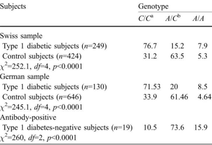
Genetic findings The SNP in which an A or a C may be present in exon 9 of PAX4 (cDNA residue 1168; Table1), corresponding to a proline or a histidine residue at position 321 in PAX4 protein, respectively (P321H), was differ-entially distributed between control subjects and subjects with type 1 diabetes. As shown in Table1, 63.5 and 61.4% of normal Swiss and German individuals, respectively, were heterozygous, whereas 76.7 and 71.5% of the Swiss and German type 1 diabetic subjects, respectively, carried the C/C genotype. Remarkably, 73% of children who had an anti-beta cell autoimmune reaction (islet cell antibody-positive) and carried the A/C genotype still remain free of type 1 diabetes after 16 years of follow-up (Table1). The relative risk of developing type 1 diabetes for carriers of the C/C genotype is 3.75-fold higher than for individuals carrying the A/C or A/A genotype. Subjects with the A/C genotype have a five-fold lower chance of developing type 1 diabetes when compared with individuals carrying the other genotypes. In agreement with previous reports [18], no association was found between the A1168C SNP (P321H) and type 2 diabetes (n=344, data not shown).
Functional implications To investigate potential functional differences between the PAX4 variants, we tested their transrepression potential in beta-like (βTC3), alpha-like (αTC1.9) and non-islet cells (NIH3T3). Compared with the A variant, the PAX4C ‘diabetic’ variant was less ef-fective at repressing the transcription of target sequences in insulinomaβTC3 cells. A combination of the two var-iants—a situation mimicking the control genotype—pro-Table 1 Genotype frequencies of the A1168C SNP in the study population
Subjects Genotype
C/Ca A/Cb A/A
Swiss sample
Type 1 diabetic subjects (n=249) 76.7 15.2 7.9
Control subjects (n=424) 31.2 63.5 5.3
χ2
=252.1, df=4, p<0.0001 German sample
Type 1 diabetic subjects (n=130) 71.53 20 8.5 Control subjects (n=646) 33.9 61.46 4.64 χ2
=245.1, df=4, p<0.0001 Antibody-positive
Type 1 diabetes-negative subjects (n=19) 10.5 73.6 15.9 χ2
=260, df=2, p<0.0001 df degrees of freedom a
Relative risk of developing type 1 diabetes in carriers of the C/C genotype vs other genotypes=3.75 (strong risk)
b
Relative risk of developing type 1 diabetes in carriers of the A/C genotype vs other genotypes=0.18 (strong protection)
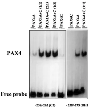
duced the greatest level of repression (Fig. 1). This effect appears to be pancreas-specific, since similar results were obtained in αTC1.9 cells but not in NIH3T3 cells (not shown). Compared with PAX4A, PAX4C appears to have an impaired ability to bind DNA target sequences (Fig.2), even though the polymorphism may not be located within the DNA-binding domains. The combination of the A and the C variants appears to increase the DNA binding ca-pacity of PAX4 (Fig. 2).
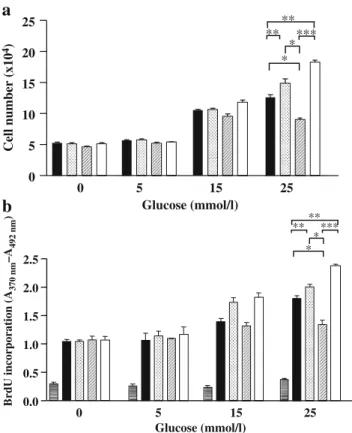
If PAX4 variants play a role in the pathogenesis of type 1 diabetes, they may influence beta cell number and/or their
differentiation. The cell count was increased by a factor of 2.5 at higher glucose concentrations (15–25 mmol/l) compared with that at lower concentrations (0 or 5 mmol/l) (Fig.3a). No further increase in the number ofβTC3 cells was seen at 30 mmol/l glucose (data not shown). The glucose-dependent increase in cell number was blunted in cells transfected with PAX4C, and this effect appeared to be overcome by the combination of A and C. The effect of transfection with PAX4A on the glucose-dependent increase in the number of βTC3 cells was not the same as that produced by transfection with the combination of PAX4A and PAX4C (Fig. 3a). The glucose-driven influence of PAX4 on cell number appears to be beta cell specific, as transfection with the PAX4 variants did not affect the cell count ofαTC1.9 cells or non-islet NIH3T3 cells (data not shown). The incorporation of BrdU into the newly syn-thesised DNA of replicating cells was significantly lower in PAX4C-transfected βTC3 cells than in cells express-ing PAX4A either alone or in combination with PAX4C (Fig. 3b). These results are consistent with those of the cell count experiments. The data suggest that the PAX4C ‘diabetic’ variant limits the proliferation of beta cells under glucose stimulation, the main physiological regulator of beta cell growth [21]. The addition of the A to the C variant (the control), restores the proliferation potential of these cells. The results indicate that the observed effects are mainly due to the overexpressed human PAX4 rather than the endogenous mouse Pax4 that is present inβTC3 cells, since the outcomes of all functional studies were more sig-nificant in the presence of human PAX4 than in its absence.
Discussion
In the present study we have demonstrated that there are differences between patients with type 1 diabetes and nor-mal individuals in terms of the genotype distribution of the A1168C SNP in PAX4. The differences were extremely striking: the majority of normal individuals were hetero-zygous, whereas most of the diabetic patients were
homozy-0.0 0.5 1.0 1.5 ** * *** ** ** ** 1 –3 –1.6 –4.5 Fold repression + + -+ + -+ -+ + -+ -+ + + + + + + -+ + + + + + + Renilla Firefly RIP PAX4 A PAX4 C Relative luciferase activity
Fig. 1 Transcriptional activity of variants of human PAX4 in transfectedβTC3 cells. Rat insulin I promoter sequences (−410 bp) were cloned upstream of a firefly luciferase cDNA (pFOXluc-410RIP1) and transfected intoβTC3 cells, together with PAX4C, PAX4A or a 1:1 mixture of the two. The same construct devoid of the target sequence was used as a control (pFOXluc). A constitutively active Renilla lucif-erase-containing construct (pRL-SV40) was used for standardisation purposes. As a control for background luminescence, the Renilla luciferase-containing vector was transfected with or without PAX4A and PAX4C. The data are expressed as relative luciferase activity (firefly/Renilla) (where no PAX4=1). The results were subjected to statistical analysis (paired t-test). The data represent the means±SD of at least three inde-pendent experiments. *p<0.05; **p<0.02; ***p<0.002; ****p<0.001. Firefly, pFOXluc; Renilla, pRL-SV40; RIP, pFOXluc-410RIP1
Fig. 2 The binding of the PAX4 variants to the human insulin promoter was analysed using an electrophoretic mobility shift assay. The oligonucleotides C2 and DH1 (corresponding to PAX4 target sequences on the human insulin promoter) were incubated with extracts fromβTC3 cells transfected with PAX4A and PAX4C, either alone or in combination at different molar ratios. The resultant protein–DNA complexes were visualised by electrophoresis
gous for the C allele. The same results were demonstrated in two distinct populations, thus rendering a bias in the choice of the cohort less likely. The fact that a significant association between PAX4 haplotypes and diabetes was recently described in a family-based Scandinavian study indicates that this is not an isolated phenomenon [19]. To investigate possible functional differences between the two variants, we tested their DNA binding capacity and trans-repression potential. It appears that the PAX4C ‘diabetic’ variant is less active than the PAX4A variant in terms of binding and repressing target sequences. Transcription is repressed to the greatest extent when the two variants are
combined (the situation mimicking the normal genotype). Although PAX4A seems to be the main regulator in alpha-like cells, its role in beta cell function is at present unclear. The combination of the two variants seems to slightly increase their DNA binding capacity, which may explain the apparent heterozygote advantage. The basis of such phenomenon is at present unclear, since members of the Pax family of transcription factors are not known to form dimers. If PAX4 variants play a role in the pathogenesis of type 1 diabetes, they may influence the proliferation and/or dif-ferentiation of beta cells. The PAX4C ‘diabetic’ variant limits the proliferation of beta cells under glucose stimu-lation, the main physiological regulatory mechanism of beta cell growth [25]. The addition of the A variant to the C variant restores the proliferation potential of these cells. These data provide evidence for the loss of function of the ‘diabetic’ genotype (C/C) and the protective function of the ‘control’ genotype (A/C).
One of the implications that can be drawn from this study concerns possible preventive approaches for type 1 diabetes. Although the clinical onset of type 1 diabetes is acute, beta cell damage precedes clinical diabetes by sev-eral months or years. Two large randomised trials that aimed to control the destruction of beta cells have, un-fortunately, yielded negative results. The Diabetes Preven-tion Trial-1 (DPT-1) failed to show any protective effect of insulin treatment [26], and in the European Nicotinamide Diabetes Intervention Trial (ENDIT), the administration of nicotinamide, which was hypothesised to attenuate DNA and beta cell damage, did not have any effect [27]. If beta cells cannot be protected from destruction, their regener-ation potential could be used to counteract the ongoing attack. This seems to be achievable in the mature pancreas, since differentiated beta cells have been shown to have significant proliferative potential in vivo [28]. Further stud-ies are required to improve our understanding of factors that predispose or protect against diabetes. This would allow the development of new assays that could be used in combination with those for previously identified markers in order to identify susceptible individuals. Our results in-dicate that the C/C genotype of the A1168C polymorphism in PAX4 is a diabetes susceptibility factor and might be used to detect individuals prone to develop type 1 diabetes. On the basis of our results we conclude that the com-bination of the two PAX4 variants may be involved in a process that has a protective effect against the onset of type 1 diabetes. This hypothesis is supported by the intriguing fact that children who tested positive for antibodies to beta cells and carried the A/C genotype did not develop type 1 diabetes. Our results indicate a link between beta cell regenerative capacity and susceptibility to type 1 diabetes, which could explain the fact that only a certain proportion of individuals who develop autoimmunity against beta cells actually contract the disease.
a
0 5 15 0.0 0.5 1.0 1.5 2.0 2.5 Glucose (mmol/l) Glucose (mmol/l) BrdU incorporation (A 370 nm –A 492 nm ) 25 *** * * ** **b
0 5 10 15 20 25 * **** ** ** Cell number (x10 4) 0 5 15 25Fig. 3 a Effect of the overexpression of the PAX4 variants onβTC3 cell number in the presence of increasing concentrations of glucose. The results were subjected to statistical analysis (paired t-test). *p<0.05; **p<0.02; ***p<0.01. The data represent the means±SD of at least three independent experiments. b Effect of the expression of the PAX4 variants on the proliferation of transfectedβTC3 cells, as measured by BrdU Cell Proliferation ELISA. The graph shows the incorporation of BrdU in adherentβTC3 cells transfected with PAX4A, PAX4C, neither, or a combination of the two and cultured in 96-well microtitre plates under different concentrations of glucose (0–25 mmol/l). The rate of proliferation at higher glucose con-centrations (30 mmol/l) is comparable to that at 25 mmol/l. The data represent duplicate measurements of experiments performed in triplicate. The results were subjected to statistical analysis (paired t-test paired). *p<0.05; **p<0.02; ***p<0.01. Blank, horizontal stripes; no PAX4, black fill; PAX4A, dotted fill; PAX4C, diagonal stripes; combination of PAX4A and PAX4C, white fill
Acknowledgements This work was supported by the Swiss Na-tional Science Foundation (grant number 32-063629.0) and by a grant from Novo Nordisk Switzerland. B. Boehm was supported by the German Research Foundation (grant numbers DFG-GRK 1041, DFG-SFB 518) and by the Kröner-Fresenius Stiftung. We are grateful to M.S. German (University of California, San Francisco, CA, USA), for the generous gift of the reporter constructs pFOXluc-410RIP1 and pFOXluc1. We thank T. Torresani, B. Schäfer, B. Thöny and D. Konrad for critical discussion of the work, and M. Steigert and M. Gartenmann-Benz for their contributions. We are indebted to R. Zollinger and E. Alig for excellent technical assistance. We also ac-knowledge the continuous support of C.H. Heizmann.
References
1. Powers AC, Eisenbarth GS (1985) Autoimmunity to islet cells in diabetes mellitus. Annu Rev Med 36:533–544
2. Freiesleben De Blasio B, Bak P, Pociot F, Karlsen AE, Nerup J (1999) Onset of type 1 diabetes: a dynamical instability. Dia-betes 48:1677–1685
3. Cooke A (1990) An overview on possible mechanisms of de-struction of the insulin-producing beta cell. Curr Top Microbiol Immunol 164:125–142
4. Bonner-Weir S (2000) Life and death of the pancreatic beta cells. Trends Endocrinol Metab 11:375–378
5. Servitja JM, Ferrer J (2004) Transcriptional networks control-ling pancreatic development and beta cell function. Diabetolo-gia 47:597–613
6. Mansouri A, St-Onge L, Gruss P (1999) Role of Pax genes in endoderm-derived organs. Trends Endocrinol Metab 10:164– 167
7. Sosa-Pineda B, Chowdhury K, Torres M, Oliver G, Gruss P (1997) The Pax4 gene is essential for differentiation of insulin-producing beta cells in the mammalian pancreas. Nature 386: 399–402
8. Smith SB, Ee HC, Conners JR, German MS (1999) Paired-homeodomain transcription factor PAX4 acts as a transcrip-tional repressor in early pancreatic development. Mol Cell Biol 19:8272–8280
9. Fujitani Y, Kajimoto Y, Yasuda T et al (1999) Identification of a portable repression domain and an E1A-responsive activation domain in Pax4: a possible role of Pax4 as a transcriptional repressor in the pancreas. Mol Cell Biol 19:8281–8291 10. Ueda Y (2000) Activin A increases Pax4 gene expression in
pancreatic beta cell lines. FEBS Lett 480:101–105
11. Demeterco C, Beattie GM, Dib SA, Lopez AD, Hayek A (2000) A role for activin A and betacellulin in human fetal pancreatic cell differentiation and growth. J Clin Endocrinol Metab 85:3892–3897
12. Heremans Y, Van De Casteele M, in’t Veld P et al (2002) Recapitulation of embryonic neuroendocrine differentiation in adult human pancreatic duct cells expressing neurogenin 3. J Cell Biol 159:303–312
13. Kojima H, Fujimiya M, Matsumura K et al (2003) NeuroD
-betacellulin gene therapy induces islet neogenesis in the liver and reverses diabetes in mice. Nat Med 9:596–603
14. Zalzman M, Gupta S, Giri RK et al (2003) Reversal of hyperglycemia in mice by using human expandable insulin-producing cells differentiated from fetal liver progenitor cells. Proc Natl Acad Sci U S A 100:7253–7258
15. Brun T, Franklin I, St-Onge L et al (2004) The diabetes-linked transcription factor PAX4 promotes beta-cell proliferation in rat and human islets. J Cell Biol 167:1123–1135
16. Shimajiri Y, Sanke T, Furuta H et al (2001) A missense mutation of Pax4 gene (R121W) is associated with type 2 diabetes in Japanese. Diabetes 50:2864–2869
17. Smith SB, Watada H, Scheel DW, Mrejen C, German MS (2000) Autoregulation and maturity onset diabetes of the young transcription factors control the human PAX4 promoter. J Biol Chem 275:36910–36919
18. Dupont S, Vionnet N, Chevre JC et al (1999) No evidence of linkage or diabetes-associated mutations in the transcription factors BETA2/NEUROD1 and PAX4 in Type II diabetes in France. Diabetologia 42:480–484
19. Holm P, Rydlander B, Luthman H, Kockum I (2004) Interac-tion and associaInterac-tion analysis of a type 1 diabetes susceptibility locus on chromosome 5q11–q13 and the 7q32 chromosomal region in Scandinavian families. Diabetes 53:1584–1591 20. Report of the expert committee on the diagnosis and
classifi-cation of diabetes mellitus (2003) Diabetes Care 26 [Suppl 1]: S5–S20
21. Schlosser M, Strebelow M, Wassmuth R et al (2002) The Karlsburg type 1 diabetes risk study of a normal schoolchild population: association of beta-cell autoantibodies and human leukocyte antigen–DQB1 alleles in antibody-positive individ-uals. J Clin Endocrinol Metab 87:2254–2261
22. Biason-Lauber A, Lang-Muritano M, Vaccaro T, Schoenle EJ (2002) Loss of kinase activity in a patient with Wolcott– Rallison syndrome caused by a novel mutation in the EIF2AK3 gene. Diabetes 51:2301–2305
23. Schreiber E, Matthias P, Müller MM, Schaffner W (1989) Rapid detection of octamer binding proteins with‘mini-extracts’, pre-pared from a small number of cells. Nucleic Acids Res 17:6419 24. Campbell SC, Cragg H, Elrick LJ, Macfarlane WM, Shennan KIJ, Docherty K (1999) Inhibitory effect of Pax4 on the human insulin and islet amyloid polypeptide (IAPP) promoters. FEBS Lett 463:53–57
25. Hellerstrom C, Swenne I (1985) Growth pattern of pancreatic islet in animals. In: Wolk BW, Arquilla ER (eds) The diabetic pancreas. Plenum Press, New York, pp 53–79
26. Diabetes prevention trial—type 1 diabetes study group (2002) Effects of insulin in relatives of patients with type 1 diabetes mellitus. N Engl J Med 346:1685–1691
27. Gale EA, Bingley PJ, Emmett CL, Collier T (2004) European nicotinamide diabetes prevention trial (ENDIT): a randomised controlled trial of intervention before the onset of type 1 diabe-tes. Lancet 363(9413):925–931
28. Dor Y, Brown J, Martinez O, Melton DA (2004) Adult pancreatic β-cells are formed by self-duplication rather than stem-cell dif-ferentiation. Nature 429:41–46