Altered patterns of senescence and ripening in gf, a
stay-green mutant of tomato (Lycopersicon esculentum Mill.)
Muhammad Shaheen Akhtar
1, Eliezer E. Goldschmidt2, Isaac John3, Simona Rodoni4,
Philippe Matile
4 and Don Grierson1,5
1 Plant Science Division, School of Biological Sciences, The University of Nottingham, Sutton Bonington
Campus, Loughborough LE12 5RD, UK
2 The Kennedy-Leigh Centre for Horticultural Research, Faculty of Agriculture, The Hebrew University of
Jerusalem, Rehovot 76100, Israel
3 Department of Biology, The University of Michigan, Ann Arbor, MI 48109–1048, USA
4 Department of Plant Biology, University of Zurich, Switzerland
Received 14 October 1998; Accepted 22 Februar y 1999
Abstract
Key words: Carotenoids, chlorophyll, ripening,senes-cence, tomato. The gf tomato mutant, which retains chlorophyll during
ripening, has been found to be affected in leaf
senes-Introduction
cence. The leaves of the gf mutant show an absolutestay-green phenotype. As leaf senescence and fruit The leaf senescence syndrome involves a large number of ripening proceed, there is a marked difference in biochemical processes, many of which relate to the disin-chlorophyll content between wild-type and gf. In both tegration of the photosynthetic apparatus. As with other attached and detached leaf studies, or after treatment developmental phenomena, natural as well as genetically with ethylene, the leaves withered and abscised in gf engineered mutants have become increasingly important with only slight loss of chlorophyll and carotenoids. for the analysis of leaf senescence and its regulation by Total protein content declined and free amino acids environmental and hormonal stimuli (Canfield et al., increased during leaf senescence in wild-type and gf, 1995; Gan and Amasino, 1995; Grbic and Bleecker, 1995; but Western analysis showed that LHCII polypeptides John et al., 1995). ‘Stay-green’ mutants, in particular, were retained at higher levels in gf. Expression of have been highlighted as valuable tools for dissection of senescence-related mRNAs increased normally in gf senescence and distinguishing between the degradation of whereas those for cab, rbcS and rbcL declined in both different thylakoid constituents (Guiamet et al., 1991; mutant and wild-type. The mutant possesses enzyme Thomas and Smart, 1993).
activity for chlorophyllase, the formation of phaeo- While tomato has been used as a major plant system phorbide a by the action of Mg-dechelatase and the for the study of fruit ripening, it has not been used oxygenolytic opening of the porphyrin macrocycle. extensively in early studies of leaf senescence (McGlasson Analysis of chlorophyll breakdown products in fruit et al., 1975; Mizrahi et al., 1975). The molecular charac-indicated that gf, like other stay-green mutants, accu- terization of tomato leaf senescence has been approached mulates chlorophyllides a and b, but phaeophorbide a more recently (Davies and Grierson, 1989; John et al., does not accumulate in vivo. This may indicate that, 1995, 1997; Drake et al., 1996). The green flesh ( gf ) in the mutant, in vivo the action of phaeophorbide a- mutant of tomato was described long ago ( Kerr, 1956), oxygenase is somehow prevented, either by altered and shown to be located on chromosome 8 ( Kerr, 1957). accessibility or transport of components required Only the altered fruit-ripening characteristics were for thylakoid disassembly or the absence of another described, however (Ramirez and Tomes, 1964; Grierson et al., 1987; Cheung et al., 1993), which include retention factor.
5 To whom correspondence should be addressed. Fax:+44 115 951 6334. E-mail: [email protected] © Oxford University Press 1999
1116 Akhtar et al.
Detached leaf senescence studies
of chlorophyll, thylakoids, and some thylakoid proteins
Whole leaves were placed on three layers of wet Whatmann
as the chloroplasts accumulate lycopene and are converted
No. 1 filter paper in 14 cm Petri dishes ( Fig. 2). The dishes,
to chromoplasts. In the present study, the leaf senescence
wrapped in parafilm and aluminium foil and incubated in the
pattern of gf plants and the degradation of chlorophyll dark at 23°C were opened for inspection and sampling every in both leaves and fruit were examined. The leaves of the 2–3 d. The effect of ethylene was tested by placing the Petri gf mutant appear to contain all the enzyme activities dishes in a glass desiccator with 20ml l−1 ethylene.
required for chlorophyll catabolism yet exhibit an
abso-Determination of chlorophyll, catabolites, and catabolic enzymes
lute, stay-green phenotype. General proteolysis of leaf
proteins continues in gf during senescence, but the LHCII Chlorophyll content was determined spectrophotometrically in
N,N-dimethylformamide extracts of leaf discs (Moran, 1982). polypeptide was preferentially retained.
Chlorophyllase activity was determined from fresh leaf extracts according to Trebitsh-Sitrit et al. ( Trebitsh-Sitrit et al., 1993) in 0.05 M phosphate buffer pH 7.4, 0.01% Triton X-100. Phaeophorbide a oxygenase activity was assayed as outlined in
Materials and methods
Hortensteiner et al. ( Hortensteiner et al., 1995). Dephytylated chlorophyll derivatives estimation was conducted by a phase Plant material
separation assay as previously described (Amir-Shapira et al., Wild-type and green flesh plants were grown in 7 cm pots in
1987), or by HPLC (Moser and Matile, 1997). John Innes M2 compost in a growth room under controlled
white fluorescent light (100mmol m−2 s−1) with a day/night
Determination of protein content, free amino acids and proteolytic period and temperature of 16/8 h, and 22/18°C, respectively.
activity The plants were given tap water every day and grown till they
showed all the stages of leaf senescence (mature green, onset, Total protein content was determined in leaf disc extracts in mid and advanced ) at about 6–7 weeks. Leaves at different 0.05 M phosphate buffer pH 7.4, 0.01% Triton X-100 according stages of senescence were harvested as described previously by to Bradford (Bradford, 1976). Free amino acids were determined John et al. (1995) and immediately frozen in liquid nitrogen, in leaf disc extracts in 80% ethanol by the ninhydrin reagent
and stored at−70 °C. Solution of Sigma (Moore, 1968). Proteolytic activity of leaf
disc extracts was determined in 0.05 M phosphate buffer pH 7.4, 0.01% Triton X-100 by the release of acid-soluble products Protein extraction and Western analysis
from radioactively labelled casein (a-casein, cold and 14C-Protein samples were extracted from tomato leaves and fruits methylated from Sigma) as given by Katayama-Fujimura et al. according to the protocol of Meyer (1988). For Western blot ( Katayama-Fujimura et al., 1987).
analysis 50mg of each protein sample was resolved on 14% SDS/PAGE and blotted onto nitro-cellulose membranes
(Bio-Pigment measurement in senescent leaves after treatment with Rad) according to the manufacturer’s instructions. Antibodies
8-hydroxyquinoline against LHCPII, Rubisco (SSU and LSU ) were obtained from
Mature leaves were cut in half along the midrib and cut surfaces Drs Kate Griffith and P Scott, respectively. The membranes
were placed on solutions in tap water of 1 mM ACC with or were probed with antibodies as described by Cornelius et al.
without addition of 8-hydroxy quinoline (1 mM ) to block (Cornelius et al., 1996).
phaeophorbide a oxygenase. The tissue were allowed to senesce for 3.5 d in permanent darkness and then analysed for pigments. RNA extraction and Northern analysis
Total RNA was extracted from leaves at different leaf senescence
stages by the method of Wadsworth et al. ( Wadsworth et al.,
Results
1988). For RNA gels 5mg of total RNA for each sample wasloaded for electrophoresis. After electrophoresis the samples Phenotype of green flesh (gf) were capillary blotted onto GeneScreen membrane (Du Pont)
and fixed using a Stratalinker UV-crosslinker 2400 (Stratagene) The early development of plants and their general
mor-according to the manufacturer’s instructions. DNA probes were phology did not appear to be altered by the gf mutation radiolabelled using random primers by the method of Feinberg and the etiolation and de-etiolation behaviour of seedlings and Vogelstein ( Feinberg and Vogelstein, 1983).
was similar. However, examination of mature gf plants ( Fig. 1A) revealed striking differences in leaf senescence
Extraction and quantitation of leaf and fruit chlorophyll and
patterns. Unlike the wild-type, leaves on the lowermost
carotenoids
nodes of gf plants did not lose their chlorophyll. The very
The method of Tomes (1963) was modified for carotenoids
old leaves eventually withered and abscised without any
extraction. One gram of leaf tissue was ground with 5 ml of
visible loss of chlorophyll. The gf fruit retained substantial
hexane:acetone (60540, v/v) and a small amount of acid-washed
sand. The aqueous phase was extracted many times until it amounts of chlorophyll during ripening ( Fig. 1B), but
became colourless. The total volume of the organic phase was other ripening changes, such as ethylene synthesis (data measured and 1 ml was taken for scanning from 350–700 nm not shown) and lycopene accumulation ( Table 1) were in a cuvette with a 1 cm path length. The amount of carotenoids
normal. Leaves of gf also retained chlorophyll when
(Davies, 1976) in 1 ml of sample was calculated by the following
detached and treated with ethylene, or kept in the dark
equation:mg of carotenoids=A450×4. Carotenoids were finally
Fig. 1. Leaf senescence and ripening in wild-type and gf plants. (A). Five-week-old wild-type and green flesh plants grown in similar conditions of
light and temperature. The wild-type plants show typical yellowing of leaves, while green flesh leaves wither and abscise without any sign of yellowing, giving it a stay green character. (B). Ripe wild-type (upper) and green flesh ( lower) fruit. Retention of chlorophyll in the ripening fruit gives it a rusty red/dirty red phenotype (Clayberg et al., 1960). (C ). Detached wild-type and green flesh leaves exposed to air or ethylene. (D). Detached wild-type and green flesh leaves kept in the dark for 9 d.
Table 1. Chlorophyll and carotenoids in leaves and fruits of intact wild-type and green flesh plants
In wild-type plants either fully expanded green leaves (MG) or those of the advanced senescence (yellow) stage (AD) were harvested. In gf leaves of a similar age and from a similar position to the wild type were taken for comparison, although there was little change in leaf colour. Fruits were harvested at the mature green (MG), and breaker plus 3 (B+3) and breaker plus 10 (B+10) stages, frozen in liquid nitrogen and 2.0 g of fruit from each stage was ground for pigment extraction.
Sample Stage Leaf pigments (mg g−1 fr. wt.)
Chlorophyll Chl% Carotenoids Car%
Wild type MG 897.30 100 185.00 100
AD 145.39 16 110.50 60
Green flesh MG 1226.76 100 209.00 100
AD 905.7 74 183.10 88
Sample Stage Fruit pigments (mg g−1 fr. wt.)
Chlorophyll Chl% Total carotenoids Lycopene
Wild type MG 32.44 100 9.23 0.82 B+3 0.74 2 27.60 21.99 B+10 0.16 0.5 36.06 28.62 Green flesh MG 30.04 100 8.38 0.71 B+3 25.37 84 32.83 21.69 B+10 8.27 28 46.27 36.64
1118 Akhtar et al.
Table 2. Chlorophyll and metabolites in leaves and fruits
(A) Chlorophylls and metabolites in ripe fruit of wild-type and gf plants.
Chlorophyll Chlorophyllide Phaeophorbide a
(nmol g−1 fr. wt.) (nmol g−1 fr. wt.) (nmol g−1 fr. wt.)
a b a b
Wild type 0.36 0.21 0.0 0.0 0.0
Green flesh 12.22 5.07 0.67 0.008 0.0
(B) Chlorophylls, chlorophyllide a and phaeophorbide a in senescent leaves treated with 8-hydroxyquinoline.
Sample Treatment with chelator Chlorophyll a+b Chlorophyllide a Phaeophorbide a (mmol g−1 fr. wt.) (nmol g−1 fr. wt.) (nmol g−1 fr. wt.)
Wild type − 0.20 0.5 0.0
+ 0.76 11.7 10.5
Green flesh − 0.96 1.4 0.0
+ 1.06 15.0 6.7
after 7 d compared to 30% for control leaves (data in the presence of 8-hydroxyquinoline ( Table 2B). Both genotypes were also competent with regard to the enzymic not shown).
conversion of phaeophorbide a into a primary
tetrapyr-Pigment content rolic product of macrocycle cleavage (pFCC-2),
sug-gesting that they contain functional phaeophorbide a Quantitative determination of pigments supported the
oxygenase and RCC reductase ( Table 3). visual observations. A decline of only 26% in chlorophyll
and 12% in carotenoids was found in ageing gf leaves, as
Protein content and proteolysis during leaf senescence compared with 84% loss of chlorophyll and 40% loss of
carotenoids in comparable wild-type leaves ( Table 1). In Total soluble protein determinations showed that senesc-fruit, almost all chlorophyll was lost from the wild-type ing leaves from both genotypes retained more than 60% 3 d after the start of colour change (B+3) and caro- of their presenescence protein level ( Table 4). Detached tenoids accumulated. Carotenoid production in gf was wild-type and gf leaves showed a prominent increase in normal, but at a comparable stage (B+3) 84% of chloro- free amino acids during senescence (3-fold and 5-fold, phyll was retained. After a further week this had dropped respectively, data not shown), indicating that protein to 28% ( Table 1). degradation takes place in both genotypes. The in vitro proteolytic activity of tomato leaf extracts was found Chlorophyll degrading enzymes
mainly at pH 7.0, with little or no activity at pH 9.0 in either fresh or senescent leaf material ( Vera and Conejero, Chlorophyllase activity was present in leaves and fruit of
both wild-type and gf. Whereas chlorophyllides were not 1990). A senescence-associated increase in proteolytic activity (1.7-fold to 2.6-fold, data not shown) was consist-detectable in the mature fruit of the wild-type, significant
amounts of chlorophyllides a and b had accumulated in ently observed with both wild-type and gf.
Analysis of total leaf protein by gel electrophoresis did the fruit of the mutant ( Table 2). Phaeophorbide a was
not detectable in mature fruit of either genotype. It was not disclose major qualitative differences in components, but some quantitative differences between the wild-type accumulated, however, in senescing leaves of both
geno-types, when the ring opening oxygenase was inhibited and gf genotypes were evident ( Fig. 2). In both genotypes
Table 3. Activities of chlorophyll catabolic enzymes in leaves and fruits of wild type and gf genotypes
Fully mature green leaves or ripe fruit from wild-type and gf plants were freshly harvested and used for in vitro chlorophyllase assay, with chlorophyll extracted from spinach leaves as substrate, or determination of phaeophorbide a oxygenase.
Sample Chlorophyllase activity Phaeophorbide a oxygenase
(mmol chlorophyllide h−1 g−1 fr. wt.) (RCC-reductase)
(pFCC-2 Fu min−1 g−1 fr. wt.)
Leaves Green fruit Ripe fruit Ripe fruit
Wild type 2.50 0.12 1.08 0.53
Green flesh 2.22 0.05 5.10 0.30
Fig. 3. Western analysis of protein in senescing leaves of wild-type and
green flesh plants. Proteins were extracted from leaves at different senescence stages and 5mg of total protein was loaded on a 12% polyacrylamide gel. G, O, M, A refer to the stages green, onset, mid, and advanced colour change. For green flesh stages are shown in italics because there is no colour change and leaves at similar positions to those in wild-type were harvested. After electrophoresis the gels were blotted onto membranes and probed with Rubisco SSU (A), Rubisco LSU (B) and LHCII (C ) antibody.
All three polypeptides showed a gradual decline during senescence in wild-type, with an indication of a slower decline in Rubisco LSU in gf. A major difference was
Fig. 2. Changes in proteins during senescence of wild-type and green
observed for LHCII, which persisted at a much higher
flesh leaves. Aliquots of total protein (50mg) from different stages of
senescence were fractionated on a 14% polyacrylamide gel. The proteins relative level in gf compared to the control. marked with arrows show differences in green flesh leaves compared to
wild-type. Leaves were harvested from 6-week-old plants grown in
Expression of senescence-related mRNAs similar conditions of light and temperature. G, O, M, A, refer to stages
green, onset, mid, and advanced colour change (for green flesh, these
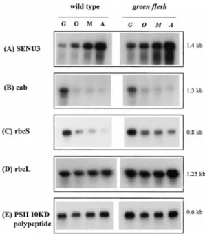
Expression of several senescence-related mRNAs was are shown in italics because there is no colour change). Green flesh
examined in senescing leaves of wild-type and gf ( Fig. 4). leaves were harvested from equivalent position to those of wild-type
leaves. Markers, position of molecular weight markers (kDa). SENU3 mRNA ( Fig. 4A), which encodes a protease which has been shown to be up-regulated during tomato leaf senescence (Drake et al., 1996), increased in both numerous polypeptides were reduced in abundance with
genotypes. Other mRNAs tested also showed a similar the advent of senescence, while others appeared to remain
increase in wild-type and gf (data not shown), including unchanged. A few seemed to increase, at least with respect
SENU2 (another senescence related protease, Drake et al., to other polypeptides, particularly two 24–26 kDa
poly-1996) and ACC oxidase. The accumulation of mRNA peptides in gf.
for the chlorophyll a/b binding protein Cab (LHCII ) was Western analysis of photosynthesis associated proteins strongly reduced during senescence in both genotypes ( Fig. 4B). The expression of the ribulose bisphosphate The abundance of the photosynthesis-related proteins,
carboxylase/oxygenase small subunit (rbcS ) mRNA Rubisco small subunit (Rubisco SSU ), Rubisco large
( Fig. 4C ) was also strongly reduced in wild-type leaves, subunit (Rubisco LSU ) and the major light-harvesting
but less dramatically in gf leaves, whereas rbcL (Fig. 4D) LHCII protein was assessed by Western analysis (Fig. 3).
remained at a relatively high level in both genotypes. The mRNA for the oxygen-evolving PSII 10 kDa polypeptide
Table 4. Soluble protein amounts in attached leaves: the selection
remained at similar levels throughout senescence in both of leaves for analysis was as described in Table 1
genotypes ( Fig. 4E ). Sample Stage (mg protein g−1 fr. wt.) Protein (%)
Wild type MG 5.91 100
Discussion
AD 4.04 68
Green flesh MG 3.92 100 The gf mutation was reported 40 years ago ( Kerr, 1956) AD 2.42 61 but only its effects on tomato fruit were described. This seems to be the first study in which the effects of the gf Each measurement is the average from two leaf samples. MG, mature
1120 Akhtar et al.
Using immunological detection ( Fig. 3), only small differences were found between wild-type and gf in the persistence of the Rubisco SSU and Rubisco LSU poly-peptides. There was some indication of a greater decline in Rubisco LSU in the wild-type, but this is difficult to measure quantitatively using antibodies. The persistence of these chloroplast polypeptides in ageing leaves is not necessarily inconsistent with the decline in their transcript levels, as previously indicated by Bate et al. (1991). The most notable difference was in the LHCII polypeptides which were retained at much higher relative levels in gf, although the mRNA abundance declined dramatically.
Northern analysis of several senescence-related mRNAs revealed identical trends for both genotypes, in all cases examined ( Fig. 4). The up-regulation of SENU3 and others on one hand, and the down-regulation of cab and rbcS, on the other, suggest that the gf mutation does not modify the transcriptional activities of these genes during senescence. A similar conclusion has been drawn pre-viously for another stay-green mutant ( Thomas et al.,
Fig. 4. Expression of mRNA during senescence in wild-type and green
1992).
flesh leaves. Total RNA was extracted from leaves at the green (G ),
senescence onset (O), mid-senescence (M ) and advanced senescence These results indicate that the LHCII polypeptides in (A) stages (5mg) of each sample was loaded on to a 1% agarose gel leaves must somehow be protected from senescent and probed with mRNAs encoding senescence protease SENU3 (A),
degradation. LHCII is a major component of PSII and Cab (B), RbcS (C ), RbcL (D), oxygen-evolving PSII 10 kDa protein
(E ). The molecular sizes of the mRNAs are indicated in kb. retention is correlated with preservation of chlorophyll and carotenoids in the thylakoids. It seems plausible that factors which maintain overall membrane integrity results show clearly that gf leaves exhibit an absolute stay
green phenotype. The behaviour of gf leaves appears to (including retention of LHCII protein) are responsible for this (as suggested by Cheung et al., 1993, for fruit). be different from that of gf fruit, which lose a considerable
portion of their chlorophyll during ripening (Ramirez Comparison with results from other stay-green mutants indicates that the precise relationship between chlorophyll and Tomes, 1964; see also Fig. 1B and Table 1).
Chlorophyllase activity was found to be similar in both and protein breakdown is still a matter of conjecture. In the cytG stay-green soybean mutant, Guiamet and genotypes ( Table 3). This confirms the early report by
Ramirez and Tomes (1964) who measured similar chloro- co-workers assumed that the mutation preferentially affects the breakdown of the Cab (LHCPII) protein, phyllase activities in fruit tissues of wild-type and gf.
According to this study’s observations, the gf mutation thereby holding back chlorophyll catabolism (Guiamet et al., 1991). On the other hand, in the BF 993 Festuca does not alter the normal course of greening, etiolation
and de-etiolation and does not seem, therefore, to interfere mutant, it was hypothesized that a lesion in the chloro-phyll catabolic pathway prevents normal degradation of with chloroplast development. The slight differences in
height between gf and wild-type plants evident from Fig. 1 the chlorophyll binding proteins (Nock et al., 1992). The loss of chlorophyll in senescent leaves is due to were not consistently found. Thus, the effect of the gf
mutation seems to be confined to the senescence phase, the stepwise degradation by chlorophyllase (producing chlorophyllides), Mg-dechelatase (yielding phaeophor-which includes numerous degradative events mostly
asso-ciated with the disintegration of the photosynthetic appar- bide) and phaeophorbide a oxygenase which cleaves the porphyrin macrocycle oxygenolytically into a colourless atus. Among the most important biochemical changes are
the breakdown of chlorophyll and the loss of protein. In fluorescent catabolite (Matile et al., 1996). This pathway has also been demonstrated to be responsible for the intact or detached ageing gf leaves, the almost complete
arrest of chlorophyll breakdown and retention of caroten- breakdown of chlorophyll in ripening fruits of Capsicum annuum (Moser and Matile, 1997). The gf mutant clearly oids is most conspicuous (Fig. 1; Table 1). However,
differences between wild-type and gf in protein degrada- contains enzymes capable of catalysing these reactions, including chlorophyllase, Mg-dechelatase and phaeophor-tion during senescence were not so obvious ( Table 4).
The increase in free amino acids and the upsurge of bide a oxygenase ( Tables 2, 3). Senescent leaves of the stay-green mutants of Festuca pratensis ( Vicentini et al., proteolytic activity (data not shown)) suggest that
senes-cent proteolysis occurs in both genotypes to a similar 1995) and of Pisum sativum ( Thomas et al., 1996) have been found to be deficient with regard to the third extent.
that accumulate during fruit ripening and leaf senescence in
catabolic step, phaeophorbide a oxygenase, for which the
response to ethylene. Planta 179, 73–80.
green flesh mutant of tomato is clearly competent. The
Drake R, John I, Farrell A, Cooper W, Schuch W, Grierson D.
conversion of phaeophorbide a into the primary fluo- 1996. Isolation and analysis of cDNAs encoding tomato rescent catabolite, pFCC-2, indicates that not only the cysteine proteases expressed during leaf senescence. Plant
Molecular Biology 30, 755–767. oxygenase but also the second enzyme of the channelled
Feinberg AP, Vogelstein B. 1983. A technique for radiolabelling
reaction, RCC reductase (Rodoni et al., 1997), is present
DNA restriction endonuclease fragments to high specific
in gf. Analysis of chlorophyll breakdown products in ripe
activity. Analytical Biochemistry 132, 6–13.
fruit showed (Table 2A) that gf accumulates chloro- Gan S, Amasino RM. 1995. Inhibition of leaf senescence phyllides a and b but phaeophorbide a does not accumu- by autoregulated production of cytokinin. Science 270,
1986–1988.
late in vivo. This may indicate that the action of
Grbic V, Bleecker AB. 1995. Ethylene regulates the timing of
phaeophorbide a oxygenase is somehow prevented in the
leaf senescence in Arabidopsis. The Plant Journal 8, 595–602.
mutant in vivo, thereby blocking the breakdown of
chloro-Grierson D, Purton ME, Knapp JE, Bathgate B. 1987. Tomato
phyll. Since the phaeophorbide a oxygenase system is ripening mutants. In: Thomas H, Grierson D, eds. present in the tissues, the lesion of gf is probably associ- Developmental mutants in higher plants. Cambridge University
Press, 73–94.
ated with altered accessibility or transport of components
Guiamet JJ, Schwartz E, Pichersky E, Noode´n LD. 1991.
or the absence of another factor required for the reaction.
Characterization of cytoplasmic and nuclear mutations affecting chlorophyll and chlorophyll-binding proteins during senescence in soybean. Plant Physiology 96, 227–231.
Acknowledgements
Hortensteiner S, Vicentini F, Matile P. 1995. Chlorophyll
breakdown in senescent cotyledons of rape, Brassica napus Muhammad Shaheen Akhtar was sponsored by The Ministry
L.: enzymatic cleavage of phaeophorbide a in vitro. New of Education, Government of Pakistan. The work was supported
Phytologist 129, 237–246.
by the Biotechnology and Biological Sciences Research Council
John I, Drake R, Farrell A, Cooper W, Lee P, Horton P,
and the Swiss National Science Foundation. EEG gratefully
Grierson D. 1995. Delayed leaf senescence in
ethylene-acknowledges the financial support by the Royal Society–Israel
deficient acc-oxidase antisense tomato plants: molecular and Visiting Research Professorship Programme. We thank Drs
physiological analysis. The Plant Journal 7, 483–490. Kate Griffith and P Scott for antibodies raised against LHCII
John I, Hacket R, Cooper W, Drake R, Farrell A, Grierson D.
and Rubisco (LSU and SSU ), respectively.
1997. Cloning and characterization of tomato leaf senescence related cDNAs. Plant Molecular Biology 33, 641–651.
Katayama-Fujimura Y, Gottesman S, Maurizi MR. 1987. A
References
multiple component, ATP-dependent protease from
Escherchia coli. Journal of Biological Chemistry 262, Amir-Shapira D, Goldschmidt EE, Altman A. 1987. Chlorophyll
catabolism in senescing plant tissues: in vivo breakdown 4477–4485.
Kerr EA. 1956. Green flesh, gf. Tomato Genetics Cooperative
intermediates suggest different degradative pathways for
citrus fruit and parsley leaves. Proceedings of the National Reports 6, 17.
Kerr E A. 1957. Linkage relations of gf. Tomato Genetics
Academy of Sciences, USA 84, 1901–1905.
Bate NJ, Rothstein SJ, Thompson JE. 1991. Expression of Cooperative Reports 8, 21.
Matile P, Hortensteiner S, Thomas H, Krantler B. 1996.
nuclear and chloroplast photosynthesis-specific genes during
leaf senescence. Journal of Experimental Botany 42, 801–811. Chlorophyll breakdown in senescent leaves. Plant Physiology
112, 1403–1409. Bradford MM. 1976. A rapid and sensitive method for the
quantitation of microgram quantities of proteins utilising the McGlasson WB, Poovaiah BW, Dostal HC. 1975. Ethylene
production and respiration in ageing leaf segments and in principles of protein–dye binding. Analytical Biochemistry
72, 248–254. discs of fruit tissue of normal and mutant tomatoes. Plant
Physiology 56, 547–549.
Canfield MR, Guiamet JJ, Noode´n LD. 1995. Alteration of
soybean seedling development in darkness and light by the Meyer Y. 1988. Preparation by two-dimensional electrophoresis
of proteins for antibody production: antibodies against stay-green mutation cytG and Gd-1d2. Annals of Botany
75, 143–150. proteins whose synthesis is induced by auction in tobacco mesophyll protoplast. Electrophoresis 9, 704–712.
Cheung AY, McNellis T, Piekos B. 1993. Maintenance of
chloroplast components during chromoplast differentiation Mizrahi Y, Dostal HC, Cherry JH. 1975. Ethylene-induced
ripening in attached rin fruits, a non ripening mutant of in the tomato mutant green flesh. Plant Physiology 101,
1223–1229. tomato. HortScience 10, 414–415.
Moore S. 1968. Amino acid analysis: aqueous dimethyl Clayberg CD, Butler L, Rick CM, Young PA. 1960. Second list
of known genes in the tomato. Journal of Heredity 51, sulphoxide as solvent for the ninhydrin reaction. Journal of
Biological Chemistry 243, 6281–6283.
167–174.
Cornelius SB, Blume B, Bouzayen M, Cooper W, Hamilton AJ, Moran R. 1982. Formulae for determination of chlorophyllous
pigments extracted with N, N-dimethylformamide. Plant
Grierson D. 1996. Differential expression of the
1-aminocyclopropane-1-carboxylase oxidase gene family of Physiology 69, 1376–1381.
Moser D, Matile P. 1997. Chlorophyll breakdown in ripening
tomato. The Plant Journal 9, 525–535.
Davies BH. 1976. Carotenoids. In: Goodwin TW, ed. Chemistry fruits of Capsicum annuum. Journal of Plant Physiology
150, 759–761.
and biochemistry of plant pigments, London, 38–165.
Davies KM, Grierson D. 1989. Identification of cDNA clones Nock LP, Rogers LJ, Thomas H. 1992. Metabolism of protein
and chlorophyll in leaf tissue of Festuca pratensis during for tomato (Lycopersicon esculentum Mill.) messenger-RNAs
1122 Akhtar et al.
chlorophyll assembly and senescence. Phytochemistry 31, Tomes ML. 1963. Temperature inhibition of carotene synthesis
in tomato. Botanical Gazette 124, 180–185. 1465–1470.
Ramirez DA, Tomes ML. 1964. Relationship between chloro- Trebitsh-Sitrit T, Goldschmidt EE, Riov J. 1993. Ethylene
induces de novo synthesis of chlorophyllase, a chlorophyll phyll and carotenoid biosynthesis in dirty-red ( green flesh)
mutant in tomato. Botanical Gazette 125, 22–226. degrading enzyme, in Citrus fruit peel. Proceedings of the
National Academy of Sciences, USA 90, 9441–9445.
Rodoni S, Mu¨hlecker W, Anderl M, Kra¨utler B, Moser D,
Thomas H, Matile P, Hortensteiner S. 1997. Chlorophyll Vera P, Conejero V. 1990. Effect of ethephon on protein
degradation and the accumulation of pathogenesis-related breakdown in senescent chloroplasts—cleavage of
phaeo-phorbide a in two enzymic steps. Plant Physiology 115, (PR) proteins in tomato leaf discs. Plant Physiology 92, 227–233.
669–676.
Thomas H, Ougham HJ, Davies E. 1992. Leaf senescence in a Vicentini F, Hortensteiner S, Schellenberg M, Thomas H, Matile P. 1995. Chlorophyll breakdown in senescent leaves:
identi-non-yellowing mutant of Festuca pratensis. Transcripts and
translation products. Journal of Plant Physiology 139, fication of the biochemical lesion in a stay-green genotype of
Festuca pratensis Huds. New Phytologist 129, 247–252.
403–412.
Thomas H, Schellenberg M, Vicentini F, Matile P. 1996. Gregor Wadsworth GJ, Redinbaugh MG, Scandalios JG. 1988. A
procedure for the small-scale isolation of RNA suitable for Mendel’s green and yellow pea seeds. Botanica Acta 109, 3–4.
Thomas H, Smart CM. 1993. Crops that stay green. Annals of RNA blot analysis. Analytical Biochemistry 172, 279–283.