Effect of obesity and site of surgery on perioperative lung
volumes
B. S. von Ungern-Sternberg*, A. Regli, M. C. Schneider, F. Kunz and A. Reber
Department of Anaesthesia, University of Basel/Kantonsspital, CH-4031 Basel, Switzerland
*Corresponding author. E-mail: [email protected]
Background. Although obese patients are thought to be susceptible to postoperative pulmonary complications, there are only limited data on the relationship between obesity and lung volumes after surgery. We studied how surgery and obesity affect lung volumes measured by spirometry.
Methods. We prospectively studied 161 patients having either breast surgery (Group A, n=80) or lower abdominal laparotomy (Group B, n=81). Premedication and general anaesthesia were standardized. Spirometry was measured with the patient supine, in a 30° head-up position. We measured vital capacity (VC), forced vital capacity, peak expiratory ¯ow and forced expiratory volume in 1 s at preoperative assessment (baseline), after premedication (before induction of anaesthesia) and 10±20 min, 1 h and 3 h after extubation.
Results. Baseline spirometric values were all within the normal range. All perioperative values decreased signi®cantly with increasing body mass index (BMI). The greatest reduction of mean VC (expressed as percentage of baseline values) occurred after extubation, and was more marked after laparotomy than after breast surgery (23 (SD 14)% vs 20 (14)%). Considering
patients according to BMI (<25, 25±30, >30), VC decreased after surgery by 12 (7)%, 24 (8)% and 40 (10)%, respectively. VC recovered more rapidly in Group A.
Conclusion. Postoperative reduction in spirometric volumes was related to BMI. Obesity had more effect on VC than the site of surgery.
Br J Anaesth 2004; 92: 202±7
Keywords: anaesthesia; complications, obesity; lung, respiratory function; monitoring, spirometry; pharmacokinetics, body mass index
Accepted for publication: August 20, 2003
About one third of the population of industrial countries are at least 20% overweight and the prevalence of obesity is increasing.1Obese patients have reduced respiratory func-tion2 and are considered to be more likely to develop postoperative pulmonary complications.3 4 General anaes-thesia and surgery reduce lung volumes and this effect may be greater in the obese.2 5 6
Clinically, obese patients appear to have worse respira-tory function after surgery but only limited data show that obesity causes reduced lung volumes after surgery.3 7 In normal subjects the site of surgery affects respiratory function: impairment is greater after abdominal surgery than after non-abdominal surgery.6 8Since there are no studies assessing the in¯uence of body mass index (BMI) and the site of surgery on perioperative lung volumes, we carried out the present study.
We considered that patients with a normal BMI (<25) would be less affected compared with obese patients (BMI>30) who would be more affected. We performed perioperative spirometry in non-obese and obese patients undergoing lower abdominal surgery or breast surgery.
Material and methods
Study population
The study was approved by the Ethics Committee of the University of Basel, Switzerland. Informed written consent was obtained from each patient before inclusion. We studied 161 women (ASA physical status I±II) scheduled for either breast surgery (Group A, n=80) or lower abdominal laparotomy with a transverse incision (Group B, n=81).
We excluded patients who were pregnant, those who had bronchial asthma requiring regular therapy, cardiac disease associated with dyspnoea >NYHA II, or severe psychiatric disorders. The expected duration of surgery was 90± 150 min.
Anaesthesia
Premedication consisted of oral midazolam 7.5 mg, 30± 60 min before surgery. General anaesthesia was standar-dized and induced with propofol 2 mg kg±1 and fentanyl 2 mg kg±1 i.v. Tracheal intubation was facilitated by atracurium 0.5 mg kg±1 i.v. A laryngeal mask was not used. Anaesthesia was maintained with nitrous oxide 66% in oxygen and propofol by infusion using the Bristol formula (10 mg kg±1h±1for the ®rst 10 min of general anaesthesia, 8 mg kg±1 h±1 for another 10 min and thereafter 6 mg kg±1 h±1 or adjusted to individual needs).9 Ventilation was controlled using an ADU ventilator (Datex Ohmeda, S/5 ADU Helsinki, Finland) with a circle system. Repeated doses of fentanyl were given during surgery as necessary based on clinical signs (heart rate, arterial pressure, pupil size and sweating) but not within 30± 60 min of the estimated end of the operation. To have the patient fully alert and compliant for spirometry, we substituted sevo¯urane for propofol 30±60 min before the estimated end of surgery as this was considered, on the basis of clinical observations, to give more rapid recovery. Increments of atracurium 5 mg i.v. were given to maintain muscle relaxation, which was monitored by train-of-four (TOF) stimulation. Neostigmine 2.5 mg and glycopyrrolate 0.5 mg were given i.v. as needed to antagonize neuro-muscular block. Before extubation, four equal twitches in the TOF without tetanic fade (50 Hz over 5 s) were required, as well as recovery of consciousness (eye opening on demand), protective airway re¯exes and adequate spontaneous ventilation.
For postoperative pain relief, we gave methadone 2 mg i.v. to achieve a pain score of <20 mm while coughing, assessed on the 100 mm visual analogue scale (VAS; where 0 mm represented no pain or no dyspnoea and l00 mm was worst possible pain or dyspnoea). The total dose of methadone given to each patient was neither limited nor weight adjusted. Basic analgesia consisted of paracetamol 1000 mg rectally or orally every 6 h starting directly after the operation. We did not administer any local anaesthetic into the wound.
Spirometry
Before the operation we measured the weight and height of each patient to obtain the exact BMI. A vitalograph (Vitalograph 2120, Vitalograph, Hamburg, Germany) was used for spirometric measurements. Measurements were made with the patient in a 30° head-up position.10After a thorough demonstration of the correct use during the
preanaesthetic visit, spirometry was measured and taken as baseline (T0). When the patient arrived in the operating theatre (30±60 min after premedication), spirometry was repeated before induction of anaesthesia (T1). After extubation, as soon as the patient was alert and fully cooperative, pain and dyspnoea were assessed using the VAS before and, if necessary, after analgesic (methadone) was given. As soon as a VAS pain score <20 mm was achieved (all patients within 20 min of extubation), spirometry was performed for the third time (T2). We use the VAS routinely to score pain and dyspnoea in our clinical practice in the postanaesthetic care unit. Spirometric measurements were repeated in the postanaesthetic care unit 1 h (T3) and 3 h (T4) after extubation. Methadone dosage was recorded at each assessment.
We measured vital capacity (VC), forced vital capacity (FVC), forced expiratory volume in 1 s (FEV1) and peak expiratory ¯ow (PEF) and calculated the FEV1/FVC ratio. At each measurement time, two spirometry manoeuvres were done, one for VC and the other for FVC, PEF and FEV1; each was done at least three times to meet the European Respiratory Society (ERS) criteria for reproduci-bility11and the best measurement was recorded. For VC, the patient was asked to inhale fully and then exhale slowly but completely. For FVC, PEF and FEV1, the patient was asked to expire as forcefully as possible.
Statistical analysis
To allow comparison between the patients and the two groups, the values were calculated as percentage change from the value measured at baseline (preoperative assess-ment). We used repeated-measures analysis of variance (ANOVA) to compare data within groups. We used a
Wilcoxon rank sum test to compare measurements between the groups. A Bonverroni test was used for post hoc comparisons. The Spearman rank correlation test was used to assess the relationship between spirometric measure-ments and BMI. P<0.05 was considered signi®cant. For statistical calculations, we used StatView for windows (SAS Institute Inc., Cary NC, USA, Version 5.0.1).
Results
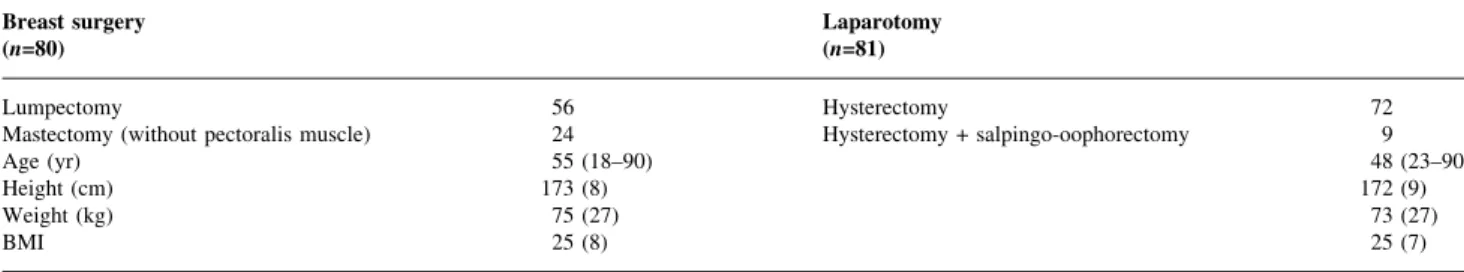
We recruited 187 women. In eight, the planned surgery was altered, 13 patients declined to continue, and measurements were unsatisfactory in ®ve. We therefore present data for 161 patients (Table 1). The patients with unsatisfactory spirometric measurements did not differ in age or weight from those with acceptable measurements, and they did not have extreme values of BMI. The distribution of non-smokers between the groups was similar with 59 (74%) in Group A and 60 (74%) in Group B. The smokers (2±15 pack-years) were evenly distributed over the BMI range, with a minor tendency towards smaller BMI. Antagonism of muscle relaxation was necessary in only three patients in
each group. All patients met the extubation criteria completely. The duration of surgery was 120 (SD18) min
and the maximum 150 min.
Vital capacity
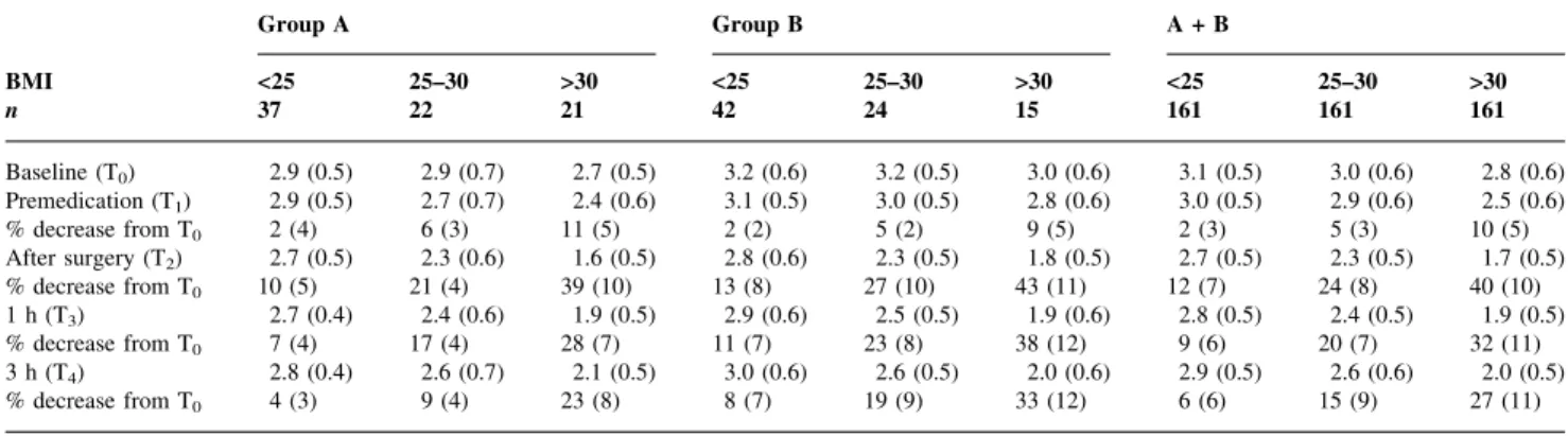
The baseline VC values were all within the normal range (Table 2). After premedication, the values decreased by 5 (5)% in both groups (Table 2, Fig. 1). The decrease was greater in those with a greater BMI, although the effect in normal-weight patients was minimal (Table 3). In both groups, the greatest decrease was directly after extubation (Group A: 20 (14)%; Group B: 23 (14)%; Table 2). Laparotomy caused a signi®cantly greater decrease in VC and recovery was slower than after breast surgery (10 (9)% vs 16 (13)% in Groups A and B, respectively, 3 h after surgery; Table 2, Fig. 2). At each postoperative measure-ment time there was a signi®cant negative correlation between BMI and VC (Table 3 and Fig. 2).
Other spirometric values
The baseline values for all other variables (FVC, FEV1and PEF) were within the normal ranges. During the periopera-tive period they changed in parallel with VC (Table 2). The FEV1/FVC ratio did not change in either group throughout the study period.
Pain scores and pain relief
There were no differences in pain score between the two groups when spirometry was performed. In both groups, a
maximum VAS for pain value of 20 mm was recorded and a comparable dose of methadone was administered directly after extubation (0.78 (1.3) mg in Group A and 0.96 (1.2) mg in Group B). However, at 1 and 3 h after extubation, the doses of methadone given to the patients after laparotomy were signi®cantly greater than those given after breast surgery: 4.2 (2.2) mg vs 2.7 (1.9) mg and 4.4 (2.6) mg vs 2.8 (2.1) mg, respectively (P<0.001). The total amounts of methadone given during the ®rst 3 h after surgery were 9.6 (3.3) mg vs 6.3 (3.5) mg for groups A and B, respectively (P<0.001). None of the patients complained of dyspnoea.
Table 2 Absolute and relative values of vital capacity (VC), forced vital capacity (FVC), forced expiratory ¯ow rate in 1 s (FEV1) and peak expiratory ¯ow
(PEF) for patients undergoing breast surgery (Group A) and laparotomy (Group B). Data are mean (SD). T0, baseline (preoperative) value; *signi®cant
difference between groups (ANOVA)
VC FVC FEV1 PEF Group A B A B A B A B Baseline (T0) 2.8 (0.6) 3.2 (0.6)* 2.8 (0.5) 3.1 (0.6)* 2.4 (0.5) 2.7 (0.5)* 345 (62.9) 374 (53.1)* Premedication (T1) 2.7 (0.6) 3.0 (0.6)* 2.6 (0.5) 2.9 (0.6)* 2.2 (0.5) 2.5 (0.5)* 325 (61.8) 352 (51.7)* % decrease from T0 5 (6) 5 (5) 6 (6) 5 (5) 6 (5) 6 (5) 6 (7) 6 (5) After surgery (T2) 2.3 (0.6) 2.5 (0.7)* 2.2 (0.6) 2.4 (0.7)* 1.9 (0.6) 2.0 (0.6) 273 (69.7) 279 (58.5) % decrease from T0 20 (14) 23 (14)* 21 (14) 23 (14)* 22 (14) 24 (14)* 21 (13) 26 (12)* 1 hr (T3) 2.4 (0.6) 2.6 (0.7)* 2.3 (0.6) 2.5 (0.7)* 2.0 (0.5) 2.1 (0.6) 289 (66.5) 284.3 (57.0) % decrease from T0 15 (10) 20 (13)* 16 (10) 20 (13)* 16 (11) 21 (13)* 17 (11) 24 (12)* 3 h (T4) 2.6 (0.6) 2.7 (0.7) 2.5 (0.6) 2.6 (0.6) 2.1(0.5) 2.2 (0.6) 306 (65.8) 296 (59.4)* % decrease from T0 10 (9) 16 (13)* 11 (10)* 17 (13)* 11 (9) 17 (13)* 12 (10) 21 (13)
Table 1 Details of patients. Data are median (range) for age, or median (interquartile range)
Breast surgery Laparotomy
(n=80) (n=81)
Lumpectomy 56 Hysterectomy 72
Mastectomy (without pectoralis muscle) 24 Hysterectomy + salpingo-oophorectomy 9
Age (yr) 55 (18±90) 48 (23±90)
Height (cm) 173 (8) 172 (9)
Weight (kg) 75 (27) 73 (27)
BMI 25 (8) 25 (7)
Fig 1 Percentage change in vital capacity (VC) in all 161 patients in relation to BMI after premedication (r=±0.703, P<0.001).
Discussion
Baseline spirometric values in non-obese and obese
patients
The excess body fat in obese patients affects chest wall mechanics. The compliance of the respiratory system is less (mass loading)5 12and lung volumes such as FRC and VC are reduced. Although VC increases in parallel with the BMI within the normal weight range, VC decreases progressively in more obese patients.12±15 The effect of obesity on other spirometric measurements is less clear.
Our initial spirometric measurements were in line with these observations: they were within the normal ranges for non-obese and for obese patients. There were no signs of airway obstruction as FEV1and the FEV1/FVC ratio were not, or only minimally, in¯uenced by obesity.16The values obtained in the group scheduled for breast surgery were lower than in the group scheduled for laparotomy, which could be explained by their greater age, as VC decreases with age.17 To compare the groups, we used percentage change from baseline.
Effect of premedication
Premedication reduced VC, with no signi®cant difference between the groups scheduled for breast surgery or laparotomy. We found, unexpectedly, that this reduction in VC was related to BMI. Normal-weight patients showed a minimal effect compared with obese patients (BMI >30; 10% decrease, Table 3). There was, however, a compara-tively wide range of individual responses to premedication (Fig. 2).
Premedication may affect the activity of the respiratory muscles. VC is a good index of respiratory muscle strength in patients with neuromuscular disorders.18 19 Benzo-diazepines have a spinally mediated muscle relaxant effect that can affect the respiratory muscles,20so premedication could affect respiration. Obese patients might be more affected by these agents because they have a greater work of breathing.12Another explanation could be that sedation by midazolam interfered with the performance of spirometry, although completely reproducible tracings at all measure-ments were obtained for all patients. This effect should have affected both obese and non-obese patients equally.
The ®nding that VC was markedly reduced in obese patients after premedication with benzodiazepines might imply that these patients should receive supplemental oxygen before the operation.
Anaesthesia and immediate postoperative respiratory
function
In our study, the smallest spirometric values were found immediately after extubation. The decrease in VC, FVC, FEV1and PEF followed the same trends (Table 2), and the FEV1/FVC ratio did not change. This suggests a restrictive pattern of respiratory compromise in the postoperative period, which has been described previously.6 21±23
The postoperative impairment of respiratory function was probably not caused by insuf®cient cooperation, since all patients were alert and fully compliant within 10±20 min of extubation and produced normal spirometric tracings that completely met the ERS criteria.11 Adequate patient cooperation was achieved by strictly following a
Table 3 Absolute values and changes of vital capacity (VC) for patients undergoing breast surgery (Group A) or laparotomy (Group B) according to BMI. Data are mean (SD). T0, baseline (preoperative) value. All differences between BMI <25 and >30 were statistically signifcant (ANOVA)
Group A Group B A + B BMI <25 25±30 >30 <25 25±30 >30 <25 25±30 >30 n 37 22 21 42 24 15 161 161 161 Baseline (T0) 2.9 (0.5) 2.9 (0.7) 2.7 (0.5) 3.2 (0.6) 3.2 (0.5) 3.0 (0.6) 3.1 (0.5) 3.0 (0.6) 2.8 (0.6) Premedication (T1) 2.9 (0.5) 2.7 (0.7) 2.4 (0.6) 3.1 (0.5) 3.0 (0.5) 2.8 (0.6) 3.0 (0.5) 2.9 (0.6) 2.5 (0.6) % decrease from T0 2 (4) 6 (3) 11 (5) 2 (2) 5 (2) 9 (5) 2 (3) 5 (3) 10 (5) After surgery (T2) 2.7 (0.5) 2.3 (0.6) 1.6 (0.5) 2.8 (0.6) 2.3 (0.5) 1.8 (0.5) 2.7 (0.5) 2.3 (0.5) 1.7 (0.5) % decrease from T0 10 (5) 21 (4) 39 (10) 13 (8) 27 (10) 43 (11) 12 (7) 24 (8) 40 (10) 1 h (T3) 2.7 (0.4) 2.4 (0.6) 1.9 (0.5) 2.9 (0.6) 2.5 (0.5) 1.9 (0.6) 2.8 (0.5) 2.4 (0.5) 1.9 (0.5) % decrease from T0 7 (4) 17 (4) 28 (7) 11 (7) 23 (8) 38 (12) 9 (6) 20 (7) 32 (11) 3 h (T4) 2.8 (0.4) 2.6 (0.7) 2.1 (0.5) 3.0 (0.6) 2.6 (0.5) 2.0 (0.6) 2.9 (0.5) 2.6 (0.6) 2.0 (0.5) % decrease from T0 4 (3) 9 (4) 23 (8) 8 (7) 19 (9) 33 (12) 6 (6) 15 (9) 27 (11)
Fig 2 Differences (%) in vital capacity (VC) between the groups according to BMI. Group A vs Group B: *P<0.05; n.s.=not signi®cant
standardized anaesthetic regimen based on short-acting anaesthetic agents, a prerequisite for collecting representa-tive data.
The reduced spirometric volumes in our study may have been caused by impaired respiratory mechanics as well as atelectasis formation promoted by general anaesthesia in the supine position.24±27A reduction in VC could be caused by a reduction in both inspiratory and expiratory reserve volumes.23 28A reduced inspiratory capacity could reduce the ability to cough effectively and may predispose to respiratory complications.3 6 23
BMI and immediate postoperative respiratory
function
Data on the impairment of postoperative respiratory func-tion in obese patients were previously sparse. There are two small studies3 7 but no controlled clinical trials and none relate the changes of postoperative lung volumes to obesity. We found a strong negative correlation between lung volumes and BMI: the smallest values of VC occurred in grossly obese patients (BMI>30) 20 min after extubation compared with non-obese patients (BMI<25) (reduction in VC 41% vs 11%, Table 3). The greatest decline in postoperative lung volumes occurred in patients with BMI>40 (Groups A and B, 51 (5)%). Three h after extubation, patients of normal weight had only a small residual reduction in lung volumes or had made a complete recovery whereas obese patients still had signi®cantly smaller lung volumes (reduction in VC 6% vs 28%) irrespective of the site of surgery.
As the expiratory reserve volume is reduced in obese patients, obesity is associated with a decrease in FRC and VC2 12 16 and thus an enhanced response to general anaesthesia compared with normal-weight patients.5 Obesity predisposes to the formation of atelectasis per se and even more so after induction of general anaesthesia,29 which could signi®cantly reduce postoperative lung volumes. Our ®ndings support previous small studies investigating the effects of BMI on lung volumes;3 6 7 24±27 most of them, however, did not start their measurements before the ®rst postoperative day.
Site of surgery, postoperative pain and respiratory
function
In studies of non-obese patients, the magnitude of the reduction in VC, tidal volume and FRC is related to the site of surgery. Ali and colleagues6 reported that abdominal surgery resulted in a greater reduction of VC than super®cial surgery (42% vs 29%) 4 h after the operation, and Diament and Palmer8observed a larger reduction of FVC after lower abdominal surgery than after non-abdominal surgery (25% vs 8%) on the ®rst day after surgery. In our study, the reduction in VC was more pronounced after laparotomy than after breast surgery but not as marked as in other
studies, although in those studies there was a longer time between surgery and spirometry.6 7 23 30In those studies, the decrease in lung volume was thought to be related to pain and abdominal muscle spasm. The greater impairment of postoperative respiratory function in these studies might be that shorter acting anaesthetic agents and pain relief given according to VAS scores were not used in these earlier studies.31It is crucial for a patient to be as free from pain as possible during spirometry and to be as close to the preoperative baseline conditions, in order to avoid factors that affect test performance. Nevertheless, in our study, even though VAS scores <20 mm were achieved while coughing in obese and non-obese patients, differences in nociception (visceral compared with somatic pain) might still have caused some differences in lung volumes. The greater sedation from larger doses of methadone required for analgesia after laparotomy might have interfered with spirometry, despite meeting the ERS criteria. Even assum-ing such an effect, this should have affected obese and normal-weight patients equally, as when midazolam was used as premedication. Other body changes caused by surgery, anaesthesia and postoperative analgesia may also have meant that laparotomy had a greater effect on respiratory function.32
Duration of observation of the perioperative
respiratory function
In contrast to most other studies of the later postoperative period,6±8 28 33we focused on the immediate postoperative period when lung volumes could be most severely affected. We limited the observation period to 3 h, corresponding to the time patients stay in our postanaesthetic care unit. The patients were mobilized in the unit immediately before discharge to the ward where further mobilization was encouraged, as this improves postoperative lung function.34 No patients developed pulmonary complications during the ®rst 24 h.
We did not assess the effect of the duration of surgery on postoperative respiratory function. Respiratory function could possibly be more affected after longer surgical procedures.
We conclude that premedication with midazolam caused a moderate reduction of lung volumes, more in the obese. Postoperative respiratory function was signi®cantly more impaired in obese patients. Respiratory impairment after laparotomy persisted well into the recovery period and was more pronounced than after breast surgery. In non-obese patients, impairment of lung function after surgery was minor and independent of the site of operation. Obesity impaired lung volumes more than the effect of surgery.
Acknowledgements
The authors are indebted to the recovery room nurses for their great help. The authors also thank J. Etlinger for editorial assistance. This study was
supported by the Department of Anaesthesia, University of Basel/ Kantonsspital, Basel, Switzerland.
References
1 Kuczmarski RJ, Flegal KM, Campbell SM, Johnson CL. Increasing prevalence of overweight among U.S. adults: the National Health and Nutrition Surveys.1960±1991. JAMA 1994; 272: 205±11 2 Luce JM. Respiratory complications of obesity. Chest 1980; 78:
626±31
3 Hansen G, Drablos PA, Steinert R. Pulmonary complications. Ventilation and blood gases after upper abdominal surgery. Acta Anaesthesiol Scand 1977; 21: 211±15
4 Blouw EL, Rudolph AD, Narr BJ, Sarr MG. The frequency of respiratory failure in patients with morbid obesity undergoing gastric bypass. AANA J 2003; 71: 45±50
5 Waltemath CL, Bergman NA. Respiratory compliance in obese patients. Anesthesiology 1974; 41: 84±5
6 Ali J, Weisel RD, Layug AB, Kripke BJ, Hechtman HB. Consequences of postoperative alterations in respiratory mechanics. Am J Surg 1974; 128: 376±82
7 Eriksen J, Andersen J, Rasmussen JP. Postoperative pulmonary function in obese patients after upper abdominal surgery. Acta Anaesthesiol Scand 1977; 21: 336±41
8 Diament ML, Palmer KNV. Postoperative changes in gas tensions of arterial blood and in ventilatory function. Lancet 1966; 2: 180±2
9 Roberts FL, Dixon J, Lewis GT, Tackley RM, Prys-Roberts C. Induction and maintenance of propofol anaesthesia. A manual infusion scheme. Anaesthesia 1988; 43 Suppl: 14±17
10 Gudmundson G, Cerveny M, Shasby DM. Spirometric values in obese individuals, effect on body position. Am J Respir Crit Care Med 1997; 155: 998±9
11 Standardized lung function testing. Of®cial statement of the European Respiratory Society. Eur Respir J Suppl 1993; 16: 1±100 12 Ray C, Sue D, Bray G, et al. Effects of obesity on respiratory
function. Am Rev Respir Dis 1983; 128: 501±6
13 Sue DY. Obesity and pulmonary function: more or less? Chest 1997; 111: 844±5
14 Sahebjami H, Gartside PS. Pulmonary function in obese subjects with a normal FEV1/FVC ratio. Chest 1996; 110: 1425±9 15 Gibson GJ. Obesity, respiratory function and breathlessness.
Thorax 2000; 55: S41±4
16 Barrera F, Reidenberg M, Winters W. Pulmonary function in the obese patient. Am J Med Sci 1967; 254: 785±96
17 Ulmer WT, Reichel G, Nolte D, Islam MS. Die Lungenfunktion. Stuttgart: Thieme Verlag, 1991
18 Lumb AB. Nunn's Applied Respiratory Physiology 5th Edn. Oxford: Butterworth±Heinemann, 2000
19 Polkey MI, Green M, Moxham J. Measurement of respiratory muscle strength. Thorax 1995; 50: 1131±5
20 Dretchen K, Ghoneim MM, Long JP. The interaction of diazepam with myoneural blocking agents. Anesthesiology 1971; 34: 463±8 21 Meyers JR, Lembeck L, O'Kane H, Baue AE. Changes in functional residual capacity of the lung after operation. Arch Surg 1975; 110: 576±82
22 Alexander JL, Spence AA, Parikh RK, Stuart B. The role of airway closure in postoperative hypoxemia. Br J Anaesth 1973; 5: 34±40 23 Craig D. Postoperative recovery of pulmonary function. Anesth
Analg 1981; 60: 46±52
24 Pelosi P, Croci M, Ravagnan I, et al. The effects of body mass on lung volumes, respiratory mechanics, and gas exchange during general anaesthesia. Anesth Analg 1998; 87: 654±60
25 Strandberg A, Tokics L, Brismar B, et al. Constitutional factors promoting development of atelectasis during anaesthesia. Acta Anaesthesiol Scand 1987; 31: 21±4
26 Rigg JR. Pulmonary atelectasis after anaesthesia: pathophysiology and management. Can Anaesth Soc J 1981; 28: 305±13
27 Rothen HU, Sporre B, Engberg G, Wegenius G, Hedenstierna G. Airway closure, atelectasis and gas exchange during general anaesthesia. Br J Anaesth 1998; 81: 681±6
28 Logan DA, Spence AA, Smith G. Postoperative pulmonary function. Anaesthesia 1977; 32: 3±7
29 Eichenberger A, Proietti S, Wicky S, et al. Morbid obesity and postoperative pulmonary atelectasis: an underestimated problem. Anesth Analg 2002; 95: 1788±92
30 Chodoff P, Margand PM, Imbembo AL. Applied pulmonary physiology: morbid obesity and pulmonary function. Crit Care Med 1974; 2: 123±8
31 Spence AA, Smith G. Postoperative analgesia and lung function: a comparison of morphine with extradural block. Br J Anaesth 1998; 81: 984±8
32 Kehlet H, Holte K. Effect of postoperative analgesia on surgical outcome. Br J Anaesth 2001; 87: 62±72
33 Spence AA, Smith G. Postoperative analgesia and lung function: A comparison of morphine with extradural block. Br J Anaesth 1971; 43: 144±8
34 Basse L, Kehlet H. Accelerated postoperative recovery programme after colonic resection improves physical performance, pulmonary function and body composition. Br J Surg 2002; 89: 446±53