ORIGINAL PAPER
Do synovial leptin levels correlate with pain
in end stage arthritis?
Anne Lübbeke&Axel Finckh&Gabor J. Puskas&
Domizio Suva&Alexandre Lädermann&Sylvette Bas&
Daniel Fritschy&Cem Gabay&Pierre Hoffmeyer
Received: 6 June 2013 / Accepted: 12 June 2013 / Published online: 9 July 2013 # Springer-Verlag Berlin Heidelberg 2013
Abstract
Purpose We evaluated whether synovial fluid (SF) leptin concentrations correlate with pain severity in patients with hip or knee endstage osteoarthritis (OA) and whether they mediate the association between increased joint pain and (1) female gender and (2) obesity.
Methods We conducted a cross-sectional study including patients with primary hip and knee OA undergoing joint replacement between January and December 2010. SF leptin concentrations obtained on the day of surgery were assessed. Main outcome was pain severity measured pre-operatively using WOMAC and VAS pain scales.
Results A total of 219 patients were included, 123 hip and 96 knee arthroplasties. Mean age was 72 years, 59 % were women. Mean SF leptin levels were 22.9 (±25.6) ng/ml in women and 5.4 (±5.9) ng/ml in men. Levels >19.6 ng/ml (highest quartile) were significantly associated with increased pain on both WOMAC (mean difference−9.6, 95 % CI −15.1 to −4.0) and VAS scale (mean difference 0.8, 95 % CI 0.2–1.3). Associations remained unchanged after adjusting for age, co-morbidities, contra-lateral arthritic joint, OA site, and disability. The
associations observed between increased pain and female gen-der or obesity were substantially reduced after adjusting for SF leptin.
Conclusion Joint pain is associated with SF leptin concen-trations. Increased pre-operative pain observed in women and obese may be related to high intra-articular leptin levels.
Introduction
Joint pain is the key clinical feature of osteoarthritis (OA) [1–3] and plays a major role in the decision for a total joint arthroplasty, but the causes of OA pain are not well established [4] and correlation between pain and structural joint damage is weak [5]. A possible link between joint pain and the adipokine leptin, which is considered as a key medi-ator in the well established association between obesity and osteoarthritis (OA) [6–13], has been indirectly suggested in studies on weight loss. Weight reduction consistently resulted in decreased joint pain in patients with OA [14, 15] and simultaneously decreased blood leptin levels [16–18]. Fur-thermore, leptin is particularly elevated in women and in obese patients [10,19], and in these two patient groups, higher OA pain levels have been reported [20–26]. Studies from outside the musculoskeletal field have lent further support to a possible association between pain and leptin concentrations. Leptin levels in peritoneal fluid correlated positively with pain severity in endometriosis patients [27]. Furthermore, recent studies in rodents [28–30] suggested that spinal leptin might be involved in the pathogenesis of neuropathic pain.
So far two clinical studies have evaluated the association between adipokines and OA pain. Gandhi et al. [31] did not find a clear association between intra-articular leptin concen-trations and pre-operative pain, but they reported that the synovial fluid (SF) adiponectin/leptin ratio predicted pain. Massengale et al. [32] showed that leptin serum concentration
A. Lübbeke (*)
:
G. J. Puskas:
D. Suva:
A. Lädermann:
D. Fritschy
:
P. HoffmeyerDivision of Orthopaedics and Trauma Surgery, University of Geneva and Geneva University Hospitals, Rue Gabrielle-Perret-Gentil 4, 1211 Genève, Switzerland
e-mail: anne.lubbekewolff@hcuge.ch
A. Finckh
:
C. GabayDivision of Rheumatology, University of Geneva and Geneva University Hospitals, Geneva, Switzerland
S. Bas
Department of Medical Specialties and Department of Genetics and Laboratory Medicine, University of Geneva and Geneva University Hospitals, Geneva, Switzerland
was associated with the intensity of pain in patients with chronic hand OA.
The objective of this study was to investigate if leptin may influence joint pain in OA patients. We hypothesized that SF leptin concentrations correlate with pain severity, and medi-ate the association between joint pain and (1) female gender and (2) obesity.
Materials and methods
Study design and patient population
We conducted a cross-sectional study including patients with end-stage OA of the hip and knee who presented for a total joint arthroplasty at a large orthopaedic centre between Jan-uary and December 2010. Only patients with primary hip or knee OA were eligible. The study was approved by the local ethics committee and informed consent was obtained from all patients. Overall, synovial fluid (SF) was collected from 250 patients. Of those 219 had completed the pain scores and they were included in the final analysis.
Exposures of interest
Exposure of interest was the intra-articular (SF) concentration of leptin obtained at the time of joint replacement surgery (hip or knee). It was measured by a sandwich ELISA technique using the DuoSet ELISA Development Systems (R&D Sys-tems, Abingdon, UK) according to the manufacturer’s proto-cols. The minimum detectable cytokine concentration for these assays was estimated to be 31 pg/ml for leptin.
Sex and body mass index (BMI) were the clinical factors of interest. BMI was analysed as continuous and dichotomised variable (BMI<30 kg/m2= non-obese vs.≥ 30 kg/m2= obese). Outcome of interest
One outcome of interest was the pre-operative pain level mea-sured with the Western Ontario McMaster Universities Osteoar-thritis Index (WOMAC) pain score [33]. We employed the 5-point Likert scale version of the WOMAC 3.0. We analysed the score both as continuous (0–100, 100 = no pain) and as dichot-omized variable (> 25 vs.≤ 25 = quartile with greatest pain). We also used the visual analog scale (VAS) for pain assessment. The scale was evaluated as continuous (0–10, 0 = no pain) and as a dichotomized variable (< 7 vs.≥ 8 = quartile with greatest pain). Both patient-assessed instruments are validated and widely used. In addition, the physician assessed pain item of the Harris hip score (HHS) (0–44, 44 = no pain) [34] was evaluated in patients with hip OA, and the physician-assessed pain item of the Amer-ican Knee Society score (AKSS) (0–50, 50 = no pain) [35] was evaluated in patients with knee OA.
Co-variates and potential confounders
The following variables were assessed: (a) age; (b) American Society of Anesthesiologists (ASA) score investigated as binary variable (ASA <3 vs.≥ 3); (c) OA site (hip vs. knee); (d) presence of diabetes; (e) Medical Outcomes Study Short Form-12 (SF-12), a patient-administered generic health-related quality of life measure comprising a mental (mcs) and physical component score (pcs) [36]; the summary mea-sures range from 0 to 100 (100 = best); (f) presence of a contra-lateral arthritic or operated joint; (g) Charnley disability category C for the hip and AKSS disability category C for the knee; the category C is defined as presence of multiple joint disease or other disabilities leading to difficulties in ambu-lation [35, 37]; (h) radiological severity of OA (Kellgren-Lawrence grades 0–4 and presence of osteophytes, subchondral sclerosis and minimal joint space width [minJSW]) [38]; (i) serum (blood) leptin concentration in ng/ml; and (j) synovial fluid adiponectin concentration inμg/ml.
Data collection
Synovial fluid (1 ml) was taken during the intervention by the operating surgeon by direct aspiration through the joint capsule, after skin incision, in order to minimize blood contamination. Blood samples (5 ml) were obtained during the operative examination, at the same time as the pre-operative blood draw. A medical secretary was responsible for SF and blood sample transport. Patients’ samples were made anonymous prior to transport to the laboratory. All blood and SF samples were immediately centrifuged, aliquoted and frozen at−80 °C, until they were measured.
Since 1996 all patients undergoing total hip arthroplasty (THA) and since 1998 all those undergoing total knee arthroplasty (TKA) in the Geneva University Hospitals have been routinely enrolled in a prospective hospital-based co-hort. The patients included in this study are part of the two cohorts. Information about baseline characteristics including medical and orthopaedic co-morbidities is rou-tinely documented on specifically designed data collec-tion forms. Pre-operative score and quescollec-tionnaire assess-ment is routinely performed for all patients undergoing joint arthroplasty surgery. Harris hip score in case of hip OA and AKSS score in knee OA were assessed by the orthopaedic surgeon in charge of the patient the day before surgery. The WOMAC and the SF-12 were sent to all patients seven to ten days prior to surgery. The medical secretary was involved in sending and collecting scores and questionnaires and in data entry. The assessment of the radio-graphic severity of OA and identification of specific OA fea-tures was performed on pre-operative radiographs by two or-thopaedic surgeons (GP, DS) blinded to the patient’s laboratory and clinical results.
Statistical analysis
The distribution of leptin concentrations (synovial fluid and serum) was right-skewed. To allow for comparison with previous literature on leptin and OA, we reported both mean (standard deviation, ± SD) and median (interquartile range, IQR) concentrations.
The association between SF leptin and preoperative pain levels was evaluated (1) as continuous variable using the Spearman’s correlation coefficient and (2) as categorical variable using quartiles. In particular, we compared pain levels in the 4th quartile (> 19.6 ng/ml) to those in the combined 1st to 3rd quartiles (≤ 19.6 ng/ml). First, we evaluated the percentage of patients with severe pain (defined as WOMAC≤ 25 and VAS ≥
Table 1 Pre-operative patient characteristics and radiological features according to leptin concentration (in quartiles) in synovial fluid
Characteristic Leptin 1st quartile
(n=56) ≤ 3.0 ng/ml Leptin 2nd quartile (n=54) 3.01–8.7 ng/ml Leptin 3rd quartile (n=56) 8.71–19.6 ng/ml Leptin 4th quartile (n=53)>19.6 ng/ml p–valuea Men (%) 39 (69.6) 34 (63.0) 13 (23.2) 3 (5.7) <0.001 Women (%) 17 (30.4) 20 (37.0) 43 (76.8) 50 (94.3) Hip OA (%) 31 (55.4) 36 (66.7) 33 (58.9) 23 (43.4) 0.160 Knee OA (%) 25 (44.6) 18 (33.3) 23 (41.1) 30 (56.6) BMI≥ 30 kg/m2(%) 4 (7.1) 11 (20.4) 15 (26.8) 31 (58.5) <0.001 Diabetes (%) 9 (16.1) 9 (16.7) 8 (14.3) 2 (3.8) 0.056 ASA score 3–4 (%) 9 (16.1) 10 (18.5) 8 (14.3) 12 (22.6) 0.515 Contra-lateral joint arthritic (%) 28 (50.0) 28 (51.9) 29 (51.8) 35 (66.0) 0.117 Disability category C (%)b 14 (25.0) 17 (31.5) 22 (39.3) 20 (37.7) 0.104 Age, mean, SD 72.5 (±9.1) 71.6 (±9.4) 72.3 (±9.3) 70.9 (±8.2) 0.448 BMI, mean, SD 24.6 (±3.5) 27.6 (±4.0) 27.9 (±3.6) 31.9 (±5.3) <0.001 SF-12 mcs, mean, SD 46.7 (±10.6) 42.6 (±12.1) 42.8 (±11.0) 39.6 (±10.6) 0.002 SF-12 pcs, mean, SD 35.6 (±8.6) 33.8 (±8.4) 35.0 (±8.2) 30.9 (±6.3) 0.010
Leptin serum, median, IQR 3.7 (2.0–5.3) 10.5 (6.5–16.0) 23.9 (15.6–34.3) 57.1 (36.9–82.2)
Adiponectin (µg/ml), SF, median, IQR
0.2 (0.1–0.6) 0.3 (0.1–0.7) 0.3 (0.2–0.6) 0.4 (0.1–0.7)
Radiological features Leptin 1st quartile
≤ 3.0 ng/ml Leptin 2nd quartile3.01–8.7 ng/ml Leptin 3rd quartile 8.71–19.6 ng/ml Leptin 4th quartile> 19.6 ng/ml p-valuea Hip OA group n=31 n=35 n=30 n=22 Kellgren-Lawrence (%) 2 2 (6.5) 3 (8.6) 7 (23.3) 3 (13.6) 0.343 3 12 (38.7) 10 (28.6) 9 (30.0) 6 (27.3) 4 17 (54.8) 22 (62.9) 14 (46.7) 13 (59.1) Osteophytes (%) Acetabular 25 (80.6) 30 (85.7) 25 (83.3) 13 (59.1) 0.096 Femoral 22 (71.0) 30 (85.7) 23 (76.7) 14 (63.6) 0.492 Subchondral sclerosis (%) 25 (80.6) 30 (85.7) 22 (73.3) 14 (63.6) 0.094 Minimal JSW, mm (SD) 0.96 (±1.1) 0.91 (±1.2) 1.28 (±1.4) 0.79 (±1.2) 0.979 Knee OA group n=24 n=18 n=21 n=30 Kellgren-Lawrence (%) 2 1 (4.2) – 1 (4.8) 1 (3.3) 0.825 3 10 (41.7) 10 (55.6) 10 (47.6) 12 (40.0) 4 13 (54.2) 8 (44.4) 10 (47.6) 17 (56.7) Osteophytes (%) Femoral 19 (79.2) 17 (94.4) 20 (95.2) 27 (90.0) 0.245 Tibial 22 (91.7) 18 (100.0) 20 (95.2) 26 (86.7) 0.370 Subchondral sclerosis (%) 19 (79.2) 16 (88.9) 20 (95.2) 26 (86.7) 0.383 Minimal JSW, mm (SD) 1.37 (±1.0) 1.50 (±0.9) 1.58 (±1.1) 1.35 (±1.4) 0.943
SF-12 mcs SF-12 mental health component score, SF-12 pcs physical health component score, IQR interquartile range, SF synovial fluid a
p-value for linear trend obtained with chi-square linear trend test for categorical variables and with ANOVA test for linearity for continuous variables
b
8) in the various leptin categories and computed relative risks (RR) of severe pain by leptin category. Second, we calculated mean pain score differences using linear regression analysis. Because differences in pain levels could be related to other factors (confounding factors), we adjusted the anal-ysis for age, diabetes, ASA score, contra-lateral arthritic joint, OA site and disability category C. In a second model we additionally adjusted for the SF-12 mental component score. This was done to separately show the influence of mediating psychological factors (mental health) on pain
expression. We first analysed the data separately in patients with hip and knee OA, but decided the priority was to com-bine the results in the absence of effect modification by OA joint. Baseline characteristics are presented according to leptin quartiles.
In the second study objective, we explored whether leptin could mediate the association between pain and (1) female gender and (2) obesity in patients with OA. We first assessed crude pain differences by gender and by BMI categories and then we examined if adjusting for SF leptin concentration in
Fig. 1 Proportion of patients
with WOMAC pain scores≤ 25
(highest pain levels) according to intra-articular leptin
concentration (in quartiles)
Fig. 2 Proportion of patients
with VAS pain scores≥
8 (highest pain levels) according to intra-articular leptin
a multivariate regression model would mitigate these univar-iate associations.
Results
Overall, 219 patients were evaluated, 123 with hip (56.2 %) and 96 with knee OA. Mean age was 72 (±9) years, and 59 % were women (n=130). Mean BMI was 28.0 (±4.9) kg/m2, 27.3 kg/m2 in the hip group and 29.1 kg/m2 in the knee group. The median SF leptin concentration was 8.7 (IQR 3.0, 19.6) ng/ml and the mean concentration 15.8 (±21.8) ng/ml. In addition, mean SF leptin concentrations were 22.9 (±25.6) ng/ml in women compared to 5.4 (±5.9) ng/ml in
men, and they were 12.4 (± 15.6) ng/ml in patients with hip OA compared to 20.1 (± 27.2) ng/ml in those with knee OA. SF leptin concentrations were divided into quartiles with the highest quartile corresponding to levels>19.6 ng/ml. The proportion of women increased strongly with rising leptin levels, up to 94 % in the highest quartile (Table1). Similar results were seen for obesity, with the greatest proportion (59 %) of obese patients in the highest quartile. In addition, patients in the highest leptin category had less often diabetes, and more often knee OA, a contralateral arthritic joint, as well as an ASA score of 3–4. The SF-12 mcs and pcs scores were the lowest in this group. The radiographic severity of hip and knee OA did not differ substantially according to SF leptin quartiles (Table1).
Synovial fluid leptin and pain
There was a weak correlation between SF leptin concentra-tions (continuous) and WOMAC pain (Spearman’s correlation coefficient r=−0.182) as well as between leptin and VAS pain (r=0.115). Patients with high SF leptin concentrations (highest quartile: >19.6 ng/ml) reported higher pain levels on both the WOMAC and VAS pain scales. In the highest leptin quartile, 45 % of patients had a WOMAC pain score≤ 25 compared to 20 % in the other quartiles (RR 2.3, 95 % CI 1.5; 3.5), and 42 % had a VAS pain score≥ 8 compared to 21 % in the other quartiles (RR 2.0, 95 % CI 1.3–3.0) (Figs.1 and 2). This association between SF leptin levels and pain (shown for WOMAC as percentage of patients≤ 25; highest quartile vs. others) was apparent both in women (46 vs. 21 %) and men (33 vs. 19 %), obese (45 vs. 23 %) and non-obese patients (46 vs. 19 %), and in hip (57 vs. 24 %) and knee OA patients (37 vs. 14 %) (Figs.3,4and5).
Fig. 3 Proportion of WOMAC pain scores≤ 25 in women and men
according to intra-articular leptin concentration (highest quartile
[leptin>19.6 ng/ml] vs. all other quartiles [≤19.6 ng/ml])
Fig. 4 Proportion of WOMAC pain scores≤ 25 in obese and non-obese
patients according to intra-articular leptin concentration (highest
quar-tile [leptin>19.6 ng/ml] vs. all other quarquar-tiles [≤19.6 ng/ml])
Fig. 5 Proportion of WOMAC pain scores≤ 25 in patients with hip and
knee osteoarthritis according to intra-articular leptin concentration
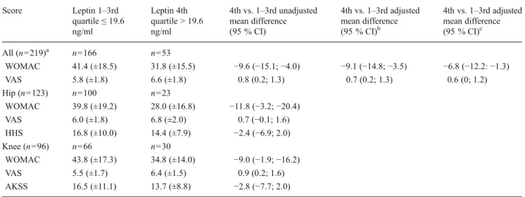
The unadjusted mean score difference between patients in the highest leptin quartile compared to those in the other quartiles was−9.6 (95 % CI −15.1; −4.0; standardized effect size 0.54) for the WOMAC and 0.8 (95 % CI 0.2; 1.3; standardized effect size 0.44) for the VAS (Table2). The association remained virtually unchanged after adjusting for potential confounders such as age, ASA score, contra-lateral arthritic joint, OA site, disability cate-gory C and diabetes (WOMAC adjusted mean difference−9.1 [95 % CI−14.8 to −3.5] and VAS adjusted mean difference 0.7 [95 % CI 0.2–1.3]). Additional adjustment for the SF-12 mental component score (WOMAC adjusted mean difference −6.8 (95 % CI−12.2 to −1.3) and VAS adjusted mean difference 0.6 [95 % CI 0–1.2]) slightly lowered the estimates, but the association remained significant. A greater degree of pain was seen in patients with high leptin levels as assessed by both the patient-assessed and the joint-specific physician-assessed scores HHS and AKSS (Table2).
Significant associations between increased joint pain as measured with WOMAC and (1) female gender and (2) obesity observed in univariate analyses were substantially
reduced after adjusting for SF leptin concentrations. The mean difference in WOMAC for women vs. men decreased from 4.7 to 1.6 and from 7.0 to 4.1 for obese vs. non-obese after correcting for leptin concentrations, suggesting that these associations might partially be mediated by leptin (Table3). Correlation with serum leptin
Synovial fluid leptin levels strongly correlated with serum leptin levels (correlation coefficient r=0.875). Overall, the median serum leptin concentration was 15.8 (IQR 5.7, 36.9) ng/ml and the mean concentration 27.9 (±34.3) ng/ml. In addition, mean serum leptin concentrations were 38.7 (±39.5) ng/ml in women compared to 11.6 (±13.2) ng/ml in men.
Discussion
We found that OA patients with high SF leptin concentra-tions reported substantially more OA pain. Furthermore, the
Table 2 Pre-operative pain levels according to leptin concentrations in synovial fluid
Score Leptin 1–3rd quartile≤ 19.6 ng/ml Leptin 4th quartile > 19.6 ng/ml 4th vs. 1–3rd unadjusted mean difference (95 % CI) 4th vs. 1–3rd adjusted mean difference (95 % CI)b 4th vs. 1–3rd adjusted mean difference (95 % CI)c All (n=219)a n=166 n=53 WOMAC 41.4 (±18.5) 31.8 (±15.5) −9.6 (−15.1; −4.0) −9.1 (−14.8; −3.5) −6.8 (−12.2: −1.3) VAS 5.8 (±1.8) 6.6 (±1.8) 0.8 (0.2; 1.3) 0.7 (0.2; 1.3) 0.6 (0; 1.2) Hip (n=123) n=100 n=23 WOMAC 39.8 (±19.2) 28.0 (±16.8) −11.8 (−3.2; −20.4) VAS 6.0 (±1.8) 6.8 (±2.0) 0.7 (−0.1; 1.6) HHS 16.8 (±10.0) 14.4 (±7.9) −2.4 (−6.9; 2.0) Knee (n=96) n=66 n=30 WOMAC 43.8 (±17.3) 34.8 (±14.0) −9.0 (−1.9; −16.2) VAS 5.5 (±1.7) 6.4 (±1.5) 0.9 (0.2; 1.6) AKSS 16.5 (±11.1) 13.7 (±8.8) −2.8 (−7.7; 2.0) a
Values presented are means and standard deviations b
Adjusted for age, diabetes, ASA score, contra-lateral arthritic joint and disability category C (multiple joint disease or other disabilities leading to difficulties in ambulation)
c
Adjusted for age, diabetes, ASA score and contra-lateral arthritic joint, disability category C and SF-12 mental component score
Table 3 Association between WOMAC pain and (1) sex and (2) body mass index
Group WOMAC
pain mean (SD)
Mean difference unadjusted (95 % CI)
p-value Mean difference
adjusted for leptin (95 % CI) p-value Women 37.2 (±18.8) Men 41.9 (±17.2) −4.7 (−9.6; 0.2) 0.062 −1.6 (−6.9; 3.7) 0.556 Obese 34.1 (±14.6) Non-obese 41.1 (±19.2) −7.0 (−12.3; −1.6) 0.011 −4.1 (−9.8; 1.7) 0.165
increased levels of pain observed in women and in obese patients seemed to be associated with high intra-articular leptin concentrations. However, due to the cross-sectional design a causal relation between leptin and pain cannot be ascertained.
Pain is certainly the most important symptom of OA, but also the least well understood [39]. Recent models imply that OA pain consists of a nociceptive stimulation of the joint and of peripheral sensitization, which can be accompanied by spinal and central sensitization [2,40–43]. Leptin may affect the OA pain response in at least two possible ways. First, leptin appears to be a mediator of the immuno-inflammatory response in different experimental models. The pro-inflammatory cytokine-like functions of leptin in OA [10, 44–47] could explain its role in peripheral sensitization. Second, animal models suggest an involvement of leptin in the pathogenesis of pain at the spinal level and a possible role in the development of neuropathic pain [28–30]. Our clinical results support these experimental findings in patients with lower limb OA.
Our findings are in accordance with Massengale et al. [32] who, in a cohort of patients with chronic hand OA, reported an association between serum leptin levels with pain inten-sity but not with radiographic severity. Ghandi et al. [31] have investigated the relation between leptin and pain. They did not find a clear association between intra-articular leptin concentrations and pain in a linear regression analysis, but reported that the synovial fluid (SF) adiponectin/leptin ratio predicted pain. We found that the association between intra-articular leptin concentration and pain intensity was not linear. Pain intensity was similar within the lower three quartiles, but substantially higher in the highest quartile of leptin concentration.
Clinical implications of these findings might be: (1) exer-cise and weight loss programs should be particularly targeted to women, who constitute the vast majority of the high leptin level group; and (2) prospective studies are warranted to test the clinical usefulness of serum leptin as a biomarker (as a proxy for intra-articular leptin level) to identify subgroups of patients, who would particularly benefit from exercise and weight loss programs, to detect the presence of neuropathic pain reported to be common in knee OA patients [48,49], as well as to predict persistence of pain after total joint arthroplasty.
The main limitation of this analysis is its cross-sectional design, and therefore the study is not suited to establish a causal link between leptin and increased pain. Leptin is a pleiotropic cytokine with central and peripheral effects in-volved in the regulation of energy intake and expenditure, of bone metabolism and inflammatory responses as well as of stress response [50]. Despite the above mentioned evidence from experimental studies supporting a possible causal effect of leptin on pain, other explanations for our findings including
inverse causation and residual confounding need to be ex-plored in future studies.
Second, we evaluated pain with two widely used, patient-assessed validated instruments. Because physician evalua-tion of pain has been found to be less influenced by the presence of co-morbidities [51], we additionally included one hip- and one knee-specific physician-assessed pain sub-score. However, information about the quality of pain (pain on movement, pain on rest, morning stiffness) was not routinely recorded. Third, depression and comorbidities, in particular the presence of OA at multiple sites, have been identified as important factors influencing the perception and severity of pain [20,52–54].
A depression-specific instrument was not used in our study, but we collected information on mental health status using the SF-12 mental component score. Moreover, specific information about OA in other joints except for the contra-lateral joint was not systematically available. However, we assessed (and adjusted for) the presence of multiple joint disease and other medical disabilities leading to difficulties in ambulation.
Conclusion
Our results suggest that high leptin concentrations may affect the level of joint pain in OA of the lower limbs. Furthermore, elevated leptin levels may potentially explain the well established association between increased pain and female sex or obesity. However, the causal relation between leptin and joint pain needs to be confirmed in a longitudinal study.
Acknowledgment The authors would like to express their deep
ap-preciation to all the orthopaedic surgeons, the personnel of the operating room and especially to Mme Carole Bandi and Mme Madeleine Vuillet for their efforts in data collection.
Funding Internal institutional funding was received for this study.
Prof. Pierre Hoffmeyer received institutional financial support from Medacta, Johnson & Johnson, and Zimmer. The funding sources had no role in the collection, analysis, or interpretation of the data, in the preparation of the manuscript, or its submission for publication.
Conflicts of interest None declared.
Ethics committee approval The study was approved by the
institu-tional review board of the Geneva University Hospitals and the pa-tients’ informed consent was obtained (N° 09-215; NAC 09-072).
References
1. Creamer P (2004) Current perspectives on the clinical presentation of joint pain in human OA. Novartis Found Symp 260:64–74, discussion 74–8, 100–4, 277–9
2. Dieppe PA, Lohmander LS (2005) Pathogenesis and management
of pain in osteoarthritis. Lancet 365(9463):965–973
3. Hunter DJ (2009) Insights from imaging on the epidemiology and
pathophysiology of osteoarthritis. Radiol Clin North Am 47(4):539–551
4. Wenham CY, Conaghan PG (2009) Imaging the painful osteoar-thritic knee joint: what have we learned? Nat Clin Pract Rheumatol
5(3):149–158
5. Bedson J, Croft PR (2008) The discordance between clinical and radiographic knee osteoarthritis: a systematic search and summary of the literature. BMC Musculoskelet Disord 9:116
6. Aspden RM, Scheven BA, Hutchison JD (2001) Osteoarthritis as a systemic disorder including stromal cell differentiation and lipid
metabolism. Lancet 357(9262):1118–1120
7. Dumond H, Presle N, Terlain B, Mainard D et al (2003) Evidence for a
key role of leptin in osteoarthritis. Arthritis Rheum 48(11):3118–3129
8. Figenschau Y, Knutsen G, Shahazeydi S, Johansen O et al (2001) Human articular chondrocytes express functional leptin receptors.
Biochem Biophys Res Commun 287(1):190–197
9. Griffin TM, Huebner JL, Kraus VB, Guilak F (2009) Extreme obesity due to impaired leptin signaling in mice does not cause knee osteoarthritis. Arthritis Rheum 60(10):2935–2944
10. Simopoulou T, Malizos KN, Iliopoulos D, Stefanou N et al (2007) Differential expression of leptin and leptin’s receptor isoform (Ob-Rb) mRNA between advanced and minimally affected osteoarthritic carti-lage; effect on cartilage metabolism. Osteoarthr Cartil 15(8):872–883 11. de Boer TN, van Spil WE, Huisman AM, Polak AA et al (2012)
Serum adipokines in osteoarthritis; comparison with controls and relationship with local parameters of synovial inflammation and cartilage damage. Osteoarthr Cartil 20(8):846–853
12. Vuolteenaho K, Koskinen A, Moilanen T, Moilanen E (2012) Leptin levels are increased and its negative regulators, SOCS-3 and sOb-R are decreased in obese patients with osteoarthritis: a link between obesity and osteoarthritis. Ann Rheum Dis 71:1912–1913
13. Jacobsen S, Sonne-Holm S (2005) Increased body mass index is a predisposition for treatment by total hip replacement. Int Orthop 29(4):229–234
14. Christensen R, Astrup A, Bliddal H (2005) Weight loss: the treat-ment of choice for knee osteoarthritis? A randomized trial. Osteoarthr Cartil 13:20–27
15. Toda Y, Toda T, Takemura S, Wada T et al (1998) Change in body fat, but not body weight or metabolic correlates of obesity, is related to symptomatic relief of obese patients with knee osteoarthritis after
a weight control program. J Rheumatol 25:2181–2186
16. Miller GD, Nicklas BJ, Loeser RF (2008) Inflammatory biomarkers and physical function in older, obese adults with knee pain and self-reported osteoarthritis after intensive weight-loss therapy. J Am
Geriatr Soc 56:644–651
17. Monzillo LU, Hamdy O, Horton ES, Ledbury S et al (2003) Effect of lifestyle modification on adipokine levels in obese subjects with
insulin resistance. Obes Res 11:1048–1054
18. Sheu WH, Chang TM, Lee WJ, Ou HC et al (2008) Effect of weight loss on proinflammatory state of mononuclear cells in obese
wom-en. Obesity (Silver Spring) 16:1033–1038
19. Presle N, Pottie P, Dumond H, Guillaume C et al (2006) Differential distribution of adipokines between serum and synovial fluid in patients with osteoarthritis. Contribution of joint tissues to their
articular production. Osteoarthr Cartil 14:690–695
20. Greenspan JD, Craft RM, LeResche L, Arendt-Nielsen L et al (2007) Studying sex and gender differences in pain and analgesia:
a consensus report. Pain 132(Suppl 1):S26–S45
21. Creamer P, Lethbridge-Cejku M, Hochberg MC (1999)
Determinants of pain severity in knee osteoarthritis: effect of de-mographic and psychosocial variables using 3 pain measures. J
Rheumatol 26:1785–1792
22. Tonelli SM, Rakel BA, Cooper NA, Angstom WL et al (2011) Women with knee osteoarthritis have more pain and poorer
function than men, but similar physical activity prior to total knee replacement. Biol Sex Differ 2:12
23. Cho HJ, Chang CB, Yoo JH, Kim SJ et al (2010) Gender differ-ences in the correlation between symptom and radiographic sever-ity in patients with knee osteoarthritis. Clin Orthop Relat Res
468:1749–1758
24. Holtzman J, Saleh K, Kane R (2002) Gender differences in func-tional status and pain in a Medicare population undergoing elective
total hip arthroplasty. Med Care 40:461–470
25. Katz JN, Wright EA, Guadagnoli E, Liang MH et al (1994) Differences between men and women undergoing major orthopedic
surgery for degenerative arthritis. Arthritis Rheum 37:687–694
26. Lubbeke A, Duc S, Garavaglia G, Finckh A et al (2009) BMI and severity of clinical and radiographic signs of hip osteoarthritis.
Obesity (Silver Spring) 17:1414–1419
27. Bedaiwy MA, Falcone T, Goldberg JM, Sharma RK et al (2006) Peritoneal fluid leptin is associated with chronic pelvic pain but not
infertility in endometriosis patients. Hum Reprod 21:788–791
28. Lim G, Wang S, Zhang Y, Tian Y et al (2009) Spinal leptin contrib-utes to the pathogenesis of neuropathic pain in rodents. J Clin Invest 119:295–304
29. Maeda T, Kiguchi N, Kobayashi Y, Ikuta T et al (2009) Leptin derived from adipocytes in injured peripheral nerves facilitates development of neuropathic pain via macrophage stimulation. Proc Natl Acad Sci USA 106:13076–13081
30. Tian Y, Wang S, Ma Y, Lim G et al (2011) Leptin enhances NMDA-induced spinal excitation in rats: a functional link between adipocytokine and neuropathic pain. Pain 152:1263–1271 31. Gandhi R, Takahashi M, Smith H, Rizek R et al (2010) The
synovial fluid adiponectin-leptin ratio predicts pain with knee os-teoarthritis. Clin Rheumatol 29:1223–1228
32. Massengale M, Lu B, Pan JJ, Katz JN et al (2012) Adipokine hormones and hand osteoarthritis: radiographic severity and pain. PLoS One 7(10):e47860
33. Bellamy N, Buchanan WW, Goldsmith CH, Campbell J et al (1988) Validation study of WOMAC: a health status instrument for mea-suring clinically important patient relevant outcomes to antirheu-matic drug therapy in patients with osteoarthritis of the hip or knee. J Rheumatol 15:1833–1840
34. Harris WH (1969) Traumatic arthritis of the hip after dislocation and acetabular fractures: treatment by mold arthroplasty. An end-result study using a new method of end-result evaluation. J Bone Joint
Surg Am 51(4):737–755
35. Insall JN, Dorr LD, Scott RD, Scott WN (1989) Rationale of the Knee Society clinical rating system. Clin Orthop Relat Res
248:13–14
36. Ware J Jr, Kosinski M, Keller SD (1996) A 12-item short-form health survey: construction of scales and preliminary tests of
reli-ability and validity. Med Care 34:220–233
37. Charnley J (1979) Numerical grading of clinical results. In: Charnley J (ed) Low friction arthroplasty of the hip: Theory and
practice. Springer Verlag, Berlin, pp 20–24
38. Kellgren JH, Lawrence JS (1957) Radiological assessment of
osteo-arthrosis. Ann Rheum Dis 16(4):494–502
39. Goldring MB (2009) The link between structural damage and pain in a genetic model of osteoarthritis and intervertebral disc
degeneration: a joint misadventure. Arthritis Rheum 60(9):2550–
2552
40. Hunter DJ, McDougall JJ, Keefe FJ (2008) The symptoms of osteoarthritis and the genesis of pain. Rheum Dis Clin North Am
34:623–643
41. Im HJ, Kim JS, Li X, Kotwal N et al (2010) Alteration of sensory neurons and spinal response to an experimental osteoarthritis pain
model. Arthritis Rheum 62:2995–3005
42. Schaible HG, Ebersberger A, Von Banchet GS (2002) Mechanisms
43. Schaible HG, Richter F, Ebersberger A, Boettger MK et al (2009)
Joint pain. Exp Brain Res 196:153–162
44. Bernotiene E, Palmer G, Gabay C (2006) The role of leptin in innate and adaptive immune responses. Arthritis Res Ther 8:217
45. Otero M, Lago R, Gomez R, Dieguez C et al (2006) Towards a pro-inflammatory and immunomodulatory emerging role of leptin.
Rheumatol (Oxford) 45:944–950
46. Hoff P, Buttgereit F, Burmester GR, Jakstadt M et al (2013) Osteoarthritis synovial fluid activates pro-inflammatory cytokines
in primary human chondrocytes. Int Orthop 37(1):145–151
47. Vuolteenaho K, Koskinen A, Kukkonen M, Nieminen R et al (2009) Leptin enhances synthesis of proinflammatory mediators in human osteoarthritic cartilage-mediator role of NO in leptin-induced PGE2, IL-6, and IL-8 production. Mediat Inflamm 2009:345838
48. Hochman JR, French MR, Bermingham SL, Hawker GA (2010) The nerve of osteoarthritis pain. Arthritis Care Res (Hoboken)
62:1019–1023
49. Hochman JR, Gagliese L, Davis AM, Hawker GA (2011) Neuropathic pain symptoms in a community knee OA cohort.
Osteoarthr Cartil 19:647–654
50. Roubos EW, Dahmen M, Kozicz T, Xu L (2012) Leptin and the hypothalamo-pituitary-adrenal stress axis. Gen Comp Endocrinol
177(1):28–36
51. Dawson J, Fitzpatrick R, Murray D, Carr A (1996) The problem of ‘noise’ in monitoring patient-based outcomes: generic, disease-specific and site-disease-specific instruments for total hip replacement. J
Health Serv Res Policy 1:224–231
52. Hawker GA, Gignac MA, Badley E, French MR et al (2011) A longitudinal study to explain the pain-depression link in older adults
with osteoarthritis. Arthritis Care Res (Hoboken) 63:1382–1390
53. Mallen CD, Peat G, Thomas E, Dunn KM et al (2007) Prognostic factors for musculoskeletal pain in primary care: a systematic
review. Br J Gen Pract 57:655–661
54. Wylde V, Hewlett S, Learmonth ID, Dieppe P (2011) Persistent pain after joint replacement: prevalence, sensory qualities, and

![Fig. 3 Proportion of WOMAC pain scores ≤ 25 in women and men according to intra-articular leptin concentration (highest quartile [leptin>19.6 ng/ml] vs](https://thumb-eu.123doks.com/thumbv2/123doknet/14836621.622671/5.892.454.819.77.356/proportion-womac-scores-according-articular-concentration-highest-quartile.webp)