HAL Id: hal-00561108
https://hal.univ-brest.fr/hal-00561108
Submitted on 31 Jan 2011HAL is a multi-disciplinary open access archive for the deposit and dissemination of sci-entific research documents, whether they are pub-lished or not. The documents may come from teaching and research institutions in France or abroad, or from public or private research centers.
L’archive ouverte pluridisciplinaire HAL, est destinée au dépôt et à la diffusion de documents scientifiques de niveau recherche, publiés ou non, émanant des établissements d’enseignement et de recherche français ou étrangers, des laboratoires publics ou privés.
Hellea balneolensis gen. nov., sp. nov., a prosthecate
alphaproteobacterium from the Mediterranean Sea
Karine Alain, Brian J. Tindall, Laurent Intertaglia, Philippe Catala, Philippe
Lebaron
To cite this version:
Karine Alain, Brian J. Tindall, Laurent Intertaglia, Philippe Catala, Philippe Lebaron. Hellea bal-neolensis gen. nov., sp. nov., a prosthecate alphaproteobacterium from the Mediterranean Sea. International Journal of Systematic and Evolutionary Microbiology, Microbiology Society, 2008, 58 (Pt 11), pp.2511-2519. �10.1099/ijs.0.65424-0�. �hal-00561108�
Hellea balneolensis gen. nov., sp. nov., 1
a novel prosthecate alphaproteobacterium from the Mediterranean Sea
2 3
Karine Alain1†, Brian J. Tindall2, Laurent Intertaglia1, Philippe Catala1 4
and Philippe Lebaron1. 5
6
1 Université Pierre et Marie Curie-Paris6, UMR7621, F-66650 Banyuls-sur-Mer, France ; CNRS, UMR7621, F-66650
7
Banyuls-sur-Mer, France. 8
2 DSMZ Deutsche Sammlung von Mikroorganismen und Zellkulturen GmbH, Inhoffenstraße 7B, 38124 Braunschweig,
9
Germany. 10
† Present address : UMR6197, Laboratoire de Microbiologie des Environnements Extrêmes, IUEM, Technopôle Brest-11
Iroise, F-29280 Plouzané, France. 12
13
Correspondence : Philippe Lebaron 14
philippe.lebaron@obs-banyuls.fr 15
Phone number : +33-(0)4-68-88-73-00 Fax : +33-(0)4-68-88-16-99 16
17
Running title: Hellea balneolensis gen. nov. sp. nov. 18
19
Category: New taxa, Proteobacteria 20
21
Footnote: The GenBank/EMBL/DDBJ accession number for the 16S rRNA gene sequence of strain 22
26III/A02/215T is AY576758. 23
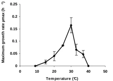
The graph showing the effect of temperature on the maximum growth rate (µmax) of strain 24
26III/A02/215T (Fig. S1) is available in IJSEM online. 25
26
A novel aerobic, heterotrophic, prosthecate bacterium designated 26III/A02/215T, was isolated from
27
surface waters of the north-western Mediterranean sea. Cells were Gram-negative, straight to slightly
28
curved rods, forming red colonies on agar plates. The strain grew at 15-37°C inclusive (optimum:
30°C), and optimally at seawater salinity. Growth on organic acids, amino-acids and complex organic
30
substrates was observed. The fatty acids (> 5%) detected in strain 26III/A02/215T were C17:1ω6c,
31
C18:1ω7c, and C17:0. The lipid pattern indicated the presence of phosphatidylglycerol,
32
glucuronopyranosyldiglyceride, monoglycosyldiglyceride, an unidentified glycolipid and three
33
unidentified phospholipids. Phosphatidylethanolamine and diphosphatidylglycerol were absent.
34
Ubiquinone Q10 was the only respiratory lipoquinone. The G+C content of the genomic DNA was
35
46.8 mol%.
36
Comparative 16S rRNA gene sequence analysis indicated that strain 26III/A02/215T belonged to the
37
Hyphomonas-Hirschia-Robiginitomaculum branch of the order Caulobacterales. This affiliation was 38
consistent with the results of polar lipid analyses. Among this group, the novel isolate was most closely
39
related to Robiginitomaculum antarcticum (93.9% 16S rDNA sequence similarity). On the basis of
40
genotypic, chemotaxonomic and phenotypic distinctness, we propose a novel genus, Hellea gen. nov.,
41
with Hellea balneolensis sp. nov. as the type species. The type strain is 26III/A02/215T (= DSM 19091T
42
= CIP 109500T = OOB 269T).
43 44
The phylum Proteobacteria is one of the 24 phyla of the domain Bacteria, described in Bergey’s Manual of 45
Systematic Bacteriology, 2nd edn (Garrity & Holt, 2001). To date, more than 200 genera have been 46
described, making this phylum one of the largest bacterial phyla. Representative members of this group are 47
widely distributed in nature and are physiologically and metabolically diverse. The phylum Proteobacteria 48
is currently divided into 5 classes, called the Alphaproteobacteria, Betaproteobacteria, 49
Gammaproteobacteria, Deltaproteobacteria and Epsilonproteobacteria, all of which have been defined 50
exclusively on the basis of 16S rRNA gene sequence analysis (Garrity & Holt, 2001). At present, the class 51
Alphaproteobacteria Garrity et al. 2006 (Validation List 107, Garrity et al., 2005a) is composed of seven 52
orders: Caulobacterales Henrici & Johnson, 1935 (Henrici & Johnson, 1935), Kordiimonadales Kwon et al. 53
2005 (Kwon et al., 2005), Rhodobacterales Garrity et al. 2006 (Validation List 107, Garrity et al., 2005b) 54
Rhodospirillales Pfenning & Trüper, 1971 (Pfenning & Trüper, 1971), Rickettsiales Gieszczykiewicz, 1939 55
(Gieszczykiewicz, 1939), Rhizobiales Kuykendall 2006 (Validation List 107, Kuykendall, 2005) and 56
Sphingomonadales Yabuuchi & Kosako, 2006 (Validation List 107, Yabuuchi & Kosako, 2005). Marine 57
species make up more than half of the Alphaproteobacteria described to date. 58
Some confusion is being caused at present by the different taxonomic placement of the “stalked” bacteria. 59
While Lee et al. (2005) place the members of the genera Hyphomonas, Oceanicaulis, Hirschia, and 60
Maricaulis in a new family, the Hyphomonadaceae, within the order Caulobacterales (which includes 61
members of the family Rhodobacteraceae), Garrity et al. (2005a) have placed members of these genera 62
within the family Rhodobacteraceae, within the order Rhodobacterales, leaving the members of the family 63
Caulobacteraceae within the order Caulobacterales. This situation is particularly unsatisfactory since use of 64
the name of the order Caulobacterales alone does not give unambiguous information on which taxa are to be 65
included within it. Furthermore, Lee et al. (2005) dealt with the taxonomy of the family Rhodobacteraceae 66
before the name was validly published (Validation List 107). Paradoxically Lee et al. (2005) created a new 67
family, the family Hyphomonadaceae, with the type defined as the genus Hyphomonas, a taxon specifically 68
included in the taxon proposed by Garrity et al. (2005b) as the family Rhodobacteraceae. Based on the 69
principle of priority, the family proposed by Garrity et al. (2005b) must be named after the earliest validly 70
published family name, which is the family Hyphomonadaceae. The family name Rhodobacteraceae Garrity 71
et al. 2006 may only be used if specifically defined to exclude the type genus of the family 72
Hyphomonadaceae. It should be noted that members of the genera Woodsholea (Abraham et al., 2004) and 73
Robiginitomaculum (Lee et al., 2007) should be included in the family Hyphomonadaceae Lee et al. 2007. 74
The members of the families Hyphomonadaceae and Caulobacteraceae contain organisms that share the 75
particular feature of being appendaged (Poindexter, 1981; Abraham et al., 1999; Weiner et al., 2000; 76
Strömpl et al., 2003). As indicated by their vernacular name (in Latin, caulis means stalk), these 77
‘caulobacteria’ bear one or several stalks, so-called prosthecae. These stalks are cytoplasm extrusions that 78
undoubtedly play a role in attachment. As a result, they increase significantly the surface to volume ratio of 79
the cells. Consequently, they have often been interpreted as an evolutionary adaptation to life in oligotrophic 80
waters. Most genera of the families Hyphomonadaceae and Caulobacteraceae (i.e. members of the genera 81
Hyphomonas, Caulobacter, Asticcacaulis, Phenylobacterium, Hirschia, Robiginitomaculum, Woodsholea, 82
Maricaulis, Oceanicaulis and Brevundimonas) are widely distributed in marine environments (Anast & 83
Smit, 1988), and especially (but not exclusively) in oligotrophic waters. They are believed to play an 84
important role in the mineralization of dissolved organic matter (Abraham et al., 1999).
85 86
In this study, a novel marine caulobacterium is described. Based on the results of a polyphasic taxonomic 87
analysis, the strain 26III/A02/215T represents a novel species and genus, Hellea balneolensis gen. nov., sp. 88
nov. 89
90
In September 2001, coastal waters were collected in the bay of Banyuls-sur-mer (42°29’N, 3°08’E), in the 91
Mediterranean Sea, France. A sea sample from the surface microlayer was spread on marine agar 2216 (MA; 92
Difco) plate, and then incubated at 25°C. After 2 weeks, a red-coloured colony was picked, purified by 93
repeated streaking on MA plates, and referenced as strain 26III/A02/215T (Agogué et al., 2005). Stock 94
cultures were stored at ─80°C in marine broth 2216 (MB; Difco) supplemented with 5% (v/v) DMSO or 95
35% (v/v) glycerol, until characterization. 96
97
Both strands of the almost complete 16S rRNA gene (1412 bp) of the strain were sequenced from one single 98
colony, as described elsewhere (Agogué et al., 2005). This sequence was compared to those in available 99
databases by use of the BLAST program (Altschul et al., 1990) and then aligned to its nearest neighbours 100
using the CLUSTALX program (Thompson et al., 1997). Alignments were refined manually using the 101
SEAVIEW program (Galtier et al., 1996). Phylogenetic trees were constructed by the PHYLIP (PHYlogeny 102
Inference Package) version 3.63 software (http://evolution.genetics.washington.edu/phylip/getme.html) on 103
the basis of evolutionary distance (neighbour-joining method with Jukes and Cantor corrections) (Saitou and 104
Nei, 1987) and maximum likelihood (Felsenstein, 1981). The robustness of the inferred topologies was 105
assessed by bootstrap analyses based on 1000 bootstrap resamplings for the neighbour-joining and 100 106
replications for the maximum likelihood method (Felsenstein, 1985). The 16S rRNA gene-based analysis 107
located the strain 26III/A02/215T within the class Alphaproteobacteria, in thebacterial domain. The results 108
of different phylogenetic reconstructions performed with different treeing algorithms located the novel 109
isolate within the Hyphomonas-Hirschia-Robiginitomaculum branch, amongst the marine caulobacteria of 110
family Hyphomonadaceae (Lee et al., 2005), order Caulobacterales (Fig. 1). Within this branch, the novel 111
isolate clustered with the recently described genus Robiginitomaculum (Lee et al., 2007) sharing 93.9% 16S 112
rDNA sequence similarity with the only species of this genus. The level of 16S rRNA gene sequence 113
similarity between strain 26III/A02/215T and representative of the genera Hyphomonas and Hirschia ranged 114
from 89 to 92%. 115
The DNA G+C content was determined, by the Identification Service of the DSMZ (Deutsche Sammlung 116
von Mikroorganismen und Zellkulturen GmbH, Braunschweig
117
Germany), by HPLC analysis of deoxyribonucleosides as described by Mesbah et al. (1989). The G+C 118
content of strain 26III/A02/215T was 46.8 mol%. Thus, it differed by more than 10 mol% from the DNA 119
G+C content of its closest relative Robiginitomaculum antarcticum (60.3 mol%). Clearly, this large 120
difference in the DNA base ratio, together with the 16S rDNA level of similarity, suggest that strain 121
26III/A02/215T belongs to a novel genus (Rosselló-Mora and Amann, 2000). 122
123
Colonies on MA were circular, smooth, brilliant, convex, with an entire edge and intensely pigmented brick-124
red. After 1 week incubation, colonies were about 1 mm in diameter. Morphological characteristics of the 125
cells were determined by light microscopy (Olympus AX70) and by transmission electron microscopy 126
(Hitachi H-7500) after negative staining with uranyl acetate (Raguénès et al., 1997). Briefly, cells of strain 127
26III/A02/215T were Gram-negative, thin straight to curved rods bearing one polar stalk (Fig. 2). Cells 128
bearing several lateral stalks were occasionally observed. Mid-exponential phase cells were 2.70-5.60 µm in 129
length (mean 3.58 ± 0.88 µm, n=15), 0.28-0.48 µm in width (mean 0.42 ± 0.06 µm, n=15), some of which 130
produced a stalk(s). When present, the stalk was more generally cylindrical and extended centrally along the 131
cell axis from one pole. This stalk showed constriction sites distributed equally all along the tube, but which 132
not corresponded to a compartmentalization. This type of constriction has been observed previously in 133
Oceanicaulis alexandrii (Strömpl et al., 2003). Stalked-cells were non-motile, while non-stalked cells were 134
motile by means of a polar flagellum. Cells divided by budding. 135
136
In order to analyse respiratory quinones and polar lipids, strain 26III/A02/215T was grown for 3 days on MB 137
medium at 30°C, and checked for purity. Initial analyses of the polar lipids and respiratory quinones were 138
carried out by the Identification Service, DSMZ, Braunschweig, Germany. Ubiquinone (Q10) was 139
determined to be the sole respiratory quinone. The thin layer chromatogram obtained with cell extracts from 140
the novel isolate was very characteristic (Fig. 3). The polar lipid pattern showed the presence of 141
phosphatidylglycerol (PG), monoglycosyldiglyceride (MGDG), glucuronopyranosyldiglyceride (GUDG), 142
one unidentified glycolipid (Gl) and three phospholipids (PL1, PL2, PL3). The presence of the polar lipids 143
monoglycosyldiglyceride (MGDG) and glucuronopyranosyldiglyceride (GUDG) appears to be a 144
characteristic signature for other members of the families Hyphomonadaceae and Caulobacteraceae, 145
together with the absence of phosphatidylethanolamine, phosphatidylcholine and diphosphatidylglycerol. 146
The presence of two unidentified phospholipids PL2 and PL3, together with an unidentified glycolipid 147
appeared to be a characteristic feature of the lipid pattern of this taxon. The determination of the whole-cell 148
fatty acid composition was performed on cultures grown at 30°C for 72h on marine agar 2216. The analysis 149
was carried out at the DSMZ according to the standard protocol of the Microbial Identification System 150
(MIDI Inc., Del. USA, 2001). Extracts were analysed using a Hewlett Packard model HP6890A gas 151
chromatograph equipped with a flame-ionization detector as described by Kämpfer & Kroppenstedt (1996). 152
Results are summarized in Table 1. The fatty acids in strain 26III/A02/215T comprised C16:0, C17:0, C18:0,
153
C19:0, C17:1ω8c, C17:1ω6c, C18:1ω9c, C18:1ω7c, C20:1ω7c, 3-OH C10:0, 3-OH C11:0, 3-OH C12:1, 2-OH C18:1, TBSA
154
10-methyl C18:0, Summed feature 3, Summed feature 7. The presence of C18:1ω7c, together with Q10 is
155
typical of the vast majority of taxa within the Alphaproteobacteria. Although the polar lipid composition of 156
the recently described Robiginitomaculum antarcticum was not reported, there were clear differences in the 157
fatty acid patterns, in particular the distribution of the 3-hydroxy fatty acids, which are probably derived 158
from lipopolysaccharide. A number of recent publications are also not complete with regard to the 159
chemotaxonomic data. In papers on the genera Oceanicaulis (Strömpl et al., 2003), Woodsholea (Abraham 160
et al., 2004) and some Maricaulis (Abraham et al., 2002) species, the quinone composition has not been 161
reported. In the case of Oceanicaulis, 3-OH fatty acids are not reported, probably because only fatty acids 162
from extracted lipids have been reported. Reports on the polar lipid composition may be incomplete because 163
emphasis has been placed on the presence of phosphate and sulfonic acid containing lipids (Strömpl et al., 164
2003, see also Abraham et al., 1997). The glycolipids that are otherwise characteristic for this evolutionary 165
group are not mentioned. 166
Unless stated otherwise, physiological characterization was carried out aerobically in marine broth medium 168
(MB 2216; Difco), in triplicate, and cell suspension incubated with agitation in the dark. Growth was 169
routinely monitored by measuring the increase in optical density at 600 nm using a spectrophotometer. Cell 170
numbers were determined by flow cytometry (Marie et al., 2000) in order to calculate calibration curves 171
‘Cell numbers = f(OD600)’. Growth rates were calculated using linear regression analysis from five to nine
172
points along the logarithmic portions of the resulting growth curves. Growth temperature was tested over the 173
range 9-44°C (i.e. 9, 15, 20, 25, 30, 33, 37, 44°C). The novel isolate was found to be mesophilic, growing at 174
15-37°C; optimal growth yields occurred at 30°C (see Supplementary Fig. S1 in IJSEM online). The 175
optimum pH for growth was tested at 30°C in buffered MB medium and was found to be around pH 6.0-8.0. 176
Salt tolerance was tested at 30°C in MB medium prepared with various concentrations of NaCl (0.02, 0.5, 1, 177
2, 3, 4, 5, 6, 7 and 9% w/v). Results indicated that the strain was a general typical marine-type halophile. 178
Growth was observed in media containing 0.02% (w/v) to 5% (w/v) NaCl, but it was better in media 179
containing half- to full-strength seawater salinity. The optimal NaCl concentration for growth was around 180
3% (w/v) NaCl. 181
182
Strain 26III/A02/215T was found to be aerobic. Conventional phenotypic tests including those for oxidase, 183
catalase, tween esterase and nitrate reductase were performed according to standard methods (Smibert & 184
Krieg, 1994). The results are given in Table 2. Biochemical tests were performed at 30°C using api®ZYM 185
(bioMérieux) and Biolog GN2 microplates (Oxoid). These tests were inoculated with cells grown on MA 186
plates, swabbed from the surface of the agar plates and then suspended in ASW ½ (diluted artificial 187
seawater) to the density specified by the manufacturer. Supplementary biochemical tests were also 188
performed using api®20NE strips (bioMérieux), following the manufacturer’s instructions. The data 189
obtained are given in Table 2. Testing for oxidation of carbon sources with Biolog GN2 plates indicated that 190
the strain was able to oxidize a wide range of organic acids and amino acids. To confirm these results and to 191
test for the capability of the strain to catabolize different substrates as sole carbon and energy source, with 192
oxygen as a terminal electron acceptor, the strain was grown aerobically, in the dark, on a mineral medium 193
supplemented with one substrate. The defined medium (modified from Widdel et al. 2004) had the 194
following composition (l─1): phosphate buffer, 30 mM; NaCl 20 g, MgCl2.6H2O 3 g, CaCl2.2H2O 1.0 g,
NH4Cl 0.3 g, KCl 0.5 g, Na2SO4 3 g, NaNO3 1 g; trace element solution, 1 ml; selenite-tungstate solution, 1
196
ml; vitamin solution, 1 ml. The strain was found to grow heterotrophically on a wide range of substrates. It 197
catabolized organic acids, amino acids, and complex substrates for energy and growth (Table 2). The 198
carbohydrates tested were unable to support growth when provided alone in the medium. 199
Antibiotic sensitivity tests were performed by using susceptibility discs (Biorad) or filter-paper discs 200
impregnated with different antibiotics. Discs were placed on MA plates spread with a culture of the isolate 201
and were then incubated at 30°C for one week. Susceptibility was scored as positive at zone diameters above 202
10 mm. The results are summarized in Table 2. 203
204
During the course of this work, we also have had cause to re-examine the taxonomy of members of the 205
families Hyphomonadaceae and Caulobacteraceae. The placement of members of the genera Hyphomonas, 206
Hirschia, Maricaulis, and Oceanicaulis, in the family Rhodobacteraceae (Garrity et al. 2005a) has been 207
called into question by Lee et al. (2005). Independent work on the genome of Hyphomonas neptunium has 208
indicated that the 16S rDNA sequence based interpretation may be prone to error (Badger et al., 2005; 209
Badger et al., 2006). This conclusion is also in accord with the chemical composition reported for members 210
of these genera, which share a number of distinctive similarities with members of the genera Caulobacter, 211
Brevundimonas, Asticcacaulis and Phenylobacterium. Similarly, extensive chemotaxonomic work on 212
additional taxa within the family Rhodobacteraceae, as defined by Garrity et al. (2005a) would also indicate 213
inconsistencies (Biebl et al., 2005a, 2005b, 2006, 2007; Martens et al., 2006; Labrenz et al., 1999, 2000) 214
with the proposal of Lee et al. (2005) to unite members of the families Rhodobacteraceae (as defined by Lee 215
et al. (2005)), Hyphomonadaceae and Caulobacteraceae (as defined by Lee et al., 2005 and Garrity et al., 216
2005a). Clearly the family Hyphomonadaceae should comprise the genera Hirschia, Hyphomonas, 217
Maricaulis, Oceanicaulis, Woodsholea, Robiginitomaculum, and the new taxon proposed here. It is 218
interesting to note that this family may be subdivided into two groups, one with cells that divide by budding, 219
the other by binary fission. In addition, there is some evidence that there may also be a correlation between 220
the two groups and the polar lipid patterns, although additional work is needed to test this hypothesis. When 221
such work is completed it would be appropriate to emend the description and circumscription of the family 222
Hyphomonadaceae (Lee et al. 2005) in the light of chemotaxonomic data, bringing it into line with 223
recommendations dating back to the ad hoc committee reports of Wayne et al. (1987) and Murray et al. 224
(1990). A similar treatment of the family Caulobacteraceae would be appropriate, which comprises the 225
genera Caulobacter, Brevundimonas, Asticcacaulis and Phenylobacterium. The order Caulobacterales 226
should also be restricted to include only the members of the families Caulobacteraceae and 227
Hyphomonadaceae and emended accordingly. A further consequence would be that the members of the 228
family Rhodobacteraceae as defined by Lee et al. (2005), should be formally assigned to a family that 229
excludes the type of the family Hyphomonadaceae. Based on published chemotaxonomic data it would also 230
be prudent to test whether members of that taxon should be further divided into several families and all 231
included in the order Rhodobacterales. 232
233
Briefly, the results of our genotypic, chemotaxonomic, morphological and physiological investigations, 234
together with the phylogenetic analyses, revealed that strain 26III/A02/215T is distinct from other members 235
of the family Hyphomonadaceae. The main characteristics differentiating the novel isolate from its closest 236
phylogenetic neighbours are summarized in Table 2. In brief, the novel taxon can be distinguished from all 237
its closest relatives, with the exception of members of the genus Hirschia, by its significantly lower G+C 238
content. The fatty acid composition and polar lipid composition represent other distinctive criteria between 239
the new taxon and other members of the family Hyphomonadaceae. Although much emphasis is put on the 240
“major fatty acids” in the majority of recent taxonomic papers we emphasise here, the fact that the large 241
amounts of 18:1ω7c (together with the presence of Q10) only indicate that this genus is a member of the 242
Alphaproteobacteria and cannot be described as “characteristic” of this, or any other genus. On the contrary 243
the sum of chemotaxonomic data, not only clearly place it within the family Hyphomonadaceae, order 244
Caulobacterales, but also provides a unique signature for with taxon within these higher taxa. In terms of 245
other phenotypic features, differences in morphological characteristics such as the fine structure of the stalk, 246
its position, the flagellation of the cells, the colonial pigmentation and the mode of division of the cells can 247
also be use to distinguish the novel isolate from members of the genera Robiginitomaculum, Hyphomonas, 248
Hirschia, Woodsholea, Oceanicaulis and Maricaulis (Table 2). 249
In conclusion, on the basis of the phylogenetic position and of genotypic, chemotaxonomic and 250
physiological, biochemical and morphological differences, we propose that the isolate 26III/A02/215T 251
should be assigned as the type strain of a novel genus and species, for which the name Hellea balneolensis 252
gen. nov., sp. nov. is proposed. 253
254
Description of Hellea gen. nov. 255
Hellea (He.lle’a. L. fem. n. Helle a sea goddess in Greek mythology; N. L. fem. n. Hellea, named after Helle in
256
reference to the marine origin of the strain). Cells are Gram-negative, non-spore forming, rod-shaped to vibrioid, and 257
dimorphic: usually, they possess one polar stalk (prostheca) and are non-motile or / they are non-stalked and motile by 258
means of a polar flagellum. Aerobic and heterotrophic. Mesophilic. Neutrophilic. Grows best at salt concentrations close 259
to marine salinity. The predominant quinone is Q10. Polar lipids comprise glucuronopyranosyldiglyceride, 260
monoglycosyldiglyceride, phosphatidylglycerol, and unidentified glycolipid and phospholipids. Fatty acids comprise 261
C16:0, C17:0, C18:0, C19:0, C17:1ω8c, C17:1ω6c, C18:1ω9c, C18:1ω7c, C20:1ω7c, 3-OH C10:0, 3-OH C11:0, 3-OH C12:1, 2-OH
262
C18:1, TBSA 10-methyl C18:0, Summed feature 3, Summed feature 7 (percentage compositions are given in Table 1). The
263
G+C content of the DNA is close to 47 mol%. The genus Hellea belongs to the class Alphaproteobacteria, order 264
Caulobacterales, family Hyphomonadaceae, showing a distant relatedness to prosthecate bacteria of marine origin,
265
namely members of the genera Hyphomonas, Robiginitomaculum, Hirschia, Woodsholea, Maricaulis and Oceanicaulis. 266
The type species is Hellea balneolensis. This is the only species within the genus. 267
268
Description of Hellea balneolensis sp. nov. 269
Hellea balneolensis (bal.ne’o.len.sis. M. L. n. Balneola, the ancient name of Banyuls-sur-mer; N. L. fem. adj.
270
balneolensis, pertaining to Balneola from where the strain was isolated). In addition to the characters described for the
271
genus, the species is characterised by the following properties. Colonies on MA medium are round, convex, brilliant and 272
pigmented a brick red colour. Optimal growth occurs at 30°C, with a growth range from 15 to 37°C. pH optimum is 273
close to neutrality. Grows at NaCl concentrations from 0.02% to 5% (w/v), with a clear optimum at 3% (w/v) NaCl. 274
Growth occurs on acetate, citrate, propionate, pyruvate, succinate, aspartate, glutamate, alanine, asparagine, L-275
histidine, L-proline, casamino acids, peptone, tryptone, yeast extract and D-mannitol. Substrates positive in Biolog GN2 276
plates are all the substrates cited above and as well as cis-aconitic acid, D-glucuronic acid, β-hydroxybutyric acid, γ-277
hydroxy butyric acid, α-ketoglutaric acid, methyl-pyruvate, quinic acid, urocanic acid, L-pyroglutamic acid, hydroxyl-L-278
proline, putrescine, n-acetyl glucosamine, D-arabitol, m-inositol and xylitol. Does not reduce NO3
-. Catalase positive, 279
oxidase negative. Tween 40 and tween 80 hydrolysis activities are positive. 280
The G+C content is 46.8 mol%. 281
The type strain, 26III/A02/215T (DSM 19091T, CIP 109500T= OOB 269T), was isolated from the surface microlayer of 282
coastal waters, in the bay of Banyuls-sur-mer, north-western Mediterranean sea, France (42°29’N, 3°08’E). 283
284
ACKNOWLEDGEMENTS
285
We thank Marie-Line Escande for assistance with the transmission electron microscopy and Hélène Agogué 286
for her contribution to the initial sequencing work. We acknowledge Prof. J. P. Euzéby for support in the 287
Latin etymologies of the genus and species names. This work was financially supported by the Equipe Mixte 288
de Recherche linking the University Pierre et Marie Curie and the Centre National de la Recherche 289
Scientifique to the Pierre Fabre Laboratories. The project was also carried out in the framework of the 290
MarBEF Network of Excellence ‘Marine Biodiversity and Ecosystem Functioning’ which is funded by the 291
Sustainable Development, Global Change and Ecosystems Program of the European Community’s Sixth 292
Framework Program (contract no. GOCE-CT-2003-505446). This publication is contribution number MPS-293
07059 of MarBEF. It was also partly funded by the French program ‘Bio-diversité et Changement Global – 294
project: development of a coastal microbial observatory’ from the ‘Institut Français de la Biodiversité’. 295
296
REFERENCES 297
Abraham, W.-R., Meyer, H., Lindholst, S., Vancanneyt, M. & Smit, J. (1997). Phospho- and sulfolipids as 298
biomarkers of Caulobacter, Brevundimonas and Hyphomonas. Syst Appl Microbiol 20, 522-539. 299
Abraham, W.-R., Strömpl, C., Meyer, H., Lindholst, S., Moore, E. R. B., Christ, R., Vancanneyt, M., Tindall, B. 300
–J., Bennasar, A., Smit, J. & Tesar, M. (1999). Phylogeny and polyphasic taxonomy of Caulobacter species. Proposal 301
of Maricaulis gen. nov. with Maricaulis maris (Poindexter) comb. nov. as the type species, and emended description of 302
the genera Brevundimonas and Caulobacter. Int J Syst Bacteriol 49, 1053-1073. 303
Abraham, W.-R., Strömpl, C., Bennasar, A., Vancanneyt, M., Snauwaert, C., Swings, J., Smit, J. & Moore, E. R. 304
B. (2002). Phylogeny of Maricaulis Abraham et al. 1999 and proposal of Maricaulis virginensis sp. nov., M. 305
parjimensis sp. nov., M. washingtonensis sp. nov. and M. salignorans sp. nov. Int J Syst Evol Microbiol 52, 2191-2201.
306
Abraham, W.-R., Strömpl, C., Vancanneyt, M., Bennasar, A., Swings, J., Lünsdorf, H., Smit, J. & Moore, E. R. 307
B. (2004). Woodsholea maritima gen. nov., sp. nov., a marine bacterium with a low diversity of polar lipids. Int J Syst 308
Evol Microbiol 54, 1227-1234.
309
Agogué, H., Casamayor, E. O., Bourrain, M., Obernosterer, I., Joux, F., Herndl, G. J. & Lebaron, P. (2005). A 310
survey on bacteria inhabiting the sea surface microlayer of coastal ecosystems. FEMS Microbiol Ecol 54, 269-280. 311
Altschul, S., Gish, W., Miller, W., Myers, E. & Lipman, D. (1990). Basic local alignment search tool. J Mol Biol 312
215, 403-410. 313
Anast, N. & Smit, J. (1988). Isolation and characterization of marine caulobacters and assessment of their potential for 314
generic experimentation. Appl Environ Microbiol 54, 809-817. 315
Badger, J.H., Eisen, J. A. and Ward, N.L (2005). Genomic analysis of Hyphomonas neptunium contradicts 16S 316
rRNA gene-based phylogenetic analysis: implications for the taxonomy of the orders 'Rhodobacterales' and 317
Caulobacterales. Int J Syst Evol Microbiol, 55: 1021-1026.
318
Badger, J.H., Hoover, T.R., Brun, Y.V., Weiner, R.M., Laub, M.T., Alexandre, G., Mrázek, J., Ren, Q., Paulsen, 319
I.T., Nelson, K.E., Khouri, H.M., Radune, D., Sosa, J., Dodson, R.J., Sullivan, S.A., Rosovitz M.J., Madupu, R., 320
Brinkac, L.M., Durkin, A.S., Daugherty, S.C., Kothari, S.P., Giglio, M.G., Zhou, L., Haft, D.H., Selengut, J.D., 321
Davidsen, T.M., Yang, Q., Zafar, N., Ward, N.L. (2006). Comparative genomic evidence for a close relationship 322
between the dimorphic prosthecate bacteria Hyphomonas neptunium and Caulobacter crescentus. J Bacteriol 323
188(19):6841-6850. 324
Biebl, H., Allgaier, M., Tindall, B. J., Koblizek, M., Lünsdorf, H., Pukall, R., and Wagner-Döbler, I. (2005) 325
Dinoroseobacter shibae gen. nov., sp. nov., a new aerobic phototrophic bacterium isolated from dinoflagellates Int J
326
Syst Evol Microbiol, 55: 1089–1096.
327
Biebl, H., Allgaier, M., Lünsdorf, H., Pukall, R., Tindall, B. J., and Wagner-Döbler, I. (2005) Roseovarius 328
mucosus sp. nov., a novel member of the Roseobacter clade with trace amounts of bacteriochlorophyll a, Int. J. Syst.
329
Evol. Microbiol. 55: 2377-2383.
330
Biebl, H., Tindall, B.J., Pukall, R., Lünsdorf, H., Allgaier, M. and Wagner-Döbler, I. (2006) Hoeflea 331
phototrophica sp. nov., a novel marine aerobic alphaproteobacterium that forms bacteriochlorophyll a. Int J Syst Evol
332
Microbiol 56: 821-826. 333
Biebl, H., Pukall, R., Lünsdorf, H., Schulz, S., Allgaier, M., Tindall, B.J. & Wagner-Döbler, I. (2007) Description of 334
Labrenzia alexandrii gen. nov., sp. nov., a novel alphaproteobacterium containing bacteriochlorophyll a, and a proposal
335
for reclassification of Stappia aggregata as Labrenzia aggregata comb. nov., of Stappia marina as Labrenzia marina 336
comb. nov. and of Stappia alba as Labrenzia alba comb. nov., and emended descriptions of the genera Pannonibacter,
337
Stappia and Roseibium, and of the species Roseibium denhamense and Roseibium hamelinense. Int J Syst Evol
338
Microbiol, 57: 1095 - 1107. 339
Felsenstein, J. (1981). Evolutionary trees from DNA sequences: a maximum likelihood approach. J Mol Evol 17, 368-340
376. 341
Felsenstein, J. (1985). Confidence limits on phylogenies: an approach using the bootstrap. Evol 30, 783-791. 342
Galtier, N., Gouy, M. & Gautier, C. (1996). SEAVIEW and PHYLO_WIN: two graphic tools for sequence alignment 343
and molecular phylogeny. CABIOS 12, 543-548. 344
Garrity, G. M. & Holt, J. G. (2001). The road map to the Manual. In Bergey’s Manual of Systematic Bacteriology, 2nd 345
edn, pp. 119-166. Edited by D. R. Boone, R. W. Castenholz & G. M. Garrity. New-York: Springer. 346
Garrity, G. M., Bell, J. A. & Lilburn, T. (2005a). Class I. Alphaproteobacteria class nov. In Bergey’s Manual of 347
Systematic Bacteriology, 2nd edn, vol. 2, The Proteobacteria, part C, The Alpha-, Beta, Gamma-, Delta-, and 348
Epsilonproteobacteria, p. 1. Edited by D. J. Brenner, N. R. Krieg, J. T. Staley & G. M. Garrity. New-York: Springer.
349
Garrity, G. M., Bell, J. A. & Lilburn, T. (2005b). Order III. Rhodobacterales ord nov. In Bergey’s Manual of 350
Systematic Bacteriology, 2nd edn, vol. 2, The Proteobacteria, part C, The Alpha-, Beta, Gamma-, Delta-, and 351
Epsilonproteobacteria, p. 161. Edited by D. J. Brenner, N. R. Krieg, J. T. Staley & G. M. Garrity. New-York: Springer.
352
Gieszczykiewicz, M. (1939). Zagadniene systematihki w bakteriologii – Zür Frage der Bakterien-Systematic. Bull Acad 353
Pol Sci Sér Sci Biol 1, 9-27.
354
Henrici, A. T. & Johnson, D. (1935). Stalked bacteria, a new order of schizomycetes. J Bacteriol 29, 3-4. 355
Kämpfer, P. & Kroppenstedt, R. M. (1996). Numerical analysis of fatty acid patterns of coryneform bacteria and 356
related taxa. Can J Microbiol 42, 989-1005. 357
Kuykendall, L. D. (2005). Order VI. Rhizobiales ord. nov. In Bergey’s Manual of Systematic Bacteriology, 2nd edn, 358
vol. 2, The Proteobacteria, part C, The Alpha-, Beta, Gamma-, Delta-, and Epsilonproteobacteria, p. 324. Edited by D. 359
J. Brenner, N. R. Krieg, J. T. Staley & G. M. Garrity. New-York: Springer. 360
Kwon, K. K., Lee, H.-S., Yang, S. H. & Kim, S.-J. (2005). Kordiimonas gwangyangensis gen. nov., sp. nov., a marine 361
bacterium isolated from marine sediments that forms a distinct phyletic lineage (Kordiimonadales ord. nov.) in the 362
Alphaproteobacteria. Int J Syst Evol Microbiol 55, 2033-2037.
363
Labrenz, M., Collins, M.D., Lawson, P. A., Tindall, B.J., Schumann, P., and Hirsch, P. (1999). Roseovarius 364
tolerans gen. nov., sp. nov., a budding bacterium with variable bacteriochlorophyll a production from hypersaline Ekho
365
Lake (Antarctica). Int J Syst Bacteriol 49, 137-147. 366
Labrenz, M., Tindall, B.J., Lawson, P. A., Collins, M.D., Schumann, P., and Hirsch, P. (2000). Staleya guttiformis 367
gen. nov., sp. nov. and Sulfitobacter brevis sp. nov. a budding bacterium with variable bacteriochlorophyll a production 368
from hypersaline, heliothermal and meromictic antarctic Ekho Lake. Int J Syst Bacteriol 50, 303-313. 369
Lee, K.-B., Liu, C.-T., Anzai, Y., Kim, H., Aono, T. & Oyaizu, H. (2005). The hierarchical system of the 370
Alphaproteobacteria: description of Hyphomonadaceae fam. nov., Xanthobacteraceae fam. nov. and
371
Erythrobacteraceae fam. nov. Int J Syst Evol Microbiol 55, 1907-1919.
Lee, K., Lee, H.-K., Choi, T.-H. & Cho, J.-C. (2007). Robiginitomaculum antarcticum gen. nov., sp. nov., a member 373
of the family Hyphomonadaceae, from Antarctic seawater. Int J Syst Evol Microbiol 57, 2595-2599. 374
Marie, D., Simon, N., Guillou, L., Partensky, F. & Vaulot, D. (2000). Flow cytometry analysis of marine 375
picoplankton. In Living Color : Protocols in Flow Cytometry and Cell sorting. Edited by R.A. Diamond & S. 376
DeMaggio, pp. 421-454. Springer-Verlag: Berlin, Heidelberg. 377
Martens, T, Heidorn, T, Pukall, R, Simon, M, Tindall, BJ, Brinkhoff, T. (2006) Reclassification of Roseobacter 378
gallaeciensis Ruiz-Ponte et al. 1998 as Phaeobacter gallaeciensis gen. nov., comb. nov., description of Phaeobacter
379
inhibens sp. nov., reclassification of Ruegeria algicola (Lafay et al. 1995) Uchino et al. 1999 as Marinovum algicola
380
gen. nov., comb. nov., and emended descriptions of the genera Roseobacter, Ruegeria and Leisingeria. Int J Syst Evol 381
Microbiol 56, 1293-1304.
382
Mesbah, M., Premachandran, U. & Whitman, W. (1989). Precise measurement of the G+C content of 383
deoxyribonucleic acid by high performance liquid chromatography. Int J Syst Bacteriol 39, 159-167. 384
MIDI Inc. (2001). Sherlock Microbial Identification System. Newark, Del. MIDI Inc. 385
Moore, R. L., Weiner, R. M. & Gebers, R. (1984). Genus Hyphomonas Pongratz 1957 nom. rev. emend., 386
Hyphomonas polymorpha Pongratz 1957 nom. rev. emend., and Hyphomonas neptunium (Leifson 1964) comb. nov.
387
emend. (Hyphomicrobium neptunium). Int J Syst Bacteriol 34, 71-73. 388
Murray, R. G. E., Brenner, D. J., Colwell, R. R., De Vos, P., Goodfellow, M., Grimont, P. A. D., Pfennig, N., 389
Stackebrandt, E., & Zavarzin, G. A. (1990). Report of the ad-hoc-committee on approaches to taxonomy within the 390
Proteobacteria. Int J Syst Bacteriol 40, 213-215. 391
Poindexter, J. S. (1981). The caulobacters: ubiquitous unusual bacteria. Microbiol Rev 45, 123-179. 392
Raguénès, G., Christen, R., Guézennec, J., Pignet, P. & Barbier, G. (1997). Vibrio diabolicus sp. nov., a new 393
polysaccharide-secreting organism isolated from a deep-sea hydrothermal vent polychaete annelid, Alvinella pompejana. 394
Int J Syst Bacteriol 47, 989-995.
395
Rosselló-Mora, R. and Amann, R. (2000). The species concept for prokaryotes. FEMS Microbiol Rev 25, 39-67. 396
Saitou, N. & Nei, M. (1987). The neighbour-joining method: a new method for reconstructing phylogenetic trees. Mol 397
Biol Evol 4, 406-425.
398
Schlesner, H., Bartels, C., Sittig, M., Dorsch, M. & Stackebrandt, E. (1990). Taxonomic and phylogenetic studies 399
on a new taxon of budding hyphal Proteobacteria, Hirschia baltica gen. nov., sp. nov. Int J Syst Bacteriol 40, 443-451. 400
Sittig, M. & Hirsch, P. (1992). Chemotaxonomic investigations of budding and or hyphal bacteria. System. Appl 401
Microbiol 15, 209-222.
Smibert, R. M. & Krieg, N. R. (1994). Phenotypic characterization. In Methods for General and Molecular 403
Bacteriology, pp. 607-655. Edited by P. Gerhardt, R. G. E. Murray, W. A. Wood & N. R. Krieg. Washington DC:
404
American Society for Microbiology. 405
Strömpl, C., Hold, G. L., Lünsdorf, H., Graham, J., Gallacher, S., Abraham, W. –R., Moore, E. R. B. & Timmis, 406
K. N. (2003). Oceanicaulis alexandrii gen. nov., sp. nov., a novel stalked bacterium isolated from a culture of the 407
dinoflagellate Alexandrium tamarense (Lebour) Balech. Int J Syst Evol Microbiol 53, 1901-1906. 408
Pfenning, N. & Trüper, H. G. (1971). Higher taxa of the phototrophic bacteria. Int J Syst Bacteriol 21, 17-18. 409
Thompson, J.D., Gibson, T.J., Plewniak, F., Jeanmougin, F. & Higgins, D.G. (1997). The ClustalX windows 410
interface: flexible strategies for multiple sequence alignment aided by quality analysis tools. Nucleic Acids Res 24, 411
4876-4882. 412
Validation List No 107. (2006). Int J Syst Evol Microbiol 56, 1-6. 413
Wagner-Döbler, I., Rheims, H., Felske, A., Pukall, R. and Tindall, B.J. (2003). Jannaschia helgolandensis gen. 414
nov., sp. nov., a novel abundant member of the marine Roseobacter clade from the North Sea. Int J Syst Evol Microbiol 415
53, 731-738. 416
Wagner-Döbler, I., Rheims, H., Felske, A., El-Ghezal, A., Flade-Schröder, D., Laatsch, H., Lang, S., Pukall, R., 417
and Tindall, B.J. (2004). Oceanibulbus indolifex gen. nov., sp. nov., a North Sea alpha-proteobacterium that produces 418
bioactive metabolites. Int J Syst Evol Microbiol 54, 1177-1184. 419
Wayne, L.G., Brenner, D.J., Colwell, R.R., Grimont, P.A.D., Kandler, O., Krichevsky, M.I., Moore, L.H., Moore, 420
W.E.C., Murray, R.G.E., Stackebrandt, E., Starr, M.P. & Trüper, H.G. (1987). Report of the ad hoc committee on 421
the reconciliation of approaches to bacterial systematics. Int J Syst Bacteriol 37, 463-464. 422
Weiner, R. M., Devine, R. A., Powell, D. M., Dagasan, L. & Moore, E. R. (1985). Hyphomonas oceanitis sp. nov., 423
Hyphomonas hirschiana sp. nov., and Hyphomonas jannaschiana sp. nov. Int J Syst Bacteriol 35, 237-243.
424
Weiner, R. M., Melick, M., O’Neill, K. & Quintero, E. (2000). Hyphomonas adhaerens, sp. nov., Hyphomonas 425
johnsonii sp. nov. and Hyphomonas rosenbergii sp. nov., marine budding and prosthecate bacteria. Int J Syst Evol
426
Microbiol 50, 459-469.
427
Widdel, F., Boetius, A. & Rabus, R. (2004). Anaerobic biodegradation of hydrocarbons including methane. In The 428
Prokaryotes, electronic edition. Edited by M. Dworkin, S. Falkow, E. Rosenberg, K.-H. Schleifer & E. Stackebrandt.
429
New-YorK: Springer. 430
Yabuuchi, E. & Kosako, Y. (2005). Order IV. Sphingomonadales ord. nov. In Bergey’s Manual of Systematic 431
Bacteriology, 2nd edn, vol. 2, The Proteobacteria, part C, The Alpha-, Beta, Gamma-, Delta-, and 432
Epsilonproteobacteria, pp. 230-233. Edited by D. J. Brenner, N. R. Krieg, J. T. Staley & G. M. Garrity. New-York:
433
Springer. 434
435
TABLES and FIGURES
436
Table 1. Whole cell fatty acid profile of strain 26III/A02/215 cultivated on marine agar.
437
Values are percentages of total fatty acids. The nomenclature is as follows: the first number indicates the 438
number of carbon atoms in the molecule. The prefixes ‘iso’, ‘OH’ and ‘cyclo’ indicate isobranched, hydroxy 439
or cyclic fatty acids. The second number following the colon indicates the number of double bonds present. 440
The position of the double bond is indicated by the carbon atom position starting from the methyl (ω) end of 441
the molecule. The suffix c indicates the cis isomer. Summed feature contain one or more of each fatty acid. 442
Summed features: 3, C16:1ω7c and/or 2-OH iso-C15:0; 7, C19:0 cyclo ω10c/C19:1 ω6c and/or unknown ECL
443
18.846. ECL, equivalent chain length. TBSA, tuberculostearic acid (10-methyloctadecanoic acid). Major 444
fatty acids are indicated by bold values. Only 62% of the fatty acid peaks could be assigned to the fatty acids 445
listed in the peak naming table of the MIS database (MIS, Microbial identification System; MIDI, Del. 446
USA). Unknown ECL that were detected are: 16.760, 17.608, 18.116, 18.585, 18.797 and 19.347. 447
448
Fatty acid Strain 26III/A02/215T grown on marine agar
Saturated fatty acids
C16:0 0.98
C17:0 5.63
C18:0 1.81
C19:0 0.79
Monounsaturated fatty acids
C17:1ω8c 2.18
C17:1ω6c 6.60
C18:1ω9c 1.06
C18:1ω7c 67.22
C20:1ω7c 0.68
Hydroxy fatty acids
2-OH C18:1 2.51
3-OH C10:0 2.23
3-OH C11:0 0.92
3-OH C12:1 1.23
Methyl substituted fatty acid
TBSA 10-methyl C18:0 1.39 Summed features Summed feature 3 0.66 Summed feature 7 4.11 449 450
451 452
Fig. 1. Phylogenetic tree based on 16S rRNA gene sequences showing the position of strain
453
26III/A02/215T within the order Caulobacterales (as outlined in this article), class Alphaproteobacteria.
454
The alignment was performed with 16S rDNA sequences of related species. Sequence data of reference 455
strains were obtained from the GenBank/EMBL and/or RDP databases. Accession numbers are indicated in 456
parentheses. The topology shown corresponds to an unrooted tree obtained by the neighbour-joining 457
algorithm, established using the PHYLIP package. Bootstrap values (from 1000 replicates) are indicated at 458
the branch nodes. The positioning of the novel isolate was confirmed by the maximum likelihood method. 459
The scale bar indicates 1.0 nt substitutions per 100 nt. 460
461
462 463
Fig. 2. Transmission electron micrograph of a budding cell of strain 26III/A02/215T negatively stained
464
with uranyl acetate. It can be observed that the products of the cell division are unequal (simple budding).
465
The cell bears a polar stalk (black arrow) which is an open ring regularly constricted on its length (open 466
arrows). Bar, 0.5 µm. 467
469
Fig. 3. Polar lipids of strain 26III/A02/215T. Legend: PG, phosphatidylglycerol; PL1, PL2, PL3,
470
phospholipids; GL, unidentified glycolipid; MGDG, monoglycosyldiglyceride; GUDG, 471
glucuronopyranosyldiglyceride. 472
473
Table 1. Phenotypic and genotypic characteristics of strain 26III/A02/215T. Legend: +, positive; ─,
474
negative; W, weakly positive; ND, not determined; VS, very susceptible (diameter of inhibition zone > 20 475
mm); S, susceptible (diameter of inhibition zone: 10-20 mm). 476
477
Characteristic Strain 26III/A02/215T
Temperature range for growth (°C) – [Optimum] 15-37 [30] NaCl range for growth (%) – [Optimum] 0.02-5 [3]
Catalase activity +
Oxidase activity ─
Hydrolysis of tween 40 +
Hydrolysis of tween 80 +
API ZYM / API 20NE
Alkaline phosphatase + Esterase + Esterase lipase + Naphtol-AS-BI-phosphohydrolase + β-galactosidase + Urease activity ─
Nitrate reductase activity ─
Hydrolysis of aesculin (β-glucosidase) +
Hydrolysis of gelatin ─
Glucose fermentation ─
Oxidation of (Biolog) / Utilization as sole carbon and energy source (minimal mineral medium)
Acetic acid + / +
cis-aconitic acid + / ND
Citric acid + / +
D-Glucuronic acid + / ND
β-Hydroxy butyric acid + / ND
γ-Hydroxy butyric acid + / ND
α-keto glutaric acid + / ND
Propionic acid + / + Pyruvic acid + / + Methyl-pyruvate + / ND Quinic acid + / ND Succinic acid + / + Urocanic acid + / ND L-aspartic acid + / + L-glutamic acid + / + L-pyroglutamic acid + / ND PG PL2 GL MGDG GUDG PL3 PL1
L-alanine + / + L-asparagine + / + L-histidine + / + Hydroxy-L-proline + / ND L-proline + / + Putrescine + / ND Casamino acids ND / + Peptone ND / + Tryptone ND / + Yeast extract ND / + n-acetyl glucosamine + / ND D-arabitol + / ND m-inositol + / ND D-mannitol + / + xylitol + / ND Dextrin w / ND D-mannose w / ND D-cellobiose w / ND α-D-glucose w / w Starch w / w Susceptibility to :
Ciprofloxacin (100 µg per disc) VS
Oxacillin (5 µg per disc) S
Penicillin (6 µg per disc) S
Rifampicin (100 µg per disc) VS
Tetracyclin (100 µg per disc) S
Vancomycin (100 µg per disc) VS
DNA G+C content (mol%) 46.8
Table 2. Characteristics differentiating Hellea from the related genera of the family Hyphomonadaceae. 479
480
Genus Characteristic
Hellea Robiginitomaculum¶ Hyphomonas* Hirschia† Oceanicaulis‡ Maricaulis§ Woodsholea#
Colony colour Brick red Rusty-orange Grey or
colourless may produce a water
soluble brown/red-brown
pigment
Yellow colourless colourless colourless
Prostheca(e) One, polar One, polar One to two, polar One to two, polar One, polar One, polar One, polar
Stalk cross wall
+ – ─ ─ + ─ +
Mode of division
Budding Binary fission Budding Budding Binary fission Binary fission Binary fission
Flagellation Monotrichous,
polar
Absent One to three,
polar Monotrichous, polar Monotrichous, polar Monotrichous, polar Monotrichous, polar Nitrate reduction ─ + + ─ + ± – Growth at 6% NaCl – – V ND + ± + Polar lipid(s)¦# PG, MGDG, GUDG, GL, PL1, PL2, PL3 ND MGDG, GUDG, PG, Tau MGDG, GUDG, PG, GL5 (PG), SQDG [MGDG, GUDG1] PG, SQDG, Tau MGDG, GUDG SQDG, Tau, MGDG, GUDG Major fatty acids 3-OH C10:0, 3-OH C12:1, 3-OH C11:0, C17:1ω6c, C17:1ω8c, C18:1ω7c, C17:0 3-OH C9:0, 3-OH C10:0, 3-OH C11:0, C15:1ω8c, C15:1ω6c, C15:0, C16:0, C16:1ω9c, C16:1ω7c, C17:1ω8c, C17:0, C17:1ω6c, C18:0, C18:1ω7c, C18:1ω9c 3-OH C12:0, 3-OH C12:1, (C15:0) C16:0, C17:1ω83, (C17:1 ω6), C17:0, C18:1ω7, 11-Me-C18:1ω6, C19:1ω8 3-OH C12:0, 3-OH C14:1 C16:1ω11c, C16:1ω7c , C16:0, C18:0, C18:1ω7c, C18:2ω7 3-OH FAME2*, C16:0, C17:1ω64, C17:0, C18:1ω7, C18:0, 7-Me-C18:1ω6, C19:0 3-OH iso C11:0, C16:0, C17:0, C16:1ω7c*, C17:0, iso C17:0, iso C17:1ω9c, C17:1ω6c, C17:1ω8c, C18:1ω7c*, C18:1ω9c 3-OH C12:0, C16:0, C17:0, C18:0, C18:1ω7c Quinones Q10 ND Q10 or Q11 Q10 ND Q10 ND DNA G+C content (mol%) 47 60 57-64 45-47 61-62 62-65 65
481 ¶
Data from Lee et al. (2007) 482
*Data from Weiner et al. 1985, 2000; Moore et al., 1984 483
†Data from Schlesner et al. (1990) 484
‡
Data from Strömpl et al. (2000) 485
§Data from Abraham et al. (2002), Sittig and Hirsch (1992) 486
#Data from Abraham et al. (2004) 487
1 The lipids MGDG and GUDG are not specifically mentioned, but the lipid fraction containing them was not investigated. 488
2 Hydroxylated fatty acids not mentioned, but only the fatty acids from a polar lipid fraction was examined 489
3
Fatty acid nomenclature used in the original paper was the ∆ nomenclature – this has been converted to the ω nomenclature in this table. Cis- or trans- isomers not specified 490
4 Cis- or trans- isomers not specified 491
5 Tindall, unpublished 492
Hydoxy fatty acids (which probably originate for the lipopolysaccharide are underlined 493
¦ PG, Phosphatidylglycerol; MGDG, monoglycosyldiglyceride; SQDG, 1,2-diacyl-3-O-sulfoquinovosylglycerol; GUDG, glucuronopyranosyldiglyceride; SQDG, sulfo-quinovosyl 494
diacylglycerol; Tau, 1,2-diacyl-3-α-D-glucuropyranosyl-sn-glycerol taurineamide; PL, unidentified phospholipid; GL, unidentified glycolipids. 495
Legend: V, variable; ND Not determined 496 497 498 499 500 501 502 503 504 505 506 507 508 509 510 511 512 513 514 515
Fig. S1. Effect of temperature on the maximum growth rate of strain 26III/A02/215T. The strain was grown in MB medium. Growth rates were calculated by 516
performing linear regression analysis along the logarithmic part of the growth curves.
517 0 0.05 0.1 0.15 0.2 0.25 0 10 20 30 40 50 Tem perature (°C) M a x im u m g ro w th r a te µ m a x ( h -1)