BASIC SCIENCE
Total IgE and eotaxin (CCL11) contents in tears of patients
suffering from seasonal allergic conjunctivitis
Simone Eperon&Marouen Berguiga&Pierluigi Ballabeni&
Catherine Guex-Crosier&Yan Guex-Crosier
Received: 18 March 2014 / Revised: 14 May 2014 / Accepted: 20 May 2014 / Published online: 12 June 2014 # The Author(s) 2014. This article is published with open access at Springerlink.com
Abstract
Background To prospectively investigate patients with sea-sonal allergic conjunctivitis (SAC) during the pollen season and test associations between tears total IgE, eotaxin concen-trations, and SAC severity.
Methods Enrolled patients presented ocular symptoms and clinical signs of SAC at the time of presentation. Ocular itching, hyperaemia, chemosis, eyelid swelling, and tearing were scored, and the sum of these scores was defined as the clinical score. Conjunctival papillae were separately graded. We measured eotaxin concentration in tears by an enzyme-linked immunosorbent assay (ELISA) and total tear IgE by Lacrytest strip.
Results Among thirty patients (30 eyes), 11 showed neither tear IgE nor tear eotaxin, while 15 out of 19 patients with positive IgE values presented a positive amount of eotaxin in their tears (Fisher’s test: p<0.001). The mean eotaxin concen-tration was 641 ± 154 (SEM) pg/ml. In patients with no amount of tear IgE, we observed a lower conjunctival papilla grade than in patients whose tears contained some amount of IgE (trend test: p = 0.032). In the 15 patients whose tear eotaxin concentration was null, tear IgE concentration was
5.3±3.5 arbitrary units; in the other 15 patients whose eotaxin was positive, IgE reached 21±4.3 arbitrary U (Mann–Whit-ney: p<0.001). We measured 127±47 pg/ml eotaxin in pa-tients with no history of SAC but newly diagnosed as suffer-ing from SAC, and 852±218 pg/ml eotaxin in patients with a known SAC (p=0.008). In contrast, tear IgE concentrations of both groups did not differ statistically significantly (p=0.947).
Conclusions If IgE and eotaxin secreted in tears are major contributors in SAC pathogenesis, they however act at differ-ent steps of the process.
Keywords Tear . Allergic conjunctivitis . IgE . Eotaxin . Conjunctival papillae
Introduction
Allergic diseases, such as asthma, dermatitis, food allergy, and allergic conjunctivitis, affect one third of the population [1,2]. These diseases have an important impact on patient quality of life and comfort, and result in a burden on the economy through healthcare costs and productivity reduction. Among ocular allergies, seasonal allergic conjunctivitis (SAC) affects a great number of patients. Understanding better the patho-genesis of SAC could help in treating patients [3,4].
Tear film plays an important role in maintaining a barrier defence at the ocular surface. During SAC, specific Immuno-globulin E (IgE) molecules are secreted in tears and combine with Fc receptors on mast cells [2, 5, 6]. In cases of re-exposure to allergen, the specific IgE on the surface of mast cells catch the allergen, leading to mast cell degranulation and mediator release, such as histamine, causing chemosis and ocular itching. Concentrations of many cytokines and chemokines increase in tears, among them, eotaxin which S. Eperon (*)
:
C. Guex-Crosier:
Y. Guex-CrosierOcular Immunology, Jules Gonin Eye Hospital, University of Lausanne, 15, Av. de France, 1000 Lausanne 7, Switzerland e-mail: [email protected]
Y. Guex-Crosier e-mail: [email protected] M. Berguiga
Ophthalmology Department, Percy University Hospital, Clamart, France
P. Ballabeni
Institute of Social and Preventive Medicine, Centre Hopitalier Universitaire Vaudois, University of Lausanne, Lausanne, Switzerland
plays a major role in eosinophil and lymphocyte recruitment [2,5,6].
In ocular allergic reactions, some amounts of IgE were measured in tears of patients suffering from SAC, perennial allergic conjunctivitis (PAC), vernal (VKC) and atopic kera-toconjunctivitis (AKC) [7–10].
Eotaxin, also called CC chemokine ligand 11 (CCL11), was found in basal tears of healthy subjects [11,12] and was constitutively expressed in their conjunctiva [13]. A signifi-cant increase in eotaxin level has been demonstrated in pa-tients suffering from either SAC during the pollen season or VKC [14,15]. Eotaxin concentration in tears of SAC patients was significantly higher in season than out of season, when patients were tested at both seasons [14].
Our goal was to analyse the relationship between total IgE level in tears, eotaxin concentration in tears, the clinical score of SAC and papilla grade in the upper tarsal conjunctiva. To our knowledge, this is the first time that both eotaxin and IgE levels have been concomitantly measured in tears of SAC patients during the in season.
Materials and methods Patients and anamnesis
Sixty patients were recruited by the Ocular Immuno-Infectiology Unit of our hospital in this prospective study from March 2007 till August 2007 and from March 2008 till August 2008, which corresponds to the pollen season.
The study was performed in accordance with ethical stan-dards (Declaration of Helsinki, Human Research Ethics Com-mittee in Lausanne). All participants gave their informed consent. A sheet form with questions made it possible to draw patient medical history and both general and ocular anamne-sis. Each patient presented with ocular symptoms of SAC, such as itching, a mandatory symptom, redness, tearing, or ocular pain and clinical signs, such as mucous discharge, chemosis, or palpebral papillae. Some patients were suffering from a known SAC diagnosed at least 1 year ago, i.e., pollen allergy. The others had not been diagnosed yet as suffering from SAC at the time of presentation, but were diagnosed as new SAC cases after their visit to the hospital. Ocular exclu-sion criteria were PAC, VKC, AKC, parasitic, bacterial or viral conjunctivitis, glaucoma, rosacea, dry eye syndrome, and ocular wound. Systemic exclusion criteria were infec-tions, atopic dermatitis, eczema, nettle rash, and systemic diseases. Patients were asked to abstain from taking anti-inflammatory and anti-histaminic drugs. A wash out period of 10 days was considered for these drugs before the enrol-ment. None of the subjects wore contact lenses, and none of them used topical drugs or nasal drops. Given all these criteria, it was possible to enrol 41 patients for tear collection.
Each individual underwent complete ophthalmic slit-lamp examination by the same ophthalmologist (MB). Among retained patients, some were suffering from a known SAC; the others presented with no known ocular pathology, but their pathology was finally diagnosed as SAC.
Ocular itching, conjunctival hyperaemia, conjunctival chemosis, eyelid swelling, and tearing were scored according to a semi-quantitative scale from 0 to 3. The clinical score was the sum of the scores for each criterion, where 0 represented no symptom/sign and 15 the maximum. Papillae were sepa-rately graded in the upper tarsal conjunctiva by slit-lamp examination according to a semi-quantitative scale from 0 to 4 based on standard photos [16].
Eotaxin in tears
Around 8μl of tear samples of each patient was necessary for analysis. To obtain unstimulated basal tears, the tear samples were collected with glass microcapillary tubes in the temporal part of the fornix of the inferior eyelid. No anaesthetic was used. Tear samples were immediately centrifuged at 4ºC to remove cells, transferred to new tubes and frozen at–80 °C.
Eotaxin concentrations were measured by a home-made enzyme-linked immunosorbent assay (ELISA) [14]. A plate was coated with a capture antibody, the purified mouse anti-human eotaxin monoclonal antibody 3C7 (Becton Dickinson: Pharmingen, # 23051D). A serial dilution of recombinant human eotaxin between 16 and 500 pg/ml and dilutions of samples were prepared in 0.05 % Tween and 1 % Bovine Serum Albumin in PBS. After washing and blocking of non-specific binding, eotaxin dilutions in duplicate and samples were added. The plate was washed and a detecting antibody, the biotinylated mouse human eotaxin monoclonal anti-body 10C11 (BD Pharmingen, # 23252D), was added. The plate was washed and streptavidin-alkaline phosphatase con-jugate (BD Pharmingen # 554065) was added. Alkaline phos-phatase yellow liquid substrate (Sigma, # A3469) was applied, and the OD was read at 405 nm with a reference filter at 490 nm. Eotaxin concentration in the samples was read from the standard curve of eotaxin. The limit of the sensitivity of the test was 60 pg/ml of eotaxin. Samples containing <60 pg/ml eotaxin were considered eotaxin-negative. Samples≥60 pg/ml were considered eotaxin-positive.
Total IgE in tears
After tear collection with the microcapillary, the Lacrytest (Adiatec SA, Diagnostic and Biotechnologies, Nantes, France) strip was set in the external third of the inferior lid, away from the cornea, until a red line appeared in the control field. The strip was then soaked in the commercial vial for at least 10 min, and removed. Internal control on the strip made it possible to ensure that the result was valid. Patients presenting
invalid results were withdrawn from the study. The result was read in the IgE reactive field where the signal intensity de-pends on total IgE level. No red line in this field was an indication that the sample was negative, IgE level being <2.5 kUI/ml. A red line indicated a positive result, IgE being ≥2.5 kUI/ml. We obtained semi-quantitative IgE concentra-tions to which we attributed arbitrary units (U): <2.5 kUI/l (=0 arbitrary U), between 2.5 and 10 kUI/l (=5 arbitrary U), between 10 and 40 kUI/l (=20 arbitrary U) and >40 kUI/l (=50 arbitrary U).
Statistics
To compare the proportion of patient tears, we drew a contin-gency table and used the Fisher’s exact test. We compared the parameters by means of the Wilcoxon-Mann–Whitney test (Figs.1,2,3and4). Values are means ± SEM. Differences were considered to be statistically significant at p<0.05. The statistical package Stata, version 12 (StataCorp LP, College Station, TX, USA) was used.
Results
Thirty patients (30 eyes, Table1) could be included in this study for data analysis after exclusion of 11 patients
presenting either invalid IgE results or too few tears to allow eotaxin measurement. All patients disclosed ocular symptoms and signs compatible with SAC. None of the patients suffered from PAC, VKC, AKC, parasitic, bacterial, or viral conjunc-tivitis. Eight patients were men and 22 women. The mean age was 40.8 ± 2.9 (SEM) years (range 18–72 years). For 12 patients, we had a record and history of known SAC, diag-nosed at least 1 year ago. Three patients were also suffering from asthma (# 5, 11, and 24).
The contingency Table 2 presents patients whose tears contained zero arbitrary U or a positive value of IgE (5, 20, or 50 arbitrary U), and no significant amount of eotaxin or a positive value of eotaxin (≥60 pg/ml). One hundred percent of patients with tear IgE = 0 showed tear eotaxin = 0, whereas among patients with a positive value of IgE, 79 % patients showed a positive value of eotaxin (Fisher’s exact test, two-tailed; p<0.001).
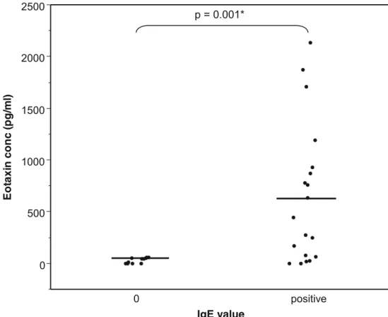
In Fig.1, mean eotaxin concentration reached 29±8 pg/ml (under the threshold of test sensitivity) (range 0–60 pg/ml) in tears without IgE and 641±154 pg/ml (0–2,132 pg/ml) in tears where IgE was positive (p=0.001).
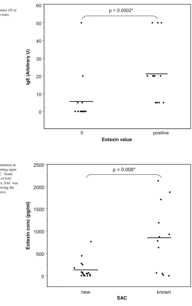
Tear IgE value was 5.3±3.5 arbitrary U in the 15 patients whose eotaxin concentration was under the threshold of sen-sitivity (Fig. 2), and 21±4.3 arbitrary U in the 15 patients whose tears contained a positive amount of eotaxin. The difference between the two groups of patients is significant (p<0.001). 0 500 1000 1500 2000 2500 0 positive p = 0.001* Fig. 1 Tear eotaxin concentration
and absence (0) or presence of IgE in tears. Bars = means
0 10 20 30 40 50 60 0 positive p = 0.0002* Fig. 2 Total tear IgE
concentration and absence (0) or presence of eotaxin in tears. Bars = means
0
1000
1500
2000
2500
new
known
p = 0.008*
500
Fig. 3 Eotaxin concentration in tears of patients presenting signs and symptoms of SAC. Some patients had a history of SAC (known); for the others, SAC was newly diagnosed following the visit at the hospital (new). Bars = means
Eotaxin concentrations were compared in tears of patients with a history of SAC and patients who had no history of SAC, but were newly diagnosed as suffering from SAC, according to symptoms and signs and after excluding other allergies (Fig.3). Eotaxin concentration reached 127±47 pg/ ml (0–758 pg/ml) in patients with newly diagnosed SAC, and 852±218 pg/ml (0–2,132 pg/ml) in tears of patients where SAC was already known. The difference between both groups is statistically significant (p=0.008).
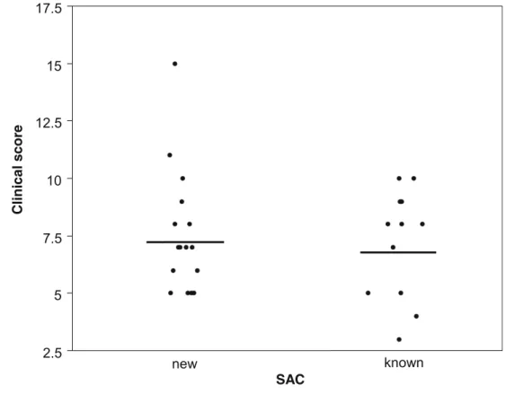
Concentrations of IgE were not significantly different in patients with a newly diagnosed SAC and those with SAC history (not shown) (p=0.947). Similarly, the clinical score was not significantly different in both groups of patients (Fig.4) (p=0.797).
Patients which had no IgE in their tears showed a lower conjunctival papilla grade than patients whose tears contained some IgE (Fig.5) (p=0.0318). On the other hand, there was no correlation between papilla grade and eotaxin presence in tears (not shown) (p=0.2194).
Discussion
We focused on patients suffering from SAC during the pollen season. SAC mechanism was demonstrated to be different from the mechanisms of more severe allergy cases such as VKC and AKC [3]. For instance, SAC induces penetration of conjunctiva by mast cells and later on neutrophils and
eosinophils, whereas VKC and AKC mainly involve T cells and eosinophils [3].
We wanted to specifically study the relation between IgE and eotaxin in tears of patients suffering from SAC during the in season. The relation between IgE and eotaxin has been investigated in plasma of asthmatic patients [17]. Plasma eotaxin values correlated with the total IgE values in asthmatic and non-asthmatic patients, suggesting that eotaxin may play a role in asthma severity [17]. To our knowledge, such compar-ison was not studied yet in ocular pathologies.
Detection and quantification of IgE in serum is the standard method for diagnosis of seasonal allergy [10]. Some authors have demonstrated that total tear IgE was correlated to serum IgE [7,10,18]. On the other hand, the largest contributor to the severity of SAC is the locally produced IgE [19, 20]. Measurement of IgE in tears may be therefore a quicker way to diagnose SAC than measurement of the serum-specific IgE. A few tests exist to measure total IgE in tears. A test in Japan made it possible to detect high levels of total IgE mean in tears of SAC and PAC patients in comparison to normal patients [7]. A Phadezym–PRIST test was able to show that
tears of SAC patients contain more IgE than control tears [19]. The Allerwatch test was used by Japanese groups; tears of an autumnal allergic group of patients contained a higher value of IgE than tears of a control group [21].
For detection of specific IgE in tears, a Japanese group has been evaluating a commercial immunochromatographic test for the semi-quantitative measurement of three specific IgE in 2.5 5 7.5 10 12.5 15 17.5 new known
Fig. 4 Clinical score of allergic conjunctivitis in patients presenting signs and symptoms of SAC. Some patients had a history of SAC (known); for the others, SAC was newly diagnosed following the visit at the hospital (new). Bars = means, p=0.797
tears [22]. For instance, Cedar pollen-specific IgE were sig-nificantly higher in tears of allergic conjunctivitis patients than in control subjects.
We chose the Lacrytest since it is an easy and rapid method to estimate total IgE levels in a very small volume of tears. The Lacrytest was previously used in a clinical study on subjects presenting, or not, signs of ocular allergy [23]. The test showed very good results, with a sensitivity of 93.8 % and a specificity of 89.7 %. In another study, if Lacrytest specificity reached 100 % [24], sensitivity was only 20 %. This was probably due to the fact that some patients enrolled in that study did not show ocular symptoms and signs of allergic conjunctivitis at the moment of the visit. Indeed, when pa-tients were submitted to conjunctival provocation test with the suspected allergen, sensitivity raised to 66.7 % [24].
The presence of IgE in tears of patients affected by SAC has already been demonstrated [7,19]. However, there were always a few patients presenting SAC but where no IgE could be revealed in their tears [19,25]. For instance, presence of IgE in tears was revealed in 23 out of 28 SAC patients [6]. Why, in our cohort of 30 patients, did only 19 demonstrate Table 1 Patient characteristics of
the prospective study. Patients presented seasonal allergic con-junctivitis (SAC) symptoms. For some patients, SAC had already been diagnosed (+); for the others, SAC was unknown at time of presentation (−)
Patient # Age Gender History of SAC Clinical score Papilla grade
1 49 F – 6 1 2 33 F + 10 2 3 49 F – 5 1 4 54 F + 4 2 5 36 F + 5 2 6 47 F + 9 2 7 54 F + 9 2 8 44 F + 7 2 9 39 M – 7 2 10 18 M + 3 3 11 23 F + 8 2 12 18 M – 6 2 13 27 F – 7 2 14 61 M – 5 1 15 72 M – 6 1 16 24 F + 5 1 17 62 F – 9 3 18 35 F – 7 3 19 23 F – 5 1 20 40 F – 8 0 21 18 M – 8 2 22 60 M – 11 3 23 18 M – 15 3 24 24 F – 5 2 25 55 F + 10 2 26 60 F – 10 2 27 35 F – 7 2 28 55 F + 8 0 29 31 F – 5 1 30 59 F + 8 2
Table 2 Frequencies of patients whose tears contain, or do not contain IgE and eotaxin. Number of patients (% of patients). Fisher’s exact test, two-tailed: p<0.001
IgE value Eotaxin value
0 Positive Total 0 11 0 11 100 % 0 % 100 % Positive 4 15 19 21 % 79 % 100 % Total 15 15 30 50 % 50 % 100 %
significant amounts of total tear IgE? It may be that, by chance, we recruited patients with lower SAC symptoms than in other studies. Therefore, their IgE levels were too low to be revealed by our test. On the other hand, we observed a fair interindividual variance in tear IgE values, phenomenon which was already described [18,19].
In the present study, we detected 127±47 pg/ml of eotaxin in tears of patients whose pathology was newly described as SAC, and 852±218 pg/ml in tears of patients with a history of SAC. These values are comparable to those reported in tears of SAC patients by Leonardi [15].
Eotaxin has been detected in tears of patients presenting various allergic reactions [15,26,27]. This chemokine was also revealed in healthy patients, but to a lesser extent [11]. Eotaxin values in our study are higher than in some other studies. This could be explained by the use of different con-ditions during the detecting method. An important study com-pared the amounts of some cytokines and chemokines detect-ed in human tears using the cytometric bead-basdetect-ed assay in different conditions [12]: with or without sample dilution before storage, with or without detergent, with or without bovine serum albumin. In the same initial sample, the authors measured eotaxin values ranging from 170 to 750 pg/ml after applying eight different conditions [12].
We showed, to some extent, a concomitant presence of total tear IgE and tear eotaxin in SAC patients. The absence of IgE in tears necessarily implies the absence of significant
amounts of eotaxin. But the presence of tear IgE implies the presence of eotaxin in only 79 % of patients.
In our study, there was no difference in total tear IgE levels between patients with a known SAC and patients with a newly diagnosed SAC. Moreover, the two groups of patients did not differ with regard to their clinical score mean. In contrast, patients with a known SAC exhibited significantly higher amounts of tear eotaxin than patients with a newly diagnosed SAC. Our findings confirm that IgE and eotaxin, if principal actors in the pathology of SAC, act separately along the allergic reaction [1].
Both IgE and eotaxin intervene at different phases of the pathogenesis of SAC. During the phase of sensitization, B lymphocytes of the conjunctiva produce IgE against the aller-gen, which will link to mast cells. During the challenge phase, the allergen binds with specific IgE on the conjunctival mast cells, leading to mast cell degranulation and mediator release, characteristic of the acute phase allergic reaction. Eotaxin is a major contributor which induces inflammatory cell invasion, driving the late and chronic phase of allergy [2, 3]. This chemokine specifically stimulates eosinophils chemotaxis and aggregation through the chemokine receptor 3 (CCR3), which is expressed in high numbers on eosinophils [28,29]. Eotaxin induces chemotaxis of other cells wearing CCR3, for instance lymphocytes and basophils [27]. But this CC chemo-kine also plays a role in mast cell priming, since it provides a co-stimulatory signal for conjunctival mast cells [30]. -0.5 0 0.5 1 1.5 2 2.5 3 3.5 0 positive p = 0.0318* Fig. 5 Conjunctival papilla grade
and absence (0) or presence of IgE in tears. Bars = means
Extensive studies have proved that experimental allergic conjunctivitis is ablated in mice deficient in eotaxin [1,3]. After induction of allergic inflammation in the conjunctiva of these mutant mice, the authors counted normal numbers of tissue mast cells in conjunctiva, and measured normal levels of IgE. However, these eotaxin-deficient mice showed a significant impairment of mast cell degranulation and a suppression of clinical symptoms in the acute phase reaction [1]. As mast cell degranulation does not depend on eotaxin, the authors hypothesized that this chemokine might provide a co-stimulatory signal, and that CCR3 plays a major role in activation of mature connective tissue-type mast cells in ocular tissue. Indeed, when CCR3 was blocked, allergen-mediated hypersensitivity re-action and IgE-mediated mast cell degranulation were suppressed [1]. Therefore, the eotaxin/CCR3 axis is the main control of mast-cell-mediated allergy.
The role of CCR3 in allergic conjunctivitis has been previously demonstrated in mice subjected to allergic sensitization [31]. Anti-CCR3 suppressed both clinical signs of allergic inflammation and mast cell degranula-tion, but could not reduce the serum levels of specific IgE in mice [31].
Presence of conjunctival papillae is characteristic of AKC, VKC, and contact lens wear [13,16,32,33]. We were able to demonstrate that our SAC patients had a low to mod-erate conjunctival papilla grade during the in season. We observed that patients with a positive amount of IgE in their tears were characterized by a higher conjunctival papilla grade than patients with no IgE. A mild papillary hypertrophy was previously observed in the upper tarsal conjunctiva of patients suffering from seasonal allergic rhinoconjunctivitis [4].
In conclusion, we investigated a cohort of patients suffer-ing from SAC dursuffer-ing the pollen season. We are aware of some limitations of interpretation of our data; Lacrytest is a semi-quantitative test with an arbitrary endpoint to determine pos-itive or negative results, and the detecting test for eotaxin has a sensitivity threshold of 60 pg/ml. However, the data suggest that when patient tears contained IgE, they were often likely to contain eotaxin too. Patients with a history of SAC were compared to patients with a newly diagnosed SAC. Both groups of patients demonstrated the same level of total tear IgE in mean, and the same clinical score. In contrast, patients with a known SAC had a signifi-cantly higher level of eotaxin in their tears than patients with a newly SAC. This confirms that, if IgE and eotaxin are princi-pal actors in IgE-mediated SAC, they contribute separately to the reaction.
Conflict of interest The authors declare that they do not have any conflict of interest.
Financial interests None.
Open Access This article is distributed under the terms of the Creative Commons Attribution License which permits any use, distribution, and reproduction in any medium, provided the original author(s) and the source are credited.
References
1. Miyazaki D, Nakamura T, Ohbayashi M, Kuo CH, Komatsu N, Yakura K, Tominaga T, Inoue Y, Higashi H, Murata M, Takeda S, Fukushima A, Liu FT, Rothenberg ME, Ono SJ (2009) Ablation of type I hypersensitivity in experimental allergic conjunctivitis by eotaxin-1/CCR3 blockade. Int Immunol 21:187–201. doi:10.1093/ intimm/dxn137
2. Ono SJ (2000) Molecular genetics of allergic diseases. Annu Rev Immunol 18:347–366. doi:10.1146/annurev.immunol.18.1.347
3. Offiah I, Calder VL (2009) Immune mechanisms in allergic eye diseases: what is new? Curr Opin Allergy Clin Immunol 9:477–481 4. Friedlaender MH (1993) Conjunctivitis of allergic origin: clinical presentation and differential diagnosis. Surv Ophthalmol 38(Suppl): 105–114
5. Cook EB (2004) Tear cytokines in acute and chronic ocular allergic inflammation. Curr Opin Allergy Clin Immunol 4:441–445 6. Mimura T, Usui T, Mori M, Aixinjueluo W, Funatsu H, Noma H,
Amano S (2010) Immunochromatographic assay for measurement of total IgE in tears, nasal mucus, and saliva of patients with allergic rhinoconjunctivitis. J Asthma 47:1153–1160
7. Nomura K, Takamura E (1998) Tear IgE concentrations in allergic conjunctivitis. Eye (Lond) 12(Pt 2):296–298
8. Pokharel S, Shah DN, Joshi SN, Choudhary M (2009) Tearfilm immunoglobulin E (IgE) level in vernal keratoconjunctivitis by ELISA. Kathmandu Univ Med J 7(26):104–108
9. Wakamatsu TH, Satake Y, Igarashi A, Dogru M, Ibrahim OM, Okada N, Fukagawa K, Shimazaki J, Fujishima H (2012) IgE and eosinophil cationic protein (ECP) as markers of severity in the diagnosis of atopic keratoconjunctivitis. Br J Ophthalmol 96:581–586. doi:10. 1136/bjophthalmol-2011-300143
10. Mimura T, Usui T, Mori M, Funatsu H, Noma H, Yamamoto H, Aixinjueluo W, Amano S (2011) Relationship between total tear and serum IgE in allergic conjunctivitis. Int Arch Allergy Immunol 154: 349–352
11. Carreno E, Enriquez-de-Salamanca A, Teson M, Garcia-Vazquez C, Stern ME, Whitcup SM, Calonge M (2010) Cytokine and chemokine levels in tears from healthy subjects. Acta Ophthalmol 88(7):e250–e258 12. LaFrance MW, Kehinde LE, Fullard RJ (2008) Multiple cytokine anal-ysis in human tears: an optimized procedure for cytometric bead-based assay. Curr Eye Res 33:525–544. doi:10.1080/02713680802190085
13. Abu El-Asrar AM, Struyf S, Al-Karashi SA, Missoten L, Van Damme J, Geboes K (2000) Chemokines in the limbal form of vernal keratoconjunctivitis. Br J Ophthalmol 84:1360–1366
14. Eperon S, Sauty A, Lanz R, Leimgruber A, Lurati F, Guex-Crosier Y (2004) Eotaxin-1 (CCL11) up-regulation in tears during seasonal aller-gic conjunctivitis. Graefes Arch Clin Exp Ophthalmol 242:966–970 15. Leonardi A, Curnow SJ, Zhan H, Calder VL (2006) Multiple
cyto-kines in human tear specimens in seasonal and chronic allergic eye disease and in conjunctival fibroblast cultures. Clin Exp Allergy 36: 777–784
16. Moschos MM, Eperon S, Guex-Crosier Y (2004) Increased eotaxin in tears of patients wearing contact lenses. Cornea 23:771–775 17. Chang HS, Kim JS, Lee JH, Cho JI, Rhim TY, Uh ST, Park BL,
polymorphism on the promoter of eotaxin1 associates with its mRNA expression and asthma phenotypes. J Immunol 174: 1525–1531
18. Inada N, Shoji J, Kato H, Kiely S, Mulyanto SM (2009) Clinical evaluation of total IgE in tears of patients with allergic conjunctivitis disease using a novel application of the immunochromatography method. Allergol Int 58:585–589
19. Aalders-Deenstra V, Kok PT, Bruynzeel PL (1985) Measurement of total IgE antibody levels in lacrimal fluid of patients suffering from atopic and non-atopic eye disorders. Evidence for local IgE produc-tion in atopic eye disorders? Br J Ophthalmol 69:380–384 20. Batellier L, Moens C, Poilane C, Mavier C, Goldschmidt P, Chaumeil
C (2008) Measurement of total IgE in tears by ultrasensible technique « 0–100 » on the Immunocap®100 (Phadia): Interest for the etiology diagnosis of the eye surface allergic achievements. Immuno Spec Biol 23:89–94
21. Mimura T, Usui T, Yamagami S, Miyai T, Amano S (2013) Relationship between total tear IgE and specific serum IgE in au-tumnal allergic conjunctivitis. Cornea 32:14–19. doi:10.1097/ICO. 0b013e318253214b
22. Mimura T, Usui T, Mori M, Funatsu H, Noma H, Amano S (2011) Rapid immunochromatographic measurement of specif-ic tear immunoglobulin E in moderate to severe cases of allergic conjunctivitis with Immfast Check J1 in the spring. Cornea 30:524–527
23. Sirbikaite L, Ehlers N (2007) The use of Lacrytest in ocular allergy. Acta Ophthalmol Scand 85:117
24. Monzon S, Arrondo E, Bartra J, Torres F, Basagana M, SanMiguel MM, Alonso R, Cistero-Bahima A (2009) Conjunctivitis and total IgE in lacrimal fluid: Lacrytest screening. J Allergy (Cairo). doi:10. 1155/2009/518903, Article ID 518903
25. Mimura T, Yamagami S, Kamei Y, Goto M, Matsubara M (2013) Specific IgE in tear fluid and features of allergic conjunctivitis. Curr Eye Res 38:917–925. doi:10.3109/02713683.2013.794248
26. Sarac O, Erdener U, Irkec M, Us D, Gungen Y (2003) Tear eotaxin levels in giant papillary conjunctivitis associated with ocular prosthe-sis. Ocul Immunol Inflamm 11:223–230
27. Wallace GR, John CS, Wloka K, Salmon M, Murray PI (2004) The role of chemokines and their receptors in ocular disease. Prog Retin Eye Res 23:435–448
28. Rankin SM, Conroy DM, Williams TJ (2000) Eotaxin and eosinophil recruitment: implications for human disease. Mol Med Today 6:20– 27
29. Fukagawa K, Okada N, Fujishima H, Nakajima T, Tsubota K, Takano Y, Kawasaki H, Saito H, Hirai K (2002) CC-chemokine receptor 3: a possible target in treatment of allergy-related corneal ulcer. Invest Ophthalmol Vis Sci 43:58–62
30. Ono SJ, Abelson MB (2005) Allergic conjunctivitis: update on pathophysiology and prospects for future treatment. J Allergy Clin Immunol 115:118–122. doi:10.1016/j.jaci.2004.10.042
31. Komatsu N, Miyazaki D, Tominaga T, Kuo CH, Namba S, Takeda S, Higashi H, Inoue Y (2008) Transcriptional analyses before and after suppression of immediate hypersensitivity reactions by CCR3 block-ade in eyes with experimental allergic conjunctivitis. Invest Ophthalmol Vis Sci 49:5307–5313. doi:10.1167/iovs.08-2154
32. Fukagawa K, Nakajima T, Tsubota K, Shimmura S, Saito H, Hirai K (1999) Presence of eotaxin in tears of patients with atopic keratocon-junctivitis with severe corneal damage. J Allergy Clin Immunol 103: 1220–1221
33. Tran HV, Eperon S, Guex-Crosier Y (2011) Persistence of increased eotaxin-1 (CCL11) level in tears of patients wearing contact lenses: a long term follow-up study. Klin Monatsbl Augenheilkd 228:326–329