HAL Id: hal-02193218
https://hal-udl.archives-ouvertes.fr/hal-02193218
Submitted on 8 Jul 2020HAL is a multi-disciplinary open access archive for the deposit and dissemination of sci-entific research documents, whether they are pub-lished or not. The documents may come from teaching and research institutions in France or abroad, or from public or private research centers.
L’archive ouverte pluridisciplinaire HAL, est destinée au dépôt et à la diffusion de documents scientifiques de niveau recherche, publiés ou non, émanant des établissements d’enseignement et de recherche français ou étrangers, des laboratoires publics ou privés.
Surfaces based on amino acid functionalized
polyelectrolyte films towards active surfaces for enzyme
immobilization
Ximena Briones, Valeria Villalobos, Yves Queneau, Caroline Silva Danna,
Rodrigo Muñoz, Hernán Ríos, Jorge Pavez, Maritza Páez, Ricardo Cabrera,
Laura Tamayo, et al.
To cite this version:
Ximena Briones, Valeria Villalobos, Yves Queneau, Caroline Silva Danna, Rodrigo Muñoz, et al.. Surfaces based on amino acid functionalized polyelectrolyte films towards active surfaces for en-zyme immobilization. Materials Science and Engineering: C, Elsevier, 2019, 104, pp.109938. �10.1016/j.msec.2019.109938�. �hal-02193218�
Amino Acid Functionalized Polyelectrolyte Films as
Bioactive Surfaces for Cell Adhesion
M.S. Leal,† X. Briones,† V. Villalobos,◊ Y. Queneau,‡ A. Leiva,§ H.E. Ríos,† J. Pavez,| C.P. Silva,| C. Carrasco,|| Andrónico Neira-Carrillo,∞ A. Roth,|| L. Tamayo,*† M.D. Urzúa*†
† Depto. de Química, Facultad de Ciencias, Universidad de Chile, Las Palmeras 3425, Santiago, Chile
◊ Universidad Autónoma de Chile, Instituto de Ciencias Químicas Aplicadas, Facultad de Ingeniería, El Llano Subercaseaux 2801, San Miguel, Chile
‡ Université de Lyon, ICBMS, UMR 5246, CNRS, UCBL, INSA Lyon, CPE Lyon, Bât. Lederer, 1 Rue Victor Grignard, 69622 Villeurbanne Cedex, France
§ Depto. Química Física, Facultad de Química, Pontificia Universidad Católica de Chile, Macul, Santiago 7820436, Chile
| Depto. de Química de los Materiales, Fac. de Química-Biología, Universidad de Santiago de Chile, Av. B. O’Higgins 3363, Santiago, Chile
|| Departamento de Biología, Facultad de Ciencias, Universidad de Chile, P. C. 780-0023, Santiago, Chile
∞ Fac. de Ciencias Veterinarias y Pecuarias, Universidad de Chile, Av. Sta. Rosa 11735, Santiago, Chile
KEYWORDS: Polyelectrolytes, amino acid, functionalization, cell adhesion, SH-SY5Y
ABSTRACT
Surfaces were prepared with polyelectrolyte derivatives of poly(styrene-alt-maleic anhydride)
(PSMA) functionalized with amino acids of different hydropathy indices, with the aim of
evaluating the effect of the chemical functionality of polyelectrolytes on SH-SY5Y
neuroblastoma cell adhesion. Functionalizing PSMA derivatives with L-glutamine, L-methionine
and L-tyrosine yielded PSMA-Gln, PSMA-Met and PSMA-Tyr polyelectrolytes, respectively.
We first studied the adsorption behavior of PSMA functionalized with amino acids on silicon
wafer surfaces modified with 3-aminopropyltriethoxysilane (APTES) at pH 4.0 and 7.0 and at
low and high ionic strength. The highest rate of polyelectrolyte adsorption was at pH 4.0 and
high ionic strength, and was higher with the glutamine and tyrosine films. The advance contact
angles (qA) of the polyelectrolyte surfaces showed a moderate effect of ionic strength and pH on polyelectrolyte film wettability, with PSMA-Tyr being slightly more hydrophobic. AFM images
of the polyelectrolyte surfaces showed two types of morphology: the well-defined globular
nanostructure of PSMA-Met and PSMA-Tyr, and the densely packed nanofibrous-like structure
of PSMA-Gln. The highest level of ionic strength caused a slight decrease in size of the
nanostructure that formed the surface domains, which was reflected in the degree of surface
roughness. Cell adhesion assays with polyelectrolyte film showed that SH-SY5Y neuroblastoma
cells cultured on PSMA-Met present a well-extended morphology characterized by a stellate
shape, with five or more actin-rich thin processes, while SH-SY5Y cells that where seeded on
PSMA-Gln and PSMA-Tyr have a round morphology, with fewer and shorter processes. These
results indicate that it is possible to modulate the surface characteristics of polyelectrolyte films
based on their chemical functionality and environmental parameters such as pH and ionic
polyelectrolytes functionalized with amino acids are an attractive and simple platform for cell
adhesion, which can be used in developing biomaterials with modulated surface properties.
1. INTRODUCTION
Important advances have been made in designing biomaterials for medical implants, supports
for cell regeneration and biosensors for disease diagnosis, but there are still challenges to be
overcome. Of particular interest are the processes and interactions that occur in the
biomaterial-biological system interface, in order to achieve a favorable response of the latter (eukaryotic cell,
bacterium, bone or blood). In the search for biological selectivity, it is key to understand
biomaterial surface properties and how to modulate them and the responses they elicit from
different cell types. Among the most important surface variables are wettability, topography,
energy and surface charge. 1 It has been observed that positive surface charge and hydrophilicity
enhance 3T3 fibroblast adhesion. 2 Other surface variables like roughness also play an important
role, and in general some characteristics has been observed that favors the adhesion of each cell
type. In the case of neuronal cell grown on silicon, surface roughness at a nanoscale of 50 to 70
nm favors adhesion. 3
Given the need to modulate the chemical properties of biomaterial surfaces, polyelectrolytes
offer important advantages due to the variety of their chemical functionality and ionizable
groups, which allows for controlling their specificity, mainly by electrostatic, dipole-dipole,
hydrophobic and hydrogen bonding interactions. 4 The biological selectivity of surfaces is an
important characteristic in biological applications, which can be enhanced by modifying
polyelectrolytes with biomolecules like carbohydrates, amino sugars, peptides, and others. 5-7
Arginine-rich peptides have been shown to promote cellular internalization, which has led to
peptides. 8,9 It has been reported that modification of chitosan grafting with hydrophobic amino
acids improves blood compatibility and increases cell viability. These effects are proportional to
the increase in the hydrophobicity of chitosan modified with amino acids of different hydropathy
indices.
Studies have evaluated the osteogenic capacity of films of chitosan modified with anionic
carbohydrates, and have found that the presence of 4-chondroitin sulfate (chondroitin 4-sulfate)
promotes BMSC cell proliferation. 10 The modification of chitosan with integrin-binding
consensus peptides common to cell matrix proteins like RGD (Arg-Gly-Asp) and GRGD
(Gly-Arg-Gly-Asp) increases the adhesion of multiple cell types (e.g. osteosarcoma, osteoblasts and
fibroblasts). 11-13 However, functionalization methods for cell adhesion peptides are expensive
and often require multiple stages of conjugation. While several studies have demonstrated the
advantages of modifying chitosan to promote cell interaction selectivity, chitosan has the
disadvantages of low water solubility, in vivo de-polymerization and hemo-incompatibility. 14
The ideal polyelectrolyte would be stable over a wide pH range, and would require simple and
inexpensive modification methods.
Polyelectrolytes like maleic anhydride derivatives have the advantage of a high degree of
water solubility over a wide pH range, in addition to being biologically active. 15 The high degree
of reactivity of the co-monomer maleic anhydride allows the synthesis of alternating copolymers
that can be used as platforms for functional modulation, providing a high degree of chemical
versatility that can be enhanced by modification with biomolecules that favor the required
biological response. 16 Given the reactivity of the maleic anhydride co-monomer and the easiness
of synthesizing alternating copolymers, in addition to their water solubility and excellent
acids, allows to obtain functionalized polyelectrolytes with enhanced properties such as, their
biocompatibility and therefore would allow for the development of active surfaces that promote
cell adhesion.
Polymeric films were developed in this work based on poly(maleic anhydride-alt-styrene)
[PSMA] modified with amino acids of different hydropathic indices (H): L-glutamine (H = -3.5),
L-tyrosine (H = -1.3) and L-methionine (H = 1.9), 17 with the aim of evaluating the effect of modifying film surface properties (wettability, surface energy, hysteresis, roughness and charge
density) on the adhesion of human SH-SY5Y neuroblastoma cells. The human neuroblastoma SH-SY5Y has served as a robust in vitro model of multiple neuronal events such as migration, differentiation and neuritogenesis. 18,19
They are sensible to different stimuli which has made it
particularly well suited for study the neuron-to-substrates interactions. 20
Thus, taking advantage
of the sensibility of SH-SY5Y cells we evaluated their response the physicochemical
characteristics of modified PSMA-substrates.
EXPERIMENTAL SECTION 2.1 Materials
The monomers maleic anhydride (MA) and styrene (S); the amino acids L-glutamine (reagent grade >99% TLC), L-methionine (>98% TLC) and L-tyrosine (reagent grade >98%, TLC); and 3-aminopropyltriethoxysilane (APTES,> 98%) were obtained commercially from
Sigma-Aldrich. The NaCl and NaHCO3 salts and all the solvents used, including benzene,
dimethylsulfoxide (DMSO), ethanol, 25% ammonium hydroxide, hydrogen peroxide and
deuterated dimethylsulfoxide (DMSO-D6) were obtained commercially from Merck. Silicon
Deionized water of 0.055 µS/cm conductivity, 18.2 MΩcm resistivity and TOC (total organic
carbon) content below 10 ppb obtained from a LabStar 4-DI deionizer was used to prepare the
aqueous solutions.
2.2 Poly(maleic anhydride-alt-styrene) Synthesis
Poly(maleic anhydride-alt-styrene) copolymer, PSMA, with a molecular weight of
Mv=6.3x10⁴ g/mol, was synthesized by radical polymerization by mixing equimolar amounts of
previously purified styrene and maleic anhydride monomers, obtaining an alternate copolymer
given by the value of the reactivity ratio of the co-monomers which is approximately equal to
zero. 21 Anhydrous benzene was used as a solvent, and 8% of α,α-azobisisobutyronitrile (AIBN)
was used as the initiator. The synthesis was carried out at 60 °C in an N2 atmosphere for 2 h.
2.3 Determining Molecular Weight
The average viscosimetric molecular weight (Mv) of the PSMA polymer of 63.000 g/mol was
determined by measuring the viscosity of PSMA solutions with THF as a solvent at 30 °C. The
weight was calculated with the Mark-Houwink-Sakurada equation, using the constants a=0.81
and k=5.07x10⁻⁵. 22,23 A polydispersity value of 1.7 was obtained. Polydispersity was obtained
using size exclusion chromatography (SEC) using a Waters Styragel HMW-6E column, solutions
of 1 mg/mL in tetrahydrofuran as solvent and polystyrene standards (18200, 42300, 83000 and
189000 Da) with a flow of 0.3 mL/min.
2.4 Functionalizing PSMA with Amino Acids
The PSMA copolymer was functionalized by mixing equimolar amounts of PSMA with the
amino acids L-glutamine (Gln), L-methionine (Met) and L-tyrosine (Tyr), using DMSO as the solvent and 0.3 % triethylamine (TEA) as a base at a constant temperature of 80 °C, under an N2
spectroscopy. The final stage of the reaction was established when the bands at 1854 cm-1 and
1779 cm-1, which belong to the two carbonyl groups of maleic anhydride, disappeared and bands
that belong to the carbonyls of the amide and carboxylic acid groups were observed at ~1660 cm -1 and ~ 1710 cm-1, respectively. 24
The disodium salts of the modified polymers were obtained by treating them with aqueous
solutions of NaHCO3 (in a 1:2 polymer:NaHCO3 ratio) for 10 days. The solutions were then
ultrafiltered and lyophilized. The salts of the polyelectrolytes obtained with the amino acids, L -glutamine, L-methionine and L-tyrosine, were respectively labeled PSMA-Gln, PSMA-Met and PSMA-Tyr. Their structures are represented in Scheme 2.
Scheme 1. Reactions to obtain salts from polyelectrolytes derived from PSMA modified with
amino acids. R represents the side groups of the amino acids: a) glutamine, b) methionine and c)
2.5 FT-IR Spectroscopy
KBr discs were prepared with 2 mg of polymer and 100 mg of KBr to characterize pure PSMA
and copolymers functionalized with amino acids. The FT-IR spectra were obtained with a Bruker
Vector Model 22 at 25°C. The spectra were collected at 4 cm−1
data intervals (scanner velocity:
10 kHz; background: 32 scans; sample: 32 scans). The calibration model for PSMA and
functionalized PSMA was built with Bruker OPUS version 6.5 software (Bruker, Ettlingen,
Germany) based on PLS (Partial Least Squares) regression.
2.6 Magnetic Resonance of Proton and Carbon-13 (1H-NMR and 13C-NMR)
To study 1H-NMR and 13C-NMR, ~80 mg of PSMA and of each copolymer functionalized
with amino acids were dissolved in DMSO-D6 (0.5 mL). The 1H-NMR and 13C-NMR spectra
were examined using an Advance-400 spectrometer at 25°C.
2.7 Quantitative Elemental Analysis
Quantitative elemental compositions were determined in an elemental analyzer (CE
Instruments, Eager 200 model). The samples were prepared in quantities of 2 mg in tin crucibles,
Scheme 2. Polyelectrolyte structures: a) PSMA-Gln, b) PSMA-Met, and c) PSMA-Tyr
2.8 Polyelectrolyte Adsorption on Solid Surfaces
Polymeric solutions were prepared by dissolving the polyelectrolytes PSMA-Gln, PSMA-Met
and PSMA-Tyr in 0.001 M and 0.1 M solutions of NaCl. The pH was adjusted to pH 4.0 and pH
7.0 by adding HCl or NaOH as appropriate. The concentration of the polyelectrolyte solutions
varied between 1.0x10⁻⁵ g/L and 1.0 g/L.
The silicon wafers used as substrates were chemically modified with
3-aminopropyltrimethoxysilane (APTES) by oxidative washing 25 in order to obtain a
homogeneous amino acid-terminated monolayer covalently bound to the silicon wafer. 26
Subsequently, the modified silicon wafers were immersed in 2 mL of the polyelectrolyte
solutions for 3 h, and then washed with deionized water and dried with an N2 flow. The quantity
2.9 Adsorption Isotherms
The adsorption isotherms were obtained by ex-situ ellipsometry in a variable angle
ellipsometer L116S300 STOKES (Gaertner Scientific Corporation, USA) equipped with a
He-Ne laser with a wavelength of λ = 632.8 nm and an angle of incidence, Φ of 70° at a temperature
of 25±1 °C. The quantity of polyelectrolyte adsorbed Γ was determined according to the
relationship between the thickness of the adsorbed layers (dpoly), the concentration (cpoly) and the
variation of the refractive index of the polyelectrolyte solutions (dn/dc). 27 The thickness of the
adsorbed polyelectrolyte layer was calculated from the ellipsometric angles Δ and Ψ using a
multilayer model composed of substrate-medium or substrate-unknown layer (APS or
polyelectrolyte film-medium), using the Elli computer program, based on the ellipsometry
equation and iterative calculations with Jones matrices. 28 Thus, it is possible to determine only the thickness of the adsorbed polyelectrolyte, dpoly. The small differences in refraction indices n
of the substrate, polyelectrolyte, and solution made an independent determination of npoly and dpoly
impossible. Therefore, npoly was kept constant as 1.50 and dpoly was calculated. 22
It is important to
remember here that, if the index of refraction assumed for the adsorbing polymer layer lies in a
reasonable range (between 1.40 and 1.60), the product npoly dpoly should be a constant value. 22, 27
This product is the parameter necessary to calculate the adsorbed amount Γ from equation 1:
Г =#$%&' (*$%&'+*,)
#./#0 = 𝑑2345𝑐2345 (1)
where n0 is the refraction index of the solution measured with an Abbe refractometer AR4,
dn/dc is the increment of refractive index determined with an interferometric refractometer, and
cpoly is the average polyelectrolyte concentration within the layer. 22,27
For the present systems, n0
± 1 °C. The dn/dc measurements were performed using an OPTILAB DSP interferometric
refractometer from Wyatt Technology instruments of sensitivity 0.002 Refractive Index Units.
The refractive indices n = 3.88 - i0.018 for pure silicon, n = 1.00 for air and n = 1.462 for thin
layers of SiO2 were used to determine the thickness of the SiO2 layer. The average thickness
obtained after several measurements was 1.2±0.2 nm. To determine the APS monolayer, it is
necessary to know the refractive indices n = 1.424 for APS and n = 1.00 for air. 29 The thickness
obtained for this layer after several measurements in air was 0.9 - 1.2 nm.
2.10 Polymeric Surface Wettability
The wettability of the polymer surfaces was determined by contact angle measurements on the
PSMA, PSMA-Gln, PSMA-Met and PSMA-Tyr films using a Drop Shape Analyzer DSA25S
(KRUSS, Germany) using the Sessile Drop method at a temperature of 25 °C. Sessile water
drops of 8 µL were used for determine the advancing contact angle (qADV), and then the volume was reduced to 4 µL to measure the receding contact angle (qREC). The hysteresis in the contact angle measurements (Dq = qADV -qREC) was calculated from the difference between (qADV) and (qREC). The measurements were performed automatically and controlled by Advance® software (KRUSS, Germany) obtaining images of drop for 1 min, where no changes were observed
associated to drop evaporation. At least three spots were done in each sample to make sure we
get the same result. Additionally, measurements of contact angle were made using drops of 5 µL
2.11 Polymeric Surface Energy
Droplets of 5 µL of water, diiodomethane or ethylene glycol were deposited on PSMA-Gln, PSMA-Met and PSMA-Tyr films to determine surface energy. At least three measurements were
made for each type of polymer surface with each type of solvent. The surface energy was
calculated automatically by Advance® software (KRUSS, Germany) from the water,
diiodomethane and ethylene glycol contact angles (θ) obtained once thermodynamic equilibrium
was reached (as described by the Young-Laplace equation and the harmonic mean equation
proposed by Wu). The latter equation was developed from the surface tension (γtotal) of a
material, considered as the sum of its dispersive (γD) and polar components (γP) that best
describe the interactions between a liquid and a polymeric surface with respect to other methods. 30-32
2.12 Polyelectrolytes Film Topography and Surface Potential
The surfaces of the polyelectrolyte films were analyzed by atomic force microscopy (AFM)
and Kelvin force microscopy (KFM) with an AFM/SPM Controller 9500 Series System,
(Keysight Technologies, CA, USA) with a 7500 scanner. The surfaces were scanned in the
magnetic-AC mode (MAC-Mode® Keysight) with a scan rate of 0.3 Hz, using commercial
AC200TS three-sided silicon probes (Olympus). The height and amplitude of AFM images were
determined at room temperature (23-25 ºC). Commercial KS-HQ:XSC11/Pt umasch probes were
used for the KFM. All Image analysis and root mean square (RMS) roughness was determined
with Keysight PicoView software.
2.13 Cells Culture and Cell Viability
Human SH-SY5Y neuroblastoma cells (CRL-2266, American Type Culture Collection,
supplemented with 10% FBS, non-essential amino acids, antibiotic–antimycotic mixture, and 20
mM HEPES buffer, at pH 7.2. The cell culture medium was replaced every two days. The effect
of the polyelectrolytes on cell viability was quantified by MTT assay following the
manufacturer’s instructions. Briefly, 5,000 cells were plated in each well of a 96-well microplate
and cultured for 24 hours in the presence of different concentrations of polyelectrolyte solutions
(in triplicate). The cell culture medium was replaced with 110 µL of fresh medium supplemented
with 1,1 mM MTT (3-(4,5-dimethylthiazol-2-yl)-2,5-diphenyltetrazolium bromide) and
incubated for 4 hours. Mitochondrial dependent formation of a colored product was terminated
by adding 100 µL of SDS-HCl solution, followed by 12 hours of solubilization at 37 ºC.
Absorbance as a measure of product formation was read at 570 nm. 33
2.14 4-HNE Immunofluorescence
Oxidative stress induced peroxidation of cell membrane lipids was assayed through
immunohistochemistry against 4-HNE. 34 AbCam, UK Poly-clonal antibody ab46545, was
diluted 1:200 as indicated by manufacturer's instructions. 35 Briefly, Human SH-SY5Y
neuroblastoma cells were seeded on top of clean and sterile acid-washed cover slips 36 or silicon
wafers (1x1 cm) coated with fixed substrates (PSMA-Gln, PSMA-MET or PSMA-Tyr) that were
UV-sterilized for 15 min. After 24 hours culture, all substrates were washed with phosphate
buffered saline (PBS) and fixed for 20 minutes in a 4% paraformaldehyde PBS solution. Cells
were permeabilized by a 10 minutes incubation in 0.2% Triton-X-100 PBS solution. Unspecific
binding sites were blocked by incubating for 90 minutes in PBS containing 5% (w/v) of bovine
serum albumin (BSA, Sigma, USA). Cells were incubated overnight at 4°C with a 1:200 dilution
of anti-4 Hydroxynonenal antibody (ab46545; AbCam, UK) in PBS+5%BSA. Substrates were
Alexa-488-conjugated goat anti-rabbit IgG (1:1000 in PBS-5%BSA), washed three times with
PBS (15 minutes) and mounted on glass slides. Glass coverslips were inverted on top of a 25 ul
drop of mounting medium (FluorSaveTM (Merk)) while silicon wafers were glued to the glass
slide, overlaid with mounting medium and covered with a glass-coverslip. Labeled cells were
observed and photographed in a Zeiss LSM 710 confocal laser scanning microscope. Statistical
significance was determined using a one-way ANOVA and Tukey post-test.
2.15 Scanning Electron Microscopy
Cells grown on substrates (PSMA-Gln, PSMA-Met or PSMA-Tyr) were fixed with 4%
paraformaldehyde, 4% sucrose in saline, permeabilized with 0.2% Triton-X-100 in PBS,
incubated overnight at 4°C. Then the samples were then rinsed twice with PBS buffer at pH 7.0
for 15 min and fixed with 1% glutaraldehyde in PBS buffer for 2 h at room temperature. The
samples were then rinsed twice in PBS buffer. Finally, the samples were dehydrated in ascending
grades of ethanol (30, 50, 70, 80, 90, and 100%) and coated with a thin film of Pt/Pd.
3. RESULTS AND DISCUSSION 3.1. Characterization of Polyelectrolytes
The FT-IR and 13C-NMR polyelectrolyte spectra were contrasted to the PSMA spectrum to
provide a complementary confirmation of the successful modification of PSMA with the
different amino acids by opening the maleic anhydride ring and forming an amide bond. The IR
spectra (Figures S1-S3 Supplementary Material) show that the characteristic bands of the C=O
maleic anhydride PSMA group at 1854 cm-1 and 1779 cm-1 disappear, while two new bands
appear at 1664 cm-1 and 1705 cm-1 for PSMA-Gln, 1660 cm-1 and 1703 cm-1 for PSMA-Met, and
carbonyl group to an amide, and the stretching of the C=O carboxylic acid group. The 13C-NMR
PSMA spectra (Figures S4-S6 Supplementary Material) show the characteristic signals of the C
atoms of the C=O group of the PSMA anhydride at 172.9 and 173.58 ppm. After modification,
these signals disappear and signals appear at 174.0 and 178.5 ppm representing the C atoms of
the C=O groups of the carboxylic acids and the C=O of the newly formed amide group. The
percentages of modification for PSMA-Gln, PSMA-Met and PSMA-Tyr obtained by elemental
analysis were respectively 81%, 98% and 80%.
3.2. Adsorption of Polyelectrolytes on Amino Acid-terminated Surfaces
Polyelectrolyte adsorption is strongly influenced by pH level and salt concentration.
Adsorption isotherms were obtained at pH 4.0 and 7.0, and at two salt concentrations, 0.1 and
0.001M, to determine the type of interaction that governs adsorption behavior. The
polyelectrolyte absorption rates at pH 4.0 were higher for PSMA-Gln and PSMA-Tyr when the
salt concentration was 0.1 M. This behavior is adjusted to what is termed "increased adsorption
by shielding", which implies that the forces that modulate the interaction between the surface and
polyelectrolyte are mainly of the hydrophobic type and can be explained by the screening of the
charged segments of the polyelectrolyte with sodium counter ions. In this case, PSMA-Gln and
PSMA-Tyr behave like a neutral polymer, while with PSMA-Met the electrolyte absorption rate
decreases with the salt concentration at 0.1 M. This behavior is determined by the "decreased
adsorption by shielding" described by Van der Steeg 37 and implies a modulating force. The
interactions between the positively charged amino acid-terminated surface and the -COOH group
The polyelectrolyte adsorption rates at pH 7.0 are lower than at pH 4.0, but similar at the two
salt concentrations (Table 1, Figure 1) except for PSMA-Tyr. The pK1 of the PSMA-Tyr
carboxylate group is 4.3 lower than the pK1 of PSMA-Gln and PSMA-Met (see Table 1). The
lower pK1 value of PSMA-Tyr is associated with a larger number of carboxylic groups than with
carboxylate groups. Considering that the propyl-amino group on the surface has a pKa close to
3.7, 38 it is expected that at pH 7.0 the absence of the amino acid-terminated surface charge
promotes H-bonding, and hydrophobic interactions between the phenol of the PSMA-Tyr side
chain and the amino acid-terminated surface, increasing the quantity of PSMA-Tyr.
On the other hand, it is well known that the adsorption process is determined by a balance
between enthalpy and entropy of the system. In fact, an important theoretical study has shown
that the balance between entropic and enthalpic contributions are the forces that drive the
adsorption of polyelectrolytes onto surfaces of opposite charge. 39 In this context, the
contribution to the enthalpy in the adsorption process implicates in part the water desolvation of
the “icelike” around the non-polar moieties of side chain of the polyelectrolyte. 40 This process is endothermic involving small values of enthalpy. However, the adsorption of polyelectrolytes, at
the same time is an exothermic process where hydrophobic interactions of the non-polar groups
of amino acid residues and of the main chain of polyelectrolytes are promoted with low enthalpy
values. Therefore, the total contribution to the enthalpy is low or close to zero and the adsorption
process should be determined by the entropy. Rubio et al. analyzed the effect of the molecular
architecture of the polyelectrolytes on the adsorption process, finding that the adsorption of the
polyelectrolyte is not driven exclusively by the electrostatic interactions, entropic contributions
being of fundamental importance, and arise mainly from the release of counterions during the
residues in the aqueous solution, results in entropy increase. In this way, the adsorption of a more
disordered polyelectrolyte to the surface, as compared with their ordered conformation in
solution, also contributes in a gain of entropy for the adsorption process.
Regarding this point, the stability of the films also was studied. The desorption experiments in
deionized water at 72 hours showed a reversible adsorption. At pH 4.0, the amount of
polyelectrolyte desorbed varied between 15-20% for both ionic strengths, while at pH 7.0 the
percent desorption was on average close to 50%. We also determined the adsorption constants
(Kads) which are summarized in Table S1 (Supplementary Materials), these values are in a range
between 3.15 x 10⁶ L/mol and 5.04 x 10⁷ L/mol which reflect a high affinity of PSMA-Gln,
PSMA-Met and PSMA-Tyr with the amino-terminal surface. At pH 4.0, when increasing the
ionic strength, an increase of one order of magnitude of Kads is observed for PSMA-Met and
PSMA-Tyr while PSMA-Gln remains constant. At pH 7.0, when increasing the ionic strength, an
opposite behavior is observed, where the Kads values remain practically constant for PSMA-Met
and PSMA-Tyr and increase in one order of magnitude for PSMA-Gln. This behavior could be
related to the entropic contribution due to the release of sodium counterions during the
adsorption process rather than the electrostatic interactions, and also due to the cooperative effect
between the first polyelectrolyte chains adsorbed on the following polyelectrolyte chains”.
Table 1. Experimental pKapp values for polyelectrolytes and reported pKa values for amino
acids
Amino acid
Polyelectrolyte pKapp Amino acids pKa
Hydropathy index pKapp1
-COO-group -COOpKapp2 -group -COOpKa1 -group -NH3pKa2 +group
Gln 4.90 8.20 2.17 9.13 -3.5
Met 4.90 7.90 2.28 9.21 1.9
Figure 1. (a) Mean quantities of adsorbed polyelectrolytes in the plateau region (Gplateau) at different levels of pH and ionic strengths. (b) Adsorption isotherms obtained for PSMA-Gln,
3.3. Polyelectrolyte Films Wettability
Table 2. Advancing (θADV) and receding (θREC) contact angles and hysteresis (Δθ) in the contact
angle for PSMA-Gln, PSMA-Met and PSMA-Tyr of 6.3x10⁴ g/mol at the 0.5 g/L adsorbed on hydrophilic surfaces at 0.1 and 0.001 mol/L NaCl and at pH 4.0 and pH 7.0
pH CNaCl
(mol/L)
Polyelectrolyte
Film θADV (º) θREC (º) Δθ(º)
4.0 0.001 PSMA PSMA-Gln 49±1 67±1 59±1 45±1 10±1 22±1 PSMA-Met 60±3 38±2 22±3 PSMA-Tyr 66±1 47±2 19±2 0.1 PSMA PSMA-Gln 51±1 66±1 44±2 54±2 7±2 12±2 PSMA-Met 67±2 59±1 8±1 PSMA-Tyr 69±1 45±1 24±1 7.0 0.001 PSMA PSMA-Gln 53±2 68±2 38±5 41±2 15±4 27±2 PSMA-Met 61±2 40±2 21±2 PSMA-Tyr 58±1 35±2 23±2 0.1 PSMA PSMA-Gln 50±2 72±1 31±3 53±1 19±3 19±1 PSMA-Met 74±3 48±1 26±2 PSMA-Tyr 73±1 50±3 23±2
The advance contact angles (θADV) of the polyelectrolyte surfaces using deionized water as a
solvent (Table 2) showed moderate effects of ionic strength and pH on the wettability of the
polyelectrolytes films. At pH 4.0 and lower ionic strength, there were a larger number of charge
groups because screening was reduced. Under this condition, the polyelectrolyte was more
extended because charges are repulsed. In this case, PSMA-Gln and PSMA-Tyr adopted
loop-like shapes that expose the phenyl group of the main polymer chain to the air, resulting in
hydrophobic domains in the polyelectrolyte film. while PSMA-Met exposed the –COO- group of
main chain, resulting in a surface with a less hydrophobic character. As was expected, the
between the amino groups of the side chain and the amino acid-terminated surfaces favored
exposing hydrophobic residues to the air at pH 4.0 and high ionic strength, so that there was
more screening of –COO- groups of lateral and main chain of polyelectrolytes and therefore the
polyelectrolytes behaved as neutral polymers when the chains are coiled due to the intra or inter
molecular interactions. Under these conditions, the PSMA-Tyr film is hydrophobic, likely
because aromatic styrene rings and the side chain are exposed to the air, thus decreasing the
wettability of the film and giving it a more hydrophobic character. In the case of PSMA-Met, the low value of the hysteresis angle obtained shows a greater chemical homogeneity in this surface
with respect to PSMA-Gln and PSMA-Tyr. This result suggests that the property of wettability
of PSMA-Met is given mainly by the exposure of a certain functional group rather than a
hydrophilic/hydrophobic balance of the main and lateral chain groups.
A slight effect of the hydropathic amino acid index on polyelectrolyte wettability is observed
at pH 7.0 and the higher ionic strength, while at the lower ionic strength, the PSMA-Gln film is
more hydrophobic, even when the Gln hydropathic index is negative (hydrophilic). This pH
value corroborates that the amino groups of the PSMA-Gln side chain interact by H-bonding
with the amino groups of the modified surface, leaving a greater number of phenyls of main
chain groups exposed to the surface.
In most cases, the polyelectrolytes adsorbed onto the amino acid-terminated surface, exposing
a mixture of hydrophilic and hydrophobic groups in their structures, which is reflected in the
high hysteresis values, and therefore the surfaces are rather heterogeneous chemically, given the
Considering that the highest amount of adsorbed polyelectrolyte occurred at pH 4.0, studies of
surface energy, topography, surface potential and cell adhesion were performed on films
prepared at pH 4.0 at low and high ionic strength.
3.4. Surface Energy of Polymeric Films
Table 3. Advance Contact angles of the water (θW), diiodomethane (θD) and ethylene glycol (θE) and surface energy with polar (γP) and dispersive components (γD) of the polymer films obtained
at different ionic strengths at pH 4.0
CNaCl
Polyelectrolyte Film θW θD θE γ
D γP γ (mol/L) (°) (°) (°) (mJ/m2) (mJ/m2) (mJ/m2) 0.001 PSMA 49 ± 1 41 ± 1 30 ± 1 41,2 ± 2,91 14,7 ± 6,52 55,9 ± 9,43 PSMA-Gln 67 ± 1 44 ± 1 47 ± 1 37,6 ± 5,76 6,3 ± 2,55 44,0 ± 8,31 PSMA-Met 60 ± 3 33 ± 1 37 ± 1 42,5 ± 5,45 8,2 ± 2,93 50,7 ± 8,38 PSMA-Tyr 66 ± 1 39 ± 1 33 ± 2 39,5 ± 7,25 17,2 ± 3,25 56,7 ± 10,5 0.1 PSMA 51 ± 1 42 ± 1 24 ± 2 40,0 ± 3,69 10,5 ± 1,69 50,4 ± 5,33 PSMA-Gln 66 ± 1 38 ± 2 40 ± 2 40,9 ± 0,48 7,1 ± 0,89 48,0 ± 1,37 PSMA-Met 67 ± 2 41 ± 2 38 ± 2 36,6 ± 10,4 11,4 ± 5,08 47,9± 15,5 PSMA-Tyr 69 ± 1 42 ± 3 36 ± 1 39,1 ± 3,81 8,0 ± 1,27 47,0 ± 5,08
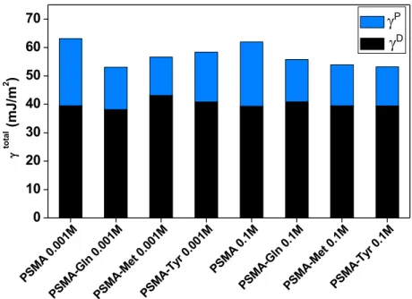
Table 3 shows the contact angles obtained using polar-non-polar solvent of water (qw), ethylene glycol (qE) and diiodomethane (qD), and the surface energy determined for the polyelectrolyte films. The surface energy values of all the polyelectrolyte films are relatively
similar (in a range of 44 to 56 mJ/m2) regardless of the amount of salt added, while the surface
energy values of the unmodified polyelectrolyte were slightly higher. The surface energy of
organic polymeric materials can be classified as low, medium and high. Low surface energy
levels were in the range of 10 to 30 mJ/m2. Medium surface energy is between 30-40 mJ/m2 and 2 42
in this work can be considered as having high surface energy. The relative contributions of the
components to surface energy (γ) are determined by the chemical composition of the surface.
The polar component (γP) is composed of several polar interactions that include hydrogen
bridges, dipole-dipole interaction and induction energy, while the dispersive component (γD) is
mainly due to London dispersion forces.
Figure 2 shows that the dispersive component of the surface energy of all the polymeric films
is much greater than the polar component, which suggests that the governing interactions are
London dispersion forces and to a lesser extent dipole-dipole interactions or hydrogen bridges.
This confirms the theory that polyelectrolytes are mainly adsorbed on the surface in a
hydrophobic/hydrophilic balance exposing of their side chains and the hydrophobic groups of the
main chain to the air, as suggested by the wettability of the films, such as shown Scheme 3.
PSM A 0. 001M PSM A-Gl n 0. 001M PSM A-M et 0.0 01M PSM A-Ty r 0.0 01M PSM A 0. 1M PSM A-Gl n 0. 1M PSM A-M et 0.1 M PSM A-Ty r 0.1 M 0 10 20 30 40 50 60 70 g tot al ( mJ /m 2 ) gP gD
Figure 2. Surface energy (γ) with the polar (γP) and dispersive components (γD) calculated for PSMA, PSMA-Gln, PSMA-Met and PSMA-Tyr films obtained at different ionic strengths
Scheme 3. Graphic representation of polyelectrolytes adsorbed onto amino-terminal surfaces
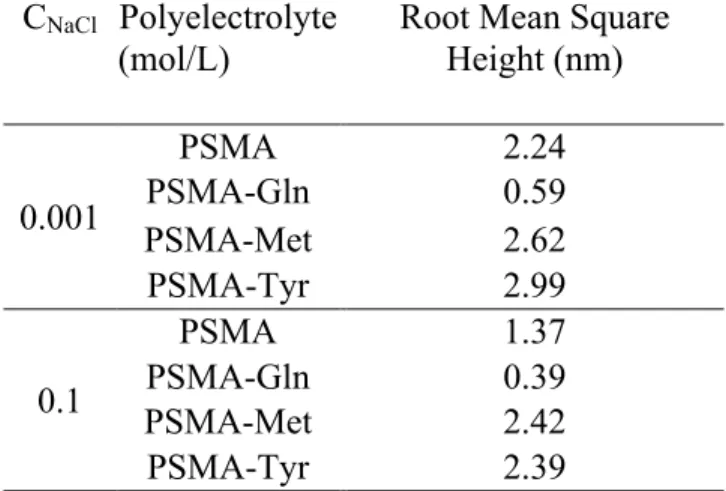
3.5. Polymeric Film Topography, Roughness and Surface Potential
Following the incubation, the grafted APTES silicon surfaces were removed from the
polyelectrolyte solution prepared at one of two levels of ionic strength; 0.001 and 0.1 M NaCl,
rinsed, and dried with N2 stream, and then immediately analyzed by the atomic force microscopy
(AFM). The AFM images in Figure 3 show the surface morphology of the polyelectrolyte films
on the APTES grafted silicon surfaces after incubation. Very homogeneous and regular polymer
deposits are observed in all cases and at both ionic strengths. Two kinds of morphology were
adopted, a well-defined globular nanostructure for PSMA-Met and PSMA-Tyr, and a densely
packed nanofibrous-like structure for PSMA-Gln. Ionic strength affected the nanostructures
forming polyelectrolyte surfaces. Although the surface morphology domains were preserved in
both cases, the higher ionic strength value caused a slight decrease in size of the nanostructures
that for all the electrolytes, surface roughness was greater with the lower ionic strength. At 1mM
NaCl, the shielding effect of the Na+ counter ion on the charged polyelectrolyte is low and
consequently there is a large number of charged groups in polymer network where electrostatic
repulsion takes place. These results in the polymer being more extended on the APTES grafted
silicon surface, with consequent increased surface roughness. These surface morphology and
roughness parameters are consistent with what is described above regarding polyelectrolyte film
wettability and surface energy.
Surface potential results using Kelvin force microscopy (frequency modulation) (KFM) for
PSMA, PSMA-Gln, PSMA-Met and PSMA-Tyr were respectively 81, 39, 23 and 14mV. These
results show higher surface potential values for PSMA and PSMA-Gln. The pKa1 and pKa2
values were respectively 2.9 and 6.4 for PSMA, and 4.3 and 8.2 PSMA-Gln. These values
indicate that at pH 4.0 there are groups, -COOH and –COO-, with the latter contributing to
electrostatic repulsion in the main and lateral chains of the polyelectrolyte. This behavior concurs
with the type of 2D nanostructure observed in the PSMA and PSMA-Gln films, which concurs
with the low RMS value obtained. It has been reported that an increase in the charge density of a
polyelectrolyte induces the adsorption of chains parallel to the surface because a more extended
shape is adopted that results in flatter films and a higher surface charge. 43 PSMA-Met and
PSMA-Tyr behaved differently where the carboxylic groups (protonated and deprotonated),
phenyl groups, -S- and –CH2- groups in side chain, generated less repulsion between the charged
segments. In this case, hydrophobic interactions and the hydrogen bridge are more important,
giving rise to globular nanostructures (3D) that contributed to increased surface roughness.
polyelectrolyte films derived from poly(maleic anhydride-alt-styrene) depends mainly on the
hydrophobic character of the polyelectrolyte side chain. 40
Table 4. RMS of the polymeric films obtained at different ionic strengths at pH 4.0
CNaCl Polyelectrolyte (mol/L)
Root Mean Square Height (nm) 0.001 PSMA 2.24 PSMA-Gln 0.59 PSMA-Met 2.62 PSMA-Tyr 2.99 0.1 PSMA 1.37 PSMA-Gln 0.39 PSMA-Met 2.42 PSMA-Tyr 2.39
Figure 3. AFM images (1.5 µm x 1.5 µm) of the polyelectrolytes adsorbed onto a solid surface at
ionic strength of 0.001 and 0.1 mol/L NaCl, at pH 4.0. The inset (0.5 µm x 0.5 µm) on
3.6. Cell Response to PSMA Modified Substrates
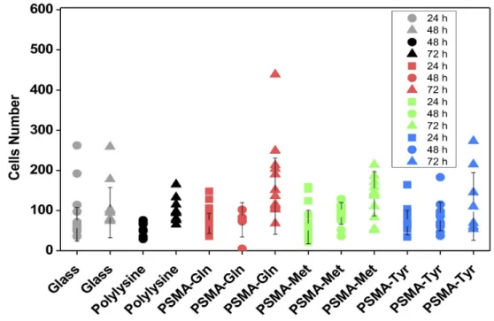
25,000 SH-SY5Y neuroblastoma cells were plated on UV-light sterilized PSMA coated silicon
surfaces (1 cm2), with aforementioned amino acid functionalization. They were compared to
cells growing on glass coverslips (in the presence or absence of polylysine). Random fields were
photographed under phase contrast microscopy and cells per field was counted in a double-blind
fashion at 24, 48 and 72 hours. As can be seen in Figure 4, the cell number per field was
consistently and proportionally higher at hour 27 hour/doubling time reported for these cells, 44
suggesting that SH-SY5Y cell proliferation was not significantly affected by the different PSMA
modified substrates.
Figure 4. SH-SY5Y neuroblastoma cell proliferation is not impaired by different PSMA
modified substrates. 25,000 SH-SY5Y neuroblastoma cells were plated and cultured for 24, 48
and 72 hours on different substrates (glass, polylysine-coated glass, PSMA-Gln, PSMA-Met,
number was determined in a double-blind fashion. Cell proliferation was not altered in response
to the different substrates.
The functionalized PSMA substrates elicited different responses from SH-SY5Y
neuroblastoma cells, likely reflecting the differences in hydrophilicity/hydrophobicity of the
exposed surface. As can be observed in Figure 5, SH-SY5Y cells spread easily over acid-washed
glass coverslips in the absence or presence of a polylysine coating. In contrast, amino-acid
functionalized PSMA-polyelectrolytes significantly diminished the spread of cells compared to
standard substrates. Only PSMA-Met appeared to induce an increase in lipid peroxidation, which
could be interpreted as a stress signal or a change in cell metabolism (Figures 5B and C
respectively). Some works have demonstrated the mechanistic aspects of the oxidation by free
radicals of sulfur in methionine, which causes lipid peroxidation and formation of reactive
oxygen species in neuronal systems. 45 This could suggest that the amino acid residue of
methionine in PSMA-Met could have the same effect on neuroblastoma cells. Nevertheless, the
treatments did not affect cell viability, as measured by the MTT assay (Figure 5D), which
Figure 5. The area covered by SH-SY5Y cells on polyelectrolyte-modified substrates were
reduced (B), but without altering cell viability. SH-SY5Y cells were plated on different
substrates (A) and cultured for 24 hours (37ºC, 5% CO2). Cells were fixed and stained for
immunofluorescence to determine the presence of 4-HNE, a membrane lipid peroxidation marker
that indicates damage to cell membranes in response to experimental toxicity (C). Cells
identified and counted by DAPI-nuclear staining (405 nm, CYAN) were imaged for 4-HNE and
found to be significantly fewer in number than the controls. Only cells plated on PSMA-Met
showed a significant increase in lipid peroxidation, which indicates increased oxidative damage.
Nevertheless, cell viability, as determined by MTT reduction, was not altered, even when cells
Figure 6. SH-SY5Y cultured on (a) glass, (b) polylysine, (c) PSMA-Gln, (d) PSMA-Met and (e)
PSMA-Tyr. SH-SY5Y cells responded to PMSA-Met substrates with significant process
extension and cell shape remodeling. Control cells a and b, glass and polylysine, respectively
had fusiform (two processes) and pyramid (three process) shapes characteristic of
undifferentiated neuroblastoma cells. Cells grown on PSMA-Met (d) had a stellate shape, with 5
or more processes, many of which extended over 5 µm from the cell body. Cells grown on PSMA-Gln (c) present fewer and shorter processes.
As can be seen in Figure 6, differences in the shape of SH-SY5Y cells reflects their sensitivity
distinct PMSA-substrate characteristics. Control cells (glass and polylysine, respectively)
consistently present fusiform (two processes) or pyramidal shapes (three processes) characteristic
of undifferentiated SH-SY5Y. These cells also present well defined actin-rich areas that are
consistent with focal adhesion. When cultured on PSMA-Met, SH-SY5Y cells present a stellate
contrast, when seeded on PSMA-Gln, SH-SY5Y cells have a round morphology, with fewer and
shorter processes. The shape suggests that these cells are in a higher state of proliferation, an
effect that did not appear to be significant when cell proliferation was assessed (Figure 4). While
suggestive, these results do not shed light on whether PSMA-Gln substrates inhibit
neuroblastoma cell proliferation or PSMA-Met substrates induce cell differentiation.
Figure 7. SEM images of SH-SY5Y cell cultures after 24 h on (a1, a2, a3) PMSA, (b1, b2, b3)
Figure 7 shows the morphology of the SH-SY5Y cells cultured on the surface of PMSA,
PMSA-Gln, PMSA-Met and PMSA-Tyr. The cells cultured on PSMA-Met have a well-extended
morphology, characterized by a much larger cell area than that of cells cultured on PSMA-Gln
and PSMA-Tyr, as can be seen in Figure 6. Cells on the surface of unmodified PSMA and
PSMA-Tyr have a spindle-like morphology characterized by a small cell area and a high aspect
ratio. The well-extended morphology that predominates on the surface of PSMA-Met is
associated with its moderate hydrophilicity and topography. Although the hydropathic index of
PSMA-Met is the most positive (hydrophobic), the orientation and conformation of PSMA-Met
allows for obtaining a surface of moderate hydrophilicity associated with the heterogeneity of the
hydrophilic domains due to its polar (-COOH and -NH2) groups in its main and lateral chains,
However, the low hysteresis value obtained for PSMA-Met suggests a greater chemical
homogeneity of its surface. According to the results of immunofluorescence 4-HNE, a greater
response in lipid peroxidation associated with sulfur group present in methionine, would indicate
that the chemical nature of the surface of PSMA-Met would be given mainly by the exposure of
the groups -S- towards the air, which would participate in the process of oxidation by free
radicals. At the same time, PSMA-Met substrates affected cell spreading, increasing the
formation of cell extensions in a manner reminiscent of cell differentiation. likely reflecting a
cellular response to the low value of hysteresis angle of the exposed surface. Homogeneous
overall structure probably provides cells with more attachment sites, thus improving their
adhesion to the substrate. While suggestive, these results do not shed light on whether
PSMA-Gln substrates inhibit neuroblastoma cell proliferation or PSMA-Met substrates induce cell
differentiation. Some works have related the optimal adhesion of neuronal cells with a greater
has been related to the ability of the surface to adsorb proteins from the serum, 47,48 what would
facilitate the cell adhesion. Another factor to consider could be related to the roughness of the
surface, it has been reported that rougher surfaces achieve a positive response in the cellular
adhesion of SH-SY5Y, while other authors have shown that roughness can play minor functions
in the response mobile. 49,50 In our case, the roughness of the surface is determined by the effects
of repulsion or formation of aggregates given by the presence of charged groups (-COO- group)
as well as the chemical functionality of polyelectrolytes. The contribution of these effects is
reflected in the surface potential values, where the highest values are associated with repulsive
effects (PSMA and PSMA-Gln) while the lowest values are related to hydrophobic or hydrogen
bridge interactions. For this last case, the associative interactions and roughness would promote
the adhesion of SH-SY5Y. In the case of surface energy, the similarity of values between PSMA-Gln, PSMA-Met and PSMA-Tyr would indicate that surface energy is not a determinant
physicochemical parameter in the behavior of cell adhesion.
Finally, polyelectrolyte films modified with amino acids of different hydropathic indices are
presented as a simple and low-cost platform, where their properties of wettability, roughness,
surface energy and surface potential can be modulated through their chemical functionalization,
as well as, the control of the pH and the ionic strength of the medium. The ability to modulate
these properties has a direct effect on cell adhesion behavior by observing significant changes in
cell processes. The advantage of modulating on simple way the surface properties has not been
CONCLUSIONS
The adsorption behavior of polyelectrolytes functionalized with amino acids on an amino
acid-terminated surface depends on the chemical nature of the polyelectrolyte side chain, its ionic
strength and the pH level of the solution. The largest quantity of polyelectrolytes adsorbed was at
pH 4.0 and high ionic strength, with more polyelectrolytes adsorbed by the glutamine and
tyrosine films than by the methionine film. The contact angle measurements of the
polyelectrolyte films showed that wettability at pH 4.0 is governed by the
hydrophilic/hydrophobic balance given by hydrophobic domains of the main polyelectrolyte
chain, and by the nature of amino acid moiety, while that at pH 7.0 there was an evident
contribution of the effect of the hydropathic amino acid index on polyelectrolyte wettability. The
AFM images of polyelectrolyte surfaces showed two kinds of morphology, a well-defined
globular nanostructure for PSMA-Met and PSMA-Tyr, and a densely packed nanofibrous-like
structure for Gln. Cell adhesion assays showed that SH-SY5Y cells cultured on
PSMA-Met had a well-extended morphology, characterized by much larger stellate shaped cells, with
five or more actin-rich thin processes, while SH-SY5Y cells that were seeded on PSMA-Gln and
PSMA-Tyr have a round morphology, with fewer and shorter processes.
These results indicate that it is possible to modulate the surface characteristics of
polyelectrolyte films based on their chemical functionality and environmental parameters such as
pH and ionic strength, in order to evaluate their effect on cell adhesion.
In conclusion, films obtained from polyelectrolytes functionalized with amino acids can be a
simple and low-cost platform for cell adhesion control aimed at developing biomaterials with
ASSOCIATED CONTENT
The following files are available free of charge.
Supporting Information: FT-IR spectra of: PSMA and PSMA-Gln, PSMA-Met and PSMA-Tyr. 13C-NMR spectra of: PSMA, PSMA-Gln, PSMA-Met and PSMA-Tyr (PDF).
AUTHOR INFORMATION Corresponding Author
† Marcela D. Urzúa, E-mail: [email protected]
† Laura Tamayo, E-mail: [email protected]
Present Addresses
†Departamento de Química, Facultad de Ciencias, Universidad de Chile, Las Palmeras 3425,
Casilla 653, Santiago 7800003, Chile.
Author Contributions
The manuscript was written through contributions of all authors. All authors have given
approval to the final version of the manuscript. These authors contributed equally. M.S. Leal,†
X. Briones,† V. Villalobos,◊ Y. Queneau,‡ A. Leiva,§ H.E. Ríos,† J. Pavez,| C.P. Silva,| C.
Carrasco,|| Andrónico Neira-Carrillo,∞ A. Roth,|| L. Tamayo,*† M.D. Urzúa*†
Funding Sources
FONDECYT 1151221 and 1100240, FONDECYT Iniciación grant 11160230, ANILLO ACT-1412, PAI-CONICYT 79170015, FONDEQUIP EQM160036.
ACKNOWLEDGMENT
The authors are grateful to FONDECYT 1151221, FONDECYT 1100240, ANILLO
ACT-1412 Project and FONDECYT Iniciación grant 11160230, PAI-CONICYT 79170015, and
FONDEQUIP EQM160036.
REFERENCES
(1) Waugh, D. G.; Toccaceli, C.; Gillett, A. R.; Ng, C. H.; Hodgson, S. D.; Lawrence, J.
Surface Treatments to Modulate Bioadhesion: A Critical Review. Rev. Adhes. Adhes. 2016, 4,
69-103. https://doi.org/10.7569/RAA.2016.097304
(2) Guo, S.; Zhu. X,; Li, M.; Shi, L., Ong, J.L.T.; Jańczewski, D.; Neoh, K.G. Parallel Control
over Surface Charge and Wettability Using Polyelectrolyte Architecture: Effect on Protein
Adsorption and Cell Adhesion. ACS Appl. Mater. Interfaces. 2016, 8, 30552-30563.
https://doi.org/10.1021/acsami.6b09481
(3) Khan, S.; Newaz, G. A Comprehensive Review of Surface Modification for Neural Cell
Adhesion and Patterning. J. Biomed. Mater. Res. A. 2010, 93, 1209-1224.
https://doi.org/10.1002/jbm.a.32698
(4) Lee, I. Molecular Self-Assembly: Smart Design of Surface and Interface Via Secondary
Molecular Interactions. Langmuir. 2013, 29, 2476-2489. https://doi.org/10.1021/la304123b
(5) Hardy, A.; Seguin, C.; Brion, A.; Lavalle, P.; Schaaf, P.; Fournel, S.; Bourel-Bonnet, L.;
Frisch, B.; De Giorgi, M. β-Cyclodextrin-Functionalized Chitosan/Alginate Compact
Polyelectrolyte Complexes (CoPECs) as Functional Biomaterials with Anti-Inflammatory
Properties. ACS Appl. Mater. Interfaces. 2018, 10, 29347-29356. https://doi.org/10.1021/acsami.8b09733
(6) Sanandiya, N. D.; Lee, S.; Rho, S.; Lee, H.; Kim, I. S.; Hwang, D. S. Tunichrome-inspired
Pyrogallol Functionalized Chitosan for Tissue Adhesion and Hemostasis. Carbohydr. Polym.
2019, 208, 77-85. https://doi.org/10.1016/j.carbpol.2018.12.017
(7) Hwang, M. P.; Ding, X.; Gao, J.; Acharya, A. P.; Little, S. R.; Wang, Y. A Biocompatible
Betaine-Functionalized Polycation for Coacervation. Soft Matter. 2018, 14, 387-395.
https://doi.org/10.1039/C7SM01763D
(8) Gao, Y.; Xu, Z.; Chen, S.; Gu, W.; Chen, L.; Li, Y. Arginine-Chitosan/DNA Self-Assemble
Nanoparticles for Gene Delivery: In Vitro Characteristics and Transfection Efficiency. Int. J.
Pharm. 2018, 359, 241-246. https://doi.org/10.1016/j.ijpharm.2008.03.037
(9) Lv, H. X.; Zhang, Z. H.; Wang, X. P.; Cheng, Q. Q.; Wang, W.; Huang, X. H.; Zou, J. P.;
Zhang, Q, Hou, L. L.; Huo, W. A Biomimetic Chitosan Derivates: Preparation, Characterization
and Transdermal Enhancement Studies Of N-Arginine Chitosan. Molecules. 2011, 16,
6778-6790. https://doi.org/10.3390/molecules16086778
(10) Park, H.; Choi, B.; Nguyen, J.; Fan, J.; Shafi, S.; Klokkevold, P.; Lee, M. Anionic
Carbohydrate-Containing Chitosan Scaffolds for Bone Regeneration. Carbohydr. Polym. 2013,
97, 587-596. https://doi.org/10.1016/j.carbpol.2013.05.023
(11) Ho, M. H.; Wang, D. M.; Hsieh, H. J.; Liu, H. C.; Hsien, T. Y.; Lai, J. Y.; Hou, L. T.
Preparation and Characterization of RGD-immobilized Chitosan Scaffolds. Biomaterials.
2005, 26, 3197-3206. https://doi.org/10.1016/j.biomaterials.2004.08.032
(12) Kim, S.; Cui, Z. K.; Fan, J.; Fartash, A.; Aghaloo, T. L.; Lee, M. Photocrosslinkable
Chitosan Hydrogels Functionalized with the RGD Peptide and Phosphoserine to Enhance
(13) Tu, H. P.; Lee, X. Q.; Lin, C. Y.; Shen, E. C.; Chen, Y. T.; Fu, E.; Chin, Y. T. Enhanced
Attachment and Growth of Periodontal Cells on Glycine-Arginine-Glycine-Aspartic Modified
Chitosan Membranes. J. Med. Sci. 2016, 36, 137. https://doi.org/10.4103/1011-4564.188898
(14) LogithKumar, R.; KeshavNarayan, A.; Dhivya, S.; Chawla, A.; Saravanan, S.;
Selvamurugan, N. A Review of Chitosan and Its Derivatives in Bone Tissue
Engineering. Carbohydr. Polym. 2016, 151, 172-188. https://doi.org/10.1016/j.carbpol.2016.05.049
(15) Jones, D. S.; Laverty, T. P.; Morris, C.; Andrews, G. P. Statistical Modelling of the
Rheological and Mucoadhesive Properties of Aqueous Poly (Methylvinylether-Co-Maleic Acid)
Networks: Redefining Biomedical Applications and the Relationship Between Viscoelasticity
and Mucoadhesion. Colloids Surf. B. 2016, 144, 125-134. https://doi.org/10.1016/j.colsurfb.2016.03.008
(16) Pompe, T.; Zschoche, S.; Herold, N.; Salchert, K.; Gouzy, M. F.; Sperling, C.; Werner, C.
Maleic Anhydride Copolymers a Versatile Platform for Molecular Biosurface
Engineering. Biomacromolecules. 2003, 4, 1072-1079. https://doi.org/10.1021/bm034071c
(17) Kyte, J.; Doolittle, R. F. A Simple Method for Displaying the Hydropathic Character of a
Protein. J. Mol. Biol. 1982, 157, 105-132. https://doi.org/10.1016/0022-2836(82)90515-0
(18) Kovalevich J, Langford D. Considerations for the use of SH-SY5Y neuroblastoma cells in
neurobiology. Methods Mol Biol. 2013;1078:9-21. doi: 10.1007/978-1-62703-640-5_2.
(19) Simpson, P. B.; Bacha, J. I.; Palfreyman, E. L.; Woollacott, A. J.; McKernan, R. M.;
Kerby, J. Retinoic Acid-evoked Differentiation of Neuroblastoma Cells Predominates Over
Growth Factor Stimulation: An Automated Image Capture and Quantitation Approach to
(20) Morelli, S.; Piscioneri, A.; Messina, A.; Salerno, S.; Al-Fageeh, M. B.; Drioli, E.; Bartolo, L. D. (2015). Neuronal Growth and Differentiation on Biodegradable Membranes. J. Tissue Eng.
Regen. Med. 2015, 9, 106-117. https://doi.org/10.1002/term.1618
(21) BC Trivedi, BM Culbertson. Maleic anhydride, Plenum Press, New York (1982)
(22) Briones, X.G.; Encinas, M.V.; Petri, D.F.S.; Pavez, J.E.; Tapia, R.A.; Yazdani-Pedram
M.; Urzúa, M.D. Adsorption Behavior of Hydrophobically Modified Polyelectrolytes onto
Amino- or Methyl-Terminated Surfaces. Langmuir. 2011, 27, 13524-13532. https://doi.org/
10.1021/la2025632
(23) Ohno, N.; Nitta, K.; Makino, S.; Sugai, S. Conformational Transition of the Copolymer of
Maleic Acid and Styrene in Aqueous Solution. J. Polym. Sci. B. 1973, 11, 413-425.
https://doi.org/10.1002/pol.1973.180110302
(24) Pretsch, E.; Buehlmann, P.; Affolter, C.; Pretsch, E.; Bhuhlmann, P.; Affolter, C. 2000.
Structure Determination of Organic Compounds (p. 108). Berlin: Springer-Verlag.
(25) Siqueira Petri, D. F.; Wenz, G., Schunk, P.; Schimmel, T. An Improved Method for the
Assembly of Amino-Terminated Monolayers on SiO2 and the Vapor Deposition of Gold
Layers. Langmuir. 1999, 15, 4520-4523. https://doi.org/ 10.1021/la981379u
(26) Motschmann, H.; Stamm, M.; Toprakcioglu, C. Adsorption Kinetics of Block Copolymers
from a Good Solvent: A Two-Stage Process. Macromolecules. 1991, 24, 3681-3688.
https://doi.org/10.1021/ma00012a032
(27) De Feijter, J.; Benjamins, D. J.; Veer, F. A. Ellipsometry as a Tool to Study the
Adsorption Behavior of Synthetic and Biopolymers at the Air–Water
(28) Azzam, R. M. A.; Bashara, N. M. Ellipsometry and Polarized Light, North-Holland Publ. Co., Amsterdam 1977. https://doi.org/10.1063/1.2994821
(29) Urzúa, M.D.; Briones, X.G.; Carrasco, L.P.; Encinas M.V.; Petri, D.F.S. Adsorption of
Anionic Amphiphilic Polyelectrolytes onto Amino-Terminated Solid Surfaces. Polymer. 2010,
51, 3445-3452. https://doi.org/10.1016/j.polymer.2010.05.054
(30) Wu, S. (1971). Calculation of Interfacial Tension in Polymer Systems. In Journal of
Polymer Science Part C: Polymer Symposia (Vol. 34, No. 1, pp. 19-30). New York: Wiley
Subscription Services, Inc., A Wiley Company. https://doi.org/10.1002/polc.5070340105
(31) Wu S. (1982) Polymer Interface and Adhesion: Taylor & Francis; 630 pp.
(32) Kosaka, P. M.; Kawano, Y.; Petri, D. F. S. Dewetting and Surface Properties of Ultrathin
Films of Cellulose Esters. J. Colloid and Interface Sci. 2007, 316, 671-677.
https://doi.org/10.1016/j.jcis.2007.07.058
(33) Mosmann, T. Rapid Colorimetric Assay for Cellular Growth and Survival: Application to
Proliferation and Cytotoxicity Assays. J. Immunol. Methods. 1983, 65, 55-63.
https://doi.org/10.1016/0022-1759(83)90303-4
(34) Khan, F.; Moinuddin, Mir A.R.; Islam S.; Alam K.; Ali A. Immunochemical Studies on
HNE-Modified HSA: Anti-HNE-HSA Antibodies as a Probe for HNE Damaged Albumin in
SLE. Int J Biol Macromol. 2016 May;86:145-54. https://doi.org/10.1016/j.ijbiomac.2016.01.053.
Epub 2016 Jan 19. PubMed PMID: 26800898.
(35) Abcam.com. (2019). Immunocytochemistry and immunofluorescence protocol | Abcam.
[online] Available at:
(36) Preparation of Slides and Coverslips for Microscopy. Andrew H. Fischer, Kenneth A.
Jacobson, Jack Rose and Rolf Zeller Cold Spring Harb Protoc 2008.
https://doi.org/10.1101/pdb.prot4988
(37) Van de Steeg, H.G.M.; Cohen Stuart M.A.; De Keizer A.; Bijsterbosch, B.H.
Polyelectrolyte Adsorption: A Subtle Balance of Forces. Langmuir. 1992, 8, 2538-2546.
https://doi.org/10.1021/la00046a030
(38) Fujimoto, J.; Petri, D.F.S. Adsorption Behavior of Carboxymethylcellulose on
Amino-Terminated Surfaces. Langmuir, 2001, 17,56-60. https://doi.org/10.1021/la000731c
(39) Fu, J., Schlenoff, J. B. Driving Forces for Oppositely Charged Polyion Association in
aqueous Solutions: Enthalpic, Entropic, but not Electrostatic. J. Am. Chem. Soc. 2016, 138,
980-990. https://doi.org/10.1021/jacs.5b11878
(40) Briones, X. G.; Urzúa, M. D.; Ríos, H. E.; Espinoza-Beltrán, F. J.; Dabirian, R.;
Yazdani-Pedram, M. Thin Films of Amphiphilic Polyelectrolytes. Soft Materials Characterized by Kelvin
Probe Force Microscopy. Polymer. 2013, 54, 5733-5740.
https://doi.org/10.1016/j.polymer.2013.07.066
(41) Guzmán, E., Ortega, F., Baghdadli, N., Luengo, G. S., Rubio, R. G. Effect of the
molecular structure on the adsorption of conditioning polyelectrolytes on solid substrates.
Colloid Surf. A. 2011, 375, 209-218. https://doi.org/10.1016/j.colsurfa.2010.12.012
(42) Wake, W. C. (1988). Adhesion and Adhesives: Science and Technology. AJ Kinloch,
Chapman and Hall, London.
(43) Llamas, S.; Guzman, E.; Ortega, F.; Baghdadli, N.; Cazeneuve, C.; Rubio, R. G.; Luengo,
Physico-Chemical Approach to a Cosmetic Challenge. Adv. Colloid Interface Sci. 2015, 222,
461-487. https://doi.org/10.1016/j.cis.2014.05.007
(44) Kovalevich, J.; Langford, D. (2013). Considerations for the Use of SH-SY5Y
Neuroblastoma Cells in Neurobiology. In Neuronal Cell Culture (pp. 9-21). Humana Press,
Totowa, NJ. https://doi.org/10.1007/978-1-62703-640-5_2
(45) Butterfield, D. A., Kanski, J. Methionine Residue 35 is Critical for the Oxidative Stress
and Neurotoxic Properties of Alzheimer’s Amyloid Β-Peptide 1–42. Peptides. 2002, 23,
1299-1309. https://doi.org/10.1016/S0196-9781(02)00066-9
(46) Lee, S. J.; Khang, G.; Lee, Y. M.; Lee, H. B. The Effect of Surface Wettability on
Induction and Growth of Neurites from the PC-12 Cell on a Polymer Surface. J. Colloid
Interface Sci. 2003, 259, 228– 235. https://doi.org/10.1016/S0021-9797(02)00163-7
(47) Wei, J.; Igarashi, T.; Okumori, N.; Igarashi, T.; Maetani, T.; Liu, B.; Yoshinari, M.
Influence of Surface Wettability on Competitive Protein Adsorption and Initial Attachment of
Osteoblasts. Biomed. Mater. 2009, 4, 45002. https://doi.org/10.1088/1748-6041/4/4/045002
(48) Arima, Y.; Iwata, H. Effect of Wettability and Surface Functional Groups on Protein
Adsorption and Cell Adhesion Using Well-Defined Mixed Self-Assembled Monolayers.
Biomaterials. 2007, 28, 3074– 3082. https://doi.org/10.1016/j.biomaterials.2007.03.013
(49) Prévôt, M. E.; Bergquist, L. E.; Sharma, A.; Mori, T.; Gao, Y.; Bera, T.; Zhu, C.; Leslie,
M. T.; Cukelj, R.; Korley, L. T. J.; Freeman, E. J.; McDonough J. A.; Clements, R. J.; Hegmann,
E. (2017, August). New Developments in 3D Liquid Crystal Elastomers Scaffolds for Tissue
Engineering: From Physical Template to Responsive Substrate. In Liquid Crystals XXI (Vol.
10361, p. 103610T). International Society for Optics and Photonics.
(50) Genchi, G. G., Ceseracciu, L., Marino, A., Labardi, M., Marras, S., Pignatelli, F.,
Bruschini, L.; Matolli, V.; Ciofani, G. (2016). P(VDF-TrFE)/BaTiO3 Nanoparticle Composite Films Mediate Piezoelectric Stimulation and Promote Differentiation of SH-SY5Y Neuroblastoma Cells. Adv. Healthc. Mater. 2016, 5, 1808-1820. https://doi.org/10.1002/adhm.201600245