HAL Id: hal-01912302
https://hal.umontpellier.fr/hal-01912302
Submitted on 5 Nov 2018
HAL is a multi-disciplinary open access
archive for the deposit and dissemination of
sci-entific research documents, whether they are
pub-lished or not. The documents may come from
teaching and research institutions in France or
abroad, or from public or private research centers.
L’archive ouverte pluridisciplinaire HAL, est
destinée au dépôt et à la diffusion de documents
scientifiques de niveau recherche, publiés ou non,
émanant des établissements d’enseignement et de
recherche français ou étrangers, des laboratoires
publics ou privés.
drug resistance in Mycobacterium tuberculosis
Quang Nguyen, Lucie Contamin, Thi Nguyen, Anne-Laure Bañuls
To cite this version:
Quang Nguyen, Lucie Contamin, Thi Nguyen, Anne-Laure Bañuls. Insights into the processes that
drive the evolution of drug resistance in Mycobacterium tuberculosis. Evolutionary Applications,
Blackwell, 2018, 11 (9), pp.1498 - 1511. �10.1111/eva.12654�. �hal-01912302�
1498
|
wileyonlinelibrary.com/journal/eva Evolutionary Applications. 2018;11:1498–1511.1 | INTRODUCTION
Tuberculosis (TB), an infectious airborne disease mainly caused by Mycobacterium tuberculosis, is one of the world’s deadliest infectious diseases. In 2015, TB affected approximately 10.4 million people and killed 1.8 million of them (WHO, 2016). The current recommended treatment for new patients with drug- susceptible TB is a 6- month regimen using a combination of four- first- line anti- TB drugs: isoniazid, rifampicin, ethambutol and pyrazinamide (WHO, 2016). This effective regimen was developed in the early 1970s and showed a high cure rate, higher than 98%, in clinical trials (STS/BMRC, 1988). The regimen
has not been changed, but now, its global treatment success rate is of 83% for new patients with TB (WHO, 2016). Indeed, its use for almost five decades has led to the emergence of first- line drug resistance. Multidrug- resistant (MDR, Box 1) TB is the most problematic first- line drug- resistant form (Nachega & Chaisson, 2003). The treatment of MDR TB requires second- line drugs that are more expensive and toxic than first- line drugs. Nevertheless, during the last 15 years, patients with extensively drug- resistant (XDR, Box 1) TB have been reported in 105 countries. In 2015, 480,000 patients with MDR TB were reported worldwide and approximately 10% of them developed XDR TB (WHO, 2016).
Received: 24 September 2017
|
Revised: 25 May 2018|
Accepted: 27 May 2018 DOI: 10.1111/eva.12654R E V I E W S A N D S Y N T H E S E S
Insights into the processes that drive the evolution of drug
resistance in Mycobacterium tuberculosis
Quang Huy Nguyen
1,2,3| Lucie Contamin
2,3,4| Thi Van Anh Nguyen
4* | Anne-Laure
Bañuls
2,3,4*
This is an open access article under the terms of the Creative Commons Attribution License, which permits use, distribution and reproduction in any medium, provided the original work is properly cited.
© 2018 The Authors. Evolutionary Applications published by John Wiley & Sons Ltd *Both authors equally contributed to the work.
1Department of Pharmacological, Medical and Agronomical Biotechnology, University of Science and Technology of
Hanoi, Vietnam Academy of Science and Technology (VAST), Hanoi, Vietnam 2Institute of Research for
Development, UMR MIVEGEC (CNRS-IRD-University of Montpellier), Montpellier, France
3LMI Drug Resistance in South East Asia (LMI DRISA), University of Science and Technology of Hanoi, Vietnam Academy of Science and Technology (VAST), Hanoi, Vietnam
4Department of Bacteriology, National Institute of Hygiene and Epidemiology (NIHE), Hanoi, Vietnam
Correspondence
Quang Huy Nguyen, Department of Pharmacological, Medical and Agronomical Biotechnology, University of Science and Technology of Hanoi (USTH), Vietnam Academy of Science and Technology (VAST), 18 Hoang Quoc Viet, Cau Giay, 10000 Hanoi, Vietnam.
Email: nguyen-quang.huy@usth.edu.vn
Abstract
At present, the successful transmission of drug- resistant Mycobacterium tuberculosis, including multidrug- resistant (MDR) and extensively drug- resistant (XDR) strains, in human populations, threatens tuberculosis control worldwide. Differently from many other bacteria, M. tuberculosis drug resistance is acquired mainly through mutations in specific drug resistance- associated genes. The panel of mutations is highly diverse, but depends on the affected gene and M. tuberculosis genetic background. The vari-ety of genetic profiles observed in drug- resistant clinical isolates underlines different evolutionary trajectories towards multiple drug resistance, although some mutation patterns are prominent. This review discusses the intrinsic processes that may influ-ence drug resistance evolution in M. tuberculosis, such as mutation rate, drug resistance- associated mutations, fitness cost, compensatory mutations and epistasis. This knowledge should help to better predict the risk of emergence of highly resist-ant M. tuberculosis strains and to develop new tools and strategies to limit the devel-opment and spread of MDR and XDR strains.
K E Y W O R D S
compensatory mutation, drug resistance mutation, epistasis, evolution, fitness cost, multidrug-resistant tuberculosis, mycobacterium tuberculosis
Mycobacterium tuberculosis is a highly clonal bacteria (ab-sence of recombination) with an extremely conserved genome and a long history of co- evolution with humans (Bañuls, Sanou, Nguyen, & Godreuil, 2015; Comas et al., 2013). At present, M. tu-berculosis consists of seven phylogenetic lineages associated with particular geographic regions and differing, among others, in vir-ulence, biological fitness and propensity to acquire drug resis-tance (Comas et al., 2013; Merker et al., 2015; Stucki et al., 2016; Warner, Koch, & Mizrahi, 2015). Besides the lineage- specific bio-logical characteristics, the remarkable capacity of adaptation and the variety of extrinsic and intrinsic processes contribute specifi-cally to the emergence and spread of highly drug- resistant strains (Coscolla & Gagneux, 2014; Müller, Borrell, Rose, & Gagneux, 2013; Trauner, Borrell, Reither, & Gagneux, 2014; Warner et al., 2015).
As example of intrinsic mechanisms, epistasis (Box 1), which plays an important role in the evolution of organisms in gen-eral, is also known to drive the evolution of antibiotic resistance (Lehner, 2011). Epistasis can occur between mutations in the same gene or in different genes and can lead to negative or posi-tive effects (Box 1) (Lehner, 2011; Wong, 2017). This mechanism may generate the combination of a set of alleles from differ-ent loci, also called linkage disequilibrium (Box 1). The spread of these sets of co- adapted alleles in the population is then favoured by the clonal reproductive mode of M. tuberculosis. Regarding the drug resistance, many studies underline that epi-static interactions can occur between different drug resistance mutations, between drug resistance mutations and compensa-tory mutations and/or the genetic background of the organism (Borrell et al., 2013; Lehner, 2011; Trindade et al., 2009; Wong, 2017).
In this review, we focus on the intrinsic factors influencing the drug resistance evolution in M. tuberculosis, particularly the mutation rate, drug resistance- associated mutations, fitness cost of resistance mutations, compensatory mutations and epistasis (Box 1). An understanding of the role of these intrinsic factors is essential to get insights into the evolutionary trajectories of drug resistance in M. tuberculosis and to help identifying the best strategies to control the emergence and spread of highly drug- resistant strains.
2 | MUTATION R ATE AND DRUG
RESISTANCE ACQUISITION
Mycobacterium tuberculosis is characterized by a low mutation rate (about 2 × 10⁻¹⁰ mutations/bp/generation) (Ford et al., 2011), with an estimated evolutionary rate of 0.4—0.5 single nucleotide poly-morphisms (SNPs)/genome/year and a divergence rarely higher than five SNPs in 3 years (Roetzer et al., 2013; Walker et al., 2013). Despite this low mutation rate, the number of drug resistant, espe-cially MDR and XDR TB cases, due to the acquisition of mutations, is progressively increasing worldwide.
Besides innate drug resistance mechanisms (for instance, the specific characteristics of the cell envelope of M. tuberculosis and the active drug efflux mechanism) (Sarathy, Dartois, & Lee, 2012), chromosomal mutations are the major mechanism of drug resistance acquisition in M. tuberculosis (Table 1) (Sandgren et al., 2009; Zhang & Yew, 2015). The rate for evolution of drug resistance to major first- and second- line drugs ranges from 10−6 to 10−10 mutations/bacterial cell/generation (McGrath, Gey van Pittius, van Helden, Warren, & Warner, 2014). This rate might also be affected by the drug concen-tration in the medium, the drug resistance profile of the strain and its genetic background (Ford et al., 2013; McGrath et al., 2014).
As the genes responsible for resistance to the various anti- TB drugs are generally not functionally related, the risk of emergence of spontaneous double, triple and quadruple drug- resistant mutants is theoretically extremely low, ranging from about 10−14 mutants (for isoniazid and rifampicin) to 10−25 mutants per population (for isoniazid, rifampicin, ethambutol and pyrazinamide). Furthermore, clinical data estimated that the population size in active pulmonary disease ranges between 107 and 1010 bacilli (Nachega & Chaisson, 2003), thus the risk of spontaneous drug resistance- associated mu-tations should be very low. In addition, drug resistance- associated mutations may impose a fitness cost because they target essential cell biological functions (Melnyk, Wong, & Kassen, 2015). Therefore, in theory the chance of drug resistance acquisition should be negli-gible when the four effective first- line drugs are used in combina-tion. However, by mathematical modelling, Colijn, Cohen, Ganesh, & Murray (2011) estimated that the probability of acquisition of re-sistance to both isoniazid and rifampicin is as high as 10−4 to 10−5 mutants/bacterial population. Similar to that, in a clinical study, Gao et al. (2016) found that 3.7% (62/1671) of pan- susceptible clinical isolates (susceptible to isoniazid, rifampicin, streptomycin and eth-ambutol) acquired different resistance patterns during the standard short- course chemotherapy according to the Directly Observed Treatment (DOT) guidelines. Among the 62 strains with acquired drug resistance, approximately 10% were resistant to four drugs, 22.6% to three drugs, 21% to two drugs and the remaining 46.8% were mono- drug resistant. These data underline that multiple drug resistance acquisition emerges at higher rate under strong drug se-lection pressure than theoretically predicted.
Regarding the genetic background, Ford et al. (2013) demon-strated that overall, the mutation rates for drug resistance acquisi-tion are higher in M. tuberculosis lineage 2 (East Asia, mainly Beijing strains) than in lineage 4 (Euro- American). In addition, the authors also demonstrated that the risk of de novo MDR acquisition before treatment is higher (approximately 22- fold) in macaques infected with M. tuberculosis strains of lineage 2 than in animals infected with lineage 4 strains. These data are consistent with the high drug resistance potential of lineage 2 observed in many epidemiological studies (Casali et al., 2014; Merker et al., 2015). Besides the effect of genetic background, Ford et al. (2013) also showed that differ-ences in target size (defined as the number of resistance- associated mutations) contribute to the two- to thirty- five- fold differences in rifampicin resistance rates that they have measured in their samples.
It is worth noting that extrinsic factors such as the economic and social situation of individuals or populations, the major political events (e.g., the fall of the Soviet Union) and the quality of TB con-trol programmes also strongly influence the speed of drug resistance spread (Eldholm et al., 2016; Klopper et al., 2013; Müller, Chihota, et al., 2013). In vivo, the drug resistance acquisition also greatly var-ies depending on the location of bacterial populations in the body and the characteristics of the drugs (Kempker et al., 2015; Warner et al., 2015). Indeed, even under an optimal treatment, the bacteria can be exposed to suboptimal drug concentrations due to variable degrees of tissue penetration linked to the tissue and/or drug. In particular, the poor penetration ability of drugs in cavitary lesions is an important risk factor for the emergence of drug resistance in TB patients under long- term treatment course (Kempker et al., 2015). Furthermore, using mathematical modelling, Moreno- Gamez et al. (2015) demonstrated that the imperfect drug penetrance leads to spatial mono- therapy and thus to a rapid evolution towards MDR. In addition, variations in drug absorption in patients (pharmaco-kinetic variability) can be also a factor of emergence of MDR TB (Pasipanodya & Gumbo, 2011).
3 | INTR A- HOST GENETIC VARIABILIT Y
OF DRUG- RESISTANT POPUL ATIONS
The intra- host evolution of bacterial resistance patterns is one of the key aspects of drug resistance emergence and spread (Eldholm et al., 2014; Meacci et al., 2005; Merker et al., 2013). The existence of genetically variable drug- resistant bacterial populations within a single patient is now acknowledged and could affect drug resistance evolution (Black et al., 2015; Eldholm et al., 2014; Müller, Borrell, et al., 2013; Shamputa et al., 2004). Meacci et al. investigated M. tu-berculosis population evolution in a noncompliant patient during more than 12 years of active disease. They identified the emergence of a MDR M. tuberculosis population from one single parental strain that was composed of discrete subpopulations with different drug resistance- associated gene variants (Meacci et al., 2005). This sug-gests that the intra- host bacterial population evolved over time by acquiring and accumulating different gene mutations associated with resistance to isoniazid, rifampicin and streptomycin. This led to the emergence, in one single patient, of different coexisting popula-tions that harbour different drug susceptibility profiles. In another BOX 1 Glossary
Acquired resistance: the ability of a bacterial population to resist the activity of a particular drug to which it was previously susceptible. Biological fitness: the capability of an individual with a certain genotype to reproduce and survive in a competitive environment.
Clonal interference: competition between lineages (“clones”) arising from different beneficial mutations to reach the fixation in asexual organisms.
Compensatory mutation: a second- site mutation acquired that arises after the acquisition of resistance mutation and that lessens or allevi-ates the fitness cost associated with the acquisition of the resistance- associated mutation.
Cross-resistance: the acquisition by a microbe of resistance to one drug through direct exposure and the gain, in parallel, of resistance to one or more other drugs to which it has not been exposed.
Epistasis: a form of interaction between genes or mutations that influences a phenotype. Epistasis occurs when the combined fitness ef-fect of multiple alleles from same locus or different loci is different from the sum of the individual allele efef-fects.
Extensively drug resistance (XDR): MDR (see below) Mycobacterium tuberculosis also resistant to at least one of the three- second- line inject-able drugs (kanamycin, amikacin and capreomycin) and one fluoroquinolone (ofloxacin, levofloxacin, moxifloxacin or gatifloxacin). Fitness cost of resistance mutation: a decrease in the relative fitness of the drug- resistant mutants in comparison with their drug- susceptible counterparts.
Genetic drift: random changes in the frequency of alleles over time usually in small populations. Innate resistance: the innate ability of a bacterial species to resist activity of a particular drug. Linkage disequilibrium: the nonrandom association of alleles at different loci.
Multidrug resistance (MDR): Mycobacterium tuberculosis resistant at least to isoniazid and rifampicin, the two more potent fist- line drugs. Mutation rate: the number of mutations per nucleotide site (bp) per generation. It is worth noting that in the case of antibiotic resistance, the mutation rate is frequently defined as the rate of resistance acquisition (see below).
Negative epistasis: if the cost of double mutant in the absence of antimicrobial use is higher than the total cost of each resistance determi-nant on its own.
Parallel evolution: evolution of a similar trait in closely related, independently evolving lineages.
Positive epistasis: if the cost of double mutant in the absence of antimicrobial use is smaller than the total cost of each resistance determi-nant on its own.
Purifying selection: selection reducing the frequency of deleterious alleles in a population.
Rate of resistance acquisition: the in vitro frequency at which detectable mutants arise in a bacterial population in the presence of a given antibiotic concentration.
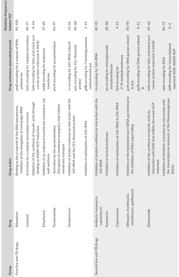
T A B LE 1 M od es o f a ct io n o f t he m ai n f irs a nd s ec on lin e a nt i- T B d ru gs , m ec ha ni sm s o f d ru g r es is ta nc e a nd m ut at io n f re qu en cy f or e ac h g en e i n c lin ic al M yc ob ac ter iu m tu ber cu lo sis is ol ate s G ro up D ru g D ru g a ct io n D ru g r es is ta nc as so ci at ed g en e( s) M ut at io n f re qu en cy i n c lin ic al is ol at es ( % ) a Fi rs lin e an TB dr ug s Ri fa m pic in B in di ng t o t he su bu ni t o f t he R N A p ol ym er as e, in hi bi tio n o f t he e lo ng at io n o f m es se ng er R N A rp oB e nc od in g f or su bu ni t o f R N A po ly mer as e 90 –1 00 Is oni az id A ct iv at io n b y a c at al as pe ro xi da se e nz ym e ka tG e nc od in g f or c at al as pe ro xi da se 40 –9 7 In hi bi tio n o f t he s yn th es is o f m yc ol ic a ci ds t hr ou gh bi nd in g t o N A D H - A C re du ct as e in hA e nc od in g f or f at ty a ci d e no yl a cy l ca rr ie r p ro te in r ed uc ta se A ( In hA ) 8– 64 Et ham but ol In hi bi tio n o f a n a ra bi no sy l t ra ns fe ra se i nv ol ve d i n c el l w al l s yn th es is em bB e nc od in g f or a ra bi no sy l tra nsf era se 47– 89 Py raz ina mi de - A ct iv at io n b y t he p yr az in am id as e - D is ru pt io n o f m em br an e e ne rg et ic s t ha t i nh ib its m emb ra ne tr an sp or t pn cA e nc odi ng fo r p yr az ina mi da se 44 –9 7 St re pt omy ci n In hi bi tio n o f p ro te in s yn th es is b y i nt er ac tio n w ith t he 16 S r RN A a nd t he S 12 r ib os om al p ro te in rr s e nc od in g f or 1 6S r RN A s ub un it 12 –2 6 rps L e nc od in g f or S 12 r ib os om al pr ot ein 40 –6 8 In hi bi tio n o f m et hy la tio n o f 1 6S r RN A gi dB enc od in g f or 7 - me th yl gu ano si ne m et hy ltra nsf era se 5–1 3 Se co nd - li ne an TB dr ug s A mik ac in , k ana m yc in , ca pr eo m yc in In hi bi tio n o f p ro te in s yn th es is b y i nt er ac tio n w ith t he 16 S r RN A rr s e nc od in g f or 1 6S r RN A 40 –9 0 K ana m yc in In hi bi tio n o f a ce ty ltr an sf er ase ei s e nc odin g f or a min ogl yc os id e ac et yl tra nsf era se 28–8 0 C ap re om yc in In hi bi tio n o f m et hy la tio n o f 1 6S r RN A & 2 3S r RN A tly A e nc od in g f or 2′ - O - m et hy ltra nsf era se 4–1 3 O flo xa ci n, le vo flo xa ci n, mo xi flo xa ci n, g ati flo xa ci n In hi bi tio n o f t he t op oi so m er as e I I ( D N A g yr as e) l ea d t o th e i nh ib iti on o f D N A s up er co ili ng gyr A e nc od in g f or D N A g yr as e s ub un it A a nd 70 –9 0 gyr B e nc od in g f or D N A g yr as e s ub un it B 0–1 1 Et hi ona mi de In hi bi tio n o f t he s yn th es is o f m yc ol ic a ci ds b y in te ra ct io n w ith N A D t ha t i nh ib its t he e no yl - A C P re du ct as e in hA e nc od in g f or f at ty a ci d e no yl a cy l ca rr ie r p ro te in r ed uc ta se A ( In hA ) 33 –62 In hi bi tio n o f me ta bo lic ac tiv at io n b y i nt er ac tio n w ith the tr an sc rip tio na l r ep re sso r o f t he M on oo xy ge na se (E th A ) eth A e nc od in g f or E th A 46 –7 2 eth R e nc odin g f or tr an scr ip tio na l re pr es so r E th R , N A D H - A C P 0–4 N ote . a Se e t he f ol lo w in g p ap er s f or d et ai ls ( C am pb el l e t a l., 2 01 1; R am ire B us by & V al af ar , 2 01 5; S an dg re n e t a l., 2 00 9; V ilc he ze & J ac ob s, 2 01 4; Z ha ng & Y ew , 2 01 5) .
study, Sun et al. (2012) described the dynamic changes of the drug resistance- associated mutation profile in M. tuberculosis populations at different stages of drug resistance acquisition. These authors found four to five transient drug resistance mutants in the same spu-tum sample, but only the fittest resistant mutant became fixed over time. Similar to that, Eldholm et al. (2014) monitored the evolution of an XDR strain from a susceptible ancestor in a single patient. They showed that drug resistance- associated mutations were acquired multiple times by individual clones, but only one expanded and re-placed the other clones. In an ultimate manner, adaptive mutants are fixed and become dominant while others are lost by competition, referred as clonal interference (Box 1, Figure 1a) (Gerrish & Lenski, 1998). In addition, recent studies also demonstrated that M. tuber-culosis populations can evolve measurably in response to selection pressures imposed by the environment within hosts (Lieberman et al., 2016; O’Neill, Mortimer, & Pepperell, 2015). This process can lead to the spatial structuring of the bacterial population within host (lungs) into related subpopulations that will evolve independently (parallel evolution, Box 1) as demonstrated previously (Gygli, Borrell,
Trauner, & Gagneux, 2017). All these studies underline the constant genome evolution due to the acquisition of multiple independent mutations in the bacterial population despite the evolutionary bot-tleneck imposed by purifying selection (Box 1) due to drug selective pressure and clonal interference (Figure 1a).
4 | CHAR ACTERISTICS AND DIVERSIT Y
OF DRUG RESISTANCE- ASSOCIATED
MUTATIONS
The mutation frequency and type vary in function of different parameters, such as the geographic region, the drug resistance pattern and genetic background (Fenner et al., 2012; Hillemann, Kubica, Rusch- Gerdes, & Niemann, 2005; Lipin, Stepanshina, Shemyakin, & Shinnick, 2007; Qian et al., 2002). Despite the large diversity of mutation patterns globally, only few specific muta-tions are predominant (Table 2) (Sandgren et al., 2009; Zhang & Yew, 2015). For instance, in the case of rifampicin resistance,
F I G U R E 1 Origin and evolution
of drug resistance in Mycobacterium tuberculosis (modified from (Chang et al., 2015; Hughes & Andersson, 2015)). The figure represents the evolution of bacteria from wild type to drug- resistant mutants with fitness advantage and illustrates several different mechanisms of fitness increase. (a) Wild type can acquire different drug resistance- associated mutations in a same gene with high or low biological cost. The bacteria with low biological cost mutations will be selected under drug pressure by clonal interference and will propagate. (b) Under drug pressure, positive epistasis may favour the acquisition of compensatory mutations to alleviate the fitness cost exerted by certain drug resistance- associated mutations. (c) Driven by positive epistasis, drug- resistant mutants are likely to be more prone to accumulate drug resistance- associated mutations at higher frequencies (Nguyen, Nguyen, et al., 2017; Trindade et al., 2009)
Clonal interference
Wild type Drug-resistantmutants Drug-resistantmutants with fitness advantage
(a) biological cost mutationAcquisition of high
Acquisition of low biological cost mutation
Bacteria with low biological cost mutation
(c) (b)
Bacteria with multiple drug resistance-associated mutations Acquisition of drug resistance-
associated mutation Acquisition of high
biological cost mutation Bacteria with high biological costmutation + compensatory mutation
Compensatory evolution
Epistasis between drug resistance mutations
Drug selection pressure and mechanisms
hundreds of rpoB mutations have been described (not all were associated with rifampicin resistance), but more than 80% of rifampicin- resistant isolates display mutations in three codons rpoB531, 526 and 516 (Campbell et al., 2011; Lipin et al., 2007; Pozzi et al., 1999; Telenti et al., 1993). Similar to that, among the approximately 300 mutations found in the katG gene, the preva-lence of katG S315T mutation can vary between 32% and 95% in isoniazid- resistant clinical isolates depending on the geographic regions and drug resistance patterns (Hazbon et al., 2006; Lipin et al., 2007; Mokrousov et al., 2002; Vilcheze & Jacobs, 2014).
In addition, different mutations in the same gene or in different genes can produce similar drug resistance phenotypes (Sandgren et al., 2009; Zhang & Yew, 2015), but can be associated with similar or different drug resistance levels (Fenner et al., 2012; Gagneux, Long, et al., 2006; Huitric, Werngren, Jureen, & Hoffner, 2006). For instance, some mutations in rpoB gene, such as rpoB S531L, H526Y, H526D and H526R, are often associated with high levels of rifampicin resistance, while mutations including rpoB L511P, H526L, H526N, L533P and I572F are generally linked to low levels of rifampicin resistance (Huitric et al., 2006; Van Deun et al., 2009). Similar to that, katG mutations are often associated with high levels of isoniazid resistance, whereas inhA mutations with low levels (Fenner et al., 2012; van Soolingen et al., 2000; Vilcheze & Jacobs, 2014).
Furthermore, mutations in different regions of the same gene can be associated with different drug resistance phenotypes. For in-stance, mutations in the 530 loop and 915 region of rrs gene are as-sociated with streptomycin resistance (Sreevatsan et al., 1996), while mutations in the 1400—1500 region are linked to resistance to kana-mycin, amikacin and capreomycin (Jugheli et al., 2009). In particular,
cross- resistance phenomena (Box 1) have also been described in M. tuberculosis (Andries et al., 2014; Jugheli et al., 2009; Vilcheze & Jacobs, 2014).
At last, the mutation type has been correlated with the genetic back-ground of M. tuberculosis lineages in several studies (Hillemann et al., 2005; Mokrousov et al., 2006; Qian et al., 2002; Ribeiro et al., 2014). For instance, the katG S315T is prevalent in lineage 2, conversely the inhA- 15 mutation is associated mostly with lineage 1 (Casali et al., 2014; Fenner et al., 2012; Gagneux, Burgos, et al., 2006; Mokrousov et al., 2002; Nguyen, Nguyen, et al., 2017). Similar to that, the rpoB S531L mutation is observed mainly in lineage 2 compared with other lineages, while the rpoB D516V mutation is more frequent in lineage 4 (LAM family) (Casali et al., 2014; Hillemann et al., 2005; Lipin et al., 2007). In particular, the prevalence of specific drug resistance- associated muta-tions also varies within the lineage, such as the frequencies of the rpoB S531L and katG S315T mutations are greater in the modern (typical) Beijing strains than in ancient (atypical) ones (Hillemann et al., 2005; Lipin et al., 2007; Qian et al., 2002). These differences could be the re-sult of epistatic interactions (see Box 1 and below) and might reflect the adaptation of M. tuberculosis sublineages to the different human populations and the efficiency of treatment and public health strategies (Bañuls et al., 2015; Comas et al., 2013; Eldholm et al., 2016).
5 | FITNESS COST OF DRUG RESISTANCE-
ASSOCIATED MUTATIONS
As the drug targets are generally involved in important biological functions, mutations in the genes encoding these factors should
TA B L E 2 The most frequent drug resistance- associated mutations found in clinical drug- resistant Mycobacterium tuberculosis isolates,
including MDR and XDR samples
Drug(s)
Drug resistance- associated gene(s)
Frequent mutation (amino acid/ nucleotide change)
Mutation frequency in clinical
drug- resistant isolates (%)b
Isoniazid katG 315 (Ser- Thr) 32–95
inhA - 15 (C- T)a 8–71
Rifampicin rpoB 531 (Ser- Leu) 41–74
526 (His- Tyr) 6–24
526 (His- Asp) 2–30
516 (Asp- Val) 5–18
Streptomycin rpsL 43 (Lys- Arg) 35–62
88 (Lys- Arg) 13–28
rrs 514 (A- C)a 3–12
Ethambutol embB 306 (Met- Val) 40–60
Fluoroquinolones gyrA 94 (Asp- Gly) 25–60
90 (Ala- Val) 12–30
Kanamycin, amikacin and capreomycin
rrs 1401 (A- G)a 30–90
Notes. aNucleotide change.
bSee the following studies for reference (Campbell et al., 2011; Duong et al., 2009; Hazbon et al., 2006; Hillemann et al., 2005; Lipin et al., 2007;
Mokrousov et al., 2002; Müller et al., 2011; Niehaus et al., 2015; Perdigao et al., 2010; Pozzi et al., 1999; Qian et al., 2002; Shi et al., 2011; van Soolingen et al., 2000; Sreevatsan et al., 1996, 1997; Telenti et al., 1993; Von Groll et al., 2009).
impart a biological cost that leads to reduced fitness of the resist-ant strains in comparison with the sensitive ones, in the absence of antibiotics (Melnyk et al., 2015). According to that, several studies showed that drug- resistant M. tuberculosis mutants are characterized by reduced fitness (Billington, McHugh, & Gillespie, 1999; Gagneux, Long, et al., 2006; Mariam, Mengistu, Hoffner, & Andersson, 2004). However, the extent of the biological cost depends on the mutation and the strain genetic background (Billington et al., 1999; Bottger, Springer, Pletschette, & Sander, 1998; Gagneux, Long, et al., 2006; Pym, Saint- Joanis, & Cole, 2002). Furthermore, in the absence of ge-netic drift (Box 1), drug- resistant mutations with low or no biological cost are more likely to be selected and maintained in the popula-tions (Farhat et al., 2013; Osorio et al., 2013). For instance, the pre-dominant mutations associated with high level of drug resistance and a low or no biological cost, such as katG S315T, rpoB S531L, rpsL K43R and gyrA D94G (conferring resistance to isoniazid, ri-fampicin, streptomycin and fluoroquinolones respectively), are more frequently found in clinical drug- resistant isolates (Billington et al., 1999; Bottger et al., 1998; Campbell et al., 2011; Casali et al., 2014; Gagneux, Burgos, et al., 2006; Gagneux, Long, et al., 2006; Mariam et al., 2004; Pym et al., 2002). Indeed, some of these resistance mu-tations do not reduce bacterial fitness in the absence of treatment (Bergval, Schuitema, Klatser, & Anthony, 2009; Huitric et al., 2006; Mariam et al., 2004). It is worth noting that MDR and XDR strains associated with outbreaks often carried these mutations explaining the successful spread of these highly drug- resistant strains in the community (Casali et al., 2014; Cohen et al., 2015; Niehaus, Mlisana, Gandhi, Mathema, & Brust, 2015; de Vos et al., 2013). All these ob-servations suggest that the prevalent mutations in clinical isolates have been positively selected because of their low biological cost (Figure 1a) (Farhat et al., 2013; Osorio et al., 2013). As demonstrated by Bergval et al. (Bergval et al., 2009), drug resistance mutation pat-terns of in vitro selected- resistant mutants do not always reflect mutation profiles obtained in clinical isolates. Indeed, mutations as-sociated with high biological cost of resistance detected in in vitro drug- resistant mutants are rarely found in clinical drug- resistant isolates (Bergval et al., 2009; Gagneux, Long, et al., 2006; Huitric et al., 2006). For example, mutations in the katG gene that lead to complete loss of the catalase- peroxidase enzyme function (confer-ring resistance to isoniazid), such as frame- shift nucleotide deletions or insertions, are found less frequently in clinical than in in vitro mu-tants (Bergval et al., 2009). Several reasons can explain the different mutation patterns obtained in in vitro mutants and clinical isolates, such as the long evolution within the human body, the clonal inter-ference, the variability in drug pressure and the parallel evolution.
The biological cost of pyrazinamide resistance mutations is of interest to investigate the evolution of pyrazinamide resistance in M. tuberculosis. The high diversity of mutations in the pncA gene detected in clinical isolates (Ramirez- Busby & Valafar, 2015) can be explained by two different hypotheses. First, pncA mutations as-sociated with pyrazinamide resistance might not cause any fitness deficit. Indeed, pncA seems to be nonessential because M. tubercu-losis can survive without this gene using other metabolic pathways
(Martinez, Holmes, Jelfs, & Sintchenko, 2015; Stoffels, Mathys, Fauville- Dufaux, Wintjens, & Bifani, 2012). Therefore, each muta-tion can have the same probability to be selected and transmitted. In an alternative way, pncA mutations could be associated with high cost of resistance that impairs M. tuberculosis transmission (den Hertog, Sengstake, & Anthony, 2015). This hypothesis is supported by the lack of pncA mutant clusters, thus reflecting a low transmis-sion potential. However, clusters of pncA mutants have been de-scribed in some specific MDR and XDR M. tuberculosis outbreaks in South Africa and in Argentina showing the successful transmission of these PZA- resistant clones (Cohen et al., 2015; Eldholm et al., 2015; Müller, Chihota, et al., 2013). The development of experimen-tal evolution studies will allow assessing the biological cost magni-tude of pncA mutations.
Several works investigated the link between M. tuberculo-sis genetic background and the cost of drug retuberculo-sistance mutations. Gagneux, Long, et al. (2006) found differences in biological cost for the rifampicin resistance- associated rpoB H526D mutation between the lineages 2 and 4, while the rpoB S531L mutants showed simi-lar costs in both lineages. The inhA- 15 and katG S315T mutations are strongly associated with lineage 1 and modern lineages, respec-tively (Casali et al., 2014; Fenner et al., 2012; Gagneux, Burgos, et al., 2006). On the contrary, katG mutations other than katG S315T that likely abrogate enzyme activity result in high biological cost and seem to be more associated with lineage 2 (Gagneux, Burgos, et al., 2006). Thus, lineage 2 could be better adapted to compensate for the loss or reduced activity of this catalase- peroxidase enzyme in the context of isoniazid resistance. This hypothesis could also ex-plain why the Beijing strains are generally strongly associated with resistance to isoniazid, regardless of the type of katG resistant- associated mutations and country (Fenner et al., 2012; Gagneux, Burgos, et al., 2006; Mokrousov et al., 2002; Ribeiro et al., 2014; van Soolingen et al., 2000).
6 | COMPENSATORY MUTATIONS
Compensatory mutations can alleviate the loss of fitness produced by drug resistance- associated mutations (Figure 1b) (Bottger et al., 1998; Brandis, Wrande, Liljas, & Hughes, 2012). In M. tuberculosis, data on compensatory mutations are still limited and mainly focused on isoniazid and rifampicin resistance (Comas et al., 2012; Sherman et al., 1996; Song et al., 2014; de Vos et al., 2013). Nevertheless, mechanisms of compensatory evolution were also proposed for other drug resistance genotypes (Table 3).
Almost all laboratory- generated mutants with a rifampicin resistance- associated mutation in the RRDR of rpoB show a signifi-cant fitness deficit compared with their drug- susceptible ancestors when grown in the absence of this drug. Therefore, it was hypothe-sized that the fitness cost linked to rifampicin resistance could be re-duced by compensatory mutations in clinical isolates (Billington et al., 1999; Comas et al., 2012; Mariam et al., 2004; de Vos et al., 2013). Nonsynonymous mutations in the rpoA and rpoC genes that encode
the α and β’ subunits of RNA polymerase, respectively, could play the role of fitness- compensatory mutations in rifampicin- resistant rpoB mutants (Comas et al., 2012; de Vos et al., 2013). Indeed, it was reported that part of rifampicin- resistant isolates with a rpoB muta-tion also carry a nonsynonymous mutamuta-tion in the rpoA or rpoC gene in South Africa (27.1%, 89/329), China (27.8%, 89/320) and Korea (39.4%, 67/170) (Comas et al., 2012; Li et al., 2016; Song et al., 2014). In addition, clinical isolates that carry mutations in the RRDR of rpoB and also in rpoA/rpoC display higher competitive fitness in vitro and in vivo compared with laboratory- generated rifampicin- resistant mutants that carry only the same rpoB RRDR mutation and that belong to the same phylogenetic lineage (Brandis & Hughes, 2013; Comas et al., 2012; Song et al., 2014). These data suggest that mu-tations in the rpoA/rpoC genes are fitness- compensatory mumu-tations that alleviate the costs of rpoB mutations. Furthermore, genetic re-constructions in a Salmonella model demonstrated that mutations not only in rpoA and rpoC, but also in rpoB are associated with higher growth rate (Brandis & Hughes, 2013; Brandis et al., 2012). In fact, many previous studies showed that rifampicin- resistant M. tuber-culosis clinical isolates carry multiple (double, triple and quadruple) mutations in the rpoB gene (Bahrmand, Titov, Tasbiti, Yari, & Graviss, 2009; Casali et al., 2014; Nguyen, Nguyen, et al., 2017; Song et al., 2014). This could be the result of compensatory mechanisms to alleviate the fitness cost exerted by specific mutations (Brandis & Hughes, 2013; Brandis et al., 2012). It is worth noting that compen-satory mutations are more commonly identified in the dominant MDR, pre- XDR and XDR clones in high MDR TB burden countries, suggesting that high drug- resistant mutants harbouring these muta-tions can be successfully transmitted in human populamuta-tions (Casali et al., 2014; Cohen et al., 2015; Comas et al., 2012; Klopper et al., 2013; Li et al., 2016; de Vos et al., 2013).
These studies also showed that the rpoC mutation is significantly associated with the rpoB S531L mutation, suggesting an interac-tion between a fitness- compensatory mutainterac-tion and a specific drug resistance- associated mutation (Casali et al., 2014; Li et al., 2016; de Vos et al., 2013). This may explain why rpoB S531L is the most common mutation observed in rifampicin- resistant clinical isolates and displays a low biological cost. The presence of compensatory mutations seems to be associated with Beijing strains, especially those harbouring the rpoB S531L variant (Casali et al., 2014; Li et al., 2016). Nevertheless, the frequency of compensatory mutations differs according to the Beijing genotype subclades, suggesting epistatic interactions (Box 1, see below) between drug resistance mutations, compensatory mutations and genetic background (Casali et al., 2014).
7 | ACCUMUL ATION OF DRUG
RESISTANCE- ASSOCIATED MUTATIONS
AND EPISTASIS
7.1 | Accumulation of drug resistance- associated
mutations
The high diversity of mutations in M. tuberculosis suggests different evolutionary trajectories towards highly resistant genotypes, in re-sponse to various selection pressures. Nevertheless, for almost all drug resistance- associated genes, the predominance of some spe-cific mutations, generally known to be associated with high level of resistance and low biological cost, has been described (see Table 2). As a result, combinations of at least two specific mutations, such as rpoB531, katG315, rpsL43, embB306 and gyrA94, are favoured (Casali et al., 2014; Cohen et al., 2015; Nguyen, Nguyen, et al., 2017).
TA B L E 3 Mechanisms of drug resistance, fitness costs and compensatory mechanisms in Mycobacterium tuberculosis
Genetic mutation(s) Mechanism of resistance Fitness cost Compensatory mechanism
katG Reduced prodrug activation Reduced protection against oxidative damage
Overexpression of ahpC by mutations in its promoter
inhA promoter NADH- ACP- reductase overexpression
No No
inhA Reduced affinity for drug Reduced fatty acid biosynthesis Secondary mutation in inhA promoter (hypothesis)
rpoB Decreased DNA polymerase
affinity for drug Reduced DNA transcription efficiency Secondary mutation in rpoA, rpoC or rpoB rpsL & rrs Reduced ribosomal target affinity
for drug
Impaired ribosome performance, Reduced protein synthesis accuracy
Unknown
embB Decreased arabinosyl transferase
affinity for drug
Reduced cell wall biosynthesis efficiency
Secondary mutation in embABC operon (hypothesis)
pncA Reduction or loss of pyrazinamide
prodrug activation
Unknown Unknown
gyrA & gyrB Reduced DNA gyrase affinity for drug
Reduced DNA supercoiling, DNA replication and transcription efficiency
Secondary mutation in gyrA or gyrB (hypothesis)
Eis Aminoglycoside acetyltransferase
overexpression
Although the quality of the treatment undoubtedly plays a role in the emergence of particular drug resistance mutations, the strains with drug resistance- associated mutations seem to have higher propen-sity to accumulate other drug resistance mutations in the same gene or in different genes (Bahrmand et al., 2009; Jagielski et al., 2014; Nguyen, Nguyen, et al., 2017; Shen et al., 2007). For instance, the katG315, embB306 or pncA mutations are more frequently observed in MDR than in non- MDR isolates (Hazbon et al., 2006; Nguyen, Contamin, et al., 2017; Salvatore et al., 2016; Shen et al., 2007). All these data suggest a cumulative effect of mutations that are specifi-cally associated with drug resistance and the occurrence of epista-sis (Figure 1c). Besides epistatic interactions, Chang et al. (2015) in their review of the causes of the excess of MDR infections suggest that the associated linkage selection can also be at the origin of the proliferation of multiple drug- resistant bacteria. This is especially the case for M. tuberculosis which follows a basic clonal evolution model (see above). This model generates a strong linkage disequilib-rium that may favour the coexistence of two or more particular drug resistance- associated alleles.
7.2 | Epistasis between drug- resistant mutations
Although little is known about epistasis between drug resistance mutations in M. tuberculosis, a finding suggests that it could play an important role in the emergence and evolution of MDR and XDR M. tuberculosis strains (Borrell et al., 2013). The interaction be-tween drug resistance mutations may restore or even increase the biological fitness of drug- resistant mutants compared with drug- susceptible strains. For example, Spies et al. demonstrated that the double mutants rpsL K43R/katG S315T, rpsL K43R/rpoB S531L and rpoB S531L/katG S315T, which are frequently detected in clini-cal isolates, grow faster than drug- susceptible strains (Spies et al., 2013). This suggests that the interaction between these mutations may offer a fitness advantage to the double mutants. Indeed, these double mutations increase the fitness of drug- resistant E. coli and drive the evolution of MDR acquisition (Trindade et al., 2009). On the contrary, Salvatore et al. found that isolates that carry the katG S315T/rpsL K43R mutations are less frequent among MDR strains that cause multiple TB cases in a household than among single- case household MDR strains, suggesting the occurrence of negative epistasis (Salvatore et al., 2016). As the katG315 mutation does not affect the virulence and transmission of isoniazid- resistant strains, the authors suggested that the rpsL43 mutation might impart a biological cost on the transmissibility of drug- resistant bacteria. Nevertheless, the combination of katG315 and rpsL43 mutations is common in drug- resistant clinical M. tuberculosis isolates, particularly in Beijing strains. This suggests that epistasis varies according to the strain genetic background (see below). Borrell et al. (2013) described epistatic interactions between mutations associated with resistance to ofloxacin and rifampicin, using M. smegmatis as model organism. The authors demonstrated that 35% (6/17) of double mutants carry-ing specific rpoB and gyrA mutations associated with rifampicin and fluoroquinolone resistance have a significant higher fitness than thecorresponding single drug- resistant mutants. In particular, the gyrA N94G mutation was associated with improved fitness in all double mutants, irrespectively of the rpoB mutation. In an interesting man-ner, the mutation combinations obtained in vitro in M. smegmatis correspond to the most common mutations detected among MDR and XDR clinical isolates in high MDR TB burden countries (Borrell et al., 2013; Casali et al., 2014; Comas et al., 2012). These authors also found some double mutants bearing higher biological cost, which can be a sign of negative epistasis. Furthermore, the acquisi-tion of a secondary mutaacquisi-tion (linked or not linked to drug resistance) in the same gene, for example rpoB, was associated with a reduction of biological cost (Brandis & Hughes, 2013; Song et al., 2014).
7.3 | Epistasis between drug resistance- associated
mutations and compensatory mutations
The progressively increasing identification of drug resistant, includ-ing MDR and XDR isolates without reduction in fitness, suggests the presence of epistatic interactions between drug resistance muta-tions and compensatory mutamuta-tions (Figure 1c). Using the M. smeg-matis model, Song et al. (2014) demonstrated higher growth rates and higher relative fitness in recombinant strains carrying both rpoB S531L and rpoC F452L or rpoC V483G mutations than in strains har-bouring only the rpoB S531L mutation. However, these interactions have not been investigated in M. tuberculosis. Nevertheless, the find-ings that many (27%–70%) clinical rifampicin- resistant mutants carry putative compensatory mutations in either rpoA or rpoC genes sup-port the hypothesis that these two mutation types interact also in M. tuberculosis (Casali et al., 2014; Comas et al., 2012; Li et al., 2016; Song et al., 2014). As example, rifampicin- resistant M. tuberculosis strains carrying the rpoB S531L mutation are often associated with putative compensatory mutations in the rpoA or rpoC genes (Casali et al., 2014; Song et al., 2014; de Vos et al., 2013).
Concerning the evolution of MDR strains, clinical and molecu-lar studies suggest that isoniazid resistance, due to the katG S315T mutation, has preceded the emergence of rpoB gene mutations lead-ing to the acquisition of rifampicin resistance (Cohen et al., 2015; Gegia, Winters, Benedetti, van Soolingen, & Menzies, 2017; Manson et al., 2017; Salvatore et al., 2016). The combination of katG315 and rpoB531 mutations with a rifampicin- resistant fitness- compensatory mutation (e.g., rpoC mutation) is favoured in clinical MDR isolates, sug-gesting that these genotypes lead to primary MDR infections. In an important way, the emergence of XDR TB seems to be caused by the transmission of XDR strains directly from person to person rather than by inadequate MDR treatment (Shah et al., 2017). Thus, compensatory evolution and epistasis could play an important role in the emergence and spread of highly resistant strains in the community.
7.4 | Epistasis between resistance determinants and
genetic background
For many bacteria, epistatic interactions have been also described between resistance determinants and their genetic background
(Chang et al., 2015). In M. tuberculosis, epidemiological and molecular data have shown the emergence and the successful spread of MDR/ XDR clones belonging to Beijing or LAM families carrying specific mutations associated with high level of drug resistance and com-pensatory mutations (Casali et al., 2014; Cohen et al., 2015; Eldholm et al., 2015). Indeed, in Beijing family, it was demonstrated that the biological costs of resistance mutations are smaller than those in other families, or the acquisition of compensatory mutations appears easier, possibly explaining the association between Beijing genotype and MDR (Casali et al., 2014; Gagneux, Long, et al., 2006).
Altogether, the interactions between different drug resistance mutations, between drug resistance mutations and compensatory mutations and between drug resistance mutations and the genetic background underline the key role of epistasis in the evolution of multiple drug resistance in M. tuberculosis.
8 | CONCLUDING REMARKS
As detailed in this review, drug resistance evolution in M. tuberculosis is driven by various factors with different effects. The mutation fre-quency and type can be affected by the drug- resistant patterns and genotypes. Different mutations can cause different levels of drug resistance and of biological fitness cost even when they are located in the same gene or on the same codon.
The most frequent drug resistance mutations in clinical isolates worldwide are often associated with high levels of resistance and low/no fitness costs, or combined with compensatory mutations to restore the bacterial fitness. This strongly suggests that epistatic interactions influence the evolution of drug resistance in M. tuber-culosis. Furthermore, all these evolutionary processes, including the basic clonal reproduction mode of M. tuberculosis, not only maintain drug- resistant strains, but also favour their transmission in host populations. Therefore, besides the well- known extrinsic factors (in-adequate treatment regimens, differences in drug pharmacokinetics- pharmacodynamics, patient adherence, etc.), intrinsic factors, such as compensatory mechanisms, reduced fitness cost, clonal interfer-ence, mutation rate and epistasis, also promote the emergence of MDR and XDR strains worldwide.
In an important way, the Beijing lineage is rapidly spreading worldwide. This lineage is associated with MDR TB as well as with high level of drug resistance and fitness- compensatory mutations (Casali et al., 2014; Manson et al., 2017). This suggests a worrying scenario in which drug resistance evolves towards very fit and highly drug- resistant genotypes and the successful transmission of deadly drug- resistant mutants. This could seriously challenge the success of TB control programmes worldwide.
It is unfortunate that, some drug resistance mechanisms remain unclear and many mechanisms of fitness- compensatory evolution and epistasis have not been investigated in M. tuberculosis. More work is needed to increase our knowledge on all the forces that drive drug resistance in M. tuberculosis for better controlling the emer-gence and rapid spread of highly drug- resistant strains. As suggested
by the levels of drug resistance reached globally, we are losing the arms race against bacteria including M. tuberculosis (Bañuls et al., 2018). M. tuberculosis, as many pathogens, has a complex ecology and evolution and is also evolving and fluctuating through time and space according to local contexts (Bañuls et al., 2015; Comas et al., 2013; Eldholm et al., 2016; Müller, Borrell, et al., 2013; O’Neill et al., 2015; Trauner et al., 2014). For instance, our review underlines that strains carrying multiple drug- resistant mutations reveal a high ability to acquire other resistances or compensatory mutations by epistatic interactions in reducing the biological cost imposed on the fitness of bacteria. These evolutionary processes suggest that, to limit the drug resistance escalation, molecules acting simultaneously on multiple bacterial targets are urgently needed to replace single- target drugs that now require to be used in more and more complex combinations (the standard treatment of TB disease is composed by a minimum of four drugs). In addition, the detailed knowledge of evolutionary mechanisms will help develop accurate models to predict the evolution of drug resistance and thus to better control it as underlined by other authors (Lehtinen et al., 2017; Schenk & de Visser, 2013).
ACKNOWLEDGEMENTS
This review was written in the framework of PHC Lotus pro-ject “Application of DNA chip technology for the development of diagnostic kits for rapid detection of drug- resistant tuberculo-sis in Vietnam, Laos and Cambodia (2014–2018)” granted by the Ministry of Science and Technology (MOST), Vietnam. We would like to thank IRD, CNRS, NIHE, USTH and LMI DRISA for their support. Nguyen Quang Huy was supported by the Vietnamese Government Scholarship Program (911 Project). We thank Elisabetta Andermacher for assistance in preparing and editing the manuscript.
CONFLIC T OF INTEREST
None declared.
ORCID
Quang Huy Nguyen http://orcid.org/0000-0002-3452-1811
REFERENCES
Andries, K., Villellas, C., Coeck, N., Thys, K., Gevers, T., Vranckx, L., … Koul, A. (2014). Acquired resistance of Mycobacterium tuberculosis to bedaquiline. PLoS ONE, 9(7), e102135. https://doi.org/10.1371/jour-nal.pone.0102135
Bahrmand, A. R., Titov, L. P., Tasbiti, A. H., Yari, S., & Graviss, E. A. (2009). High- level rifampin resistance correlates with multiple mu-tations in the rpoB gene of pulmonary tuberculosis isolates from the Afghanistan border of Iran. Journal of Clinical Microbiology, 47(9), 2744–2750. https://doi.org/10.1128/JCM.r00548-09
Bañuls, A. L., Nguyen, T. V. A., Nguyen, Q. H., Nguyen, T. N. A., Tran, H. H., & Godreuil, S. (2018). Antimicrobial resistance: The 70-year arms race between humans and bacteria. In Ecology and evolution of
infectious diseases in low-income countries. Oxford: Oxford University
Press, Chapter 6 (in press).
Bañuls, A. L., Sanou, A., Nguyen, T. V. A., & Godreuil, S. (2015).
Mycobacterium tuberculosis: Ecology and evolution of a human
bacte-rium. Journal of Medical Microbiology, 64(11), 1261–1269. https://doi. org/10.1099/jmm.0.000171
Bergval, I. L., Schuitema, A. R., Klatser, P. R., & Anthony, R. M. (2009). Resistant mutants of Mycobacterium tuberculosis selected in vitro do not reflect the in vivo mechanism of isoniazid resistance.
Journal of Antimicrobial Chemotherapy, 64(3), 515–523. https://doi.
org/10.1093/jac/dkp237
Billington, O. J., McHugh, T. D., & Gillespie, S. H. (1999). Physiological cost of rifampin resistance induced in vitro in Mycobacterium
tubercu-losis. Antimicrobial Agents and Chemotherapy, 43(8), 1866–1869.
Black, P. A., de Vos, M., Louw, G. E., van der Merwe, R. G., Dippenaar, A., Streicher, E. M., … Pain, A. (2015). Whole genome sequencing reveals genomic heterogeneity and antibiotic purification in Mycobacterium
tuberculosis isolates. BMC Genomics, 16, 857. https://doi.org/10.1186/
s12864-015-2067-2
Borrell, S., Teo, Y., Giardina, F., Streicher, E. M., Klopper, M., Feldmann, J., … Gagneux, S. (2013). Epistasis between antibiotic resistance mu-tations drives the evolution of extensively drug- resistant tuberculo-sis. Evolution, Medicine and Public Health, 2013(1), 65–74. https://doi. org/10.1093/emph/eot003
Bottger, E. C., Springer, B., Pletschette, M., & Sander, P. (1998). Fitness of antibiotic- resistant microorganisms and compensatory mutations.
Nature medicine, 4(12), 1343–1344. https://doi.org/10.1038/3906
Brandis, G., & Hughes, D. (2013). Genetic characterization of compen-satory evolution in strains carrying rpoB Ser531Leu, the rifampi-cin resistance mutation most frequently found in clinical isolates.
Journal of Antimicrobial Chemotherapy, 68(11), 2493–2497. https://
doi.org/10.1093/jac/dkt224
Brandis, G., Wrande, M., Liljas, L., & Hughes, D. (2012). Fitness- compensatory mutations in rifampicin- resistant RNA poly-merase. Molecular Microbiology, 85(1), 142–151. https://doi. org/10.1111/j.1365-2958.2012.08099.x
Campbell, P. J., Morlock, G. P., Sikes, R. D., Dalton, T. L., Metchock, B., Starks, A. M., … Posey, J. E. (2011). Molecular detection of mutations associated with first- and second- line drug resistance compared with conventional drug susceptibility testing of Mycobacterium
tu-berculosis. Antimicrobial Agents and Chemotherapy, 55(5), 2032–2041.
https://doi.org/10.1128/AAC.01550-10
Casali, N., Nikolayevskyy, V., Balabanova, Y., Harris, S. R., Ignatyeva, O., Kontsevaya, I., … Drobniewski, F. (2014). Evolution and transmis-sion of drug- resistant tuberculosis in a Russian population. Nature
Genetics, 46(3), 279–286. https://doi.org/10.1038/ng.2878
Chang, H. H., Cohen, T., Grad, Y. H., Hanage, W. P., O’Brien, T. F., & Lipsitch, M. (2015). Origin and proliferation of multiple- drug re-sistance in bacterial pathogens. Microbiology and Molecular Biology
Reviews, 79(1), 101–116. https://doi.org/10.1128/MMBR.00039-14
Cohen, K. A., Abeel, T., Manson McGuire, A., Desjardins, C. A., Munsamy, V., Shea, T. P., … Earl, A. M. (2015). Evolution of extensively drug- resistant tuberculosis over four decades: Whole genome sequenc-ing and datsequenc-ing analysis of Mycobacterium tuberculosis Isolates from KwaZulu- Natal. PLoS Medicine, 12(9), e1001880. https://doi. org/10.1371/journal.pmed.1001880
Colijn, C., Cohen, T., Ganesh, A., & Murray, M. (2011). Spontaneous emergence of multiple drug resistance in tuberculosis before and during therapy. PLoS ONE, 6(3), e18327. https://doi.org/10.1371/ journal.pone.0018327
Comas, I., Borrell, S., Roetzer, A., Rose, G., Malla, B., Kato-Maeda, M., … Gagneux, S. (2012). Whole- genome sequencing of rifampicin- resistant Mycobacterium tuberculosis strains identifies compensatory mutations in RNA polymerase genes. Nature Genetics, 44(1), 106– 110. https://doi.org/10.1038/ng.1038
Comas, I., Coscolla, M., Luo, T., Borrell, S., Holt, K. E., Kato-Maeda, M., … Gagneux, S. (2013). Out- of- Africa migration and Neolithic coex-pansion of Mycobacterium tuberculosis with modern humans. Nature
Genetics, 45(10), 1176–1182. https://doi.org/10.1038/ng.2744
Coscolla, M., & Gagneux, S. (2014). Consequences of genomic diversity in Mycobacterium tuberculosis. Seminars in Immunology, 26(6), 431– 444. https://doi.org/10.1016/j.smim.2014.09.012
Duong, D. A., Nguyen, T. H., Nguyen, T. N., Dai, V. H., Dang, T. M., Vo, S. K., … Caws, M. (2009). Beijing genotype of Mycobacterium
tuber-culosis is significantly associated with high- level fluoroquinolone
re-sistance in Vietnam. Antimicrobial Agents and Chemotherapy, 53(11), 4835–4839.
Eldholm, V., Monteserin, J., Rieux, A., Lopez, B., Sobkowiak, B., Ritacco, V., & Balloux, F. (2015). Four decades of transmission of a multidrug- resistant Mycobacterium tuberculosis outbreak strain. Nature
commu-nications, 6, 7119. https://doi.org/10.1038/ncomms8119
Eldholm, V., Norheim, G., von der Lippe, B., Kinander, W., Dahle, U. R., Caugant, D. A., … Balloux, F. (2014). Evolution of extensively drug- resistant Mycobacterium tuberculosis from a susceptible ancestor in a single patient. Genome Biology, 15(11), 490. https://doi.org/10.1186/ s13059-014-0490-3
Eldholm, V., Pettersson, J. H.-O., Brynildsrud, O. B., Kitchen, A., Rasmussen, E. M., Lillebaek, T., … Debech, N. (2016). Armed conflict and population displacement as drivers of the evolution and dispersal of Mycobacterium tuberculosis. Proceedings of the National Academy
of Sciences, 113(48), 13881–13886. https://doi.org/10.1073/
pnas.1611283113
Farhat, M. R., Shapiro, B. J., Kieser, K. J., Sultana, R., Jacobson, K. R., Victor, T. C., … Murray, M. (2013). Genomic analysis identifies targets of convergent positive selection in drug- resistant Mycobacterium
tuberculosis. Nature Genetics, 45(10), 1183–1189. https://doi.
org/10.1038/ng.2747
Fenner, L., Egger, M., Bodmer, T., Altpeter, E., Zwahlen, M., Jaton, K., … Group the Swiss Molecular Epidemiology of Tuberculosis Study. (2012). Effect of mutation and genetic background on drug resistance in Mycobacterium tuberculosis. Antimicrobial Agents and Chemotherapy,
56(6), 3047–3053. https://doi.org/10.1128/AAC.06460-11
Ford, C. B., Lin, P. L., Chase, M. R., Shah, R. R., Iartchouk, O., Galagan, J., … Lipsitch, M. (2011). Use of whole genome sequencing to estimate the mutation rate of Mycobacterium tuberculosis during latent infection.
Nature Genetics, 43(5), 482. https://doi.org/10.1038/ng.811
Ford, C. B., Shah, R. R., Maeda, M. K., Gagneux, S., Murray, M. B., Cohen, T., … Fortune, S. M. (2013). Mycobacterium tuberculosis mutation rate estimates from different lineages predict substantial differences in the emergence of drug- resistant tuberculosis. Nature Genetics, 45(7), 784–790. https://doi.org/10.1038/ng.2656
Gagneux, S., Burgos, M. V., DeRiemer, K., Encisco, A., Munoz, S., Hopewell, P. C., … Pym, A. S. (2006). Impact of bacterial genetics on the transmission of isoniazid- resistant Mycobacterium
tubercu-losis. PLoS pathogens, 2(6), e61. https://doi.org/10.1371/journal.
ppat.0020061
Gagneux, S., Long, C. D., Small, P. M., Van, T., Schoolnik, G. K., & Bohannan, B. J. (2006). The competitive cost of antibiotic resis-tance in Mycobacterium tuberculosis. Science, 312(5782), 1944–1946. https://doi.org/10.1126/science.1124410
Gao, J., Ma, Y., Du, J., Zhu, G., Tan, S., Fu, Y., … Li, Q. (2016). Later emer-gence of acquired drug resistance and its effect on treatment out-come in patients treated with Standard Short- Course Chemotherapy for tuberculosis. BMC Pulmonary Medicine, 16, 26. https://doi. org/10.1186/s12890-016-0187-3
Gegia, M., Winters, N., Benedetti, A., van Soolingen, D., & Menzies, D. (2017). Treatment of isoniazid- resistant tuberculosis with first- line drugs: A systematic review and meta- analysis. The Lancet
Infectious Diseases, 17(2), 223–234. https://doi.org/10.1016/
Gerrish, P. J., & Lenski, R. E. (1998). The fate of competing beneficial mutations in an asexual population. Genetica, 102, 127. https://doi. org/10.1023/A:1017067816551
Gygli, S. M., Borrell, S., Trauner, A., & Gagneux, S. (2017). Antimicrobial resistance in Mycobacterium tuberculosis: Mechanistic and evolution-ary perspectives. FEMS microbiology reviews, 41(3), 354–373. https:// doi.org/10.1093/femsre/fux011
Hazbon, M. H., Brimacombe, M., Bobadilla del Valle, M., Cavatore, M., Guerrero, M. I., Varma-Basil, M., … Alland, D. (2006). Population genetics study of isoniazid resistance mutations and evolution of multidrug- resistant Mycobacterium tuberculosis. Antimicrobial Agents
and Chemotherapy, 50(8), 2640–2649. https://doi.org/10.1128/
AAC.00112-06
den Hertog, A. L., Sengstake, S., & Anthony, R. M. (2015). Pyrazinamide resistance in Mycobacterium tuberculosis fails to bite? Pathogens and
Disease, 73(6), ftv037.
Hillemann, D., Kubica, T., Rusch-Gerdes, S., & Niemann, S. (2005). Disequilibrium in distribution of resistance mutations among
Mycobacterium tuberculosis Beijing and non- Beijing strains iso-lated from patients in Germany. Antimicrobial Agents and
Chemotherapy, 49(3), 1229–1231. https://doi.org/10.1128/
AAC.49.3.1229-1231.2005
Hughes, D., & Andersson, D. I. (2015). Evolutionary consequences of drug resistance: Shared principles across diverse targets and organ-isms. Nature Reviews Genetics, 16(8), 459. https://doi.org/10.1038/ nrg3922
Huitric, E., Werngren, J., Jureen, P., & Hoffner, S. (2006). Resistance levels and rpoB gene mutations among in vitro- selected rifampin- resistant Mycobacterium tuberculosis mutants. Antimicrobial Agents
and Chemotherapy, 50(8), 2860–2862. https://doi.org/10.1128/
AAC.00303-06
Jagielski, T., Bakuła, Z., Roeske, K., Kamiński, M., Napiórkowska, A., Augustynowicz-Kopeć, E., … Bielecki, J. (2014). Detection of mu-tations associated with isoniazid resistance in multidrug- resistant
Mycobacterium tuberculosis clinical isolates. Journal of Antimicrobial Chemotherapy, 69(9), 2369–2375. https://doi.org/10.1093/jac/
dku161
Jugheli, L., Bzekalava, N., de Rijk, P., Fissette, K., Portaels, F., & Rigouts, L. (2009). High level of cross- resistance between kanamycin, ami-kacin, and capreomycin among Mycobacterium tuberculosis isolates from Georgia and a close relation with mutations in the rrs gene.
Antimicrobial Agents and Chemotherapy, 53(12), 5064–5068. https://
doi.org/10.1128/AAC.00851-09
Kempker, R. R., Kipiani, M., Mirtskhulava, V., Tukvadze, N., Magee, M. J., & Blumberg, H. M. (2015). Acquired drug resistance in Mycobacterium
tuberculosis and poor outcomes among patients with multidrug-
resistant tuberculosis. Emerging Infectious Disease, 21(6), 992–1001. https://doi.org/10.3201/eid2106.141873
Klopper, M., Warren, R. M., Hayes, C., Gey van Pittius, N. C., Streicher, E. M., Muller, B., … Trollip, A. P. (2013). Emergence and spread of exten-sively and totally drug- resistant tuberculosis, South Africa. Emerging
Infectious Diseases, 19(3), 449–455. https://doi.org/10.3201/
eid1903.120246
Lehner, B. (2011). Molecular mechanisms of epistasis within and between genes. Trends in Genetics, 27(8), 323–331. https://doi.org/10.1016/j. tig.2011.05.007
Lehtinen, S., Blanquart, F., Croucher, N. J., Turner, P., Lipsitch, M., & Fraser, C. (2017). Evolution of antibiotic resistance is linked to any ge-netic mechanism affecting bacterial duration of carriage. Proceedings
of the National Academy of Sciences, 114(5), 1075–1080. https://doi.
org/10.1073/pnas.1617849114
Li, Q. J., Jiao, W. W., Yin, Q. Q., Xu, F., Li, J. Q., Sun, L., … Shen, A. D. (2016). Compensatory mutations of rifampin resistance are as-sociated with transmission of multidrug- resistant Mycobacterium
tuberculosis Beijing genotype strains in China. Antimicrobial Agents
and Chemotherapy, 60(5), 2807–2812. https://doi.org/10.1128/
AAC.02358-15
Lieberman, T. D., Wilson, D., Misra, R., Xiong, L. L., Moodley, P., Cohen, T., & Kishony, R. (2016). Genomic diversity in autopsy samples reveals within- host dissemination of HIV- associated Mycobacterium
tuberculo-sis. Nature medicine, 22(12), 1470. https://doi.org/10.1038/nm.4205
Lipin, M. Y., Stepanshina, V. N., Shemyakin, I. G., & Shinnick, T. M. (2007). Association of specific mutations in katG, rpoB, rpsL and rrs genes with spoligotypes of multidrug- resistant Mycobacterium tuberculosis isolates in Russia. Clinical Microbiology and Infection, 13(6), 620–626. https://doi.org/10.1111/j.1469-0691.2007.01711.x
Manson, A. L., Cohen, K. A., Abeel, T., Desjardins, C. A., Armstrong, D. T., Barry, C. E., … Earl, A. M. (2017). Genomic analysis of globally diverse
Mycobacterium tuberculosis strains provides insights into the
emer-gence and spread of multidrug resistance. Nature Genetics, 49(3), 395–402. https://doi.org/10.1038/ng.3767
Mariam, D. H., Mengistu, Y., Hoffner, S. E., & Andersson, D. I. (2004). Effect of rpoB mutations conferring rifampin resistance on fitness of
Mycobacterium tuberculosis. Antimicrobial Agents and Chemotherapy, 48(4), 1289–1294. https://doi.org/10.1128/AAC.48.4.1289-1294.2004
Martinez, E., Holmes, N., Jelfs, P., & Sintchenko, V. (2015). Genome sequencing reveals novel deletions associated with secondary resistance to pyrazinamide in MDR Mycobacterium tuberculosis.
Journal of Antimicrobial Chemotherapy, 70(9), 2511–2514. https://doi.
org/10.1093/jac/dkv128
McGrath, M., Gey van Pittius, N. C., van Helden, P. D., Warren, R. M., & Warner, D. F. (2014). Mutation rate and the emergence of drug resistance in Mycobacterium tuberculosis. Journal of Antimicrobial
Chemotherapy, 69(2), 292–302. https://doi.org/10.1093/jac/dkt364
Meacci, F., Orru, G., Iona, E., Giannoni, F., Piersimoni, C., Pozzi, G., … Oggioni, M. R. (2005). Drug resistance evolution of a Mycobacterium
tuberculosis strain from a noncompliant patient. Journal of Clinical Microbiology, 43(7), 3114–3120. https://doi.org/10.1128/
JCM.43.7.3114-3120.2005
Melnyk, A. H., Wong, A., & Kassen, R. (2015). The fitness costs of anti-biotic resistance mutations. Evolutionary Application, 8(3), 273–283. https://doi.org/10.1111/eva.12196
Merker, M., Blin, C., Mona, S., Duforet-Frebourg, N., Lecher, S., Willery, E., … Aleksic, E. (2015). Evolutionary history and global spread of the
Mycobacterium tuberculosis Beijing lineage. Nature Genetics, 47(3),
242. https://doi.org/10.1038/ng.3195
Merker, M., Kohl, T. A., Roetzer, A., Truebe, L., Richter, E., Rusch-Gerdes, S., … Niemann, S. (2013). Whole genome sequencing reveals complex evolution patterns of multidrug- resistant Mycobacterium
tuberculo-sis Beijing strains in patients. PLoS ONE, 8(12), e82551. https://doi.
org/10.1371/journal.pone.0082551
Mokrousov, I., Jiao, W. W., Sun, G. Z., Liu, J. W., Valcheva, V., Li, M., … Shen, A. D. (2006). Evolution of drug resistance in different sublin-eages of Mycobacterium tuberculosis Beijing genotype. Antimicrobial
Agents and Chemotherapy, 50(8), 2820–2823. https://doi.org/10.1128/
AAC.00324-06
Mokrousov, I., Narvskaya, O., Otten, T., Limeschenko, E., Steklova, L., & Vyshnevskiy, B. (2002). High prevalence of KatG Ser315Thr sub-stitution among isoniazid- resistant Mycobacterium tuberculosis clin-ical isolates from northwestern Russia, 1996 to 2001. Antimicrobial
Agents and Chemotherapy, 46(5), 1417–1424. https://doi.org/10.1128/
AAC.46.5.1417-1424.2002
Moreno-Gamez, S., Hill, A. L., Rosenbloom, D. I. S., Petrov, D. A., Nowak, M. A., & Pennings, P. S. (2015). Imperfect drug penetration leads to spatial monotherapy and rapid evolution of multidrug re-sistance. Proceedings of the National Academy of Sciences, 112(22), E2874–E2883. https://doi.org/10.1073/pnas.1424184112
Müller, B., Borrell, S., Rose, G., & Gagneux, S. (2013). The heterogeneous evolution of multidrug- resistant Mycobacterium tuberculosis. Trends in