HAL Id: hal-01889011
https://hal.univ-grenoble-alpes.fr/hal-01889011
Submitted on 24 Nov 2020
HAL is a multi-disciplinary open access
archive for the deposit and dissemination of
sci-entific research documents, whether they are
pub-lished or not. The documents may come from
teaching and research institutions in France or
abroad, or from public or private research centers.
L’archive ouverte pluridisciplinaire HAL, est
destinée au dépôt et à la diffusion de documents
scientifiques de niveau recherche, publiés ou non,
émanant des établissements d’enseignement et de
recherche français ou étrangers, des laboratoires
publics ou privés.
dependent NF-κB signalling
Erika Pellegrini, Ambroise Desfosses, Arndt Wallmann, Wiebke Manuela
Schulze, Kristina Rehbein, Philippe Mas, Luca Signor, Stephanie Gaudon,
Grasilda Zenkeviciute, Michael Hons, et al.
To cite this version:
Erika Pellegrini, Ambroise Desfosses, Arndt Wallmann, Wiebke Manuela Schulze, Kristina Rehbein,
et al.. RIP2 filament formation is required for NOD2 dependent NF-κB signalling. Nature
Commu-nications, Nature Publishing Group, 2018, 9 (1), �10.1038/s41467-018-06451-3�. �hal-01889011�
RIP2
filament formation is required for NOD2
dependent NF-
κB signalling
Erika Pellegrini
1
, Ambroise Desfosses
2
, Arndt Wallmann
3
, Wiebke Manuela Schulze
1
, Kristina Rehbein
3
,
Philippe Mas
2
, Luca Signor
4
, Stephanie Gaudon
1
, Grasilda Zenkeviciute
1,6
, Michael Hons
1
, Helene Malet
2
,
Irina Gutsche
2
, Carsten Sachse
5
, Guy Schoehn
2
, Hartmut Oschkinat
3
& Stephen Cusack
1
Activation of the innate immune pattern recognition receptor NOD2 by the bacterial
muramyl-dipeptide peptidoglycan fragment triggers recruitment of the downstream adaptor
kinase RIP2, eventually leading to NF-
κB activation and proinflammatory cytokine production.
Here we show that full-length RIP2 can form long
filaments mediated by its caspase
recruitment domain (CARD), in common with other innate immune adaptor proteins. We
further show that the NOD2 tandem CARDs bind to one end of the RIP2 CARD
filament,
suggesting a mechanism for polar
filament nucleation by activated NOD2. We combine X-ray
crystallography, solid-state NMR and high-resolution cryo-electron microscopy to determine
the atomic structure of the helical RIP2 CARD
filament, which reveals the intermolecular
interactions that stabilize the assembly. Using structure-guided mutagenesis, we
demon-strate the importance of RIP2 polymerization for the activation of NF-
κB signalling by NOD2.
Our results could be of use to develop new pharmacological strategies to treat in
flammatory
diseases characterised by aberrant NOD2 signalling.
DOI: 10.1038/s41467-018-06451-3
OPEN
1European Molecular Biology Laboratory, 71 Avenue des Martyrs, CS 90181, 38042 Grenoble, Cedex 9, France.2Univ. Grenoble Alpes, CNRS, CEA, CNRS, IBS, F-38000 Grenoble, France.3Leibniz-Forschungsinstitut für Molekulare Pharmakologie (FMP), Department for NMR-supported Structural, Biology Robert-Rössle-Straße 10, 13125 Berlin, Germany.4University Grenoble Alpes, CEA, CNRS, IBS, F-38000 Grenoble, France.5European Molecular Biology Laboratory, Structural and Computational Biology Unit, Meyerhofstraße 1, 69117 Heidelberg, Germany.6Present address: Grasilda Zenkeviciute, Department
of Pharmacology, University of Cambridge, Tennis Court Road, Cambridge CB2 1PD, United Kingdom. Correspondence and requests for materials should be addressed to S.C. (email:[email protected])
123456789
T
o respond rapidly to microbial infection, the innate
immune system uses pattern recognition receptors (PRRs)
to detect specific molecules called
pathogen-associated molecular patterns (PAMPs)
1. This primary event
leads to receptor activation and signalling via an immediate
downstream adaptor protein. Here we focus on the cytosolic PRR
NOD2 (nucleotide oligomerization domain 2)
2(#285), which
senses bacterial infection by recognizing the peptidoglycan
breakdown product MDP (muramyl di-peptide)
3,4, and the
adaptor protein RIP2 (receptor-interacting protein 2, also known
as RICK
5.
NOD2 belongs to the Nod-like receptor (NLR) family, which
are characterised by three functional domains: a C-terminal
ligand-binding domain comprising leucine-rich repeats (LRRs), a
central ATP-binding and oligomerization domain (nucleotide
oligomerization domain, NOD) and an N-terminal effector
death-domain (DD), which in the case of NOD2 is a double
CARD (caspase recruitment domain)
6,7. The downstream
adap-tor RIP2 belongs to the RIP kinase family and comprises an
N-terminal kinase domain, a C-N-terminal CARD domain and a
bridging intermediate domain
8. Upon cognate ligand binding,
NOD2 oligomerizes and recruits RIP2 via CARD-CARD
inter-actions
5,7,9–14. After RIP2 auto-phosphorylation and
ubiquitina-tion, RIP2 becomes a platform for downstream protein effectors
including several ubiquitin E3-ligases
15–17. Eventually, the
NOD2 signalling pathway triggers an inflammatory response
through NF-κB, MAPK activation and autophagy as well as the
production of anti-bacterial peptides, which protect gut epithelial
cells from both residual
flora and pathogen invasion
7,8,18,19.
Excessive or absent NOD2–RIP2 signalling is associated with
several genetic and non-genetic inflammatory diseases, which
lack specific and effective therapies. Loss-of-function single
nucleotide polymorphisms (SNPs) in NOD2, which result in
impaired epithelial mucosal barrier function, are one of the major
genetic susceptibility factors for Crohn’s disease (CD), an
increasingly frequent disorder in the western world
20–23. On the
other hand, gain-of-function SNPs of NOD2 can cause Blau
syndrome and early-onset sarcoidosis (EOS), which are
sys-tematic granulomatous inflammatory diseases
24–27. Aberrant
overactive NOD2-RIP2 signalling might also be involved in
inflammatory arthritis, asthma, colorectal cancer and multiple
sclerosis, as suggested by animal models and association studies
9.
Despite the importance of the NOD2 signalling pathway in health
and disease, there is still an incomplete understanding of its
molecular basis. In particular, obtaining insight into the
mechanism by which the ligand-induced oligomerization of
NOD2 induces RIP2 activation is an important goal since it could
lead to the development of new therapies for these clinical
conditions.
Recent studies of other intracellular innate immune signalling
pathways have shown that ligand-induced PRR oligomerization
promotes the polymerization of the adaptor protein through their
DDs
28,29. This interaction leads to the formation of
fibrillar
protein assemblies, called signalosomes, which link the upstream
danger signal to the downstream enzyme-driven pathway.
Examples are the anti-viral pathway mediated by RIG-I and
downstream adaptor MAVS
30–32, the inflammasome pathway
involving NLRP3, ASC and caspase-1
33,34, and the recently
described T-cell/B-cell signalosome CARMA1-BLC10-MALT1
35.
Several groups have investigated the interaction between
NOD2CARDS and RIP2CARD
12,14, but the question remains
whether this hetero-CARD interaction also leads to formation of
a higher-order signalosome?
Here we present biophysical and structural data, showing that
full-length RIP2 can form
filaments that are mediated by
RIP2-CARD oligomerization. We report the atomic structure of
RIP2CARD
filaments, solved by high-resolution cryo-electron
microscopy (cryo-EM), which reveals the molecular interactions
underlying its assembly. We show that NOD2CARDS can bind to
one end of the RIP2CARD
filaments, suggesting that NOD2
activation could nucleate RIP2
filament formation. Consistent
with this, we use structure-guided mutants, designed to
specifi-cally disrupt RIP2
filament formation, to demonstrate, in vitro
and in cell based assays, the relevance of RIP2 polymerization for
the activation of NF-κB signalling following NOD2 stimulation.
Results
RIP2 forms
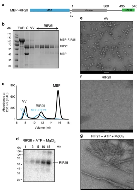
filaments in vitro via its CARD domain. Using the
baculovirus system in sf21 cells, we expressed and purified
recombinant full-length human RIP2 with a cleavable
maltose-binding protein (MBP) tag at the N-terminus
36(MBP-RIP2fl)
(Fig.
1
a–c). Negative-stain EM of MBP-RIP2fl eluting from the
amylose resin shows that the sample is a mixture of aggregates
and oligomeric protein (Supplementary Fig. 1a). Addition of ATP
and magnesium promotes elongation of the aggregates into a
filamentous structure (Supplementary Fig. 1b). After MBP tag
cleavage, the protein was further purified by size exclusion
chromatography yielding a void volume (VV) fraction and a
soluble, non-aggregated fraction denoted RIP2fl (Fig.
1
b–c,
Sup-plementary Fig. 2). Analysis of the non-aggregated RIP2fl by
electrospray ionization (ESI) mass spectrometry confirmed that
the protein is highly phosphorylated (Supplementary Fig. 1c) and
an in vitro phosphorylation assay showed that it is capable of
further self-phosphorylation and is thus functionally active
(Fig.
1
d). Negative-stain EM of the VV fraction shows irregular
aggregates with some short
filaments displaying a central
fila-mentous core (Fig.
1
e, Supplementary Fig. 1d-e), whereas imaging
the RIP2fl fraction confirms that it is non-aggregated and
pre-sumably dimeric (Fig.
1
c, f and Supplementary Fig. 2). When
ATP and magnesium were added to the non-aggregated RIP2fl
fraction, the protein oligomerized into long
filaments of diameter
30–40 nm and variable length (0.1–1 μm) and which have a
tendency to side-by-side aggregation (Fig.
1
g). Extended
filaments
could also be obtained from the VV fraction by adding
non-aggregated RIP2fl, ATP and magnesium, with the VV aggregates
acting as seeds (Supplementary Fig. 1f).
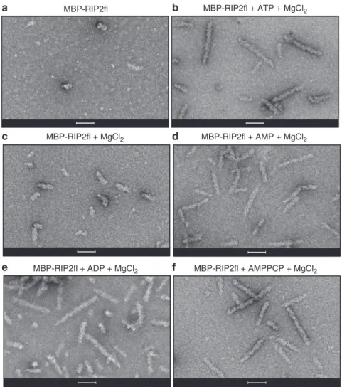
We then investigated the importance of different nucleotides in
promoting RIP2fl polymerization. For this, we used the uncleaved
MBP-RIP2fl fusion protein (Fig.
2
a) rather than tag-free RIP2fl.
This is because when ATP and magnesium are added,
MBP-RIP2fl polymerizes into short filaments (0.1–0.2 μm) that
aggregate less compared to those made with tag-free protein,
making them easier to visualise by negative-stain EM (Fig.
2
b).
MBP-RIP2fl polymerization was imaged in the presence of
magnesium alone (Fig.
2
c) or together with either ATP (Fig.
2
b),
AMP (Fig.
2
d), ADP (Fig.
2
e) or the ATP analogue AMPPCP
(Fig.
2
f). The micrographs reveal that MBP-RIP2fl begins to
polymerize in the presence of magnesium and the
filaments
elongate when any adenosine nucleotide is provided. Given that
the recombinant RIP2fl is active and already highly
phosphory-lated (Fig.
1
d and Supplementary Fig. 1c), these data suggest that
ATP (or other adenosine nucleotide) boosts RIP2fl
polymeriza-tion by stabilizing the kinase domain in a pro-filament
conformation,
rather
than
by
promoting
further
auto-phosphorylation.
Interestingly, the polymerized RIP2fl filaments appear to have
an inner core decorated with surface projections (Fig.
1
g,
Supplementary Fig. 1e). In analogy to other adaptor proteins
capable of polymerization via their DDs
30,32–34,37, we
hypothe-sized that the RIP2CARD forms the
filament core, whilst the
outer projections correspond to the kinase domain. Therefore, we
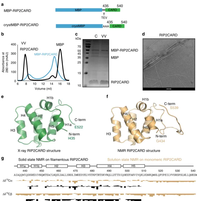
expressed and purified from E. coli recombinant RIP2CARD
(residues 435–540) with a cleavable MBP tag at the N-terminus
(MBP-RIP2CARD, Fig.
3
a–c). Upon MBP tag cleavage by the
Tobacco Etch Virus (TEV) protease, RIP2CARD mainly migrated
in the void volume (VV) of the size-exclusion chromatography
column (Fig.
3
b). Negative-stain micrographs revealed that
RIP2CARD from the VV forms long
filaments (Fig.
3
d), which
have similar length to the RIP2fl filaments, but a smaller diameter
of less than 10 nm.
The structure of monomeric RIP2CARD. To aid structural
analysis of the RIP2CARD
filaments, we determined the X-ray
crystal structure of RIP2CARD (residues 435–540), using a
con-struct with crystallisable MBP
38fused at the N-terminus
(crystMBP-RIP2CARD, Fig.
3
a, e). This construct crystallised in
space group P2
1with four molecules per asymmetric unit
(Sup-plementary Fig. 3). The structure was solved by molecular
replacement using NLRP1 CARD domain with a crystallisable
MBP at the N-terminus (PDB accession code 4IFP,
39) as search
model and refined at 3.3 Å resolution (Supplementary Table 1).
RIP2CARD has the typical CARD fold comprising a Greek key
helical bundle with the N- and C-termini oriented in the same
direction and with helix H1 broken into two shorter helices: H1a
and H1b (Fig.
3
e, Supplementary Fig. 3–4). The RIP2CARD
crystal structure is very similar to the previously reported solution
NMR structure (Fig.
3
f, PDB code: 2N7Z)
40, with a
root-mean-square deviation (RMSD) of all Cα positions of 0.95 Å.
Interest-ingly H6 is absent in both the crystal and NMR structures and
replaced by a long C-terminal loop, visible only in the NMR
structure, which contains putative phosphorylation sites
41–43.
MBP Kinase CARD TEV
g
RIP2fl MBP-RIP2fl RIP2fl + ATP + MgCl2 300 1 435 540 MBPb
c
d
f
e
MBP-RIP2fla
RIP2fl VV kDa 170 130 100 70 55 45 35 25 EAR C VV RIP2fl 1 0 300 600 900 6 8 10 12 14 16 18 RIP2fl VV Volume (ml) Absorbance at 280 nm (mAU) MBP MBP-RIP2fl VV Min kDa 130 100 70 55 45 35 25 RIP2fl RIP2fl + ATP + MgCl2 15 10 5 3Fig. 1 Full-length RIP2 formsfilaments in vitro. a Domain organization of the MBP-RIP2fl construct used for expression and purification of recombinant RIP2fl from sf21 insect cells. b 12.5 % SDS-PAGE showing recombinant RIP2fl at consecutive purifications steps. c Typical size exclusion chromatography profile for tagged (blue) and tag-free RIP2fl (black). d In vitro kinase activity of RIP2fl. Complete auto-phosphorylation is achieved after 15 min. e–g Negative-stain micrographs of (e) VV RIP2fl, (f) RIP2fl and (g) RIP2fl with added ATP and magnesium chloride. Scale bars are 100 nm. EAR: eluate from amylose resin, C: sample after tag cleavage as loaded on size-exclusion chromatography, VV: size exclusion chromatography void volume, Min: minutes
The structure of RIP2CARD within
filaments. We used
solid-state NMR to study the structure of RIP2CARD within the
fila-ment. In order to obtain backbone resonance assignments, we
recorded
1H-detected (H)CANH, (HCO)CA(CO)NH, (HCA)CB
(CA)NH, (HCA)CB(CACO)NH, (H)CONH and (H)CO(CA)NH
spectra on
2H,
13C,
15N-labeled and 100% back-exchanged
RIP2CARD samples at 60 kHz magic angle spinning (MAS)
44.
This data was evaluated together with
13C-detected
13C-
13C
DARR correlations on protonated samples, that were either
uniformly
13C-labelled or selectively-labelled using [2-
13C]- or
[1,3-
13C]-glycerol as carbon source during protein
expres-sion
45,46. The analysis of the
1H-detected data yielded the
sequence specific assignment for residues Q441 to Q507
(Sup-plementary Table 2), except for the loop residues 448–451 and
497. The chemical shifts of the assigned residues of
filamentous
RIP2CARD closely match many chemical shifts of monomeric
RIP2CARD in solution showing that the overall conformation is
maintained upon
filament formation (Fig.
3
g). We were not able
to assign any cross-peaks to the C-terminal 29 residues that were
reported to be
flexible by solution NMR investigations
40. To
check whether these signals are absent in our MAS NMR spectra,
we inspected
13C-
13C correlation spectra of the samples with a
2-or 1,3-glycerol labelling pattern. At sh2-ort mixing times, the amino
acids Leu, Pro, Thr and Val lead to characteristic cross-peak
pattern that allow for a counting of signals. We observed signals
corresponding to 9 of 14 leucine residues, 6 of 6 Thr, 5 of 7 Val
and only 1 of 4 proline residues (Supplementary Fig. 5). Relying
on the distribution of the respective amino acid types in the
sequence, this strongly suggests that the missing signals concern
residues in the C-terminal segment from 512 to 540. Especially
the absence of three proline signal sets, only one being detected,
indicates strong structural heterogeneity or mobility in that
region where they cluster. Furthermore, the number of missing
Leu and Val signal sets corresponds to the number present in the
C-terminus and thus corroborates the lack of an ordered
struc-ture there, indicating that H6 is also absent in
filamentous
RIP2CARD.
NOD2CARDS bind to one end of the RIP2CARD
filament. We
have shown that both RIP2fl and RIP2CARD samples form
filaments in vitro. However, in the cellular context, we expect that
such polymerization is initiated by NOD2 oligomerization in
response to cognate ligand binding. To recapitulate the core
elements of this process, we set out to reconstitute in vitro a
filamentous sample comprising the CARDS of both proteins that
would be suitable for high-resolution structure determination by
cryo-EM.
We
first investigated whether NOD2 could be detected by
immuno-gold labelling in RIP2CARD
filaments formed in the
presence of NOD2, following what was previously done for both
c
d
f
e
b
MBP-RIP2fl MBP-RIP2fl + MgCl2 MBP-RIP2fl + ATP + MgCl2 MBP-RIP2fl + AMPPCP + MgCl2 MBP-RIP2fl + AMP + MgCl2 MBP-RIP2fl + ADP + MgCl2a
Fig. 2 Full-length RIP2filaments are promoted by nucleotide binding. a–c Negative-stain micrographs of RIP2fl filaments obtained from (a) MBP-RIP2fl alone, (b) MBP-RIP2fl plus ATP dissolved in magnesium chloride buffer, (c) MBP-RIP2fl plus magnesium, 5 mM nucleotide dissolved in buffer containing magnesium chloride: (d) AMP, (e) ADP and (f) AMPPCP. Scale bars are 100 nm
the AIM2-ASC or NLRP3-ASC complexes
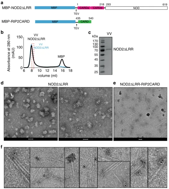
33. Using the
baculo-virus insect cell system we expressed and purified a truncated
form of NOD2 with a TEV cleavable MBP tag, comprising the
CARDS and NOD, but lacking the LRR domain
(MBP-NOD2ΔLRR, residues 1–619) (Fig.
4
a). This construct is
presumed to be derepressed with the CARDS available for
interaction. Indeed, a similar NLRP3 construct proved to be a
more powerful ASC polymerization promoter compared with the
full-length receptor
33.
Purified and tag-free NOD2ΔLRR eluted mainly in the void
volume of a size-exclusion chromatography column (Fig.
4
b–c)
and consistent with this, negative-stain images showed that
NOD2ΔLRR forms soluble aggregates (Fig.
4
d). We mixed
MBP-RIP2CARD with a less aggregated fraction of NOD2ΔLRR
(Fig.
4
b, d) and induced
filament polymerization by addition of
TEV. We then applied immuno-gold labelling against NOD2. As
a control, we applied the same immuno-gold labelling to the
NOD2ΔLRR sample in the absence of RIP2CARD (Fig.
4
d). The
control demonstrates that with the protocol used, NOD2ΔLRR
can be specifically labelled, although with a heterogeneous
number of gold-particles bound per aggregate. Micrographs of
the NOD2ΔLRR sample mixed with RIP2CARD showed
crystMBP AAA CARD540 435 Δδ13Cα Δδ13Cβ 440 450 460 470 480 490 500 510 520 530 540 H1a H1b H2 H3 H4 H5 MBP CARD
a
435 540b
c
TEV RIP2CARD MBP-RIP2CARD MBP VV kDa 70 55 45 35 25 15 10 C RIP2CARDSolution state NMR on monomeric RIP2CARD Solid state NMR on filamentous RIP2CARD
g
d
X-ray RIP2CARD structure NMR RIP2CARD structure
C-term N-term H1a H1b H2 H3 H4 H5 G434 S539
e
f
MBP-RIP2CARD crystMBP-RIP2CARD C-term N-term H1a H1b H2 H3 H4 H5 E522 I435 0 100 200 300 400 6 8 10 12 14 16 Absorbance at 280 nm (mAU) Volume (ml) MBP-RIP2CARD VV 18 RIP2CARD MBPFig. 3 Structure of monomeric RIP2CARD. a Domain organization of MBP-RIP2CARD constructs used for expression and purification of recombinant RIP2CARD fromE. coli Rosetta 2. MBP-RIP2CARD was used for characterising the polymerization ability of the CARD domain and for NMR experiments. CrystMBP-RIP2CARD was used for crystallization.b Typical size exclusion chromatography profiles of RIP2CARD purification showing both tagged (blue) and tag-free RIP2CARD (black).c 17 % SDS-PAGE showing typical sample obtained from RIP2CARD purification. The SDS-PAGE indicates that tag cleavage by TEV is incomplete.d Negative-stain electron micrograph of RIP2CARD VV. Scale bar is 100 nm. e Ribbon diagram of the RIP2CARD crystal structure reported in this paperf Ribbon diagram of the solution NMR structure of RIP2CARD (PDB: 2N7Z). g Comparison of secondary chemical shifts of RIP2CARD as a monomer in solution (yellow; BMRB entry: 25828) and in thefilament, determined by proton-detected solid-state NMR (black). Experimental13Cα and13Cβ chemical shifts were subtracted from the respective random coil values for each amino acid type (ΔδCα, ΔδCβ)79,80. C:
gold-particles
on
individual
NOD2ΔLRR aggregates or
NOD2ΔLRR aggregates bound to RIP2CARD filaments, mostly
at one
filament-end (Fig.
4
e–f).
We then investigated by co-purification and immuno-gold
labelling whether the NOD2CARDS are sufficient to interact with
the RIP2CARD
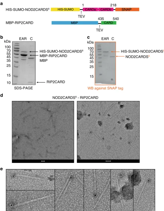
filament. For this, we used NOD2CARDS
(residues 1–218) expressed with a cleavable N-terminal
HIS-SUMO tag and a C-terminal SNAP tag (HIS-HIS-SUMO-NOD2-
(HIS-SUMO-NOD2-CARDS
S) together with cleavable MBP-RIP2CARD (Fig.
5
a). Due
to the different requirement for optimal expression of these two
constructs, we expressed them separately in E. coli and then
mixed the pellets and co-purified the proteins. After clarification
of the crude extract by centrifugation, the supernatant was
applied to amylose resin and the eluate was analysed by
SDS-PAGE and western blot (WB), using a specific antibody against
the SNAP tag (Fig.
5
b–c). The results showed that
HIS-SUMO-NOD2CARDS
Sco-elute with MBP-RIP2CARD (Fig.
5
b–c). The
diameter of
filaments observed by negative-stain EM after
HIS-MBP tag cleavage was the same as the homo RIP2CARD
filaments, while their length ranged from 50 to 500 nm (Fig.
5
d).
We then applied immuno-gold labelling against the SNAP tag.
The results showed single gold-particles mostly sitting on
filament-ends (Fig.
5
d–e, Supplementary Fig. 6). In order to
evaluate the binding position of NOD2CARDS
Son RIP2CARD
filament, two more controls were performed with the same
immuno-gold labelling protocol: immuno-gold labelling on
RIP2CARD
filament without NOD2CARDS
S(Control 1, C1)
and immuno-gold labelling with only the secondary antibody on
NOD2CARDS
S-RIP2CARD
filament (Control 2, C2)
(Supple-mentary Fig. 6c, d). Control 1 was used to evaluate the specificity
of the primary antibody, whilst Control 2 was used to judge the
specificity of secondary antibody. We collected 20 random images
on two different grids for each condition at a magnification of
×16,000 and evaluated the number and position of gold-particles
MBP-RIP2CARD MBP CARD 435 540 NOD MBP 0 50 100 150 6 8 10 12 14 16 18 MBP VV NOD2ΔLRR Absorbance at 280 nm (mAU) volume (ml) VV NOD2ΔLRR
a
b
c
e
MBP-NOD2ΔLRR NOD2ΔLRRf
CARDb CARDa TEV TEV 1 218 293 619 kDa 170 130 100 70 55 45 35 25 VVd
NOD2ΔLRR NOD2ΔLRR-RIP2CARDFig. 4 NOD2ΔLRR binds RIP2CARD filaments. a Domain organization of NOD2ΔLRR and RIP2CARD constructs used for immuno-gold labelling experiments. Expression of recombinant NOD2ΔLRR was done in sf21 insect cell. b, c Size exclusion chromatography profile (b) and (c) corresponding 12.5% SDS-PAGE showing typical sample obtained from NOD2ΔLRR purification. The size exclusion chromatography profile shows that both tagged (blue) and tag-free NOD2ΔLRR (black) elute in the VV. The dashed rectangular shape indicated the protein fraction used for immuno-gold labelling experiments. d Negative-stain images of VV NOD2ΔLRR, corresponding to the fraction used reconstitution with RIP2CARD, unlabelled (left) and after immuno-gold labelling (right).e, f Example negative-stain micrograph of RIP2CARDfilaments with NOD2ΔLRR bound (e) and zoom showing gold-particles (black dots) after immuno-gold labelling against NOD2 (f). Scale bars are 50 nm. VV: size exclusion chromatography void volume
in each micrograph (Supplementary Fig. 6f). Final statistics
revealed that 70.9% of gold-particles are found on
filaments, of
which 91.7 % are at one end and we never observe gold-particles
at both ends. This shows that NOD2CARDS
Sare preferentially
bound at one end of the RIP2CARD
filament. These data are
consistent with the hypothesis that under physiological
condi-tions activated NOD2 nucleates RIP2
filament formation yielding
a polar assembly.
Cryo-EM of RIP2CARD
filament. To elucidate the architecture
of the RIP2CARD
filament by cryo-EM, we optimized the
pro-tocols for production of both the RIP2CARD and
NOD2-CARDS
S-RIP2CARD
filaments (Supplementary Fig. 7). For this
we used a different RIP2CARD construct encoding for
RIP2-CARD (residues 431
– 540) with a P3C (human rhinovirus 3C
protease) cleavable HIS-MBP tag at the N-terminus
(HIS-MBP-RIP2CARD) (Supplementary Fig. 7a). This new RIP2CARD
construct dramatically increased the tag cleavage efficiency
(compare Fig.
3
c with Supplementary Fig. 7c, f). We optimised
the purification protocol for NOD2CARDS
S-RIP2CARD
fila-ments, by introducing a size exclusion chromatography step
before tag cleavage (Supplementary Fig. 7b). This allows
aggre-gates to be discarded and the tagged NOD2CARDS
s-RIP2CARD
complex to be separated from monomeric HIS-MBP-RIP2CARD.
NOD2CARDS
s-RIP2CARD complexes and RIP2CARD were
then recombined as described in the Methods. After cleavage,
MBP CARD 435 540 CARDb SNAP CARDa HIS-SUMO TEV 1 218a
b
c
d
e
TEV 100 70 55 45 35 25 15 10WB against SNAP tag
NOD2CARDSs EAR C EAR C NOD2CARDSs - RIP2CARD RIP2CARD HIS-SUMO-NOD2CARDSs MBP-RIP2CARD MBP MBP-RIP2CARD HIS-SUMO-NOD2CARDSs HIS-SUMO-NOD2CARDSs kDa 100 70 55 45 35 25 15 kDa SDS-PAGE
Fig. 5 NOD2CARDS bind at one end of the RIP2CARDfilaments. a Domain organization of HIS-SUMO-NOD2CARDSsand RIP2CARD constructs used for immuno-gold labelling experiments. Expression of recombinant of HIS-SUMO-NOD2CARDSswas carried out inE. coli Rosetta 2. b, c 17% SDS-PAGE (b) and corresponding WB (c) of NOD2CARDSs-RIP2CARD co-purification at the amylose elution step (EAR) and after tag cleavage (C) . For clarity, only relevant lanes are labelled.d Negative-stain image of RIP2CARDfilaments with NOD2CARDSsbound, unlabelled (left) and after immuno-gold labelling (right). Scale bars are 50 nm.e Zoom on micrographs of RIP2CARDfilaments with NOD2CARDSsbound, showing gold particles (black dots) after immuno-gold labelling against the SNAP tag attached to NOD2CARDSs
HIS-MBP, HIS-SUMO tags and HIS-tagged proteases, were
removed by affinity chromatography followed by dialysis with a
high molecular weight cut off (Supplementary Fig. 7c).
RIP2-CARD
filaments were prepared following the same protocol
(Supplementary Fig. 7f). Negative-stain and cryo-EM
micro-graphs of samples prepared under the same conditions show that
RIP2CARD
filaments with bound NOD2CARDS
sare shorter,
straighter and have a lower tendency to aggregate than
RIP2-CARD
filaments without NOD2 (Supplementary Fig. 7d, e, g, h).
Therefore, the hetero-CARD
filaments were used for cryo-EM
data collection and analysis (Supplementary Table 3).
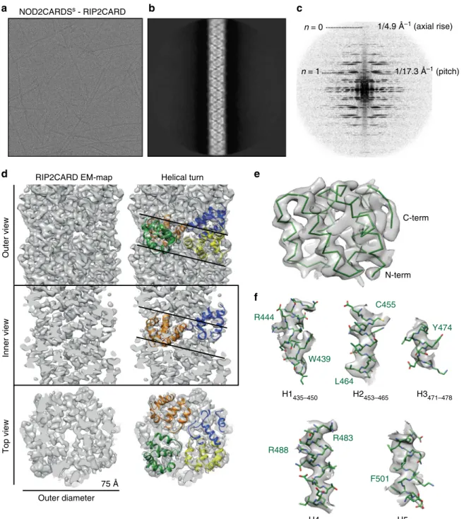
Visual inspection of the individual cryo-EM images, 2D
class-averages and corresponding power spectra indicate that the
RIP2CARD
filament has a helical symmetry (Fig.
6
a–c). Indexing
of the power spectra and symmetry refinement revealed a
left-handed helix of 3.56 subunits/turn with an axial rise of 4.848 Å/
subunit (Supplementary Fig. 8a, b). The
final cryo-EM map at
3.94 Å resolution (Supplementary Fig. 8c) shows that the
filament
has an approximate outer diameter of ~75 Å with a central
solvent channel of ~25 Å diameter (Fig.
6
d). The crystal structure
of RIP2CARD can be unambiguously
fitted into the cryo-EM
density, with both N- and C-terminal ends orientated towards the
outside of the
filament (Fig.
6
e). Manual adjustment using the
clear density for the
α-helices and the larger side chains such as
tryptophan and arginine (Fig.
6
f), followed by real space
refinement, led to a final atomic model which comprises
RIP2CARD residues P433 to Q518 (Supplementary Table 3).
This shows that, consistent with the solid-state NMR results,
there are only minor structural re-arrangements of the CARD
domain upon
filament formation (RMSD of 1.12 Å for Cα
positions of residues 433–518) and the presumed flexible
C-terminus is not observed in the EM map.
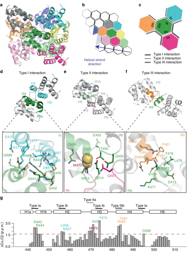
Structure of the RIP2CARD
filament. The RIP2CARD filament
has a similar helical configuration to other CARD filaments
already described, such as MAVS CARD, Caspase-1 CARD and
the recently described BCL10 CARD
filament
30,31,34,35(Supple-mentary Table 4). Following the established convention, the
RIP2CARD assembly can be described through interactions at
three major asymmetric interfaces, named type I, type II and type
III
47. Type I and II are inter-strand interactions, whilst type III is
intra-strand along the helical strand trajectory (Fig.
7
a–c).
The type I interface is defined as the interaction between
helices H1 and H4 of one molecule (type Ia surface) with H2 and
H3 of the adjacent one (type Ib surface). In the RIP2CARD
filament, the type I interaction is electrostatic in nature involving
several charged residues that form polar interactions (Fig.
7
d).
D461 and N457 from H3 (type Ib) interact with R444 and R448
from H1 and H4 (type Ia) respectively. The interaction between
H1 (type Ia) and H2 (type Ib) is further reinforced by backbone
contacts between the break in H1 and the C-terminus of H2
(Fig.
7
d, Supplementary Fig. 9a-b). Additional charged or polar
residues such as D492, D495, Q441, E445 and Q489 (type Ia) and
D467, K471 (type IIb) contribute to the type I interface (Fig.
7
d).
The type II interface is normally defined by the interaction
between the C-terminal end of H4 and the H4-H5 loop (type IIa
surface) and a groove defined by the H1 and H2 corner from one
side and H6 helix and its preceding loop on the other (type IIb
surface). In the case of the RIP2CARD, which lacks H6, we
identified a somewhat different type II interface. The type IIa
surface includes the C-terminal end of H4, the H4-H5 and
H2-H3 loops, whilst the type IIb surface comprises the H1-H2 loop,
the N-terminal of H2 and the visible part of the RIP2CARD
C-terminal (Fig.
7
e). At the type II interface, the side-chains of
M470 (type IIa) and C455 (type IIb) make van der Waals
interactions (Fig.
7
e and Supplementary Fig. 9c), these side-chains
being specific to RIP2CARD (Supplementary Fig. 4). This
interface is further reinforced by a polar interaction between
N512 and Q458 (Fig.
7
e). Moreover, Q497 (type IIa) can interact
with the side chains of Q450, T452 and Q514 (type IIb) (Fig.
7
e).
The type III interaction normally occurs between H3 (type
IIIa) and a groove formed by H1-H2 and the H3-H4 loop (type
IIIb). In the RIP2CARD
filament, type IIIb comprises the H3-H4
loop and N-terminal of H4, whilst the H1-H2 loop is only
contributing to the type IIa surface as described above (Fig.
7
f).
P481, T482 (type IIIa) and L476 (type IIIb) contribute
hydrophobic interactions, whilst E472 and E475 from H3 (type
IIIa) potentially form salt bridges with R488 and R483 (type IIIb).
Interestingly, both side chain of T482, T484 could swap from the
original position in the crystal structure to interact with type IIIa
instead of contributing to the intra-αhelical interactions (Fig.
7
f).
Figure
7
g shows that the most significant chemical-shift
differences between RIP2CARD in solution and within the
filament, as determined by NMR, map to the subunit interfaces
described above. These chemical-shift differences report on local
conformational changes due to packing effects in the
filament and
therefore independently confirm the overall architecture of the
intermolecular interfaces observed by cryo-EM. Notably, residues
K443, R444, D495, L457, L456, K471 and Y474, located close at
the type I interface, undergo strong chemical shift changes in the
filament. Residue Y474 shows the largest effect. At its Cγ
resonance, multiple contacts in
13C-
13C correlations employing
long mixing times are observed, which indicates tight packing of
Y474 in the
filament. Similarly, conformational changes in the
type IIa (M470) and type IIIb (R483, T484) residues lead to
significant chemical shift changes of the respective residues,
indicating a change in environment of these residues.
Mutational analysis of the RIP2CARD type II interface. Our
immuno-gold labelling results show that NOD2CARDS
Sbind at
one end of the RIP2CARD
filament, suggesting that an initial
hetero-CARD complex might act as a nucleation point to
pro-mote unidirectional RIP2CARD
filament growth. Available
structures or models of hetero DD complexes, such as
RIG-I-MAVS, the Myddosome, the PIDDosome and NLRP3-ASC, show
that the DD belonging to the receptor protein continues the
helical arrangement of the effector DDs, by forming a
combina-tion of the same type I–II-III interfaces. With a view to testing the
effect on signalling of site-directed mutants that would uniquely
disrupt the RIP2CARD
filament structure and not the
RIP2-NOD2 hetero-CARD interaction, we modelled the hypothetical
hetero-CARD type I, II, III interfaces and evaluated their
importance based on the available interaction and mutagenesis
studies
12,14(Supplementary Fig. 10). As the structure of
NOD2-CARDS is not yet available and the only reported direct
inter-actions are to the N-terminal NOD2 CARDa (residues 26–122),
we computed a three-dimensional NOD2CARDa model using
Swiss Modeller
48using as template the X-ray structure of the
CARD of Nucleolar Protein 3 (PDB code:4UZ0)
49, which shares
35% identity with NOD2CARDa (Supplementary Fig. 10a), The
NOD2CARDa model structure obtained is similar to RIP2CARD
(RMSD of 1.70 Å for all Cα positions), consistent with the
pre-sumed compatibility of both CARDS to form similar type I-II-III
interactions.
NOD2CARDa residues R38, R86, E69, D70 and E72 and
RIP2CARD residues E472, D473, E475, D461 and D492 residues
have been shown to be important for hetero-CARD
interac-tions
12,14. Our modelling shows that all these charged residues
occur in type I or type III interfaces and are in fact highly
conserved
between
NOD2CARDa
and
RIP2CARD
(Supplementary Fig. 10b-c, and Supplementary Fig. 4). Therefore,
mutagenesis of RIP2 residues involved in these interfaces would
affect both the interaction with NOD2 and
filament formation.
Conversely, none of the residues belonging to the observed RIP2
homo-type II interface have been explicitly implicated in the
NOD2CARD-RIP2CARD interaction. We deduced that type II
interactions within the RIP2CARD
filament, notably involving
RIP2CARD specific hydrophobic residues C455 and M470, could
specifically stabilise homo-interactions within the filament.
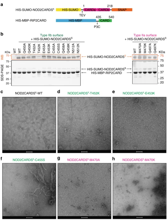
We therefore mutated the residues belonging to RIP2CARD
type IIa and IIb surfaces (type IIa: M470 and Q497; type IIb:
Q450, T452, E453, C455, Q458 and N512) by alanine and lysine
substitution (serine for C455). We then assayed the ability of each
mutated RIP2CARD construct to bind NOD2CARDS
S, using the
co-purification protocol described above (Fig.
8
a, b). Mutant
RIP2CARD domains were expressed at similar level to wild-type
RIP2CARD and all the mutants displayed unimpaired binding to
NOD2CARDS
S, except for RIP2CARD T452K, which clearly
Outer diameter
d
Outer view Inner view Top view 75 ÅRIP2CARD EM-map Helical turn
e
f
C-term N-term H1435–450 H2453–465 H3471–478 H4483–497 H5499–511 Y474 F501 R483 R444 W439 R488a
b
c
n = 0 1/4.9 Å–1 (axial rise) n = 1 1/17.3 Å–1 (pitch) C455 NOD2CARDSs - RIP2CARD L464Fig. 6 Cryo-EM structure of the RIP2CARDfilament. a Cryo-EM image of NOD2CARDSs-RIP2CARDfilaments used for structure determination. b, c 2D-class average (b) and corresponding power spectra (c) used for initial symmetry parameter estimation. d Final cryo-EM map of the three-dimensional RIP2CARDfilament at 3.94 Å resolution (FSC in Supplementary Fig. 8c). Outer, inner and top view without (left) and with (right) RIP2CARD models fitted into one helical turn.e View of the RIP2CARD monomerfitted into the EM map showing only the main chain for clarity. f Zoomed-in views of the fitting of individualα-helices into the sharpened cryo-EM density. Helices are defined as in the sequence alignment (Supplementary Fig. 4) except for H1, which starts from I435 in the RIP2CARD monomer within thefilament. Sidechains are shown as stick models. A few residues with clear EM density for sidechains are labelled
H5 H1b H2 H4 H3 H5 H1a H1b H2 H3 H4 H3 H3 H4 Type I interaction Type II interaction Type III interaction
H1b
a
b
c
d
e
f
P481 Y474 N457 D461 S465 K471 K471 R483 R488 T482T479 L476 E475 D467 S442 R444 R488 E445 E453 N512 Q514 T452 Q450 Q458 C455 M470 Q497 D495 Q441 E475 Q489 E472Type I interaction Type II interaction Type III interaction
D492 L464 D473 T484 Helical strand direction IIb IIa Ia Ib IIIb IIIa 0.0 1.5 3.0 440 450 460 470 480 490 500 Type Ib Type Ia Type IIa Type IIIb Ib Ia IIb IIa IIIa IIIb 510 Type Ib Δ C α ,C β (p.p.m.)
g
Type Ia H2 H1a H3 H4 H5 R444 K443 N457 L456 M470 K471 Y474 R483T484 D495Fig. 7 Structural analysis of the RIP2CARDfilament assembly. a Ribbon diagram of RIP2CARD filament comprising 10 subunits. b, c Schematic diagram of the helicalfilament (b) and relative orientations of type I, type II and type III interfaces (c). Each subunit is represented as a hexagon with the same colour code as in thefilament structure in (a). Each turn comprises 3.56 subunits. The fourth subunit is represented as a half-empty hexagon to highlight that it is shared with the next turn. Type I, II, III interfaces are represented as a single line, single-dashed line or double line respectively.d–f Ribbon diagram of RIP2CARD dimers interacting through (d) type I, (e) type II and (f) type III interfaces. Protein regions involved in the interface are highlighted using the colours as in (c). The insets show the interactions at relative type surface. H-bonds are represented by black dashed line. Backbone contacts are highlighted by blue dash line, respectively. Sulphurous groups involved in Van der Waals interactions in the type II surface are represented as spheres. g Sum of13Cα and13Cβ chemical shift differences between RIP2CARD in the solution and the solid state for each assigned amino acid. Residues that show a strong variation from the mean (1.2 p.p.m.; dotted red line) are labelled in the same colour code as used in (d–f). The secondary structure elements and positions of the interface surfaces are also shown
showed lower binding in comparison to wild-type and all the
other mutants. We then tested the ability of each construct to
polymerize after tag cleavage by imaging the sample with
negative-stain EM. The micrographs revealed that many
constructs can still polymerise (Supplementary Fig. 11), but with
a dramatic change in the
filament quality notably for mutants
T452K, E453K, C455S, M470A and M470K (Fig.
8
c–h).
RIP2CARD-T452K forms long
filaments but with multiple
interruptions compared to wild-type and furthermore they have
impaired binding to NOD2CARDS (Fig.
8
b–d). This indicates
that the mutation negatively affects both binding to
NOD2-CARDS
Sand
filament quality. Micrographs of
RIP2CARD-E453K and RIP2CARD-M470A show particles that might
correspond to protein aggregates with rare
filaments (Fig.
8
e,
g). RIP2CARD-C455S forms irregular, more
flexible filaments
with a high tendency to aggregate (Fig.
8
f). M470K micrographs
show protein aggregation and absence of
filaments (Fig.
8
h).
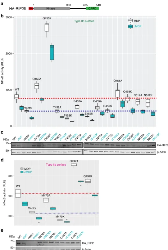
We next investigated the effect of these mutations on the
activation by NOD2 of transcription factor NF-ĸB, by using a
luciferase reporter assay (Fig.
9
and Supplementary Fig. 12). We
transiently transfected HEK293T cells with HA tagged RIP2fl
mutants (Fig.
9
a) together with a plasmid encoding
firefly
luciferase under the control of NF-ĸB promoter
50. NF-ĸB
activation was induced using the specific NOD2 activator MDP,
and cells were lysed 20 h later to record luciferase activity and
assay protein expression. In agreement with the in vitro data,
mutants Q450, Q458, Q497, N512 show unimpaired NF-κB
signalling; with the particularity that Q450K, Q497A and Q497K
HIS-MBP-RIP2CARD HIS-MBP CARD435 540 CARDb SNAP CARDa HIS-SUMO 1 218 75 KDa HIS-SUMO-NOD2CARDSS HIS-SUMO-NOD2CARDSS HIS-MBP-RIP2CARD WB SDS-PAGE HIS-SUMO-NOD2CARDSs TEV P3C
a
b
Type IIb surface+ HIS-SUMO-NOD2CARDSS
Type IIa surface
+ HIS-SUMO-NOD2CARDSS 75 75 50 KDa 37 25 20 75 50 37 25 20 WT M470A M470K Q497A Q497K WT WT Q450A Q450K T452A T452K E453A E453K C455A C455S Q458A Q458K N512A N512K
NOD2CARDSs-E453K NOD2CARDSs-T452K
NOD2CARDSs-WT
NOD2CARDSs-C455S NOD2CARDSs-M470A NOD2CARDSs-M470K
c
d
e
f
g
h
Fig. 8 Effect of type II surface mutations on RIP2CARD polymerization. a Domain organization of the NOD2CARDS and RIP2CARD constructs used for testing the behaviour of RIP2CARD mutants.b Western blot (WB, top panel) and 12.5% SDS-PAGE (bottom panel) of NOD2CARDSs-RIP2CARD type II interface mutant co-purification at the amylose elution step (EAR). For clarity, only relevant lanes are labelled. Type IIa and IIb surfaces are coloured as in Fig.7.c–h Example micrographs of (c) NOD2CARDSs-WT, (d) NOD2CARDSs-T452K, (e) NOD2CARDSs-E453K, (f) NOD2CARDSs-C455S, (g) NOD2CARDSs-M470A and (h) NOD2CARDSs-M470K, after tag cleavage. Scale bars are 100 nm
show high levels of auto-activation compared with the wild-type
RIP2 (Fig.
9
b, d). In contrast, T452K, E453K, C455S and M470K
failed to transmit the signal from NOD2 to NF-κB, as the
luciferase value is equal or lower than the control empty vector
(Fig.
9
b, d). The less drastic alanine mutants T452A, E453A,
C455A and M470A also showed lower or zero activity (Fig.
9
b, d).
All the mutants show similar protein expression to wt RIP2
(Fig.
9
c, e and Supplementary Figure 12).
cMDP WT Q450A T452A T452K E453A E453K C455A C455S Q458A Q458K N512A N512K Vector 2000 1000 0 HA_RIP2 β-Actin MDP cMDP WT Vector M470K Q497A Q497K M470A
Type IIa surface
900
600
300
HA-RIP2fl Kinase CARD
1 300 435 540
HA
HA-RIP2 β-Actin
WT cWTvector Q450A Q450K T452A T452K E453A E453K C455A C455S Q458A Q458K N512A N512K
75 50 KDa 75 50 KDa MDP Q450K
cQ450A cQ450K cT452A cT452K cE453A cE453K cC455A cC455S cQ458A cQ458K cN512A cN512K
cvector
Type IIb surface 3000 NF-κ B activity (RLU) NF-κ B activity (RLU)
WT cWTvectorcvectorM470AcM470AM470KcM470KQ497AcQ497AQ497KcQ497K
a
b
c
d
e
Fig. 9 Effect of type II surface mutations on NOD2 dependent NF-κB signalling. a Domain organization of the HA-RIP2fl construct used for the luciferase reporter assay.b, d Luciferase reporter assay in HEK293T cells showing the effect on NF-κB signalling of transfected WT and mutant RIP2fl for (b) type IIb surface and (d) type IIa surface. Each MDP value is significantly higher than that using c MDP. Average RLU values for WT RIP2fl-MDP and control-MDP are shown by dashed red and blue lines, respectively. The boxplot is the graphical representation of the summary statistics of a vector contains information in this order: minimum value (lower whisker),first quartile (25% of data lower bound box), median (50% of data centre line), third quartile (75% of data higher bound box) and maximum value (upper whisker). Black dots on boxplot are extreme values.c, e Example WB for (c) type IIb surface mutants and (e) type IIa surface mutants, showing expression of wild-type (WT) HA-RIP2fl and mutants (top) and β-actin (bottom). For each construct, the results for cells induced by either MDP or its control (cMDP,c) are reported in black and cyan respectively
Discussion
Recent studies on several innate immune systems have shown
that recognition of cognate ligands by PRRs that contain a DD
(e.g. CARD and PYD) triggers their oligomerization and
inter-action with the downstream adaptor resulting in the formation of
a higher-order
filamentous assembly called a signalosome
29,51.
Here we focus on the NOD2-RIP2 signalling pathway, a
receptor-adaptor protein combination that shares close structural
simila-rities with PRRs involved in signalosome formation. Specifically,
we investigated whether the recruitment of RIP2 by NOD2 via
CARD-CARD interactions could lead to the formation of such a
signalosome (‘nodosome’). Our biophysical, structural and
func-tional data show that RIP2, via its CARD, can form helical
fila-ments, plausibly nucleated from one end by activated NOD2.
Furthermore, we show that RIP2 polymerization is essential for
NF-ĸB activation by NOD2, presumably by favouring
recruit-ment of downstream effectors such as the RIP2 ubiquitin ligase
XIAP
17.
The starting point was our
finding that phosphorylated and
active RIP2fl forms filaments in vitro in the presence of ATP and
magnesium. The subsequent observation that RIP2CARD
also spontaneously forms more slender
filaments, suggests that
the CARD domain forms the core of the RIP2fl filaments, while
the kinase domain (RIP2K) is on the exterior. Interestingly
we observed that not only ATP, but also non-hydrolysable
adenosine nucleotides together with magnesium promote
merization of RIP2fl. This suggests that enhanced RIP2
poly-merization by nucleotide-binding results from RIP2K structure
stabilization
rather
than
any
increase
in
RIP2K
auto-phosphorylation activity. Our previously published biophysical
data
36, show that stable activation of RIP2K involves the coupling
of kinase dimerization with auto-phosphorylation of the
activa-tion loop. We therefore speculate that CARD polymerizaactiva-tion
promotes kinase dimerization, by increasing the local RIP2
con-centration. Dimerization favours the kinase domain being in the
active conformation and therefore able to bind any
adenosine-derived nucleotide, independently from the phosphorylation state
of the activation loop. In a physiological context, RIP2 could
either be already phosphorylated, as it was reported that
auto-phosphorylation contributes to protein stabilization
36,52or
fur-ther phosphorylated upon dimerization to stabilize the active
conformation. These observations are in line with a recent study
showing that an active conformation of the RIP2 kinase, rather
than necessarily a catalytically active kinase, is essential for
NOD2 signalling, since it permits interaction with the E3 ligase
XIAP
17.
In order to investigate the assembly mechanism of the RIP2
filament core, we co-purified RIP2CARD and NOD2CARDS and
used immuno-gold labelling to show that NOD2CARDS bind
preferentially at one of the two
filament-ends, forming a polar
assembly. We then successfully imaged this sample using
cryo-EM, obtaining an EM density map at 3.94 Å resolution, where we
could unambiguously
fit and refine the X-ray structure of
RIP2-CARD, also reported in this paper. Solid-state NMR confirmed
the absence of an ordered structure for the C-terminal segment in
the
filaments, involving residues beyond K510. RIP2CARD
assembles into a left-handed helical
filament with 3.56 subunits
per turn, a configuration similar to that described for filaments of
MAVS, Caspase-1 and, most recently, BCL10 CARDs
30,31,34,35.
RIP2 thus assembles differently from RIP1 and RIP3, which
polymerize through their RHIM (RIP homotopic interaction
motif) domain into amyloid
fibrils
53.
RIP2CARD assembles into a helical arrangement using the
conventional type I, II and III interfaces. Consistent with this,
Cα-Cβ chemical shift differences between the solution and solid-state
are the most significant for the residues involved at these
interfaces (Fig.
3
g). The cryo-EM structure reveals that the type I
and type III interfaces are polar (Fig.
7
d, f), mediated by
inter-actions of R488 and R444 (type Ia surface) with N457 and D461
(type Ib), and by contacts between E472 and E475 (type IIIa
surface) and R483, R488 and T484 (type IIIb). The type II
interaction is more hydrophobic for instance involving C455 and
M470 (Fig.
7
e). Y474, which has the highest chemical shift
change, is observed buried in the type I interface (Fig.
7
d),
sug-gesting that it is not accessible for phosphorylation in the
fila-ment. Nonetheless, it is reported that auto-phosphorylation of
this residue is important for NOD2 signalling
54, but might occur
at a later stage, after RIP2 ubiquitination by cIAP1
54.
We then tested the relevance of RIP2 polymerization on the
activation of NF-κB by NOD2, by designing RIP2CARD mutants
that potentially affect RIP2 polymerization but preserve the
interaction with NOD2. Based on available structures of
hetero-CARD complexes
31,47, we expect that a cluster of NOD2CARDS
from activated and oligomerized NOD2 could form a short
helical extension that preserves the same helical parameters as the
RIP2CARD
filament. By combining this premise with available
mutagenesis data, we deduced that the observed type II interface
in the
filament, unlike the type I or III interfaces, may be specific
to the RIP2 homo-CARD interaction (Supplementary Fig. 10).
Therefore, we performed mutagenesis of the RIP2 type II surface
with the aim of
finding mutants that specifically disrupt homo-,
but not hetero-CARD interactions, thus allowing assessing the
biological significance of the extended filament. Results from our
structure-guided mutagenesis, show that RIP2CARD mutations
to lysine (or serine for C455) at the type II interface can affect
either hetero- and CARD interactions or only
homo-CARD domain interactions. For instance, the T452K mutation
both decreased the interaction with NOD2 (Fig.
8
b), led to
aberrant
filament formation (Fig.
8
d) and severely affecting
sig-nalling (Fig.
9
b). On the other hand, mutations of E453, C455 and
M470 did not disrupt the interaction with NOD2 but severely
affected the quality of RIP2 polymerization and abolished
sig-nalling (Fig.
8
b, e–h and Fig.
9
b, d). The E453 side chain points
towards the central channel, suggesting that the lysine mutation
disfavours
filament elongation by affecting the inner filament
electrostatics. Similar reverse charge mutations at the type II
interfaces in MAVS CARD
filament, combined with others,
abrogate MAVS
filament elongation
31. The C455S and M470A/K
mutations would reduce the hydrophobic interaction at the
RIP2CARD type II interface, consistent with their strong effect on
the quality of
filaments or whether filaments form at all
(Fig.
8
f–h). Interestingly, some alanine mutants (e.g. T452A,
E453A and C455A) also show a significant decrease in the NF-κB
signal, despite the fact that they do not affect the ability of
RIP2CARD either to bind NOD2CARDS or to polymerize
in vitro. We speculate that alanine mutations have a milder effect,
compared to changing the charge, that cannot be detected by
negative-stain EM, except for the case of M470A, whose severe
disruptive affect highlights the importance of this RIP2 specific
residue in forming the type II interface in the
filament.
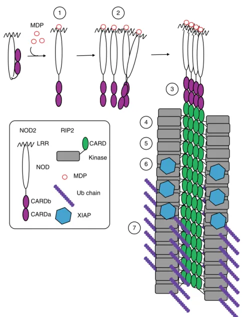
In conclusion, we provide evidence for the existence and
bio-logical importance of a NOD2-RIP2 polar
filamentous assembly,
which is likely the core of the nodosome complex. Based on the
results described here, other published data and analogy to other
signalosome systems, we propose that nodosome assembly occurs
as follows (Fig.
10
): (1) binding of MDP to NOD2 LRR domain
activates the receptor causing derepression of the CARDs; (2)
NOD2 oligomerises via its NOD and CARD domains; (3)
oli-gomerized NOD2 recruits RIP2 via its CARD domain forming
the hetero-CARD complex (4) cumulative binding of RIP2 to the
hetero-CARD complex promotes
filament elongation to form the
helical assembly here described; (5) polymerization of RIP2CARD
in the presence of ATP stabilises the active antiparallel dimeric
form of RIP2K; (6) E3 ligases, such as XIAP bind the active from
of RIP2K
17; (7) RIP2 becomes K63-ubiquitinated enabling it to
recruit downstream effector proteins.
Finally, by combining our biophysical, structural and
func-tional analysis with existing data, we provide essential
informa-tion that could potentially be used to explore new therapeutic
options for inflammatory diseases characterised by aberrant
NOD2-RIP2 signalling.
Methods
Constructs, protein expression and purification. Constructs described in this paper were generated from pcDNA3 plasmids encoding human RIP2 and human NOD250. The N-terminally HIS-tagged TEV (Tobacco Etch Virus) and P3C
(human rhinovirus 3C) proteases used in this paper were produced at the Protein Expression and Purification Core Facility at EMBL, Heidelberg, Germany.
Recombinant human RIP2fl and human NOD2ΔLRR(1–619) were produced using the baculovirus system in sf21 insect cells. The DNA sequence of RIP2fl and NOD2ΔLRR were cloned from pcDNA3 into the vector pFastBacHTB using NcoI and HindIII cloning sites. Using the In-Fusion cloning technology (Takara Clontech), the original TEV cleavable HIS tag was replaced with a TEV cleavable maltose-binding protein (MBP) tag, which improved both expression and stability of recombinant proteins during insect cells expression (MBP-RIP2fl and MBP-NOD2ΔLRR). MBP-RIP2fl and MBP-NOD2ΔLRR were expressed and purified
following the same protocol36.Virus generation and amplification, insect cell
infection and protein expression were performed at the EMBL Eukaryotic Expression Facility. Briefly, Sf21 cells at 0.6 × 10⁶cells ml−1were infected with a
virus shot able to stop cells growing in 24 h. On the fourth day post-infection, cells were harvested and re-suspended in 1 × 10⁻¹(v/v) ratio of lysis buffer 20 mM Tris pH 7.5, 50 mM NaCl, 2 mMβ-mercapto-ethanol (βMe), 0.01% NP40 supplied with protease cocktail inhibitor (Complete, Roche). Using a douncer, cells were homogenized and afterwards centrifuged at 18,000 × g for 30 min. The resulting supernatant solution was incubated for at least 2 h with amylose-affinity chromatography resin (New England Biolabs), whilst gently shaking at 4 °C. The fusion protein was then eluted using the same lysis buffer supplemented with 40 mM maltose. Upon overnight TEV cleavage, either RIP2fl or NOD2ΔLRR were applied to size exclusion chromatography, using a similar buffer composition of lysis buffer (20 mM Tris pH 7.5, 50 mM NaCl, 0.5 mM TCEP).
For crystallization purposes, RIP2CARD (435–540), was cloned into pETXM1 plasmid using the NcoI and XhoI cloning sites, resulting in a protein construct with an N-terminal crystallisable MBP (crystMBP-RIP2CARD) spaced by a three alanine linker. The construct was expressed in E. coli Rosetta 2 (Novagen) by growing the bacterial culture at 37 °C until an OD600 nmof 0.6 and inducing with
0.3 mM IPTG (isopropyl-β-D-1-thiogalactopyranoside) overnight at 16 °C. The cells where harvested, re-suspended in 1 10−1(v/v) ratio of lysis buffer 20 mM Tris pH8, 50 mM NaCl, 2 mM (β-Me) containing protease cocktail inhibitor (Complete, Roche) and lysed by sonication. The crude extract was centrifuged for 30 min at 18,000 × g and the soluble fraction was applied to amylose-affinity chromatography resin (New England Biolabs) and purified as described above. After elution, the fusion protein was applied to a prepacked anion exchange chromatography column (GE Healthcare) with a 0 to 1 M NaCl gradient. The protein was further purified on
1 2 3 6 4 5 7 MDP LRR NOD CARDb CARDa NOD2 CARD Kinase RIP2 MDP Ub chain XIAP
Fig. 10 Model of nodosome assembly. Model of nodosome assembly, based on the results described here, other published data and analogy to other signalosome systems. Steps 1–7 in the process are discussed in the main text. The legend for the components of the system is in the inset