HAL Id: hal-02341097
https://hal.archives-ouvertes.fr/hal-02341097
Submitted on 18 Nov 2020
HAL is a multi-disciplinary open access
archive for the deposit and dissemination of
sci-entific research documents, whether they are
pub-lished or not. The documents may come from
teaching and research institutions in France or
abroad, or from public or private research centers.
L’archive ouverte pluridisciplinaire HAL, est
destinée au dépôt et à la diffusion de documents
scientifiques de niveau recherche, publiés ou non,
émanant des établissements d’enseignement et de
recherche français ou étrangers, des laboratoires
publics ou privés.
The Amyloid Precursor Protein C-Terminal Domain
Alters CA1 Neuron Firing, Modifying Hippocampus
Oscillations and Impairing Spatial Memory Encoding.
Paula Pousinha, Xavier Mouska, Daniela Bianchi, Mariana Temido-Ferreira,
Joana Rajão-Saraiva, Rui Gomes, Sebastian Fernandez, Ana Rita
Salgueiro-Pereira, Carine Gandin, Elisabeth Raymond, et al.
To cite this version:
Paula Pousinha, Xavier Mouska, Daniela Bianchi, Mariana Temido-Ferreira, Joana Rajão-Saraiva,
et al.. The Amyloid Precursor Protein C-Terminal Domain Alters CA1 Neuron Firing, Modifying
Hippocampus Oscillations and Impairing Spatial Memory Encoding.. Cell Reports, Elsevier Inc,
2019, 29 (2), pp.317-331.e5. �10.1016/j.celrep.2019.08.103�. �hal-02341097�
Cell Reports
Article
The Amyloid Precursor Protein C-Terminal Domain
Alters CA1 Neuron Firing, Modifying Hippocampus
Oscillations and Impairing Spatial Memory Encoding
Paula A. Pousinha,1,5,*Xavier Mouska,1Daniela Bianchi,2Mariana Temido-Ferreira,3Joana Raj~ao-Saraiva,3Rui Gomes,3 Sebastian P. Fernandez,1Ana Rita Salgueiro-Pereira,1Carine Gandin,1Elisabeth F. Raymond,1Jacques Barik,1 Romain Goutagny,4 Ingrid Bethus,1Luisa V. Lopes,3Michele Migliore,2and He´le`ne Marie1
1Universite´ C^ote d’Azur, CNRS UMR 7275, IPMC, Valbonne, France 2Institute of Biophysics, National Research Council, Palermo, Italy
3Instituto de Medicina Molecular, Faculdade de Medicina de Lisboa, Universidade de Lisboa, Lisboa, Portugal 4Universite´ de Strasbourg, CNRS UMR 7364, LNCA, Strasbourg, France
5Lead Contact
*Correspondence:pousinha@ipmc.cnrs.fr https://doi.org/10.1016/j.celrep.2019.08.103
SUMMARY
There is a growing consensus that Alzheimer’s
dis-ease (AD) involves failure of the homeostatic
machin-ery, which underlies the firing stability of neural
circuits. What are the culprits leading to neuron firing
instability? The amyloid precursor protein (APP) is
central to AD pathogenesis, and we recently showed
that its intracellular domain (AICD) could modify
syn-aptic signal integration. We now hypothesize that
AICD modifies neuron firing activity, thus
contrib-uting to the disruption of memory processes. Using
cellular, electrophysiological, and behavioral
tech-niques, we show that pathological AICD levels
weaken CA1 neuron firing activity through a
gene-transcription-dependent mechanism. Furthermore,
increased AICD production in hippocampal neurons
modifies oscillatory activity, specifically in the
g-fre-quency range, and disrupts spatial memory task.
Collectively, our data suggest that AICD pathological
levels, observed in AD mouse models and in human
patients, might contribute to progressive neuron
ho-meostatic failure, driving the shift from normal aging
to AD.
INTRODUCTION
Alzheimer’s disease (AD) is an incurable neurodegenerative dis-order, with increasing prevalence in aging populations. Almost two decades ago, Selkoe (2002) convincingly proposed that AD begins with subtle alterations at the synapse leading to syn-aptic failure. Yet, could synsyn-aptic modifications alone account for the collapse of neural networks stability? Currently, there is a growing consensus that AD results from the failure of the homeo-static machinery, which underlies the firing stability of neural cir-cuits, hence precipitating an imbalance between neuron firing stability and synaptic plasticity (De Strooper and Karran, 2016;
Frere and Slutsky, 2018). Indeed, whole-head magnetoenceph-alography (MEG) studies show alterations in firing synchroniza-tion at gamma (30–80 Hz) frequencies in AD patients and AD mouse models (Gillespie et al., 2016; Palop et al., 2007; Stam et al., 2002; Verret et al., 2012). The molecular mechanisms un-derlying these neuron firing modifications remain to be clearly elucidated.
Human genetic evidence indicates that the amyloid precursor protein (APP) is central to the pathogenesis of AD (Jonsson et al., 2012; van der Kant and Goldstein, 2015). Comprehensive char-acterization of mutant APP-overexpressing transgenic mice have reported changes of firing homeostasis in both excitatory and inhibitory neurons of the hippocampus (Brown et al., 2011; Hazra et al., 2013; Kaczorowski et al., 2011; Kerrigan et al., 2014; Minkeviciene et al., 2009; Wykes et al., 2012), the entorhi-nal cortex (Marcantoni et al., 2014), the cortex (Hazra et al., 2013; Verret et al., 2012), and the cerebellum (Hoxha et al., 2012), but the entities behind these firing modifications are yet to be iden-tified. In these models, the contribution of individual peptides generated from abnormal or enhanced proteolytic APP cleavage cannot be dissociated. Approaches allowing to decipher the contribution of individual APP fragments to the modulation of neuron firing, are thus needed and will be crucial to fully under-stand the complexity of AD pathogenesis.
We and others have shown that the APP intracellular domain (AICD) is increased in the brain of AD patients and animal models (Ghosal et al., 2009; Lauritzen et al., 2012; Pousinha et al., 2017). Given its impact on nuclear signaling (Bukhari et al., 2017), AICD stands as a key APP fragment to shape cellular functions. Of note, this APP fragment has been linked to the neuron homeo-static machinery as AICD was shown to modulate intracellular calcium homeostasis (Hamid et al., 2007; Leissring et al., 2002), and destabilization of intracellular calcium is a key factor leading to neuronal intrinsic excitability disruption (Zhang and Linden, 2003). Notably, both AICD lack of expression (Ghosal et al., 2009; Klevanski et al., 2015) and overexpression (Ghosal et al., 2009) lead to memory deficits. Together, these data sug-gest that an imbalance in AICD levels, and therefore function, has detrimental cellular impact and could thus contribute to AD
pathogenesis. We here asked whether pathological levels of AICD could affect neuron firing and oscillatory activity, thus contributing to the disruption of memory processes.
RESULTS
AICD Weakens CA1 Pyramidal Neuron Firing in the g Frequency Range through a Gene-Transcription-Dependent Mechanism
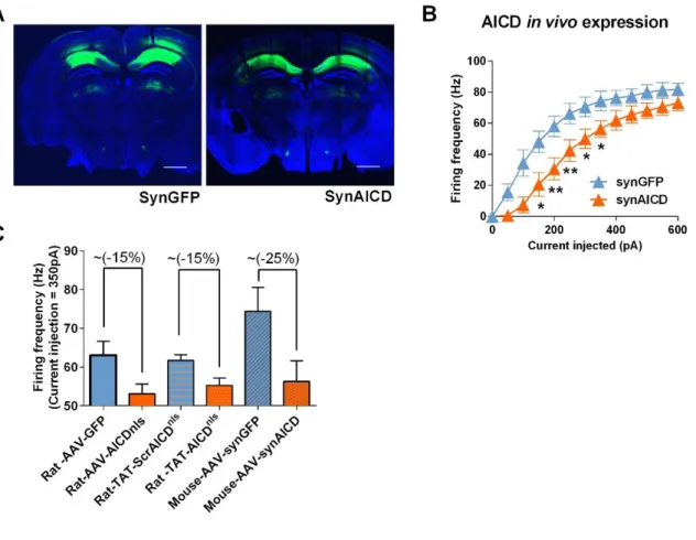
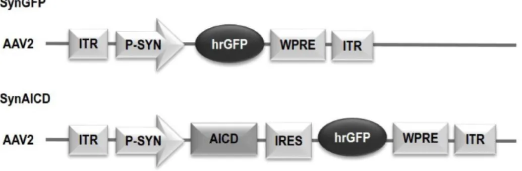
First, we examined the impact of AICD on neuron firing activity. We increased intracellular AICD levels by using in vivo transduc-tion of neurotropic recombinant adeno-associated viruses (AAVs) encoding AICD (Figures 1A and 1B), as described in detail in Pousinha et al. (2017). Briefly, the human AICD c-DNA sequence (last 50 amino acids of APP) was inserted in an AAV vector also expressing GFP (called hereafter AICD virus). We also created AAVs expressing AICD coupled to a nuclear locali-zation signal (NLS) (AICDnlsvirus) or coupled to a nuclear
export-ing signal (NES) (AICDnesvirus) to allow discrimination between
the nuclear versus cytoplasmatic roles of AICD. The AAV vector expressing only GFP was used as control (GFP virus) ( Fig-ure S1A). Whole-cell current-clamp recordings of CA1 pyramidal cells transduced with different virus (AICD, AICDnls, AICDnes, and
GFP) were performed (Figures 1A and 1B). No alterations were observed regarding the resting membrane potential (Figure S1B), nor the membrane input resistance (Figures S1C–S1E). In order to study supra-threshold CA1 pyramidal cells properties in the transduced neurons, 200-ms depolarizing current pulses, incre-menting in steps of 50 pA, were applied at a pre-stimulus potential fixed at !65 mV and AP firing properties were analyzed (Figure 1C). As illustrated (Figure 1D), when a current injection of 300 pA was applied, AICD or AICDnlsneurons fired at a lower
fre-quency than GFP or AICDnesneurons, a difference that was sys-tematically observed for current injections higher than 300 pA. Thus, increased levels of AICD disrupt firing activity at the gamma-frequency range. Moreover, this effect likely requires AICD presence in the nucleus as neurons transduced with AICDnesbehaved like GFP neurons.
Spike-frequency adaptation is a property of many neurons defined as a decreasing rate of action potential (AP) firing during prolonged excitation. This mechanism has been shown to play roles in perceptual processing and learning (Benda et al., 2005; Moyer et al., 2000; Peron and Gabbiani, 2009). To better evaluate the ability of AICD neurons to adapt their firing behavior with time, we measured the firing adaptation ratio for I = 500 pA in the transduced neurons. As shown (Figure 1E), AICD or AICDnls
neurons present higher firing adaptation than GFP neurons ("40% higher). Importantly, AICDnes neurons showed similar
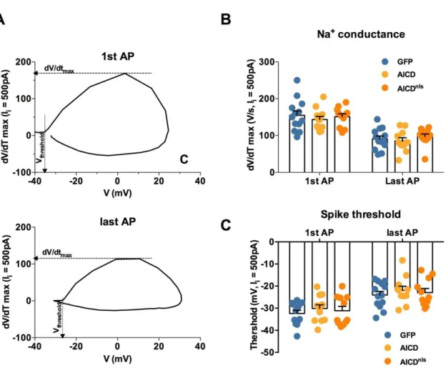
firing adaptation ratio to GFP neurons pointing again for a nu-clear role of AICD in this phenotype. Further analysis of AP shape and AP adaptation was performed in order to access the cellular mechanism linked to the AICD effect on neuron intrinsic excit-ability. Using phase-plane plots of dV/dt versus V for the first and last APs of trains generated during 500-pA current steps (Figure S2), we estimated the average dV/dtmaxas an indication
of the maximal Na+current contributing to the AP (Colbert et al., 1997; Marcantoni et al., 2014) and the voltage at which dV/dtmax
is 4% of its maximum as the threshold potential of AP generation
(Bean, 2007). As shown inFigure S2, these parameters were similar in GFP, AICD, and AICDnls neurons. We therefore
conclude that the Na+channels responsible for AP generation
are not implicated in the AICD-dependent alteration of neuronal excitability.
To confirm our finding and exclude putative overexpression-related artifacts of virus-mediated in vivo expression, we used an alternative approach; i.e., we provided ex vivo intracellular de-livery of AICD. We synthesized peptides representing AICDnlsor
a negative scrambled control peptide (scrAICDnls) linked at the
N terminus to the cell-penetrating domain TAT (Guscott et al., 2016). We pre-incubated hippocampal slices with a dose of TAT-AICDnls(100 nM), shown to induce an AICD intracellular in-crease in the range of values observed in an AD mouse model (Pousinha et al., 2017). Importantly, in this previous work, we confirmed that TAT-AICDnlsis enriched in the nucleus (Pousinha
et al., 2017). Consistent to what we observed with 2 weeks in vivo expression of virally encoded AICD, ex vivo delivery of 100 nM TAT-AICDnlsreduced the firing frequency of CA1 pyramidal cells,
an effect that was not observed with incubation of 100 nM con-trol peptide (TAT-scrAICDnls;Figure 1F). Furthermore, ex vivo
de-livery of AICD engages the same cellular mechanisms as in vivo virus-mediated AICD expression, as spike-frequency adaptation (Figure 1G) was increased in neurons from slices pre-incubated with TAT-AICDnls (100 nM). Since the data obtained by using in vivo AICD intracellular increase pointed for a nuclear
AICD-dependent mechanism, we examined the firing frequency prop-erties of neurons from slices pre-incubated with TAT-AICDnlsor
TAT-scrAICDnlsin presence of the inhibitor of gene transcription, actinomycin (25 mM). Actinomycin was added during both TAT peptides incubation and bath perfusion of slices. As shown ( Fig-ure 1H), TAT-AICDnlsfailed to alter the neuronal firing frequency
in the presence of this inhibitor, confirming a transcription-dependent effect.
AICD Decreases Neuron Firing through Enhancement of the Afterhyperpolarization
APs in hippocampal neurons are followed by a post-burst after-hyperpolarization potential (AHP) (Sah, 1996; Storm, 1990). The AHP is critical for the control of firing rate (Storm, 1990). There-fore, we hypothesized that AICD could alter neuronal intrinsic excitability through AHP enhancement. As AHP size is related to AP number during a sustained depolarization (Coulter et al., 1989), we examined the post-burst AHP elicited by a similar number of APs at a firing frequency shown to be impaired in AICD or AICDnlsneurons (12 APs at a frequency of 60 Hz;
Fig-ure 2A). We performed the post-burst AHP analysis by consid-ering its medium (mAHP) (20–100 ms post-burst) and slow (sAHP) (400 ms to 4 s post-burst) components (Figure 2A), as both components have been described to affect CA1 neuron firing activity (Chen et al., 2014; King et al., 2015; Lancaster and Nicoll, 1987; Madison and Nicoll, 1984; Sah, 1996; Storm, 1990). As shown (Figures 2B and 2C), the mAHP was slightly enhanced in AICD neurons, becoming statistically significant in AICDnlsneurons, a modification not observed in AICDnes
neu-rons. Also, both sAHP full area and sAHP amplitude were enhanced in AICDnlsneurons (Figures 2D and 2E). A similar result
Figure 1. AICD Weakens CA1 Pyramidal Neuron Firing in the Gamma-Frequency Range through a Gene Transcription-Dependent Mechanism
(A) Diagram shows schematic of in vivo viral transduction protocol.
(B) Photos show low-magnification (34; top panels; scale bars, 200 mm) and high-magnification (360; bottom panels; scale bars, 10 mm) images of CA1 region of hippocampal slice (left panels show differential interference contrast [DIC] images; right panels show GFP fluorescence) prepared from rat transduced in vivo with AICDnls.
(C) Protocol used for dissecting the supra-threshold CA1 pyramidal cells properties in the transduced neurons. 200-ms depolarizing current pulses, incrementing in steps of 50 pA, were applied at a pre-stimulus potential fixed at !65 mV.
(D, F, and H) Mean firing frequency versus injected current summary plot for (D) all tested virus (*p < 0.05, **p < 0.01 AICDnlscompared to GFP and AICDnes, fp < 0.05 AICD compared to GFP and AICDnes; two-way ANOVA with repeated measures); from slices pre-incubated in TAT-scrAICDnlsor TAT-AICDnlspeptides
(100 nM) for 2 h, alone (F), or in the presence of the gene transcription inhibitor, actinomycin (acti., 25 mM) (H) (*p < 0.05, two-way ANOVA with repeated measures). On the right of (D) and (F), samples of AP trains in response to 500-pA current step.
(E and G) Scatter dot plot graphs of spike-frequency adaptation analysis based on the ratio concerning the interval inter stimulus (ISI) observed between the two last AP and between the two first AP from a train of APs triggered by 500-pA current step, observed on in vivo viral transduced neurons (E) and on neurons from slices pre-incubated in TAT-scrAICDnls or TAT-AICDnls peptides (100 nM) for 2 h (G).
Each dot in (E) and (G) represents one neuron, also indicated by n value in each panel (**p < 0.01 and ***p < 0.001, one-way ANOVA). Error bars represent the SEM. Statistic details are fully described inTable S1.
Figure 2. AICD Decreases Neuron Firing through Enhancement of the AHP
(A) Protocol used to trigger the AHP. Neurons were submitted to a depolarization current step sufficient to induce a train of 12 action potentials at 60 Hz. (B and F) Examples of traces recorded from (B) trans-duced neurons or (F) neurons from slices pre-incubated in TAT-scrAICDnlsor TAT-AICDnlspeptides (100 nM) for
2 h, with insets where the AHP is presented on an expanded scale.
(C–E and G–I) Shown are scatter dot plot graphs of AICD-mediated effect on the mAHP amplitude (C and G), sAHP area (D and H), and sAHP amplitude (E and I) recorded in transduced neurons (C–E) or neurons from slices pre-incubated in TAT-scrAICDnls or TAT-AICDnls peptides
(100 nM) for 2 h (G and H).
Each dot represents one neuron, also indicated by n value in each panel (*p < 0.05, one-way ANOVA or un-paired Student t test). Error bars represent the SEM. Statistic details are fully described inTable S2.
cells from slices pre-incubated in TAT-AICDnlsor TAT-scrAICDnls peptides (Figures 2F–2I).
SK2 and KCa3.1 Channels Contribute to the AICD-Mediated Effect on CA1 Neuron Firing Activity
mAHP is highly sensitive to changes in intracellular Ca2+( Lan-caster and Zucker, 1994; Storm, 1987; Velumian and Carlen, 1999). In cortical neurons, the mAHP is mediated by small-conductance Ca2+-activated K+channels (SK channels) (
Villalo-bos et al., 2004), which are activated by intracellular Ca2+ions,
with submicromolar Ca2+affinity (Ko¨hler et al., 1996). In CA1
neurons, however, the mAHP is mediated by multiple ionic chan-nels, where SK channels do not seem to be the main contributor (Chen et al., 2014; Gu et al., 2005). As AICD modulates the resting (Hamid et al., 2007) and evoked (Pousinha et al., 2017) intracellular calcium levels, we asked whether it could affect the charge transfer of SK channels, and therefore trigger an increased contribution of these channels to the mAHP. To test this hypothesis, we first investigated the effect of apamin, the SK channel-specific antagonist, on mAHP amplitude. As illus-trated (Figure 3A), apamin failed to significantly alter the mAHP generation in GFP neurons. These data show that, physiologi-cally, the mAHP in CA1 neurons is not mediated by SK channels, an observation that has previously been reported by others (Gu et al., 2005), who showed that in these neurons the mAHP is mediated by a complex of other ionic channels, including Kv7 channels. Interestingly, in the presence of apamin, all trans-duced neurons displayed an identical mAHP amplitude ( Fig-ure 3A). These data suggest that in the pathological context (increased AICDnls levels), the SK channel-mediated K+current
significantly contributes to mAHP amplitude of CA1 excitatory neurons. In order to investigate whether the SK channel-medi-ated enhancement of the mAHP in AICDnls could affect neuron firing activity, we next performed the same protocol used to study supra-threshold CA1 pyramidal cells properties (200-ms depolarizing current pulses, incrementing in steps of 50 pA, applied at a pre-stimulus potential fixed at !65 mV;Figure 1A), but in the presence of apamin. We focused on AICDnlsneurons, which displayed the most significant increase in the mAHP amplitude. AP firing analysis showed that the AICDnlsfiring fre-quency could be restored to control levels by apamin (100 nM) at all current injection steps (Figure 3B), strongly suggesting that SK channels charge transfer is increased in AICDnlsneurons.
To corroborate this observation, we accessed the apamin-sensi-tive current (ISK) of the IAHP. To measure IAHP, neurons were
voltage clamped at !55 mV, and tail currents were evoked with a depolarizing step to +20 mV for 100 ms followed by a re-turn to !55 mV (Figure 3C). The ISKcomponent was obtained by
subtraction of the IAHPmeasured before and after 30 min of
apa-min application. As shown (Figures 3D and 3E), in GFP neurons the apamin-sensitive component of the IAHPwas marginal. By
contrast, the apamin-sensitive somatic current was significantly increased in AICDnlsneurons compared to GFP neurons (Figures
3D and 3E). Since SK channels are sensitive to intracellular cal-cium levels and we previously reported that AICD enhances N-methyl-D-aspartate receptor (NMDAR) conductance ( Pousi-nha et al., 2017), we examined whether these receptors could be the source of Ca2+leading to SK channels increased
perme-ability. As shown, the NMDAR-selective antagonist (2R)-amino-5-phosphonovaleric acid (APV) (50 mM) failed to prevent the increase in ISK observed in AICDnls neurons (Figure 3E).
Together, these data demonstrate that increased levels of AICD lead to over-activation of somatic SK channels, through an NMDAR-independent mechanism.
CA1 pyramidal neurons express the intermediate-conduc-tance calcium-gated potassium channel KCa3.1, which contrib-utes to the sAHP (King et al., 2015; Turner et al., 2016). Since AICD enhances both mAHP and sAHP, it is possible that AICD not only affects SK channels conductance, but also KCa3.1 channel function. Thus, we evaluated the sAHP amplitude and the firing excitability in neurons from slices pre-incubated in TAT-scrAICDnlsor TAT-AICDnlsin presence of the
KCa3.1-selec-tive inhibitor (TRAM-34). As shown (Figures 3F and 3G), the inhi-bition of KCa3.1 channels normalizes the sAHP amplitude and excitability profile of neurons from slices pre-incubated with TAT-AICDnls, suggesting that these channels also contribute to
the AICD-mediated effect on CA1 neuron firing activity.
The Effect of AICD on Neuron Firing Requires L-type Ca2+Channels Activity
We next thought to identify the source of calcium that impacts SK- and KCa3.1-dependent postburst AHP increase in AICD neurons, as we previously excluded NMDAR (Figure 3E). We focused our attention on L-type voltage-dependent Ca2+ chan-nels because these chanchan-nels co-localize with SK and KCa3.1 channels in the somatodendritic compartment, with a spatial proximity that permits rapid regulation of these channels’ conductance during APs (Marrion and Tavalin, 1998; Sahu et al., 2017). Accordingly, both L-type Ca2+channel-selective blockers, nimodipine (5 mM) and isradipine (500 nM), could restore the firing frequency of neurons pre-incubated in TAT-AICDnlsto control (TAT-scrAICDnls) values at all current injection
steps (Figures 4A and S3A). To test whether AICD-related enhancement of AHP is due to enhanced L-type Ca2+influx,
we examined AHP components magnitude in presence of nimo-dipine or isranimo-dipine in neurons pre-incubated with either TAT-AICDnlsor TAT-scrAICDnls. Blocking L-type Ca2+channels with
either drug prevented the effects of AICD on AHP (Figures 4B,
4C,S3B, andS3C). This indicates that increased AICD expres-sion leads to an increase of L-type Ca2+channels permeability, which stimulates Ca2+-activated channels, namely SK and
KCa3.1 channels, leading to enhanced AHP and, consequently, to hypoexcitability.
To directly test the influence of AICD on L-type calcium chan-nels activity, we measured the nimodipine-sensitive current during a step current injection protocol (Figures 4D and 4E) in neurons pre-incubated with either TAT-AICDnls or TAT-scrAICDnls. L-type current magnitude was measured by
subtrac-tion of the selective inhibitor-resistant current from the total current after perfusion of the antagonist (Figure 4F), confirming that this current was enhanced by AICDnls.
As SK, KCa3.1, and L-type calcium channel functions are all affected by AICD, and we show that these AICD modifications are transcription dependent, we investigated whether AICD could modify transcription of the genes encoding these chan-nels. We treated rat hippocampal neuronal cultures (post-natal
day 16 [P16] to P18) with TAT-scrAICDnls or TAT-AICDnls at a concentration of 100 nM for 2 h. We collected RNA and per-formed qPCR for quantification of SK2, KCa3.1, Cav1.2, and CaV1.3 genes. We did not observe any significant alterations in the transcription of these genes (Figures S3D–S3F), suggesting that AICD indirectly modulates their activity by promoting the transcription of other targets.
In SilicoExperiments Predict that Increased AICD Production in CA1 Pyramidal Cells Impairs Hippocampus Gamma Oscillations
In order to further support and better explain the ex vivo experi-mental findings at the cellular level under somatic current injec-tion, herein described and also recently reported (Pousinha et al., 2017), we performed in silico experiments. We used a CA1 pyra-midal cell model previously validated against experimental find-ings and used to build a network model (Bianchi et al., 2014). Since this network model was originally based on mouse neuron properties, we first performed ex vivo whole-cell patch-clamp recordings in mouse neurons with increased in vivo AICD pro-duction to confirm that AICD effects on neuron excitability are species independent. As shown in Figure S4, the effect of AICD on mouse neuron firing activity is qualitatively similar to what we have described above in rat.
We optimized the neuron model to reproduce the number of spikes observed experimentally under control conditions as a function of a somatic current injection (Figures 5A and 5C). To reproduce the AICD effects, we found that a 50-fold increase of the sAHP current and a 60% increase in the L-calcium current was required in order to reduce the number of spikes as observed in the experiment. With these parameters, we were able to obtain a good replication of the experimental profile un-der the AICD condition (Figures 5B and 5D).
We next tested in silico what would happen in an individual CA1 pyramidal cell during an in vivo excitatory synaptic activity in the gamma range (a condition that would be extremely difficult to carry out experimentally). We carried out a set of sim-ulations (n = 20) stimulating the neuron with synaptic inputs tar-geting the distal and proximal dendrites (to model excitatory EC and CA3 pathways, respectively). The results are shown in
Figure 5E. Under control conditions (Figure 5E, left), the asyn-chronous activation of the synapses resulted in a tonic firing behavior at a relatively high frequency. After AICD, under exactly the same pattern of synaptic activation as in control
(Figure 5E, right), the neurons displayed a much lower excit-ability, and the tonic firing behavior changed into a bursting ac-tivity. These effects were caused by the slow dynamics of the Ca2+-dependent K+channels.
These results are in agreement with recently reported experi-mental findings (Pousinha et al., 2017) illustrating that AICD im-pairs synaptic signal integration when the neuron is stimulated at frequencies higher than 10 Hz. The power spectrum, obtained by averaging the somatic traces from 20 simulations (Figures 5F and 5G), showed that increased AICD production in these neu-rons can strongly reduce activity in the gamma range (40– 80 Hz) without affecting theta oscillations ("4 Hz). Taken together, the model results suggest that the AICD-mediated decrease in CA1 pyramidal cell excitability can significantly impact hippocampus oscillations during synaptic activity.
In VivoProduction of AICD Elicits Spatial Memory Encoding Deficits
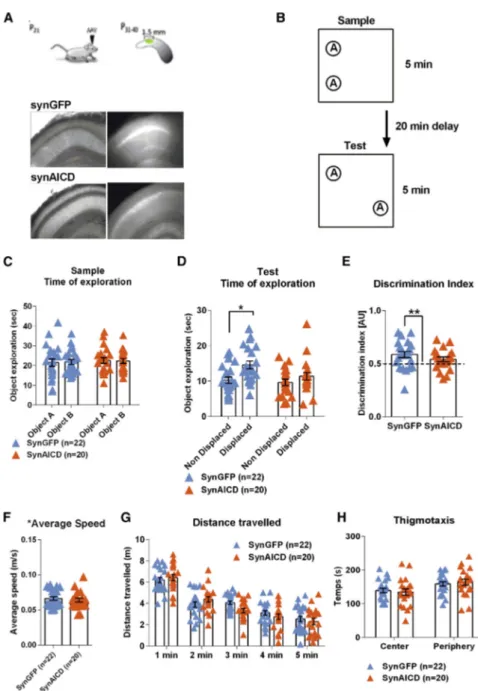
Hippocampal oscillatory activity has been described as a critical player in memory encoding, consolidation, and retrieval. We therefore asked whether animals with increased in vivo AICD production could present cognitive deficits. SynGFP or synAICD viruses (Figure S5) were injected bilaterally in dorsal hippocampi of 21-day-old male rats (Figure 6A). 15 days after in vivo virus transduction, the rats were subjected to a spatial object recogni-tion task, which was recently demonstrated to require CA1 pyra-midal cells gamma activity (Trimper et al., 2017; Zheng et al., 2016). Briefly, rats were first allowed to freely explore two iden-tical objects for 5 min in a square arena (sample trial). After 20 min of rest, rats were replaced in the same arena and were al-lowed to explore the same objects presented before, but one of the two objects was displaced to a new location (test trial) ( Fig-ure 6B). Notably, synAICD rats showed a significant decrease in the preference for the object in a new location (Figures 6D and 6E). AICD-related alterations in locomotor or visual accuracy can be excluded as both groups (synGFP and synAICD) pre-sented similar performances in the sample trial (Figure 6C), average speed (Figure 6F), and distance traveled (Figure 6G). Also, both groups presented similar thigmotaxis (Figure 6H) excluding a putative AICD effect on anxiety levels. These results clearly demonstrate that increased in vivo AICD production in neurons, in a restricted region of dorsal CA1 hippocampus of adult rats, is sufficient, per se, to impair spatial memory encoding.
Figure 3. SK2 and KCa3.1 Channels Contribute to the AICD-Mediated Effect on CA1 Neuron Firing Activity
(A) Scatter dot plot graphs showing that the selective SK channel blocker, apamin (100 nM), prevents AICD-mediated enhancement of mAHP amplitude. (B) Mean firing frequency versus injected current summary plot for neurons transduced with GFP and AICDnlsviruses in absence or presence of apamin (100 nM).
(C) Protocol used to measure the IAHP. Neurons were voltage clamped at !55 mV, and tail currents were evoked with a depolarizing step to +20 mV for 100 ms
followed by a return to !55 mV.
(D) Representative traces of the IAHP, seen as an outward tail current, from GFP and AICDnlstransduced neurons before (black trace) and after (gray trace) bath
application of apamin (100 nM).
(E) Scatter dot plot graph show the subtracted apamin-sensitive current, ISKamplitude (IAHPbefore apamin perfusion – IAHPafter apamin perfusion) observed in
GFP and AICDnlstransduced neurons. The NMDAR selective blocker, APV (50 mM) failed to prevent AICDnlseffect.
(F and G) Scatter dot plot graph of the sAHP amplitude (F) and mean firing frequency versus injected current summary plot (G) in control condition or presence of the selective KCA3.1 channel blocker, TRAM-34 (1 mM) for neurons from slices pre-incubated in TAT-scrAICDnlsor TAT-AICDnlspeptides (100 nM) for 2 h.
Each dot in (A) and (E) and (F) represents one neuron, also indicated by n value in each panel (***p < 0.0001, two-way-ANOVA; *p < 0.05, two-way or one-way ANOVA). Error bars represent the SEM.
Figure 4. The Effect of AICD on Neuron Firing Requires L-type Ca2+Channel Activity
(A) Mean firing frequency versus injected current summary plot recorded in neurons from slices pre-incubated in TAT-scrAICDnlsor TAT-AICDnlspeptides
(100 nM) for 2 h, and after recorded in the presence of the L-type Ca2+channels blocker, nimodipine (5 mM), in the bath perfusion (nim., nimodipine).
(B and C) Scatter dot plot graphs showing (B) mAHP and (C) sAHP amplitude (Vm-Vh, where Vh= !65 mV) recorded from neurons submitted to a depolarization
current step sufficient to induce a train of 12 APs at 60 Hz.
(D) Protocol used to measure the nimodipine-sensitive current. Neurons were voltage clamped at !60 mV, and tail currents were evoked with a hyperpolarizing step to !100 mV for 100 ms followed by 200-ms depolarizing current pulses, incrementing in steps of 10 pA and return to !60 mV.
(E) I-V plot of whole-cell calcium currents before and after nimodipine (5 mm) application, where I/Imaxis the current normalized to the maximal current of each cell.
On the right representative traces of whole-cell calcium current elicited by a voltage step from !100 to +10 mV, before and after nimodipine (5 mm) application. (F) Scatter dot plot graph shows the subtracted nimodipine-sensitive current in the recorded neurons (*p < 0.05, two-way ANOVA or unpaired Student t test). Each dot in (B), (C), and (F) represents one neuron, also indicated by n value in each panel. Error bars represent the SEM.
DISCUSSION
Here, we provide strong evidence that AICD, at pathological levels, is a critical trigger for disruption of CA1 neuron firing activ-ity, alteration of hippocampal network oscillations, and impair-ment in spatial memory encoding. Notably, we could unravel the cellular mechanism underlying AICD-mediated hypoexcit-ability of CA1 pyramidal cells. Briefly, when a neuron with increased AICD levels receives inputs to fire at high frequency, an alteration in the L-type Ca2+channels causes an increased Ca2+-sensitive AHP, weakening its firing frequency. We showed
evidence that this effect is mediated by Ca2+-activated K+ chan-nels, namely SK and KCa3.1 channels. Also, in silico experiments revealed an AICD-mediated alteration of hippocampal network
Figure 5. Computational Model of AICD Ef-fects
(A and B) Typical traces from somatic current injections in control (A) and AICD (B) neurons for I = 0.3 nA under experimental and in silico model situations.
(C and D) Number of spikes elicited as a function of the somatic current injection under control (C) or AICD (D).
(E) Typical traces from in silico synaptic simula-tions under control (Ctrl) (left) and AICD (right) conditions.
(F and G) Normalized power spectrum calculated from 20 simulations under control or AICD condi-tions for frequencies of 0–150 Hz (F) and same graph zoomed in for frequencies of 0–15 Hz (G); lines represent the average and the SEM. Statistic details are fully described inTable S5.
oscillations, specifically affecting gamma rhythms. In agreement, animals with increased AICD levels in the dorsal hippo-campus failed to recognize a familiar ob-ject in a novel location, a task that was recently reported to recruit CA1 pyramidal neurons firing at gamma frequency (Trimper et al., 2017; Zheng et al., 2016).
We gathered solid data advocating that AICD exerts its effect on neuron firing ac-tivity through a transcription-dependent mechanism. Besides the fact that AICD and AICDnls displayed a similar
pheno-type, albeit sometimes more pronounced in AICDnls neurons, we could prevent
these effects in the presence of a gene transcription inhibitor, actinomycin, or by expelling AICD from the nucleus (AICDnes). Accordingly, AICD is a scription factor able to modulate tran-scription of a number of genes (Beckett et al., 2012; Bukhari et al., 2017; Grimm et al., 2013; Konietzko, 2012; Pardossi-Piquard and Checler, 2012) through inter-action with cofactors/nuclear modulators (Bukhari et al., 2017; Cupers et al., 2001; Grimm et al., 2013; Kim-berly et al., 2001).
In agreement with our results, several studies demonstrate that blocking L-type Ca2+channels leads to a reduction of the
postburst AHP in hippocampal neurons (Kumar and Foster, 2002), and Santos et al. (2009) reported that expression of full-length human APP in rat cortical neuron cultures enhances the AHP, inhibiting calcium oscillations, through a similar mechanism (L-type Ca2+channels–Ca2+-activated K+channels
coupling), that we herein describe. In the past few years, several authors reported that in CA1 hippocampal neurons, SK channels play an auxiliary role in controlling the intrinsic excitability of these neurons (Gu et al., 2005; Chen et al., 2014), barely contrib-uting to the generation of the mAHP in physiological conditions,
as we could also observe in GFP neurons.Chen and colleagues (2014)showed that when the activity of the main contributors to the mAHP in CA1 neurons, the low-voltage Kv7/M channels, is compromised, SK channels take charge of reducing spike output of these neurons. Since our data and the work ofSantos et al. (2009)show a significant contribution of SK channels to the mAHP in CA1 pyramidal cells, it is plausible to consider that in pathological conditions the molecular mechanisms underlying the mAHP generation change. As corollary to enhanced SK contribution in these conditions, an additional decrease of the low-voltage Kv7/M channels contribution can thus not be excluded. Of note,Santos et al. (2009)did not address a putative APP gene-transcription-dependent mechanism. These results suggest that increased expression of the full-length APP protein
Figure 6. In Vivo Production of AICD Elicits Spatial Memory Encoding Deficits
(A) Top: diagram shows schematic of in vivo viral transduction protocol with AVV injections at P21 and post-behavioral hippocampal transduction spreading analysis at P40. Bottom: photos show low-magnification (34) images of CA1 area of hippocampal slice (left panels show IR-DIC im-ages; right panels show GFP fluorescence) pre-pared from rat injected in vivo with synGFP or synAICD, as indicated.
(B) Diagram illustrating behavioral task to test hippocampus-dependent spatial memory encod-ing, adapted fromZheng et al. (2016), in synGFP and synAICD rats.
(C and D) Scatter dot plot graphs representing: (C) total object exploration during sample trial and (D) time of exploration during the test trial, where one of the objects is presented in a new location (dis-placed) (*p < 0.05, two-way ANOVA).
(E) Discrimination index (**p < 0.01, one-sample Student’s t test).
(F) Average speed during the test trial.
(G) Distance traveled per bins of 1 min during test trial.
(H) Thigmotaxis during test trial. The blue triangles (synGFP) and orange triangles (synAICD) corre-spond to animals (22 and 20, respectively). Error bars represent the SEM.
Statistic details are fully described inTable S6.
or its C-terminal fragment AICD can trigger similar signaling cascade of events to modulate neuronal firing homeostasis. Indeed, the full-length APP-mediated inhibitory effect on calcium oscillations was shown to be dependent on APP-T668 phosphorylation (Santos et al., 2011), a residue located in the intracel-lular domain crucial for the binding of APP to other intracellular proteins such as Fe65 or Pin1 (Ando et al., 2001; Pastor-ino et al., 2006) and known to be impli-cated in AICD-nuclear signaling (Goodger et al., 2009; Grimm et al., 2015; Kimberly et al., 2001).
Conspicuously, an imbalance in AICD levels, and therefore function, seems to cause detrimental cellular impact by modi-fying calcium permeability at both synapse and soma. In another recent study (Pousinha et al., 2017), we demonstrated that, at synapses, APP deletion leads to the loss of synaptic GluN2B-NMDARs, a phenotype rescued by AICD delivery; whereas increasing AICD levels enhances synaptic GluN2B-NMDARs currents. Accordingly, N2a cells with suppressed levels of AICD present reduced endoplasmic reticulum (ER) Ca2+storage
(Hamid et al., 2007), while cells with enhanced levels of AICD show increased ER Ca2+filling (Leissring et al., 2002).
Intracel-lular Ca2+homeostasis greatly relies on the rapid redistribution of Ca2+ions into the diverse subcellular organelles, which serve
Burdakov et al., 2005; Clapham, 1995; Wuytack et al., 2002). Notably, AICD effects on calcium homeostasis were not observed when cells were incubated in Ca2+-free medium (
Ha-mid et al., 2007). Together, these previous data and our current study strongly suggest that AICD effects on Ca2+homeostasis
are due to alterations in plasma membrane calcium permeability, likely involving NMDARs and L-type Ca2+channels.
Of note, we observed common features in the cellular mech-anisms underlying the action of AICD in our other recent study, focused at the synapse (Pousinha et al., 2017), and in the pre-sent work, focused at the cell body. In both studies, the physio-pathological actions of AICD are gene-transcription-dependent and independent of in vivo chronic expression. Moreover, in both studies, calcium channels present at the plasma mem-brane are affected by AICD in a frequency-dependent manner. However, AICD alters NMDAR to modulate synapse function, whereas it modulates L-type Ca2+channels to modify firing
ho-meostasis. In both situations, AICD leads to increased calcium entry, hence rendering the membrane hyperpolarized via Ca2+
-activated K+channels overactivation. Thus, AICD specifically af-fects calcium channels that are functionally coupled to Ca2+
activated K+channels in distinct spatial regions of the neuron: spine and cell body (Ko¨hler et al., 1996; Marrion and Tavalin, 1998). Overall, it is plausible to advocate that AICD synaptic and somatic effects perturb neuron homeostatic machinery, tuning down the sensitivity of CA1 excitatory neurons to the incoming inputs, reducing its chance to participate in a given memory trace.
The mean firing rate, reflecting an average level of sponta-neous spiking activity, is under homeostatic control in central neural circuits, as recently demonstrated in both ex vivo (
Slomo-witz et al., 2015) and in vivo (Hengen et al., 2016) models. Thus, homeostatic mechanisms stabilize neural circuit function by keeping firing rates within a set-point range, by, for example, adjusting the Ca2+-sensitive AHP, which is critical for the control of neuron firing activity (Storm, 1990). All transmembrane cur-rents contribute to brain rhythms, in particular Na+ currents that are generated by APs (Buzsa´ki et al., 2012). We could show, through in silico modeling, that increased AICD produc-tion in CA1 pyramidal cells can strongly reduce their activity within the gamma-rhythm range (40–80 Hz) without affecting theta oscillations ("4 Hz). These results are in agreement with our original hypothesis: that increased levels of AICD cause CA1 pyramidal cells hypoactivity specifically at inputs of high fre-quency. Gamma oscillations have been attributed to various cognitive processes including hippocampus memory encoding (Lisman and Idiart, 1995; Zheng et al., 2016). Notably, we now show that increased AICD production in a restricted area of dor-sal hippocampus is sufficient to impair object recognition in a novel location, a task recently attributed to CA1 place cell firing at gamma frequency (Zheng et al., 2016), thus suggesting a cau-sality between AICD-associated CA1 pyramidal cells decreased firing and hippocampal cognitive deficits.
Both physiological aging and AD comprehend functional and structural alterations in the hippocampus that drive cognitive decline. Therefore, it is conceivable to hypothesize that the shift from normal aging to AD could be related to common patho-physiological events, which become exacerbated in the neuro-degenerative disorder. Intriguingly, our results highly correlate with observations reported in CA1 neurons from aged rodents. One of the well-characterized markers of physiological aging is an age-related decrease in AP firing rates of CA1 pyramidal cells,
Figure 7. Emerging Model for AICD Patho-logical Effects on Neuronal Function
(A and B) Here, we show two comparative panels illustrating two scenarios: physiology (A) and pa-thology due to increased levels of AICD (B). As indicated in (B), when AICD levels are increased to pathological levels, it acts both at the spines (3 to 8) and at the soma (9 to 14), changing the sensitivity of the neuron to the inputs. AICD translocates to the nucleus (1), where it modulates gene tran-scription (2) through yet-unknown mechanisms. At the synapse (as reported inPousinha et al., 2017), increased AICD leads to NMDAR conductance upregulation (3), leading to increased Ca2+
permeability (4), which in turn activates the nearby Ca2+activated K+channels (5). These channels are
K+permeable, causing a hyperpolarization of the
cell membrane (6), hence inhibiting NMDA re-ceptors (7) and, consequently, perturbing synaptic signal integration (8). At the soma, pathological levels of AICD trigger increased Ca2+permeability
through L-type Ca2+channels (9, 10), thus leading
to close-by Ca2+-activated K+channels opening
(11). As K+exits the cell, the membrane becomes
hyperpolarized (12), producing an increased Ca2+
-sensitive AHP (13), thus decreasing neuron firing activity (14). Together, these mechanisms contribute to neuron firing homeostasis failure, in particular at high frequencies, thus impairing the signal output and therefore the associated memory processes. The increase in ionic channel numbers in the physiopathological condition is for graphical purposes only as we do not at present hold evidence for any AICD-induced increase in cell surface expression of these channels.
with a concomitant increase in the amplitude of the AHP respon-sible for spike frequency adaptation (Disterhoft et al., 1996; Landfield and Pitler, 1984; Power et al., 2002). Moreover, although the molecular mechanisms responsible for these alter-ations in aging brains are still not fully understood, several studies suggest that it is due to L-type calcium channels dysre-gulation. Indeed, their charge transfer is enhanced in aged hip-pocampal neurons (Nu´n˜ez-Santana et al., 2014; Veng et al., 2003), and their antagonists can counteract the effects of aging on cognition (Rose et al., 2007; Veng et al., 2003). A provocative interpretation may arise from these similarities: AICD age-related progressive upregulation, likely due to either a lower degradation rate linked to reduced expression of its insulin-degrading enzyme (Cook et al., 2003; Nalivaeva and Turner, 2017) or increased production triggered by BACE1 enhanced expression (Ahmed et al., 2010; Holsinger et al., 2002), may contribute to the shift from physiological aging to AD.
Our observations extend the knowledge on AICD pathological role strengthening its contribution to AD pathogenesis. Although more work is needed to understand precisely the nuclear AICD targets/mechanisms that modify membrane calcium channel permeability, we propose an emerging model based on our recent work (Pousinha et al., 2017) and the results herein described (Figure 7). Increased AICD levels lead to enhanced membrane calcium permeability through synaptic GluN2B-NMDAR and somatic L-type Ca2+channels, which activate the nearby Ca2+-activated K+channels, rendering the neuron
hypo-active. Thus, AICD decreases neuron sensitivity to excitatory synaptic inputs and, simultaneously, reduces its ability to fire at gamma frequency when solicited. Together, these AICD-trig-gered modifications will impede normal neuron responses to experience-driven stimuli, hence impairing the underlying mem-ory tasks, like spatial memmem-ory encoding. More broadly, the cur-rent findings highlight common features between physiological aging and AD, thus suggesting that AICD might correlate with progressive neuron homeostatic failure, driving the transition from synaptic and cognitive impairments, observed in physiolog-ical aging, to neurodegeneration.
STAR+METHODS
Detailed methods are provided in the online version of this paper and include the following:
d KEY RESOURCES TABLE
d LEAD CONTACT AND MATERIALS AVAILABILITY d EXPERIMENTAL MODEL AND SUBJECT DETAILS
B Animals
B Primary neuronal cultures
d METHOD DETAILS
B Virus Constructs B TAT peptides B In Vivo AAV Injections
B Quantitative PCR in primary neuronal cultures B Pharmacological tools
B Patch-Clamp Electrophysiology B In silico modeling
B Behavioral analysis
d QUANTIFICATION AND STATISTICAL ANALYSES d DATA AND CODE AVAILABILITY
SUPPLEMENTAL INFORMATION
Supplemental Information can be found online athttps://doi.org/10.1016/j. celrep.2019.08.103.
ACKNOWLEDGMENTS
This work was supported by the ATIP/AVENIR program (Centre National de la Recherche Scientifique [CNRS]) to P.A.P. and H.M.; Fondation pour la Recherche Me´dicale (FRM; postdoctoral fellowship) to P.A.P.; the French Fon-dation Plan Alzheimer to E.F.R., X.M., and H.M.; the EU Joint Program– Neurodegenerative Disease Research (JPND) Project CIRCPROT (jointly funded by BMBF, MIUR, and EU Horizon 2020 Grant Agreement 643417) to H.M. and P.A.P., D.B., and M.M.; EU Horizon 2020 Framework Programme for Research and Innovation (Specific Grant Agreement 785907; Human Brain Project SGA2); the Universite´ C^ote d’Azur to H.M. and P.A.P.; Santa Casa da Miserico´rdia (MB-7-2018); and Fundac¸~ao para a Ci^encia e Tecnologia (PD/BD/ 135516/2018, SFRH/BD/52228/2013, and PTDC/BIMMEC/4778/2014) to L.V.L., J.R.-S., and M.T.-F. We thank Maxime Villet for behavioral data analysis and Frank Aguila for the design of illustrations presented inFigure 7and the Graphical Abstract.
AUTHOR CONTRIBUTIONS
P.A.P. and H.M. designed the study and interpreted the results. P.A.P. per-formed ex vivo electrophysiological recordings and analysis and helped with behavioral assays. P.A.P. and S.P.F. performed in vivo stereotaxic surgeries. E.F.R., X.M., and H.M. produced viruses, cloned viruses, and performed biochemical analysis. R.G. designed the primers. M.T.-F., J.R.-S., and L.V.L. prepared and performed the qPCR on hippocampal neuron cultures. I.B. and A.R.S.-P. performed behavioral assays. C.G. performed histology. R.G. and J.B. provided intellectual and technical inputs. D.B. and M.M. de-signed and performed all of the modeling. P.A.P. wrote the manuscript with input from the other authors.
DECLARATION OF INTERESTS
The authors declare no conflict of interest. Received: August 10, 2018
Revised: August 9, 2019 Accepted: August 29, 2019 Published: October 8, 2019
REFERENCES
Ahmed, R.R., Holler, C.J., Webb, R.L., Li, F., Beckett, T.L., and Murphy, M.P. (2010). BACE1 and BACE2 enzymatic activities in Alzheimer’s disease. J. Neurochem. 112, 1045–1053.
Ando, K., Iijima, K.I., Elliott, J.I., Kirino, Y., and Suzuki, T. (2001). Phosphoryla-tion-dependent regulation of the interaction of amyloid precursor protein with Fe65 affects the production of beta-amyloid. J. Biol. Chem. 276, 40353– 40361.
Bean, B.P. (2007). The action potential in mammalian central neurons. Nat. Rev. Neurosci. 8, 451–465.
Beckett, C., Nalivaeva, N.N., Belyaev, N.D., and Turner, A.J. (2012). Nuclear signalling by membrane protein intracellular domains: the AICD enigma. Cell. Signal. 24, 402–409.
Benda, J., Longtin, A., and Maler, L. (2005). Spike-frequency adaptation separates transient communication signals from background oscillations. J. Neurosci. 25, 2312–2321.
Bianchi, D., De Michele, P., Marchetti, C., Tirozzi, B., Cuomo, S., Marie, H., and Migliore, M. (2014). Effects of increasing CREB-dependent transcription on the storage and recall processes in a hippocampal CA1 microcircuit. Hippocam-pus 24, 165–177.
Brini, M., and Carafoli, E. (2009). Calcium pumps in health and disease. Physiol. Rev. 89, 1341–1378.
Brown, J.T., Chin, J., Leiser, S.C., Pangalos, M.N., and Randall, A.D. (2011). Altered intrinsic neuronal excitability and reduced Na+currents in a mouse
model of Alzheimer’s disease. Neurobiol. Aging 32, 2109.e1–2109.e14.
Bukhari, H., Glotzbach, A., Kolbe, K., Leonhardt, G., Loosse, C., and M€uller, T. (2017). Small things matter: implications of APP intracellular domain AICD nu-clear signaling in the progression and pathogenesis of Alzheimer’s disease. Prog. Neurobiol. 156, 189–213.
Burdakov, D., Petersen, O.H., and Verkhratsky, A. (2005). Intraluminal calcium as a primary regulator of endoplasmic reticulum function. Cell Calcium 38, 303–310.
Bustin, S.A., Benes, V., Garson, J.A., Hellemans, J., Huggett, J., Kubista, M., Mueller, R., Nolan, T., Pfaffl, M.W., Shipley, G.L., et al. (2009). The MIQE guide-lines: minimum information for publication of quantitative real-time PCR exper-iments. Clin. Chem. 55, 611–622.
Buzsa´ki, G., Anastassiou, C.A., and Koch, C. (2012). The origin of extracellular fields and currents—EEG, ECoG, LFP and spikes. Nat. Rev. Neurosci. 13, 407–420.
Chen, S., Benninger, F., and Yaari, Y. (2014). Role of small conductance Ca2+-activated K+ channels in controlling CA1 pyramidal cell excitability.
J. Neurosci. 34, 8219–8230.
Clapham, D.E. (1995). Intracellular calcium. Replenishing the stores. Nature
375, 634–635.
Colbert, C.M., Magee, J.C., Hoffman, D.A., and Johnston, D. (1997). Slow re-covery from inactivation of Na+ channels underlies the activity-dependent attenuation of dendritic action potentials in hippocampal CA1 pyramidal neu-rons. J. Neurosci. 17, 6512–6521.
Cook, D.G., Leverenz, J.B., McMillan, P.J., Kulstad, J.J., Ericksen, S., Roth, R.A., Schellenberg, G.D., Jin, L.-W., Kovacina, K.S., and Craft, S. (2003). Reduced hippocampal insulin-degrading enzyme in late-onset Alzheimer’s disease is associated with the apolipoprotein E-epsilon4 allele. Am. J. Pathol.
162, 313–319.
Coulter, D.A., Lo Turco, J.J., Kubota, M., Disterhoft, J.F., Moore, J.W., and Al-kon, D.L. (1989). Classical conditioning reduces amplitude and duration of cal-cium-dependent afterhyperpolarization in rabbit hippocampal pyramidal cells. J. Neurophysiol. 61, 971–981.
Cupers, P., Orlans, I., Craessaerts, K., Annaert, W., and De Strooper, B. (2001). The amyloid precursor protein (APP)-cytoplasmic fragment generated by gamma-secretase is rapidly degraded but distributes partially in a nuclear fraction of neurones in culture. J. Neurochem. 78, 1168–1178.
De Strooper, B., and Karran, E. (2016). The cellular phase of Alzheimer’s dis-ease. Cell 164, 603–615.
Disterhoft, J.F., Thompson, L.T., Moyer, J.R., Jr., and Mogul, D.J. (1996). Cal-cium-dependent afterhyperpolarization and learning in young and aging hip-pocampus. Life Sci. 59, 413–420.
Frere, S., and Slutsky, I. (2018). Alzheimer’s disease: from firing instability to homeostasis network collapse. Neuron 97, 32–58.
Ghosal, K., Vogt, D.L., Liang, M., Shen, Y., Lamb, B.T., and Pimplikar, S.W. (2009). Alzheimer’s disease-like pathological features in transgenic mice ex-pressing the APP intracellular domain. Proc. Natl. Acad. Sci. USA 106, 18367–18372.
Gillespie, A.K., Jones, E.A., Lin, Y.-H., Karlsson, M.P., Kay, K., Yoon, S.Y., Tong, L.M., Nova, P., Carr, J.S., Frank, L.M., and Huang, Y. (2016). Apolipo-protein E4 causes age-dependent disruption of slow gamma oscillations dur-ing hippocampal sharp-wave ripples. Neuron 90, 740–751.
Goodger, Z.V., Rajendran, L., Trutzel, A., Kohli, B.M., Nitsch, R.M., and Ko-nietzko, U. (2009). Nuclear signaling by the APP intracellular domain occurs
predominantly through the amyloidogenic processing pathway. J. Cell Sci.
122, 3703–3714.
Grimm, M.O., Mett, J., Stahlmann, C.P., Haupenthal, V.J., Zimmer, V.C., and Hartmann, T. (2013). Neprilysin and Ab clearance: impact of the app intracel-lular domain in NEP regulation and implications in Alzheimer’s disease. Front. Aging Neurosci. 5, 98.
Grimm, M.O., Mett, J., Stahlmann, C.P., Gro¨sgen, S., Haupenthal, V.J., Bl€umel, T., Hundsdo¨rfer, B., Zimmer, V.C., Mylonas, N.T., Tanila, H., et al. (2015). APP intracellular domain derived from amyloidogenic b- and g-secre-tase cleavage regulates neprilysin expression. Front. Aging Neurosci. 7, 77.
Gu, N., Vervaeke, K., Hu, H., and Storm, J.F. (2005). Kv7/KCNQ/M and HCN/h, but not KCa2/SK channels, contribute to the somatic medium after-hyperpo-larization and excitability control in CA1 hippocampal pyramidal cells. J. Physiol. 566, 689–715.
Guscott, B., Balklava, Z., Safrany, S.T., and Wassmer, T. (2016). A cell-perme-able tool for analysing APP intracellular domain function and manipulation of PIKfyve activity. Biosci. Rep. 15, e00319.
Hamid, R., Kilger, E., Willem, M., Vassallo, N., Kostka, M., Bornho¨vd, C., Reichert, A.S., Kretzschmar, H.A., Haass, C., and Herms, J. (2007). Amyloid precursor protein intracellular domain modulates cellular calcium homeostasis and ATP content. J. Neurochem. 102, 1264–1275.
Hammond, R.S., Bond, C.T., Strassmaier, T., Ngo-Anh, T.J., Adelman, J.P., Maylie, J., and Stackman, R.W. (2006). Small-conductance Ca2+-activated
K+channel type 2 (SK2) modulates hippocampal learning, memory, and
syn-aptic plasticity. J. Neurosci. 26, 1844–1853.
Hauck, B., Chen, L., and Xiao, W. (2003). Generation and characterization of chimeric recombinant AAV vectors. Mol. Ther. 7, 419–425.
Hazra, A., Gu, F., Aulakh, A., Berridge, C., Eriksen, J.L., and Ziburkus, J. (2013). Inhibitory neuron and hippocampal circuit dysfunction in an aged mouse model of Alzheimer’s disease. PLoS ONE 8, e64318.
Hengen, K.B., Torrado Pacheco, A., McGregor, J.N., Van Hooser, S.D., and Turrigiano, G.G. (2016). Neuronal firing rate homeostasis is inhibited by sleep and promoted by wake. Cell 165, 180–191.
Hines, M.L., and Carnevale, N.T. (2003). The NEURON simulation environ-ment. In The Handbook of Brain Theory and Neural Networks, Volume 2, M.A. Arbib, ed. (MIT Press), pp. 769–773.
Holsinger, R.M.D., McLean, C.A., Beyreuther, K., Masters, C.L., and Evin, G. (2002). Increased expression of the amyloid precursor beta-secretase in Alz-heimer’s disease. Ann. Neurol. 51, 783–786.
Hoxha, E., Boda, E., Montarolo, F., Parolisi, R., and Tempia, F. (2012). Excit-ability and synaptic alterations in the cerebellum of APP/PS1 mice. PLoS ONE 7, e34726.
Jonsson, T., Atwal, J., Steinberg, S., Snaedal, J., Jonsson, P., Bjornsson, S., Stefansson, H., Sulem, P., Gudbjartsson, D., Maloney, J., et al. (2012). A mu-tation in APP protects against Alzheimer’s disease and age-related cognitive decline. Nature 488, 96–99.
Kaczorowski, C.C., Sametsky, E., Shah, S., Vassar, R., and Disterhoft, J.F. (2011). Mechanisms underlying basal and learning-related intrinsic excitability in a mouse model of Alzheimer’s disease. Neurobiol. Aging 32, 1452–1465.
Kerrigan, T.L., Brown, J.T., and Randall, A.D. (2014). Characterization of altered intrinsic excitability in hippocampal CA1 pyramidal cells of the Ab-ove-rproducing PDAPP mouse. Neuropharmacology 79, 515–524.
Kimberly, W.T., Zheng, J.B., Gue´nette, S.Y., and Selkoe, D.J. (2001). The intra-cellular domain of the beta-amyloid precursor protein is stabilized by Fe65 and translocates to the nucleus in a notch-like manner. J. Biol. Chem. 276, 40288– 40292.
King, B., Rizwan, A.P., Asmara, H., Heath, N.C., Engbers, J.D.T., Dykstra, S., Bartoletti, T.M., Hameed, S., Zamponi, G.W., and Turner, R.W. (2015). IKCa channels are a critical determinant of the slow AHP in CA1 pyramidal neurons. Cell Rep. 11, 175–182.
Klevanski, M., Herrmann, U., Weyer, S.W., Fol, R., Cartier, N., Wolfer, D.P., Caldwell, J.H., Korte, M., and M€uller, U.C. (2015). The APP intracellular domain
is required for normal synaptic morphology, synaptic plasticity, and hippo-campus-dependent behavior. J. Neurosci. 35, 16018–16033.
Ko¨hler, M., Hirschberg, B., Bond, C.T., Kinzie, J.M., Marrion, N.V., Maylie, J., and Adelman, J.P. (1996). Small-conductance, calcium-activated potassium channels from mammalian brain. Science 273, 1709–1714.
Konietzko, U. (2012). AICD nuclear signaling and its possible contribution to Alzheimer’s disease. Curr. Alzheimer Res. 9, 200–216.
Kumar, A., and Foster, T.C. (2002). 17beta-estradiol benzoate decreases the AHP amplitude in CA1 pyramidal neurons. J. Neurophysiol. 88, 621–626.
Lancaster, B., and Nicoll, R.A. (1987). Properties of two calcium-activated hy-perpolarizations in rat hippocampal neurones. J. Physiol. 389, 187–203.
Lancaster, B., and Zucker, R.S. (1994). Photolytic manipulation of Ca2+and the
time course of slow, Ca2+-activated K+current in rat hippocampal neurones.
J. Physiol. 475, 229–239.
Landfield, P.W., and Pitler, T.A. (1984). Prolonged Ca2+-dependent
afterhyper-polarizations in hippocampal neurons of aged rats. Science 226, 1089–1092.
Lauritzen, I., Pardossi-Piquard, R., Bauer, C., Brigham, E., Abraham, J.D., Ra-naldi, S., Fraser, P., St-George-Hyslop, P., Le Thuc, O., Espin, V., et al. (2012). The b-secretase-derived C-terminal fragment of bAPP, C99, but not Ab, is a key contributor to early intraneuronal lesions in triple-transgenic mouse hippo-campus. J. Neurosci. 32, 16243–16255.
Leissring, M.A., Murphy, M.P., Mead, T.R., Akbari, Y., Sugarman, M.C., Janna-tipour, M., Anliker, B., M€uller, U., Saftig, P., De Strooper, B., et al. (2002). A physiologic signaling role for the gamma -secretase-derived intracellular frag-ment of APP. Proc. Natl. Acad. Sci. USA 99, 4697–4702.
Lisman, J.E., and Idiart, M.A. (1995). Storage of 7 +/! 2 short-term memories in oscillatory subcycles. Science 267, 1512–1515.
Madison, D.V., and Nicoll, R.A. (1984). Control of the repetitive discharge of rat CA 1 pyramidal neurones in vitro. J. Physiol. 354, 319–331.
Marcantoni, A., Raymond, E.F., Carbone, E., and Marie, H. (2014). Firing prop-erties of entorhinal cortex neurons and early alterations in an Alzheimer’s dis-ease transgenic model. Pflugers Arch. 466, 1437–1450.
Marie, H., Morishita, W., Yu, X., Calakos, N., and Malenka, R.C. (2005). Gener-ation of silent synapses by acute in vivo expression of CaMKIV and CREB. Neuron 45, 741–752.
Marrion, N.V., and Tavalin, S.J. (1998). Selective activation of Ca2+-activated
K+channels by co-localized Ca2+channels in hippocampal neurons. Nature
395, 900–905.
Minkeviciene, R., Rheims, S., Dobszay, M.B., Zilberter, M., Hartikainen, J., F€ulo¨p, L., Penke, B., Zilberter, Y., Harkany, T., Pitka¨nen, A., and Tanila, H. (2009). Amyloid beta-induced neuronal hyperexcitability triggers progressive epilepsy. J. Neurosci. 29, 3453–3462.
Moyer, J.R., Jr., Power, J.M., Thompson, L.T., and Disterhoft, J.F. (2000). Increased excitability of aged rabbit CA1 neurons after trace eyeblink condi-tioning. J. Neurosci. 20, 5476–5482.
Nalivaeva, N.N., and Turner, A.J. (2017). Role of ageing and oxidative stress in regulation of amyloid-degrading enzymes and development of neurodegener-ation. Curr. Aging Sci. 10, 32–40.
Nu´n˜ez-Santana, F.L., Oh, M.M., Antion, M.D., Lee, A., Hell, J.W., and Dister-hoft, J.F. (2014). Surface L-type Ca2+channel expression levels are increased
in aged hippocampus. Aging Cell 13, 111–120.
Palop, J.J., Chin, J., Roberson, E.D., Wang, J., Thwin, M.T., Bien-Ly, N., Yoo, J., Ho, K.O., Yu, G.-Q., Kreitzer, A., et al. (2007). Aberrant excitatory neuronal activity and compensatory remodeling of inhibitory hippocampal circuits in mouse models of Alzheimer’s disease. Neuron 55, 697–711.
Pardossi-Piquard, R., and Checler, F. (2012). The physiology of the b-amyloid precursor protein intracellular domain AICD. J. Neurochem. 120 (Suppl 1), 109–124.
Pastorino, L., Sun, A., Lu, P.J., Zhou, X.Z., Balastik, M., Finn, G., Wulf, G., Lim, J., Li, S.H., Li, X., et al. (2006). The prolyl isomerase Pin1 regulates amyloid pre-cursor protein processing and amyloid-beta production. Nature 440, 528–534.
Peron, S., and Gabbiani, F. (2009). Spike frequency adaptation mediates looming stimulus selectivity in a collision-detecting neuron. Nat. Neurosci.
12, 318–326.
Pousinha, P.A., Mouska, X., Raymond, E.F., Gwizdek, C., Dhib, G., Poupon, G., Zaragosi, L.-E., Giudici, C., Bethus, I., Pacary, E., et al. (2017). Physiolog-ical and pathophysiologPhysiolog-ical control of synaptic GluN2B-NMDA receptors by the C-terminal domain of amyloid precursor protein. eLife 6, e25659.
Power, J.M., Wu, W.W., Sametsky, E., Oh, M.M., and Disterhoft, J.F. (2002). Age-related enhancement of the slow outward calcium-activated potassium current in hippocampal CA1 pyramidal neurons in vitro. J. Neurosci. 22, 7234–7243.
Rose, G.M., Ong, V.S., and Woodruff-Pak, D.S. (2007). Efficacy of MEM 1003, a novel calcium channel blocker, in delay and trace eyeblink conditioning in older rabbits. Neurobiol. Aging 28, 766–773.
Sah, P. (1996). Ca2+-activated K+currents in neurones: types, physiological
roles and modulation. Trends Neurosci. 19, 150–154.
Sahu, G., Asmara, H., Zhang, F.-X., Zamponi, G.W., and Turner, R.W. (2017). Activity-dependent facilitation of CaV1.3 calcium channels promotes KCa3.1
activation in hippocampal neurons. J. Neurosci. 37, 11255–11270.
Santos, S.F., Pierrot, N., Morel, N., Gailly, P., Sindic, C., and Octave, J.-N. (2009). Expression of human amyloid precursor protein in rat cortical neurons inhibits calcium oscillations. J. Neurosci. 29, 4708–4718.
Santos, S.F., Tasiaux, B., Sindic, C., and Octave, J.-N. (2011). Inhibition of neuronal calcium oscillations by cell surface APP phosphorylated on T668. Neurobiol. Aging 32, 2308–2313.
Schmittgen, T.D., and Livak, K.J. (2008). Analyzing real-time PCR data by the comparative C(T) method. Nat. Protoc. 3, 1101–1108.
Selkoe, D.J. (2002). Alzheimer’s disease is a synaptic failure. Science 298, 789–791.
Slomowitz, E., Styr, B., Vertkin, I., Milshtein-Parush, H., Nelken, I., Slutsky, M., and Slutsky, I. (2015). Interplay between population firing stability and single neuron dynamics in hippocampal networks. eLife 4, e04378.
Stam, C.J., van Cappellen van Walsum, A.M., Pijnenburg, Y.A.L., Berendse, H.W., de Munck, J.C., Scheltens, P., and van Dijk, B.W. (2002). Generalized synchronization of MEG recordings in Alzheimer’s disease: evidence for involvement of the gamma band. J. Clin. Neurophysiol. 19, 562–574.
Storm, J.F. (1987). Action potential repolarization and a fast after-hyperpolar-ization in rat hippocampal pyramidal cells. J. Physiol. 385, 733–759.
Storm, J.F. (1990). Potassium currents in hippocampal pyramidal cells. Prog. Brain Res. 83, 161–187.
Trimper, J.B., Galloway, C.R., Jones, A.C., Mandi, K., and Manns, J.R. (2017). Gamma oscillations in rat hippocampal subregions dentate gyrus, CA3, CA1, and subiculum underlie associative memory encoding. Cell Rep. 21, 2419– 2432.
Turner, R.W., Asmara, H., Engbers, J.D.T., Miclat, J., Rizwan, A.P., Sahu, G., and Zamponi, G.W. (2016). Assessing the role of IKCa channels in generating the sAHP of CA1 hippocampal pyramidal cells. Channels (Austin) 10, 313–319.
Valadas, J.S., Batalha, V.L., Ferreira, D.G., Gomes, R., Coelho, J.E., Sebasti~ao, A.M., Dio´genes, M.J., and Lopes, L.V. (2012). Neuroprotection af-forded by adenosine A2A receptor blockade is modulated by corticotrophin-releasing factor (CRF) in glutamate injured cortical neurons. J. Neurochem.
123, 1030–1040.
van der Kant, R., and Goldstein, L.S. (2015). Cellular functions of the amyloid precursor protein from development to dementia. Dev. Cell 32, 502–515.
Velumian, A.A., and Carlen, P.L. (1999). Differential control of three after-hy-perpolarizations in rat hippocampal neurones by intracellular calcium buff-ering. J. Physiol. 517, 201–216.
Veng, L.M., Mesches, M.H., and Browning, M.D. (2003). Age-related working memory impairment is correlated with increases in the L-type calcium channel protein alpha1D (Cav1.3) in area CA1 of the hippocampus and both are amelio-rated by chronic nimodipine treatment. Brain Res. Mol. Brain Res. 110, 193–202.
Verret, L., Mann, E.O., Hang, G.B., Barth, A.M.I., Cobos, I., Ho, K., Devidze, N., Masliah, E., Kreitzer, A.C., Mody, I., et al. (2012). Inhibitory interneuron deficit links altered network activity and cognitive dysfunction in Alzheimer model. Cell 149, 708–721.
Villalobos, C., Shakkottai, V.G., Chandy, K.G., Michelhaugh, S.K., and An-drade, R. (2004). SKCa channels mediate the medium but not the slow cal-cium-activated afterhyperpolarization in cortical neurons. J. Neurosci. 24, 3537–3542.
Wuytack, F., Raeymaekers, L., and Missiaen, L. (2002). Molecular physiology of the SERCA and SPCA pumps. Cell Calcium 32, 279–305.
Wykes, R., Kalmbach, A., Eliava, M., and Waters, J. (2012). Changes in the physiology of CA1 hippocampal pyramidal neurons in preplaque CRND8 mice. Neurobiol. Aging 33, 1609–1623.
Zhang, W., and Linden, D.J. (2003). The other side of the engram: experience-driven changes in neuronal intrinsic excitability. Nat. Rev. Neurosci. 4, 885–900.
Zheng, C., Bieri, K.W., Hwaun, E., and Colgin, L.L. (2016). Fast gamma rhythms in the hippocampus promote encoding of novel object-place pairings. eNeuro 3, ENEURO.0001-16.2016.
STAR+METHODS
KEY RESOURCES TABLE
REAGENT or RESOURCE SOURCE IDENTIFIER
Chemicals, Peptides, and Recombinant Proteins
TAT-AICDnls: YGRKKRRQRRRVMLKKKQYT SIHHGVVEVDAA VTPEERHLSKMQQNGYENPTYKFFEQMQNPKKKRKV PSL GmbH (Heidelberg, Germany) N/A TAT-scrAICDnls: YGRKKRRQRRRVQGITQKMYHNQEGKF LNQKVNVKTHMQFEHETLVDSMKAYYRVEEPSAPKKKRKV PSL GmbH (Heidelberg, Germany) N/A
Actinomycin Sigma Cat#A9415
Apamin Calbiochem CAS 24345-16-2
TRAM-34 Abcam Cat# ab141885
D-APV Abcam Cat# ab120003
TTX Abcam Cat# ab120055
nimodipine Abcam Cat# ab120138
Isradipine Abcam Cat# ab120142
Critical Commercial Assays
RNeasy Plus Mini Kit QIAGEN, Germany Cat# 74134
SuperScript First-Strand Synthesis System Invitrogen, USA Cat# 18091050 Power SYBR Green PCR Master Mix Applied Biosystems, UK Cat#1725095 Deposited Data
CA1 pyramidal neuron model Bianchi et al., 2014 ModelDB acc.n.151126 All models and simulation files for this paper This paper https://senselab.med.yale.
edu/ModelDB, acc.n. 256388 Experimental Models: Cell Lines
AAV-293 cells Agilent Technologies, USA RRID:CVCL_6871 Experimental Models: Organisms/Strains
Rat: Sprague Dawley Janvier Labs, France RRID:RGD:5508397 Mouse: C57BL/6JRj Janvier Labs, France RRID:MGI:5752053
Oligonucleotides
PPIA peptidylprolyl isomerase A (cyclophilin A) Forward Primer: ATCTGCACTGCCAAGACTGAGTG
Invitrogen, USA N/A PPIA peptidylprolyl isomerase A (cyclophilin A)
Reverse Primer: CTTCTTGCTGGTCTTGCCATTCC
Invitrogen, USA N/A Ribosomal protein L13A
Forward Primer: GGATCCCTCCACCCTATGACA
Invitrogen, USA N/A Ribosomal protein L13A
Reverse Primer: CTGGTACTTCCACCCGACCTC
Invitrogen, USA N/A Potassium Calcium-Activated Channel Subfamily N Member 2
Forward Primer: TATGCGCTCATCTTCGGCAT
Invitrogen, USA N/A Potassium Calcium-Activated Channel Subfamily N Member 2
Reverse Primer: AGAATACAGCGACGCCTTGT
Invitrogen, USA N/A Potassium Calcium-Activated Channel Subfamily N Member 4
Forward Primer: AGTGTTTAATCACGCTGTCCACT
Invitrogen, USA N/A Potassium Calcium-Activated Channel Subfamily N Member 4
Reverse Primer: CCCGTTGTCAGTCATGAACA
Invitrogen, USA N/A Calcium voltage-gated channel subunit alpha1 C
Forward Primer: TGCCTCCGAACACTACAACC
Invitrogen, USA N/A Calcium voltage-gated channel subunit alpha1 C
Reverse Primer: CCCCGCACACAATGAAACAG
Invitrogen, USA N/A