HAL Id: hal-03035601
https://hal.archives-ouvertes.fr/hal-03035601
Submitted on 2 Dec 2020HAL is a multi-disciplinary open access archive for the deposit and dissemination of sci-entific research documents, whether they are pub-lished or not. The documents may come from teaching and research institutions in France or abroad, or from public or private research centers.
L’archive ouverte pluridisciplinaire HAL, est destinée au dépôt et à la diffusion de documents scientifiques de niveau recherche, publiés ou non, émanant des établissements d’enseignement et de recherche français ou étrangers, des laboratoires publics ou privés.
Concomitant emergence of circularly polarized
luminescence and single-molecule magnet behavior in
chiral-at-metal Dy complex
Bahjat El Rez, Jiawen Liu, Virginie Béreau, Carine Guyard-Duhayon, Yuki
Horino, Takayoshi Suzuki, Laurent Coolen, Jean-Pascal Sutter
To cite this version:
Bahjat El Rez, Jiawen Liu, Virginie Béreau, Carine Guyard-Duhayon, Yuki Horino, et al.. Concomi-tant emergence of circularly polarized luminescence and single-molecule magnet behavior in chiral-at-metal Dy complex. Inorganic Chemistry Frontiers, Royal Society of Chemistry, 2020, 7 (22), pp.4527-4534. �10.1039/D0QI00919A�. �hal-03035601�
1
Concomitant Emergence of Circularly Polarized Luminescence and
Single-Molecule Magnet behavior in chiral-at-metal Dy complex
Bahjat El Rez,a Jiawen Liu,c Virginie Béreau,a,b Carine Duhayon,a Yuki Horino,d Takayoshi Suzuki,d,e Laurent Coolen,*,c Jean-Pascal Sutter*,a
a Laboratoire de Chimie de Coordination du CNRS (LCC-CNRS), Université de Toulouse, CNRS,
Toulouse, France sutter@lcc-toulouse.fr
b Université de Toulouse, Institut Universitaire de Technologie-Département de Chimie, Castres,
France
c Sorbonne Université, CNRS, Institut de NanoSciences de Paris, INSP, F-75005 Paris, France
coolen@insp.jussieu.fr
d Graduate School of Natural Science and Technology, Okayama University, Okayama 700-8530,
Japan.
e Research Institute for Interdisciplinary Science (RIIS), Okayama University, Okayama 700-8530,
Japan
Abstract:
Circularly polarized luminescence (CPL) was evidenced for the first time in single crystals of a chiral-at-metal Dy(III) single-molecule magnet. This CPL is opposite for the enantiomers and was found to appear and become stronger at lower temperatures when the relaxation of the magnetization for the Dy centers is slower. The luminescence dissymmetry factor (glum up to +/-0.18) was found to have
little dependence on the emission angle which permitted observing a similar CPL for an ensemble of micro-crystals of random orientations. The investigated Dy(III) compounds consist of bimetallic ZnLn units assembled by a Schiff base ligand, control of the stereochemistry of the Ln center was achieved using chiral [3-(trifluoromethylhydroxymethylene)-camphorate]- as anionic ligands. CPL was not found for the Eu(III) homologues.
Graphical abstract:
Circularly polarized luminescence was evidence in solid state for a chiral-at-metal Dy(III) single-molecule magnet. This CPL is opposite for the enantiomers and develops at lower temperatures when the relaxation of the magnetization for the Dy becomes slower.
2 INTRODUCTION
Single-molecule magnets (SMM) and single-chain magnets (SCM) are the focus of a tremendous interest because of their prospective interest as active magnetic materials in future devices. These discrete and 1–D spin architectures are characterized by a possible blocking of their magnetization below a critical temperature which confers them bistability property.1 Remarkable progress has been
achieved in the design of such species and in the understanding of their physics. An emerging target concerns their implementation as active materials in demonstrators, an aspect that requires addressing the readout of the orientation of their magnetization. The synergy that can exist between magnetic and optical properties offers interesting possibilities.
The interplay between light and magnetism has remained a continuous research interest since the seminal report by Faraday in 18452 of a rotation by a magnetic field of the plane of polarization of
light propagating in a dielectric material. More generally, magneto-optical effects rely on a change of light polarization or intensity when propagating through a medium that is altered by the presence of a magnetic field.3 An interesting situation arises when the propagating medium is chiral and
magnetized which results in the emergence of novel properties called magneto-chiral dichroism (MChD) in absorption and magneto-chiral birefringence in refraction.4-7 The simultaneous breaking of space and time-inversion symmetries leads to a slight difference in the optical absorption coefficient of the medium when the magnetic polarization of the material is parallel or antiparallel to the propagation of non-polarized light, (i.e. ↑↑− ↑↓ ≠ 0 ).8 The observed magneto-chiral dichroism will
reverse sign on changing the enantiomer of the chiral medium. The same situation applies for light emitted by a luminescent chiral material.9, 10 MChD has been evidenced in chiral magnets and chiral
paramagnetic compounds, and was shown to be enhanced by the intrinsic magnetic field of the medium, especially by magnetic ordering. 11-13
Circular polarization of luminescence induced by a magnetic field (MCPL)14 is another magneto-optical effect. It corresponds to a difference in intensities of the left-handed and right-handed circularly polarized components of the emitted light. Interestingly, chiral compounds can exhibit circularly polarized luminescence (CPL) even in absence of magnetic field. 15, 16 Like for MChD, MCPL
intensity is stronger for ferromagnetic-like materials17 but the effect of intra-material magnetization
on CPL remains poorly addressed. CPL can be especially strong for chiral lanthanide(III) complexes in solution.15 Few reports have analyzed solid-state lanthanide CPL whereas some results suggest CPL
may be increased in solid-state crystals as compared to solutions.18, 19 Opto-electronic applications such as 3D-display, open-space secure communications or anti-counterfeiting may benefit from solid-state CPL sources.
Herein we report on circularly polarized luminescence exhibited by single crystals of a chiral-at-metal Dy(III) single-molecule magnet. This CPL is opposite for the enantiomers and was found to appear and become stronger at lower temperatures when the relaxation of the magnetization for the Dy centers is slower. Such CPL was not observed for the isostructural Eu(III) complexes with non-magnetic ground state.
3 RESULTS AND DISCUSSION
The Dy(III) compounds investigated consist of bimetallic ZnLn units assembled by a Schiff base ligand (H2LMe2, Scheme 1). Such ZnDy derivatives are known to exhibit SMM behavior20-23 but also to be
good luminophores.24 For an electronic transition to possess an associated CPL, it must occur in the presence of a chiral force field; in other words the luminescent metal ion must be center of chirality. This was achieved using chiral [3-(trifluoromethylhydroxymethylene)-camphorate] (camph
-hereafter) as anionic ligand for the Ln center.
O OMe N N O OMe Zn Ln(Camph)2 O O CF3 * * O O CF3 * * (R) (S) Camph Cl HOMe
Scheme 1: Sketch of the ligand system in [LMe2Zn(Cl)Ln(camph)2(MeOH)]
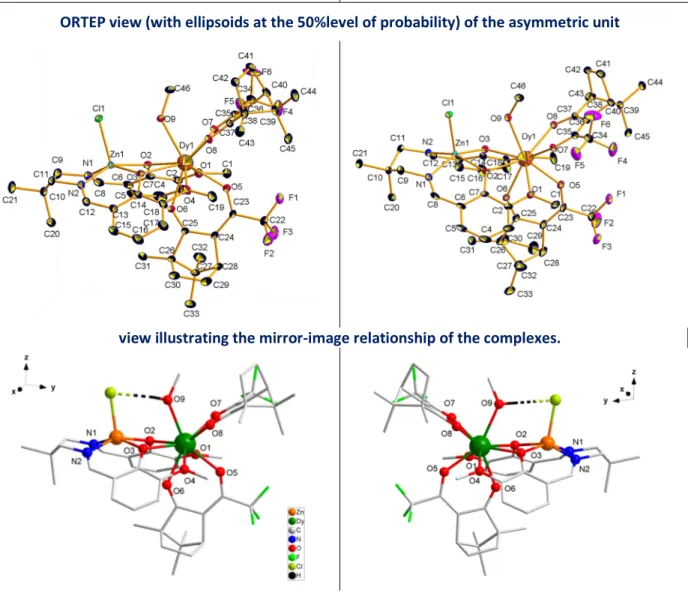
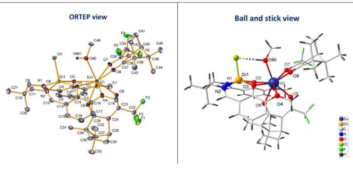
Figure 1. (top) [LMe2Zn(Cl)Dy((-)-camph)2(MeOH)], ∆-Dy, and (bottom) coordination sphere of the Dy(III)
4
[LMe2Zn(Cl)Dy((+)-camph)2(MeOH)], Λ-Dy, was formed by reaction of DyCl3(H2O)6, ZnLMe2·2H2O,
(+)-3-(trifluoroacetyl)camphor, and piperidine in 1/1/2/2 ratio in MeOH, whereas its enantiomer ∆-Dy was obtained with (-)-3-(trifluoroacetyl)camphor. Same procedure was followed to prepare the isomorphous Eu-enantiomers Λ-Eu and ∆-Eu, and the mixed metal complex Λ-Dy/Y (see Experimental section).
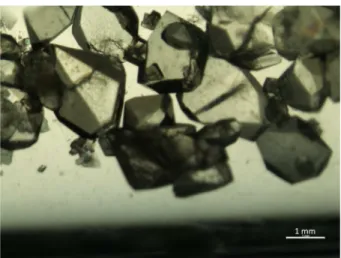
Crystals formed by slow evaporation of solvent (Picture S1) crystallize in monoclinic space group
P212121 and asymmetric unit comprises a single complex. The molecular structure for ∆-Dy is
depicted in Figure 1 (Figure S1 for stereo-isomer Λ-Dy, Figure S2 for Λ-Eu, and Figure S3 for the mixed metal Λ-Dy/Y derivative; selected bond distances are given in Table S2). It consists of a bimetallic complex made of the Schiff base accommodating a Zn(II) ion and a Dy(III) ion in the [N2O2] and [O2O2] compartments, respectively. The Zn(II) is in a square-base pyramid-like environment with two oxygen and two nitrogen atoms of the Schiff base in the basal position and one Cl atom in the apical position. The Dy center, in addition to the four oxygen atoms of the Schiff base, is associated with two Camph- ligands in chelate interaction with the Ln ion, and one MeOH molecule. An
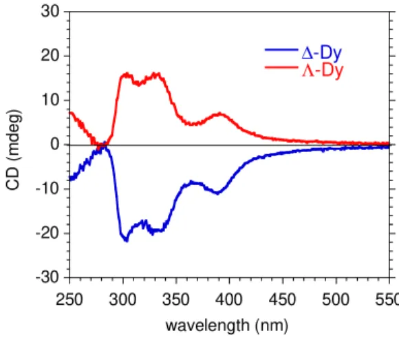
intramolecular hydrogen bond is established between the OH group of the alcohol and the Cl atom. The polyhedral shape of the Dy coordination sphere can be described either by a distorted capped square antiprism or a distorted muffin geometry (Table S3).25 The Ln is a stereogenic center and its formation is stereospecific; the Λ conformation is obtained by coordination of (+)-camph- whereas enantiomer ∆ is achieved with (-)-camph- ligands (Figure 1). The enantiomorphism of the two derivatives is confirmed by their solid state circular dichroism (CD) spectra showing symmetrical signals but of opposite sign (figure 2). The CD transitions can be assigned to π-π* intra-ligand transitions of the Schiff-base moiety (see Figure S4).26
Figure 2. Solid state CD spectra for ∆-Dy (in blue) and Λ-Dy (in red)
These complexes were found to exhibit slow relaxation of their magnetization. AC susceptibility revealed the emergence of an out-of-phase component, χM’’, below 12 K in zero field with however,
clear contribution of quantum tunneling of the magnetization (QTM) for lower temperatures. This QTM is only partly quenched by a static field (Figure S5) therefore the dilution technique was applied to investigate the relaxation characteristics. The behavior for Λ-Dy diluted (9 %) in an isostructural diamagnetic matrix of [LMe2Zn(Cl)Y((+)-camph)
2(MeOH)], i.e. Λ-Dy/Y, is given in Figures 3 and S6. AC
susceptibility data were collected in a static field of 1 kOe found to be the optimal field to reach slowest relaxation. Analysis of the Cole-Cole plots (Figure S6d) revealed a rather narrow distribution
-30 -20 -10 0 10 20 30 250 300 350 400 450 500 550 ∆-Dy Λ-Dy C D ( m d e g ) wavelength (nm)
5
width of the relaxation in the higher T range (10.5-7 K) with α = 0.15. For lower T the value of α steadily increased to 0.4 for 2, K which is indicative of a multi-process relaxation.
The temperature dependence of the relaxation time (Figure S6c) was obtained by analyzing the χM’’ = f(ν) for different temperatures with an extended Debye model. The linear variation of τ = f(1/T) between 8.5 and 4 K was fitted assuming a thermally activated process yielding Ueff/kB = 25 K with τ0
= 7.4x10⁻⁶ s. A modelling over the entire temperature range (2 to 8.5 K) was possible assuming
contributions from the Orbach, Raman and direct relaxation processes ( = / + 1/ ( ) + 1/( ). The values of the adjustment parameters are Ueff/ kB = 23 (± 3) K, τ0 = 1 (± 2)
x10-5 s, R = 0.1 (± 0.6) s-1, n = 4 (± 7), D = 2 (± 2) x104 K-1T-4s-1). The value for τ
0 is obviously too large
for an Orbach process but calculation uncertainty is large because of the number of variables. Adjustment to the experimental data failed without the Orbach contribution. These results confirm the emergence of slow relaxation of the magnetization below 12 K for probing frequency of 1 kHz.
Figure 3. AC susceptibility behavior for Λ-Dy diluted in diamagnetic [LMe2Zn(Cl)Y((+)-camph)2(MeOH)].
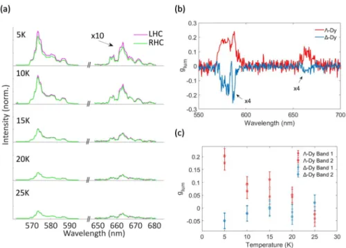
The luminescence properties of individual crystals of Λ-Dy and ∆-Dy were investigated by low temperature (5 - 30 K) micro-photoluminescence. We excited an area of 30 µm diameter so that single monocrystals could be observed (Picture S2). Figure 4 shows the emission spectra for a Λ-Dy crystal along left-handed and right-handed circular polarizations. Two groups of emission peaks appear at 573 and 663 nm and are attributed respectively to the 4F9/2-6H13/2 and 4F9/2-6H11/2 transitions
of Dy(III).27, 28 The emission intensity decreases as a function of temperature, barely any emission
being observed above 30 K. For both transitions, the left-handed circularly-polarized emission is slightly stronger than the right-handed polarization. This observation was made for all observed Λ-Dy crystals. On the other hand, for all ∆-Dy crystals the right-handed emission was consistently stronger than the left-handed emission (Figure S7). The degree of circular polarization is usually quantified by the luminescence dissymmetry factor glum (Equation 1).15
g
lum=
!" # $%( !& #)
(1)
This quantity ranges between -2 and +2 and vanishes when the emission is not circularly polarized. It is typically of the order of 10-3 – 10-2 for chiral organic molecules but can reach higher values for lanthanide complexes,15 with up to glum ≈ 1.4 reported for Eu(III).29, 30 For solutions of Dy(III) chiral
complexes, generally lower glum values of 0.0127 and up to ≈ 0.431 have been achieved. 0 0.5 1 1.5 2 2.5 3 3.5 4 2 4 6 8 10 12 1.0 Hz 3.0 Hz 5.0 Hz 7.0 Hz 10 Hz 15 Hz 20 Hz 30 Hz 40 Hz 70 Hz 130 Hz 200 Hz 320 Hz 521 Hz 781 Hz 997 Hz χM ( c m 3m o l -1) T (K) χM' χ M'' HDC = 1 kOe 0 0.5 1 1.5 2 0.1 1 10 100 1000 2.0 K 2.2 K 2.4 K 2.5 K 2.8 K 3.0 K 3.5 k 4.0 K 4.5 K 5.0 K 5.5 K 6.0 K 6.5 K 7.5 K 8.0 K 9.0 K 10 K χ M '' (c m 3m o l -1) ν (Hz) HDC = 1 kOe
6
Figure 4. Temperature dependent emission of Λ-Dy crystal. (a) Purple lines: left-handed circular (LHC)
polarization; green lines: right-handed circular (RHL) polarization for the Λ-Dy sample at different
temperatures ; (b) CPL dissymmetry factor glum as a function of the wavelength for the two samples at 5
K; (c) Temperature-dependent variation of the luminescence dissymmetry factor, glum for the two
samples and the two spectral bands.
From our spectra, using equation 1, we extract the dissymmetry factor as a function of the wavelength (fig. 4(b)) for the two samples at 5 K. Over the two emission spectral intervals, the dissymmetry factor shows unambiguous opposite signs for the two samples, demonstrating the effect of the ligand chirality on the CPL. The dissymmetry factor is quite lower in absolute value for the ∆-Dy sample, possibly because of some measurement uncertainty. We then average the dissymmetry factor over each of the two intervals and plotted its average value as a function of the temperature in figure 4(c) and S7. In spite of some uncertainty on glum values and some variation
between the individual crystals, a general trend is observed: the degree of circular polarization decreases with temperature and vanishes around 25 K. These observations were repeated for several crystals, with a distribution of glum values ranging between 0.04 and 0.18 for Λ-Dy and between -0.04
and -0.16 for ∆-Dy at 5 K.
It can be noticed that the appearance of CPL and the increase of glum with decrease of temperature
coincide with the emergence of slow relaxation of magnetization for these complexes. As mentioned above, CPL can be induced by the chiral structure of the luminophore but it can also be generated by a magnetic field whether applied or intrinsic to the material. For a SMM, the later results from the easy-axis orientation the individual magnetization. In order to provide additional information on the possible role of each of these sources, studies have been carried out on isomorphous Eu(III) complexes (see SI for crystal data). At low temperature, Eu(III) is essentially non-magnetic, which makes it possible to probe the contribution of structural origin. Moreover, Eu(III) is the lanthanide element for which the strongest CPL has been reported in the literature.15, 27, 31
7
Characteristic luminescence bands where observed below 250 K for Λ-Eu and ∆-Eu (Figure S8). It can be noticed that the most prominent emission is arising from the 5D
0 -> 7F2 transition with two main
lines (613 and 619 nm) for each enantiomer, whereas the intensity of the 5D
0 -> 7F0 transition at
585-595 nm is weak. Their relative intensity ratio is R = 19. This feature is characteristic for a low symmetry field at the Eu(III) site, in agreement with point symmetry Cs deduced from the crystal structure (Table S3).32, 33 Down to 5 K the emission is exactly the same for the two circular polarizations. The degree of circular polarization was calculated for each peak at each temperature and glum was always close to 0, fluctuating between -0.04 and +0.04. It can be concluded that CPL of
structural origin is very low for these compounds.
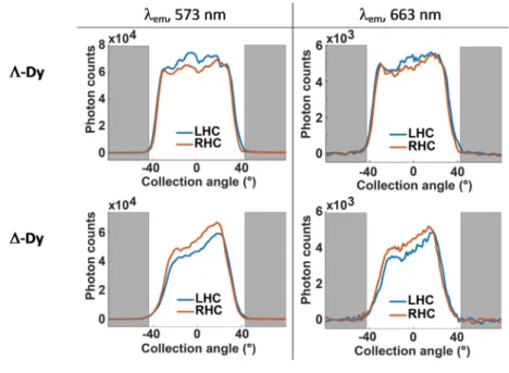
Finally, we used Fourier-plane imaging to obtain the emission angular distribution in the two circular polarizations (Figure 5) for a Λ-Dy and a ∆-Dy crystal. The overall emission is distributed rather isotropically for the first crystal and has more preferred directions for the second crystal, probably due to the different shape of the crystal boundaries, but for both crystals the angular distribution is the same for the two emission lines. In spite of some significant noise, the curves clearly show that the degree of circular polarization has little dependence on the emission angle. This is confirmed by observing an ensemble of crystals of random orientations. A powder formed by crushing a single crystal and illuminated over a 30 µm diameter area showed dissymmetry factors of the same order as for a single crystal, also with isotropic angular distribution (Figure S9). There is thus little difference between the circular-polarization of a single crystal and an ensemble of randomly-oriented crystals. Beyond the obvious advantage for processing such a material, the use of a powder also solves the problem of the amount of material needed for reliable measurement.
Figure 5. Angular distribution of the emission bands at 573-nm and 663-nm bands for a (top) Λ-Dy and
(bottom) a ∆-Dy crystal at 5 K. Angular range scanned is +/- 44°. The grey portions correspond to the
angles not collected by the objective (outside the numerical aperture)
8
Concomitant rise of circularly polarized luminescence and SMM-type behavior was evidenced in the solid state for an enantiomeric pair of chiral Dy complexes. This observation strongly suggests that magnetic anisotropy of the Dy combines with structural chirality to enhance the chiral force field acting on the luminescent Ln center. The synergy between the emission and the magnetic features is supported by the lack of CPL for the Eu derivatives. This allows excluding a purely structural effect and substantiates the essential role of the magnetization blocking in the CPL observed for the Dy complexes.
It is noticeable that the observed luminescence dissymmetry factor is large with |glum| up to 0.18.
Interestingly, the same CPL was found for singles crystals and the polycrystalline powders. This is a particularly important observation as it suggests that CPL can be exploited on a crystalline powder and is not limited to a single crystal.
EXPERIMENTAL
General procedures: Reagents and solvents were obtained commercially and used without further
purification. The Schiff base H2LMe2 and its Zn complex [ZnLMe2]·2H2O were prepared as previously
reported34 and recrystallized from MeOH solutions by slow evaporation of the solvents. Purity of the
samples was verified by 1H NMR and elemental analysis. The two enantiomers (+) and (-) of the
trifluoroacetyl-3-camphor were synthesized following the literature procedure.35 Purity of the samples was checked by 1H, 13C, 19F NMR, and by specific optical rotation measurements. Elemental C, H and N analyses were performed on a Perkin-Elmer 2400 II analyzer on freshly prepared and isolated samples. IR spectra were recorded in the 4000-700 cm-1 range with a Perkin-Elmer Spectrum
100 FTIR using the ATR mode. Optical rotation measurements were performed on a Perkin-Elmer 241 polarimeter with a sodium lamp, at room temperature. 1H NMR spectra were recorded using a
DPX300 Brüker spectrometer with working frequencies at 300 MHz for 1H. Chemical shifts were referenced to the residual proton resonance of the deuterated solvents.
Magnetic studies: Magnetic measurements were carried out with a Quantum Design MPMS 5S
SQUID magnetometer. Magnetic susceptibility, χM, between 2 and 300 K was obtained with an
applied field of 1000 Oe. Isothermal magnetization measurements were performed up to 5 T at 2 K. The ac susceptibility responses were recorded using an oscillating field of HAC = 3 Oe in zero field and
under applied static field of 0.1T. The measurements were performed on crushed crystals from freshly isolated samples mixed to grease and held in gelatin capsules. The molar magnetic susceptibilities were corrected for sample holder and for the diamagnetic contribution of all the atoms by using Pascal’s tables.36
Solid-state Circular Dichroism (CD) measurement: The solid-state CD spectra (collected in transmittance mode) were measured on a JASCO J-720 spectropolarimeter using KBr disks of samples. A disk was prepared from several crystals of a batch of either ∆-Dy or Λ-Dy enantiomer with crystalline KBr, according to the following procedure: A mixture of sample and KBr was well ground in an agate mortar then vacuum pressed to form a uniform disk. The disk was placed in a sample holder, which was then positioned in the cell holder of the spectropolarimeter.37 For each disk, the CD signals were measured from both sides, and the average was taken. A blank disk was used to measure background spectra. The data were corrected accordingly.
9
Crystallographic Studies. Data collections were performed at low temperature on a Bruker Apex2
(Mo microfocus source) or an Oxford-Diffraction Gemini (Cu source) diffractometer. The structures were solved by SUPERFLIP38 or SIR92,39 and refined by means of least-squares procedures on F or F2 using the programs of the PC version of CRYSTALS.40 For mixed metal compound Λ-Dy/Y best
agreement was obtained for ratio 0.16 Dy/0.84. Atomic scattering factors were taken from the International tables for X-ray Crystallography. Hydrogen atoms were refined using a riding model. The absolute configuration of the compounds was determined unambiguously by Flack parameter refinement. Data are gathered in table S1. Cif have been deposited at CCDC under references 2007104 (for ∆-Dy), 2007105 (for Λ-Dy), 2018680 (for Λ-Eu), and 2033959 (for Λ-Dy/Y).
CPL studies: Crystals of size 300-800 µm were attached by a silver lacquer to the cold finger of a
helium-flow cryostat (Oxford, HiRes II) and observed by a homemade microscope (0.7 numerical aperture objective, x60 magnification). The sample was illuminated through the objective by the light from a mercury lamp filtered over a 330-480 nm pass band. The fluorescence emission was collected by the same objective, filtered by a 500 nm long-pass filter and analyzed by an imaging spectrometer (Horiba Jobin-Yvon, Triax-190). Circular polarization analysis was performed by introducing a polarizer before the spectrometer entrance and comparing the +45° and -45° positions of a quarter-wave plate before the polarizer.
The polarization effect of the different setup elements was corrected by introducing compensating elements. Fluorescent polymer spheres (Thermo Fisher, 200nm, 580-605 nm emission) were used for setup polarization calibration. By circular polarization analysis, an average degree of circular polarization of 0 was measured, with fluctuation in the range of ±0.02. When introducing a polarizer below the objective to polarize the emission, a 0.99 degree of linear polarization was obtained. We thus conclude that the emission polarization was preserved by the setup with 1-3 % precision.
Syntheses:
[LMe2Zn(Cl)Dy((+)-camph)
2(MeOH)], Λ-Dy, and [LMe2Zn(Cl)Dy((-)-camph)2(MeOH)], ∆-Dy:
[ZnLMe2]·2H2O (233 mg; 0.5 mmol) and DyCl3·6H2O (188 mg; 0.5 mmol) were solubilized in 10 mL of
methanol. Either the (+)-3-(trifluoroacetyl)camphor or (-)-3-(trifluoroacetyl)camphor (250 mg; 1 mmol) was added to this solution followed by piperidine (85 mg; 1 mmol). The resulting pale yellow solution was stirred at room temperature for 30 min. and then transferred in a crystallization tube for slow evaporation of the solution. Pale yellow crystals suitable for X-Ray diffraction appeared after a few days (Picture S1). They were collected and washed with methanol.
Data for Λ-Dy: Yield 410 mg (71 %). IR (ATR): ν = 3251 (w), 2955 (m), 1652 (s), 1639 (s), 1535 (m), 1473 (m), 1424 (m), 1392 (w), 1293 (m), 1267 (m), 1243 (w), 1222 (vs), 1200 (vs), 1178 (s), 1126 (s), 1069 (s), 1053 (m), 1023 (m), 972 (m), 918 (w), 850 (m), 803 (w), 734 (s), 642 (m) cm-1. C46H56ClDyF6N2O9Zn (1158.3): calcd. C 47.70, H 4.87, N 2.42; found C 47.46, H 4.80, N 2.30.
Data for ∆-Dy: Yield 355 mg (61 %). IR (ATR): ν = 3252 (w), 2955 (m), 1652 (s), 1640 (s), 1539 (m), 1472 (m), 1423 (m), 1393 (w), 1295 (m), 1268 (m), 1244 (w), 1221 (vs), 1201 (vs), 1179 (s), 1127 (s), 1069 (s), 1054 (m), 1023 (m), 971 (m), 918 (w), 849 (m), 803 (w), 735 (s), 642 (m) cm-1.
10
[LMe2Zn(Cl)Y/Dy((+)-camph)2(MeOH)], Λ-Dy/Y: [ZnLMe2]·2H2O (93 mg; 0.2 mmol), DyCl3·6H2O (5.8 mg;
0.016 mmol) and YCl3·6H2O (56 mg; 0.183 mmol) were solubilized in 6 mL of methanol.
(+)-3-(trifluoroacetyl)camphor (97.5 mg; 0.393 mmol) was added to this solution followed by piperidine (32 mg; 0.385 mmol). The resulting pale yellow solution was stirred at room temperature for 30 min. and then transferred in a crystallization tube for slow evaporation of the solution. Pale yellow crystals (62 mg) were collected after several days and washed with methanol. Crystal structure confirmed a crystallographic phase in accordance with that of Λ-Dy (Figure S3). IR (ATR): ν = 3402 (vbr, w), 2961 (m), 1662 (s), 1628 (s), 1535 (m), 1539 (m), 1479 (m), 1437 (m), 1393 (w), 1305 (m), 1267 (s), 1243 (w), 1222 (vs), 1199 (vs), 1178 (vs), 1123 (s), 1074 (s), 1051 (m), 1004 (w), 973 (w), 921 (w), 854 (w), 803 (w), 734 (s), 644 (m) cm-1. EDX analysis performed on crystals confirmed the
presence of both Y (83.1 wt%, 93.5 mol%) and Dy (16.9 wt%, 10 mol%). A Dy content of 9 mol% was deduced from magnetic behavior (see Figure S6).
[LMe2Zn(Cl)Eu((+)-camph)
2(MeOH)], Λ-Eu, and [LMe2Zn(Cl)Eu((-)-camph)2(MeOH)], ∆-Eu : These
complexes have been obtained by same procedure as for the Dy derivatives using EuCl3, 6H2O. Yield
400 mg (70%); Elemental analysis calcd (%) for C46H56Cl1Eu1F6N2O9Zn1 (1148 gmol-1): C 48.14, H 4.92,
N 2.44; found for Λ-Eu: C 47.99, H 4.72, N 2.36; and for ∆-Eu: C 48.16, H 4.85, N 2.42. IR (ATR): as for Dy derivatives. XRPD for ∆-Eu confirmed a crystallographic phase in agreement with that Λ-Eu (Figure S10).
Conflict of Interest
The authors declare no competing financial interest. Acknowledgements
This work was supported by the French Research Agency (Agence Nationale de la Recherche, reference ANR-09-BLAN-0054-01). Authors are grateful to M. J.-F. Meunier and M. L. Réchignat (LCC) for technical assistance in magnetic data collection.
11 REFERENCES.
1. D. Gatteschi, R. Sessoli and J. Villain, Molecular Nanomagnets, Oxford University Press, Oxford, 2006.
2. M. Faraday, I. Experimental researches in electricity.-Nineteenth series, Phil. Trans. R. Soc., 1846, 136, 1-20.
3. T. Haider, A Review of Magneto-Optic Effects and Its Application, Int. J. Electromagn. & and
Appl., 2017, 7, 17-24.
4. G. Wagnière and A. Meier, The influence of a static magnetic field on the absorption coefficient of a chiral molecule, Chem. Phys. Lett., 1982, 93, 78-81.
5. L. D. Barron and J. Vrbancich, Magneto-chiral birefringence and dichroism, Mol. Phys., 1984, 51, 715-730.
6. P. Kleindienst and G. H. Wagnière, Interferometric detection of magnetochiral birefringence,
Chem. Phys. Lett., 1998, 288, 89-97.
7. M. Atzori, G. L. J. A. Rikken and C. Train, Magneto-Chiral Dichroism: A Playground for Molecular Chemists, Chem. Eur. J., 2020, 26, 9784-9791.
8. G. L. J. A. Rikken and E. Raupach, Pure and cascaded magnetochiral anisotropy in optical absorption, Phys. Rev. E, 1998, 58, 5081-5084.
9. G. L. J. A. Rikken and E. Raupach, Observation of magneto-chiral dichroism, Nature, 1997, 390, 493-494.
10. K. Taniguchi, M. Nishio, S. Kishiue, P.-J. Huang, S. Kimura and H. Miyasaka, Strong
magnetochiral dichroism for visible light emission in a rationally designed paramagnetic enantiopure molecule, Phys. Rev. Mater., 2019, 3, 045202.
11. M. Atzori, I. Breslavetz, K. Paillot, K. Inoue, G. L. J. A. Rikken and C. Train, A Chiral Prussian Blue Analogue Pushes Magneto-Chiral Dichroism Limits, J. Am. Chem. Soc., 2019, 141, 20022-20025. 12. C. Train, R. Gheorghe, V. Krstic, L.-M. Chamoreau, N. Ovanesyan, G. L. J. A. Rikken, M. Gruselle and M. Verdaguer, Strong magneto-chiral dichroism in enantiopure chiral ferromagnets,
Nature Mater., 2008, 7, 729-734.
13. R. Sessoli, M.-E. Boulon, A. Caneschi, M. Mannini, L. Poggini, F. Wilhelm and A. Rogalev, Strong magneto-chiral dichroism in a paramagnetic molecular helix observed by hard X-rays, Nat
Phys, 2015, 11, 69-74.
14. J. P. Riehl and F. S. Richardson, Circularly polarized luminescence spectroscopy, Chem. Rev., 1986, 86, 1-16.
15. F. Zinna and L. Di Bari, Lanthanide Circularly Polarized Luminescence: Bases and Applications,
Chirality, 2015, 27, 1-13.
16. R. Carr, N. H. Evans and D. Parker, Lanthanide complexes as chiral probes exploiting circularly polarized luminescence, Chem. Soc.Rev., 2012, 41, 7673-7686.
17. K. Müller, F. Fuchs and F. K. Kneubühl, Partial circular polarization of the spectral thermal emission from ferromagnetic iron, Phys. Lett. A, 1977, 64, 249-250.
18. F. Zinna, C. Resta, S. Abbate, E. Castiglioni, G. Longhi, P. Mineo and L. Di Bari, Circularly polarized luminescence under near-UV excitation and structural elucidation of a Eu complex, Chem.
Comm., 2015, 51, 11903-11906.
19. H. Tsumatori, T. Harada, J. Yuasa, Y. Hasegawa and T. Kawai, Circularly Polarized Light from Chiral Lanthanide(III) Complexes in Single Crystals, Appl. Phys. Exp., 2011, 4, 011601.
20. J. P. Costes, S. Titos-Padilla, I. Oyarzabal, T. Gupta, C. Duhayon, G. Rajaraman and E. Colacio, Effect of Ligand Substitution around the DyIII on the SMM Properties of Dual-Luminescent Zn–Dy and Zn–Dy–Zn Complexes with Large Anisotropy Energy Barriers: A Combined Theoretical and
Experimental Magnetostructural Study, Inorg. Chem., 2016, 55, 4428-4440.
21. W.-B. Sun, P.-F. Yan, S.-D. Jiang, B.-W. Wang, Y.-Q. Zhang, H.-F. Li, P. Chen, Z.-M. Wang and S. Gao, High symmetry or low symmetry, that is the question - high performance Dy(iii) single-ion magnets by electrostatic potential design, Chem. Sci., 2016, 7, 684-691.
12
22. I. Oyarzabal, J. Ruiz, J. M. Seco, M. Evangelisti, A. Camón, E. Ruiz, D. Aravena and E. Colacio, Rational Electrostatic Design of Easy-Axis Magnetic Anisotropy in a ZnII–DyIII–ZnII Single-Molecule Magnet with a High Energy Barrier, Chem. Eur. J., 2014, 20, 14262-14269.
23. J.-H. Jia, Q.-W. Li, Y.-C. Chen, J.-L. Liu and M.-L. Tong, Luminescent single-molecule magnets based on lanthanides: Design strategies, recent advances and magneto-luminescent studies, Coord.
Chem. Rev., 2019, 378, 365-381.
24. T. D. Pasatoiu, C. Tiseanu, A. M. Madalan, B. Jurca, C. Duhayon, J.-P. Sutter and M. Andruh, Study of the Luminescent and Magnetic Properties of a Series of Heterodinuclear [ZnIILnIII]
Complexes Inorg. Chem., 2011, 50, 5879-5889.
25. A. Ruiz-Martínez, D. Casanova and S. Alvarez, Polyhedral Structures with an Odd Number of Vertices: Nine-Coordinate Metal Compounds, Chem. Eur. J., 2008, 14, 1291.
26. V. Béreau, V. Jubéra, P. Arnaud, A. Kaiba, P. Guionneau and J.-P. Sutter, Modulation of the Luminescence Quantum Efficiency for Blue Luminophor {Al(salophen)}+ by Ester-Substituents Dalton Trans., 2010, 39, 2070-2077.
27. S. Petoud, G. Muller, E. G. Moore, J. Xu, J. Sokolnicki, J. P. Riehl, U. N. Le, S. M. Cohen and K. N. Raymond, Brilliant Sm, Eu, Tb, and Dy Chiral Lanthanide Complexes with Strong Circularly Polarized Luminescence, J. Am. Chem. Soc., 2007, 129, 77-83.
28. P. Babu, K. Hyuk Jang, E. Sik Kim, L. Shi and H. Jin Seo, Optical Properties and White-Light Emission in Dy3+-Doped Transparent Oxyfluoride Glass and Glass Ceramics Containing CaF2 Nanocrystals, J. Korean Phy. Soc. , 2009, 54, 1488-1491.
29. J. L. Lunkley, D. Shirotani, K. Yamanari, S. Kaizaki and G. Muller, Extraordinary Circularly Polarized Luminescence Activity Exhibited by Cesium Tetrakis(3-heptafluoro-butylryl-(+)-camphorato) Eu(III) Complexes in EtOH and CHCl3 Solutions, J. Am. Chem. Soc., 2008, 130, 13814-13815.
30. J. Kumar, B. Marydasan, T. Nakashima, T. Kawai and J. Yuasa, Chiral supramolecular polymerization leading to eye differentiable circular polarization in luminescence, Chem. Comm., 2016, 52, 9885-9888.
31. R. S. Dickins, J. A. K. Howard, C. L. Maupin, J. M. Moloney, D. Parker, J. P. Riehl, G. Siligardi and J. A. G. Williams, Synthesis, Time-Resolved Luminescence, NMR Spectroscopy, Circular Dichroism and Circularly Polarised Luminescence Studies of Enantiopure Macrocyclic Lanthanide Tetraamide Complexes, Chem. Eur. J., 1999, 5, 1095-1105.
32. R. Reisfeld, Berlin, Heidelberg, 1973.
33. B. R. Judd, Hypersensitive Transitions in Rare-Earth Ions, J. Chem. Phys., 1966, 44, 839-840. 34. J.-P. Costes, J.-P. Laussac and F. Nicodème, Complexation of a Schiff base ligand having two coordination sites (N2O2 and O2O2) with lanthanide ions (Ln = La, Pr): an NMR study, J. Chem. Soc.
Dalton Trans., 2002, 2731-2736.
35. H. L. Goering, J. N. Eikenberry, G. S. Koermer and C. J. Lattimer, Direct determination of enantiomeric compositions with optically active nuclear magnetic resonance lanthanide shift reagents, J. Am. Chem. Soc., 1974, 96, 1493-1501.
36. O. Kahn, Molecular Magnetism, VCH, Weinheim, 1993.
37. M. Matsushima, K. Wada, Y. Horino, K. Takahara, Y. Sunatsuki and T. Suzuki, Transition-metal(ii) complexes with a tripodal hexadentate ligand, 1,1,1-tris[2-aza-3-(imidazol-4-yl)prop-2-enyl]ethane, exhibiting incomplete total or absolute spontaneous resolution, CrystEngComm, 2020, 22, 458-466.
38. L. Palatinus and G. Chapuis, Superflip, J. Appl. Cryst., 2007, 40, 786-790.
39. A. Altomare, G. Cascarano, C. Giacovazzo, A. Guargliardi, M. C. Burla, G. Polidori and M. Camalli, SIR92 – a program for automatic solution of crystal structures by direct methods, J. Appl.
Cryst., 1994, 27, 435.
40. P. W. Betteridge, J. R. Carruthers, R. I. Cooper, K. Prout and D. J. Watkin, CRYSTALS, J. Appl.
13
SUPPORTING INFORMATION
CONTENTS
Picture 1. Crystals of [LMe2Zn(Cl)Dy((-)-camph)2(MeOH)], ∆-Dy.
Table S1. Crystal data for Λ-Dy and ∆-Dy.
Figure S1. Asymmetric units for Λ-Dy and ∆-Dy with numbering scheme.
Figure S2. Λ-Eu : ORTEP plot (with ellipsoids at the 50% level of probability) of the
asymmetric unit with numbering scheme.
Figure S3 Λ-Dy/Y : ORTEP plot (with ellipsoids at the 50% level of probability) of the
asymmetric unit with numbering scheme.
Table S2. Selected bond distances (Å) and angles (°) for Eu and Dy complexes
Table S3. SHAPE Analysis for the ML9 coordination sphere of the Ln ions.
Figure S4. Solid-state UV-vis spectra for ZnLMe2 and [LMe2 Zn(Cl)Dy((+/-)-camph)2(MeOH)] complexes
Figure S5. Magnetic behaviors for Λ-Dy and ∆-Dy.
Figure S6. Magnetic behaviors for [LMe2Zn(Cl)Y/Dy((+)-camph)2(MeOH)], Λ-Dy/Y.
Picture 2. Crystals (size 300-800 µm) and powders (crushed crystal) on the cold stage for micro-photoluminescence studies.
Figure S7. Temperature dependent emission and glum of a ∆-Dy crystal.
Figure S8. Temperature dependent emission and glum for a Λ-Eu and a ∆-Eu crystal.
Figure S9. Angular distribution of the emission (bands at 573-nm and 663-nm) recorded at 5K, and temperature dependent variation of degree of circular polarization for a crystalline powder of Λ-Dy and a ∆-Dy crystal. Figure S10. X-Ray Powder Diffraction (XRPD) for Λ-Dy , ∆-Dy, Λ-Eu , and ∆-Eu.
14
Picture 1. Crystals of [LMe2Zn(Cl)Dy((-)-camph)2(MeOH)], ∆-Dy.
Table S1. Crystal data for Λ-Dy, ∆-Dy, Λ-Eu, and Λ-Dy/Y.
Compound Λ-Dy ∆-Dy Λ-Eu Λ−Dy/Y
Empirical formula C46 H56 Cl Dy F6 N2 O9 Zn C46 H56 Cl Dy F6 N2 O9 Zn C46 H56 Cl Eu F6 N2 O9 Zn C46 H56 Cl Dy0.16 F6 N2 O9 Y0.84 Zn
Formula weight (g∙mol-1) 1158.28 1158.28 1147.74 1096.46
λ (Å) 0.71073 0.71073 1.54180 0.71073
Temperature (K) 100 100 100 110
Crystal system Orthorhombic Orthorhombic Orthorhombic Orthorhombic Space group P 21 21 21 P 21 21 21 P 21 21 21 P 21 21 21 a (Å) 13.0607(9) 13.0364(7) 13.0430(2) 13.0472(5) b (Å) 18.8726(11) 18.8572(11) 18.9020(2) 18.8726(7) c (Å) 19.3077(13) 19.2842(11) 19.3871(2) 19.2735(9) Volume (Å3) 4759.1(5) 4740.6(3) 4779.7(1) 4745.8(3) Z 4 4 4 4 Calc. Density (g∙cm-3) 1.616 1.623 1.595 1.534 Absorption coefficient (mm-1) 2.197 2.206 11.111 1.913
Theta range for data collection (°) 1.8-35.7 2.1-32.9 3.3-70.4 1.5-30.6
Reflections collected 115603 159374 37407 169605 Independent reflections 18888 16609 9170 14355 R(int) 0.028 0.034 0.033 0.044 Data/restraints/parameters 17290/0/596 15792/0/596 8868/0/596 9542/1/596 Goodness-of-fit 1.09 1.08 1.09 0.97 Refinement on F F F F2
Final R indices [I > nσ(I)] 0.0150 (n=3) 0.0125 (n=3) 0.0192 (n=3) 0.0163 (n=2) Final wR indices [I > nσ(I)] 0.0152 (n=3) 0.0131 (n=3) 0.0228 (n=3) 0.0433 (n=2) Flack parameter, nb Friedel-pairs -0.015(2), 8300 -0.009(2), 7333 -0.0135(8), 4090 -0.014(2), 6445 Largest diff. Peak and hole (e Å-3) 0.97/-0.91 0.45/-0.46 3.31/-2.99 0.38/-0.19
15
Figure S1. Asymmetric units for Λ-Dy and ∆-Dy with numbering scheme.
[LMe2Zn(Cl)Ln((+)-camph)
2(MeOH)], Λ-Dy [LMe2Zn(Cl)Ln((-)-camph)2(MeOH)], ∆-Dy ORTEP view (with ellipsoids at the 50%level of probability) of the asymmetric unit
16
Figure S2. Λ-Eu : ORTEP plot (with ellipsoids at the 50% level of probability) of the asymmetric unit with
numbering scheme.
[LMe2Zn(Cl)Eu((+)-camph)
2(MeOH)], Λ-Eu
ORTEP view Ball and stick view
Figure S3. Λ-Dy/Y : ORTEP plot (with ellipsoids at the 50% level of probability) of the asymmetric unit with
numbering scheme.
[LMe2Zn(Cl)Dy/Y((+)-camph)
2(MeOH)], Λ-Dy/Y
17
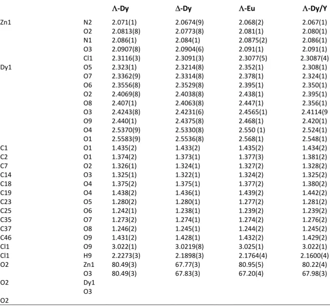
Table S2. Selected bond distances (Å) and angles (°) for ∆- and Λ-Dy, Λ-Eu, and Λ-Dy/Y complexes.
Λ-Dy ∆-Dy Λ-Eu Λ-Dy/Y
Zn1 N2 O2 N1 O3 Cl1 Dy1 O5 O7 O6 O2 O8 O3 O9 O4 O1 C1 O1 C2 O1 C7 O2 C14 O3 C18 O4 C19 O4 C23 O5 C25 O6 C35 O7 C37 O8 C46 O9 Cl1 O9 Cl1 H9 O2 Zn1 O3 O2 Dy1 O3 O2 2.071(1) 2.0813(8) 2.086(1) 2.0907(8) 2.3116(3) 2.323(1) 2.3362(9) 2.3556(8) 2.4069(8) 2.407(1) 2.4243(8) 2.440(1) 2.5370(9) 2.5583(9) 1.435(2) 1.374(2) 1.326(1) 1.325(1) 1.375(2) 1.438(2) 1.280(2) 1.242(1) 1.273(2) 1.246(2) 1.431(2) 3.022(1) 2.2273(3) 80.49(3) 80.49(3) 2.0674(9) 2.0773(8) 2.084(1) 2.0904(6) 2.3091(3) 2.3214(8) 2.3314(8) 2.3529(8) 2.4038(8) 2.4063(8) 2.4231(6) 2.4375(8) 2.5330(8) 2.5536(8) 1.433(2) 1.373(1) 1.324(1) 1.322(1) 1.375(1) 1.436(1) 1.280(1) 1.238(1) 1.274(1) 1.245(1) 1.428(1) 3.0219(8) 2.1898(3) 67.77(3) 67.83(3) 2.068(2) 2.081(1) 2.0875(2) 2.091(1) 2.3077(5) 2.352(1) 2.378(1) 2.395(1) 2.438(1) 2.447(1) 2.4565(1) 2.468(1) 2.550 (1) 2.568(1) 1.435(2) 1.377(3) 1.327(2) 1.324(2) 1.377(2) 1.439(2) 1.277(2) 1.239(2) 1.274(2) 1.244(2) 1.432(2) 3.025(1) 2.1764(4) 80.95(5) 67.20(4) 2.067(1) 2.080(1) 2.086(1) 2.091(1) 2.3087(4) 2.308(1) 2.324(1) 2.350(1) 2.395(1) 2.356(1) 2.4114(9 2.420(1) 2.524(1) 2.548(1) 1.434(2) 1.381(2) 1.328(2) 1.325(2) 1.380(2) 1.442(2) 1.281(2) 1.239(2) 1.276(2) 1.245(2) 1.429(2) 3.022(1) 2.1600(4) 80.22(4) 67.98(3)
18
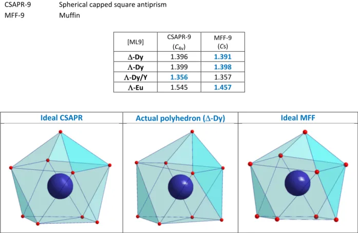
Table S3: SHAPE Analysis for the ML9 coordination sphere of the Ln ions.1
CSAPR-9 Spherical capped square antiprism
MFF-9 Muffin [ML9] CSAPR-9 (C4v) MFF-9 (Cs) ∆-Dy 1.396 1.391 Λ-Dy 1.399 1.398 Λ-Dy/Y 1.356 1.357 Λ-Eu 1.545 1.457
Ideal CSAPR Actual polyhedron (∆-Dy) Ideal MFF
Figure S4. Solid-state UV-vis spectra for ZnLMe2 and [LMe2Zn(Cl)Dy((+/-)-camph)2(MeOH)] complexes.
1
Llunell, M.; Casanova, D.; Cirera, J.; Alemany, P.; Alvarez, S.; SHAPE: Program for the stereochemical analysis of molecular fragments by means of continuous shape measures and associated tools 2.1 ed.; University of Barcelona: Barcelona, 2013.
250 300 350 400 450 500 550 ZnLMe2 Λ-Dy ∆-Dy I. ( a. u .) Wavelength (nm)
19 Figure S5. Magnetic behaviors for Dy complex.
Magnetic behavior is independent of the chirality therefore a full set of magnetic behavior has been collected only for Λ-Dy. The magnetic purity of the ∆ enantiomer was confirmed by susceptibility values at 300 K (found χMT = 14.10 cm3mol-1K) and AC versus T trace with frequency of 1 kHz (shown
below) that was found identical to Λ-Dy.
DC studies: The temperature dependence of χMT for Λ-Dy (Fig. S5a) is in agreement with the
behavior expected for an isolated Dy(III) ion. The value found at 300 K is 14.15 cm3mol-1K and reaches
11.41 cm3mol-1K at 4 K. Magnetization versus field at 2 K (b) reaches as value of 5.35 µB under 5 T.
(a) (b)
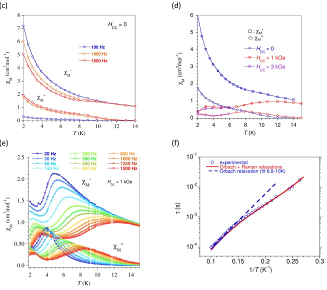
AC studies: AC magnetic susceptibility as a function of temperature recorded without an applied filed
is shown in (c). An out-of-phase component, χM’’, was found below 15 K and lower temperature
behavior indicated other relaxation processes become active. Application of an external DC field allowed to drastically reduce these contributions but not to fully cancel them even when a field of 3 kOe (d). Since the χM’’ = f(T) for different AC frequencies obtained with HDC = 1 kOe display well
defined maxima, a full data set was collected (e). The temperature dependence of the relaxation time between 3.8 and 10 K (f) was obtained by analyzing the χM’’ = f(ν) for different temperatures
with an extended Debye model. The linear variation of τ =f(1/T) between 10 and 6.5 K was fitted assuming a thermally activated process yielding Ueff/kB = 42 K with τ0 = 1.4x10⁻⁶ s (dotted blue line). A
modelling over the entire temperature range (3.8 to 10 K, red line) was possible considering contributions from the Orbach and Raman relaxation processes ( = / + 1/( ). The values of the adjustment parameters are Ueff/ kB = 53 ± 9 K, τ0 = 0.8 ± 2 x10⁻⁸ s, R = 0.2 ± 0.1 s-1, n =
4.9± 0.4. 0 5 10 15 0 50 100 150 200 250 300 χ M T ( c m 3 m o l -1 K ) T (K) H = 1 kOe 0 1 2 3 4 5 6 0 10 20 30 40 50 M ( µ B ) H (kOe) T = 2 K
20
(c) (d)
(e) (f)
Figure S5g shows the χM’’ = f(T) trace for ∆-Dy under HDC = 1 kOe and for ν = 1 kHz which confirms a
behavior identical to that of the Λ-Dy enantiomer. (g) 0 1 2 3 4 5 6 7 8 2 4 6 8 10 12 14 100 Hz 1000 Hz 1500 Hz χ M ( cm 3 m ol -1 ) T (K) χM' χM'' H DC = 0 0 1 2 3 4 5 6 2 4 6 8 10 12 14 H DC = 0 H DC = 1 kOe H DC = 3 kOe χ M ( c m 3 m o l -1 ) T (K) : χM’ : χM’’ 0.0 0.50 1.0 1.5 2.0 2.5 2 4 6 8 10 12 14 20 Hz 30 Hz 50 Hz 100 Hz 200 Hz 300 Hz 400 Hz 600 Hz 800 Hz 1000 Hz 1250 Hz 1500 Hz χ M ( cm 3 m ol -1 ) T (K) H DC = 1 kOe χM'' χM' 10-4 10-3 10-2 10-1 0.1 0.15 0.2 0.25 0.3 experimental
Orbach + Raman relaxations Orbach relaxation (fit 6.8-10K)
τ (s ) 1/T (K-1) 0 0.1 0.2 0.3 0.4 0.5 0.6 5 10 15 Λ-Dy ∆-Dy χ M '' (c m 3 m o l -1 ) T (K) HDC = 1 kOe HAC = 3 Oe Frq = 1 kHz
21
Figure S6. Magnetic behaviors for [LMe2Zn(Cl)Y/Dy((+)-camph)2(MeOH)], Λ-Dy/Y.
(a) (b)
(c) (d)
(e)
The effective composition in Dy(III) complex was estimated by comparison of the M = f(H) for the sample to that of pure Λ-Dy. The high-field magnetizations matched for 9 % molar ratio of [LMe2Zn(Cl)Dy((+)-camph)
2(MeOH)] in
[LMe2Zn(Cl)Y((+)-camph)
2(MeOH)]. The behaviors
depicted in S6 have been plotted for the corresponding amount of Dy complex. The dependence in field of the relaxation time (τ) deduced from χM’’ = f(ν) at 3.5 K (S6e) revealed
an increase of τ for small applied DC fields; therefore the subsequent AC studies were performed with HDC = 1 kOe.
8 9 10 11 12 13 14 15 0 50 100 150 200 250 300 Diluted Λ-Dy (9%) χ M T ( c m 3 m o l -1 K ) T (K) H = 1 kOe 0 1 2 3 4 5 6 0 10 20 30 40 50 Diluted Λ-Dy (9%) M ( µ B ) H (kOe) 5.34 µB T = 2 K 10-4 10-3 10-2 10-1 100 0.1 0.2 0.3 0.4 0.5 experimental
Fit for Orbach-Direct-Raman relaxations fit for T-activated relaxation (4-8.5K)
τ ( s ) 1/T (K-1) 0 0.5 1 1.5 2 0 0.5 1 1.5 2 2.5 3 3.5 4 10.5K 10K 9.5K 9K 8.5K 8K 7.5K 7K 6.5K 6K 5.5K 5K 4.5K 4K 3.5K 3K 2.5K 2K χM '' (c m 3m o l -1) χM' (cm 3mol-1) 0 1 2 3 4 5 6 7 0 2000 4000 6000 8000 1 104 tau (ms) τ ( s. 10 -3 ) H (Oe) 1000 Oe T = 3.5 K
22
Picture 2: Crystals (size 300-800 µm) and powders (crushed crystal) on the cold stage for micro-photoluminescence studies.
Figure S7. (a) Temperature dependent emission of a ∆-Dy crystal. Blue lines: left-handed circular (LHC) polarization; red lines: right-handed circular (RHL) polarization. (b) Temperature dependence of the deduced dissymmetry factor '()*.
23
Figure S8. Temperature dependent emission and glum for a Λ-Eu (a, b), and a ∆-Eu (c, d) crystal (blue
lines: left-handed circular (LHC) polarization; red lines: right-handed circular (RHL) polarization); (e) emission spectrum at 5 K (∆-Eu).
24
Figure S9. Angular distribution of the emission (bands at 573-nm and 663-nm) recorded at 5K, and temperature dependent variation of the dissymmetry factor glum for a crystalline powder of (left) Λ
-Dy and (right) a ∆-Dy crystal. The grey portions correspond to the angles not collected by the objective (outside the numerical aperture).
25
Figure S10. X-Ray Powder Diffraction (XRPD) at 295 K for Λ-Dy , ∆-Dy, Λ-Eu, and ∆-Eu.
5 10 15 20 25 30 35 40 45 Λ-Dy I (a .u .) 2 theta (°) experimental
calculated from cif
5 10 15 20 25 30 35 40 45 ∆-Dy I (a .u .) 2 theta (°) experimental
calculated from cif
5 10 15 20 25 30 35 40 45 Λ-Eu I (a .u .) 2 theta (°) experimental
calculated from cif
5 10 15 20 25 30 35 40 45 ∆-Eu I (a .u .) 2 theta (°) experimental
![Figure 3. AC susceptibility behavior for Λ-Dy diluted in diamagnetic [L Me2 Zn(Cl)Y((+)-camph) 2 (MeOH)]](https://thumb-eu.123doks.com/thumbv2/123doknet/13659830.429403/6.892.114.631.426.656/figure-ac-susceptibility-behavior-diluted-diamagnetic-camph-meoh.webp)