HAL Id: inserm-00790722
https://www.hal.inserm.fr/inserm-00790722
Submitted on 20 Feb 2013
HAL is a multi-disciplinary open access
archive for the deposit and dissemination of
sci-entific research documents, whether they are
pub-lished or not. The documents may come from
teaching and research institutions in France or
abroad, or from public or private research centers.
L’archive ouverte pluridisciplinaire HAL, est
destinée au dépôt et à la diffusion de documents
scientifiques de niveau recherche, publiés ou non,
émanant des établissements d’enseignement et de
recherche français ou étrangers, des laboratoires
publics ou privés.
Sandra Derouiche, Marine Warnier, Pascal Mariot, Pierre Gosset, Brigitte
Mauroy, Jean-Louis Bonnal, Christian Slomianny, Philippe Delcourt, Natalia
Prevarskaya, Morad Roudbaraki
To cite this version:
Sandra Derouiche, Marine Warnier, Pascal Mariot, Pierre Gosset, Brigitte Mauroy, et al.. Bisphenol A
stimulates human prostate cancer cell migration via remodelling of calcium signalling. SpringerPlus,
SpringerOpen, 2013, 2 (1), pp.54. �inserm-00790722�
R E S E A R C H
Open Access
Bisphenol A stimulates human prostate cancer
cell migration via remodelling of calcium
signalling
Sandra Derouiche
1,2, Marine Warnier
1,2, Pascal Mariot
1,2, Pierre Gosset
3, Brigitte Mauroy
4, Jean-Louis Bonnal
4,
Christian Slomianny
1,2, Philippe Delcourt
1,2, Natalia Prevarskaya
1,2and Morad Roudbaraki
1,2*Abstract
Bisphenol A (BPA), the principal constituent of reusable water bottles, metal cans, and plastic food containers, has been shown to be involved in human prostate cancer (PCa) cell proliferation. The aim of the present study was to explore the effect of BPA on PCa cell migration and the pathways involved in these processes. Using the transwell technique, we clearly show for the first time that the pre-treatment of the cells with BPA (1–10 nM) induces human PCa cell migration. Using a calcium imaging technique, we show that BPA pre-treatment induces an amplification of Store-Operated Calcium Entry (SOCE) in LNCaP cells. RT-PCR and Western blot experiments allowed the identification of the ion channel proteins which are up-regulated by BPA pre-treatments. These include the Orai1 protein, which is known as an important SOCE actor in various cell systems, including human PCa cells. Using a siRNA strategy, we observed that BPA-induced amplification of SOCE was Orai1-dependent. Interestingly, the BPA-induced PCa cell migration was suppressed when the calcium entry was impaired by the use of SOCE inhibitors (SKF96365, BTP2), or when the extracellular calcium was chelated. Taken together, the results presented here show that BPA induces PCa cells migration via a modulation of the ion channel protein expression involved in calcium entry and in cancer cell migration. The present data provide novel insights into the molecular mechanisms involved in the effects of an environmental factor on cancer cells and suggest both the necessity of preventive measures and the possibility of targeting ion channels in the treatment of PCa cell metastasis.
Keywords: Environmental factors, Bisphenol A, Cell migration, Calcium signalling, Ion channels, Orai1, Prostate cancer
Background
Prostate cancer (PCa) is the most common non-cutaneous malignancy diagnosed in men and the metastatic PCa forms represent the second cause of mortality (Gronberg 2003; Jemal et al. 2009). Early PCa requires androgen to survive and to proliferate; this dependence is exploited in the treatment of a disseminated disease, where androgen ablation is the first line of therapeutic intervention. Al-though these regimens are initially effective, tumors ultimately recur due to reactivation of Androgen
Receptor (AR) signalling, causing treatment failure and patient morbidity. Despite the importance of understanding androgen action in the prostate, little is understood about the mechanisms underlying an-drogen independence, and the means by which the androgen requirement is bypassed in relapsed tumors. As such, identifying the factors affecting the efficacy of androgen deprivation therapy is essential in order to improve the outcome of PCa treatment and thereby to increase patient survival.
Accruing evidence indicates that exposure to environ-mental compounds, “endocrine disrupting compounds”, or EDCs, may adversely impact human health through multiple mechanisms, including alterations to the hor-mone receptor function (Henley and Korach 2006; Welshons et al. 2003). In humans, a putative link has
* Correspondence:[email protected]
1
Inserm, U-1003, Equipe labellisée par la Ligue Nationale contre le cancer,
Villeneuve d’Ascq, France
2
Laboratory of Excellence, Ion Channels Science and Therapeutics; Université
Lille I Sciences et Technologies, Villeneuve d’Ascq, France
Full list of author information is available at the end of the article
© 2013 Derouiche et al.; licensee Springer. This is an Open Access article distributed under the terms of the Creative Commons Attribution License (http://creativecommons.org/licenses/by/2.0), which permits unrestricted use, distribution, and reproduction in any medium, provided the original work is properly cited.
been established between an increased abundance of EDCs in the environment and rising hormone-dependent cancer incidence (Huff et al. 1996). Thus, recent inves-tigations have placed particular emphasis on delineating the consequence of EDC exposure for various tissues in-cluding the reproductive tissues.
One such environmental factor is bisphenol A (BPA), a non-planar plasticizer leached in microgram quanti-ties from polycarbonate plastics and epoxy resins into food and water supplies (Welshons et al. 2003). Studies showed that up to 95% of adults have detectable BPA in their urine (Calafat et al. 2005), with adult serum con-centrations reported to range in nanomolar concen-trations [reviewed by (Welshons et al. 2006). Further, BPA has been shown to be involved in prostate carcino-genesis. A recent animal studies showed that perinatal exposure to BPA at low doses results in increased sensi-tivity to estrogen as the male animal ages, and to an increased risk of developing PCa (Ho et al. 2006). At en-vironmentally relevant levels (1nM), BPA has also been identified as a mitogen for a subset of PCa cell lines (Wetherill et al. 2002; Wetherill et al. 2005; Wetherill et al. 2006), in addition to accelerating tumor growth after androgen ablation (Wetherill et al. 2006). BPA was also shown to induce the growth and resistance to apoptosis of human breast cancer cells, suggesting that hormone-dependent tissues are affected by this environ-ment factor (LaPensee et al. 2010; Pupo et al. 2012).
Another aspect of tumor cell evolution is their metas-tasis, which is the major cause of death from PCa. Inva-sion of surrounding tissue and vasculature by cancer cells is an initial step in tumor metastasis. Environmen-tal factors such as BPA could impact PCa metastasis by inducing cell migration. However, data on the effects of BPA on human cancer cell migration are lacking.
Accumulating data show that cell proliferation, apop-tosis and migration are paralleled by an altered function and/or expression of ion channels involved in the signal-ling of fundamental cellular mechanisms (Lang et al. 2005; Prevarskaya et al. 2011). A ubiquitous Ca2+ influx pathway that is activated by intracellular Ca2+ store de-pletion is store-operated Ca2+ entry (SOCE), which is activated through a complex interplay between a Ca2+ channel at the cell membrane, Orai1, and a Ca2+ sensor located in the endoplasmic reticulum, STIM1 (Courjaret and Machaca 2012). Recently, a number of known mo-lecular players in cellular Ca2 homeostasis, including Orai1, STIM1 and transient receptor potential (TRP) channels have been implicated in tumor cell migra-tion and the metastatic cell phenotype (for review see Prevarskaya et al. 2011). In this context, we previously showed that TRPC1, TRPV6 and Orai1 are the main actors in SOCE in human PCa cells LNCaP (Flourakis et al. 2010; Vanden Abeele et al. 2003a,b).
Here, for the first time, we investigated the impact of BPA on human PCa cell migration and the mechanisms involved in the effects of BPA in these cells. In the latter context, we studied the impact of BPA on calcium sig-nalling, on the expression of ion channels involved in SOCE and human PCa cell migration in the absence of androgens.
Results
BPA increases the migration of prostate cancer cells
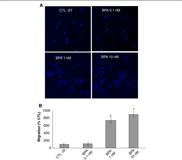
Previous works have clearly shown that BPA induces cell proliferation in androgen-dependent human PCa cells (Wetherill et al. 2005). In addition to cell growth, cancer metastasis is significantly involved in the progression of the disease and leads to death. Cancer cell migration and invasion play very important roles in cancer me-tastasis. So, we further studied the effects of BPA on migration and invasion, as well as the related calcium signalling in androgen-dependent and -independent hu-man PCa cells. Migration assay using transwell chambers showed that BPA at low concentrations of 1 and 10 nM significantly increased the migration of LNCaP cells with an increase rate of 800% and 900% respectively, after the cells were treated for 48 h with BPA prior to the migra-tion assays (Figure 1A and 1B). In these experiments, when higher concentrations of BPA (100 nM and 1 μM) were used, the BPA-induced cell migration rate was si-milar to the one observed with 10 nM (data not shown). We further studied the mechanisms involved in the effects of BPA on PCa cells.
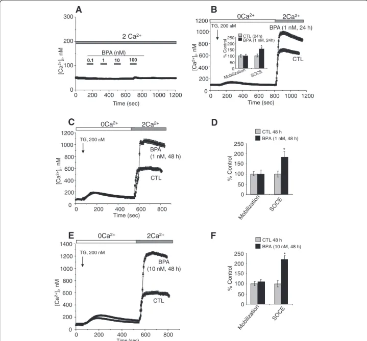
Effects of BPA on calcium signalling in prostate cancer cells
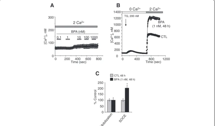
Several studies have shown that an increase in intracel-lular calcium originating from an extracelintracel-lular source greatly promotes the migration of cancer cells (Saidak et al. 2009; Yang et al. 2009). Yang et al. (2009) clear-ly showed that store-operated calcium entry channels (Orai1 and STIM1) are essential for breast tumor cell migrationin vitro and tumor metastasis in mice. In this context, BPA could induce the activation and/or over-expression of ion channels proteins involved in calcium entry and thus promotes the consequent PCa cell mig-ration. First, we examined the direct effects of BPA on the free cytosolic calcium concentration. When different concentrations of BPA were applied to LNCaP cells, an androgen-dependent PCa cell line (Figure 2A) and to LNCaP-C4.2 cells, which are a more invasive cell line derived from LNCaP cells (Figure 3A), no significant modification of the basal calcium was observed. To de-termine if BPA can induce a delayed calcium response, LNCaP cells were exposed to a range of the environ-mental factor concentrations (1nM, 10 nM, 1 μM and 10 μM) and the intracellular calcium concentrations
were continuously monitored for 2 to 3 hours. In these experiments, we observed no calcium response to the applications of BPA. These data confirm the absence of a direct effect of BPA on the activity of the ion channels in human prostate cancer cells.
We further examined the modification of the calcium signalling and remodelling of the expression of the ion channels in PCa cells after a pre-treatment with BPA. To verify the possible modification of the calcium signalling in BPA-treated cells, a test, as described in the following section, was used to compare the rate of calcium entry in control and in BPA-treated cells.
Ca2+entry, but not Ca2+release, is increased in BPA- treated LNCaP cells
We previously showed that the application of the store-depleting SERCA inhibitor thapsigargin (TG) induces a calcium mobilization from intracellular stores and a calcium entry due to SOCE in human PCa cells (Lallet-Daher et al. 2009).
The two phases of free intracellular calcium concen-tration ([Ca2+]i) changes were separated using a Ca2+ add-back protocol. The addition of the store-depleting SERCA inhibitor thapsigargin (TG, 200 nM) in nomi-nally Ca2+-free solution was followed by rapid, transient
A
BPA 1 nM M igra tion (% CTL) 0 200 400 600 800 1000*
*
B
CTL -ST BPA 10 nM BPA 0.1 nMFigure 1 BPA induces prostate cancer cells migration. Cell migration was measured via the Transwell chamber assay. (A) Following the 48 h BPA treatments, LNCaP cells were trypsinized and equal numbers of live cells were plated into inserts and allowed to migrate for 24 h. Cells in the insert (non-migrated cells) were eliminated by scraping and migrated cells were fixed and their nuclei were stained by Hoechst 33342 dye (Blue) and examined under fluorescence microscopy. At least ten different fields per condition were examined and cells were then counted. Experiments were performed in duplicate and repeated at least twice. (B) Relative migration (%) ± S.E. (Bar) is shown for the indicated BPA concentrations and the control condition (CTL-ST), where the cells were incubated in CS-RPMI alone. *, P < 0.001 relative to CTL condition.
increases in [Ca2+]i, as measured by calcium imaging
(Figure 2B) These increases are due to the mobilization of Ca2+ from internal stores. A subsequent addition of Ca2+to the extracellular bath resulted in a rapid and sus-tained increase in cytosolic Ca2+ due to SOCE. Analyses were also performed on both the rate of the TG-induced calcium mobilization and the amplitude of SOCE in CTL cellsversus those treated with different concentrations of
BPA for 24 and 48 h. In these analyses, the amplitude and the decay of the calcium mobilization were not signifi-cantly different between CTL and treated cells (Figure 2B, 2D and 2F). Interestingly, in these experiments, we ob-served that when the cells were cultured in a steroid-free medium (CS-RPMI), the amplitude of the SOCE was sig-nificantly lower (40 to 50%) than that developed in cells cultured in normal RPMI containing steroids (data not
0 200 400 600 800 1000 1200 0 100 200 300
E
F
A
B
D
2Ca2+ 0Ca2+ TG, 200 nM BPA (10 nM, 48 h) CTL 200 400 600 800 0 Time (sec) 1200 1000 800 600 400 0 200 [Ca 2+ ]i , nM % Control 0 50 100 150 200 250 CTL 48 h BPA (10 nM, 48 h) * 1400 % Control 0 50 100 150 200 250 CTL 48 h BPA (1 nM, 48 h) *C
0Ca2+ 2Ca2+ BPA (1 nM, 48 h) CTL 0 200 400 600 800 0 200 400 600 800 1000 1200 Time (sec) TG, 200 nM [Ca 2+ ]i , nM 0.1 1 10 100 2 Ca2+ BPA (nM) Time (sec) [Ca 2+ ]i , nM % Control 0 50 100 150 200 250 CTL (24h) BPA (1 nM, 24h) * 0Ca2+ 2Ca2+ TG, 200 nM 0 200 400 600 800 1000 1200 0 400 800 1200 [Ca 2+ ]i , nM Time (sec) 1000 600 200 BPA (1 nM, 24 h) CTLFigure 2 Effects of BPA on basal calcium and Store-operated Ca2+(SOC) entry (SOCE) in LNCaP cells. (A) Cells were grown on glass
coverslips and cultured in complete medium and then the direct effects of BPA at different concentrations on [Ca2+]iwere studied by calcium
imaging. (B, C, E) Cells grown on glass coverslips and cultured in CS-RPMI alone (CTL), or containing BPA at 1 or 10 nM BPA for 24 h (B) or
48 h (C, E) and then processed by calcium imaging to estimate the amplitude of SOCE. For Ca2+recording, cells were treated with 200 nM
thapsigargin (TG) in Ca2+-free bath solution (0 Ca2+) and exposed to 2 mM extracellular Ca2+(2Ca2+) as indicated. Each experiment was repeated
at least 3 times in duplicate in different cell cultures on a field of 25–40 cells and representative experiments performed on 50–80 cells as
mean ± S.E. are presented. A quantification of the SOCE for each experiment is presented in (D) and (F) (mean ± S.E., n = 50–80 cells), *P < 0.01.
shown). TG-mediated Ca2+ signalling was then studied in LNCaP treated with BPA for 24, and 48 h. At 24 h (Figure 2B), the amplitude of the Ca2+entry (SOCE) was 40 ± 15% higher than that developed in the cells cultured for the same periods in CS-RPMI alone (CTL). These effects of BPA reached 85 ± 19% and 122 ± 19% when the cells were pre-treated for 48 h by BPA 1 nM (Figure 2C and 2D) and 10 nM (Figure 2E and 2F) respectively. Simi-lar SOCE amplifications were observed for LNCaP-C4.2, a more invasive cell line derived from LNCaP cells (Figure 3B and 3C). These observations suggest that BPA modulates the expression of the ion channels involved in the SOCE in human PCa cells. The identification of these ion channels could allow a schematization of the mechanism by which BPA modulates these cancer cells migrations.
BPA up-regulated ion channels expression in prostate cancer cell lines
Our previous studies showed that of several ion chan-nels, it was mainly the calcium channels (Orai1/STIM1, TRPV6) and potassium channels (IKCa1, BKCa) that were
involved in the generation of the SOCE in human PCa cells (Flourakis et al. 2010; Lallet-Daher et al. 2009; Vanden Abeele et al. 2003a,b). Thus, experiments were designed to explore the impact of a 48 h exposure of the cells to BPA on the expression of these calcium and po-tassium channels involved in the SOCE. In the present study, in order to eliminate the impact of steroids, and androgens in particular, the experiments were performed in Phenol red-free RPMI 1640 containing charcoal-stripped Foetal Calf Serum (FCS) (CS-RPMI or -ST), a steroid-deprived medium. We have previously shown that the expression of Orai1 was at least decreased by 90% in this steroïds-free medium (Flourakis et al. 2010).
As shown in Figure 4, the rate of Orai1 transcript (Figure 4A) and protein (Figure 4B) was significantly lower when the cells were incubated in CS-RPMI, sug-gesting that Orai1 expression is steroid-dependent. Inte-restingly, when the cells were treated with 0.1 to 10 nM BPA, the Orai1 protein level was much higher at the mRNA (Figure 4A) and protein (Figure 4B) levels. Orai1 protein appears as a doublet band representing different
2 Ca2+ 0 Ca2+ 400 800 1200 0 200 400 600 800 1000 1200 1400 0 Time (sec) [Ca 2+ ]i , nM BPA (1 nM, 48 h) CTL TG, 200 nM
A
B
% Control 0 50 100 150 200 250 CTL 48 h BPA (1 nM, 48 h)C
2 Ca2+ 0.1 1 10 100 1000 BPA (nM) 0 200 400 600 800 0 100 200 300 Time (sec) [Ca 2+ ]i , nM *Figure 3 Effects of BPA on basal calcium and Store-operated Ca2+(SOC) entry (SOCE) in LNCaP C4.2 cells. (A) Cells were grown on glass
coverslips and cultured in complete medium and then the direct effects of BPA at different concentrations on [Ca2+]iwere studied by calcium
imaging. (B) Cells grown on glass coverslips and cultured in CS-RPMI alone (CTL), or containing BPA at 1 nM BPA for 48h and then processed by
calcium imaging to estimate the amplitude of SOCE. For Ca2+recording, cells were treated with 200 nM thapsigargin (TG) in Ca2+-free bath
solution (0 Ca2+) and exposed to 2 mM extracellular Ca2+(2 Ca2+) as indicated. Each experiment was repeated at least 3 times in duplicate in
different cell cultures on a field of 25–40 cells and representative experiments performed on 50–80 cells as mean ± S.E. are presented. A
quantification of the SOCE for experiment (B) is presented in (C) (mean ± S.E., n = 50–80 cells), *P < 0.01. The TG application is shown by an arrow
and extracellular Ca2+ increases from 0 (0 Ca2+) to 2 mM (2 Ca2+) are marked by horizontal bars. The graphs represent the mean peak amplitudes
CT L +St CT L -St DHT 1 nM BP A 1 nM Orai1 -actin Orai1 -actin M (bp) H2 O CT L +St CT L -S t BP A 0.1 nM BP A 1nM BP A 10nM CTL +ST CTL -ST BPA 1 nM STIM1 -actin CT L +St CT L – St BP A 1n M BP A 10 nM CT L +St CT L – S t BP A 1 nM BP A 10 nM -actin
D
C
A
B
E
AF
506/517 396 344 290 200 290 S S 1 4.4 2 3.5 2 1 4.1 3 0.7 1 0.7 0.7 1 12.8 6.9 9.1 15.8 A A A A S S Orai1Figure 4 Effects of BPA on Orai1 and STIM1 expression in prostate cancer cells. (A), Total RNA was extracted and semi-quantitative RT-PCR
experiments performed to study the expression of Orai1 (406 bp) mRNA using specific primers.β-actin mRNA expression (212 bp) was used as an
internal standard. PCRs were carried out as described in Methods using 40 ng ARN equivalent cDNA from CTL and treated samples. H2O and samples without reverse transcriptase (not shown) were used as negative controls. Amplified fragments were resolved on 1.5% agarose gel by electrophoresis and visualized by EtBr staining.M, 1-kb DNA ladder, a molecular weight marker. (B, C, D), Effects of BPA on the ion channels'
protein expression in LNCaP cells. Following the 48 h BPA treatments, total proteins were extracted and 10μg proteins were analysed on 10%
acrylamide gel (SDS-PAGE), transferred to PVDF membranes. Immuno-blots were then performed as described in Methods for the detection of
Orai1 and STIM1.β-actin expression was used for the loading control of the samples. Experiments were performed at least twice in two
independent cell cultures and representative figures are presented. The fold variation of the Orai1 and STIM1 proteins in different samples
normalized toβ-actin expression is shown under the panel. (E), Immunofluorescence studies of the effects of BPA on the expression of Orai1 in
LNCaP. Cells were cultured for 48 h in the complete medium containing steroids (CTL + ST) or in the same medium without steroids (CS-RPMI, CTL -ST) or in CS-RPMI containing 1 nM BPA. (F), Expression of Orai1 protein in human PCa tissue. PCa tissue Cryosections were processed by Immunofluorescence for the detection of Orai1. S, Stroma; A, Acinus.
glycosylated forms of the protein, as previously described by Gwack et al. (2007). We have previously shown that the expression of Orai1 was modulated by the AR path-ways (Flourakis et al. 2010). In the present work, the AR agonist (DHT) was used as a positive CTL for the induction of Orai1 protein expression, as observed by western-blotting. As shown in Figure 4C, the expression of Orai1 induced by the activation of the Androgen Re-ceptor by 1 nM DHT was mimiked by BPA. This would suggest that the effects of BPA on Orai1 expression could be at least partly mediated by activation of the AR. In these experiments, BPA failed to significantly affect the expression of the Orai1 partner, STIM1 (Figure 4D). The up-regulation of Orai1 protein by BPA is confirmed by immunofluorescence studies performed on LNCaP cells. The incubation of the cells in CS-RPMI (−ST) re-duced Orai1 staining, whereas the 1 nM BPA treatments of cells for 48 h induced intense Orai1 protein staining (Figure 4E). As these experiments on Orai1 proteins were performed on the LNCaP PCa cell line, we studied the expression of Orai1 protein in 3 grade 3 human PCa tissues using immunofluorescence. As shown in Figure 4F, Orai1 immunostaining was observed in epithelial cells of the acini, as well as in stromal cells, suggesting a role for the protein in both the epithelial and stromal cells com-partments of PCa tissues.
Further experiments dealing with the modulation of other calcium and potassium channels potentially invol-ved in SOCE in LNCaP cells showed a clear modification of their expression in BPA-treated cells. In the same manner, the expression of the TRPV6 calcium channel (Figure 5A) and those of BKCa and IKCa1 Ca2+-activated
potassium channels (Figure 5B) were clearly induced. The
up-regulated potassium channels (BKCaand IKCa1), by
hy-perpolarizing the membrane potential, may be implicated in the BPA-induced amplification of SOCE, by promoting significant calcium entry through Orai1 and TRPV6.
Involvement of Orai1 in BPA-induced modification of calcium signalling
It has previously been shown that Ca2+influx is essential for the migration of various cell types, including tumor cells (Komuro and Rakic 1993; Marks and Maxfield 1990; Nishiyama et al. 2003; Yang and Huang 2005). In addition, recent data has clearly shown that Orai1 and STIM1, both of which involved in store-operated calcium entry, are es-sential for tumor cell migrationin vitro and tumor metas-tasis in mice (Yang et al. 2009). Thus, the BPA-induced up-regulation of Orai1 expression might be involved in the effects of the environmental factor on cell migration. In this context, we studied the involvement of the Orai1 protein in the effects of BPA on calcium signalling in LNCaP cells. The cells were treated with Orai1 targeting siRNA (siOrai1, 20 nM) for 48 h during the BPA treat-ments of the cells. Then, calcium imaging experitreat-ments were performed using the protocol described in Figure 2. As shown (Figure 6A-D), the treatment of the cells by siOrai1 suppressed the BPA-induced SOCE amplification by 60 to 80%. As described above, in the present study, in order to eliminate the impact of steroids, and andro-gens in particular, the experiments were performed in CS-RPMI (−ST), a steroid-deprived medium. In this me-dium, the expression of Orai1 was at least decreased by 90% (Flourakis et al. 2010). As expected, the siOrai1 treat-ments of the cells without BPA treattreat-ments did not modify the amplitude of the TG-induced calcium entry. In these
A
B
TRPV6 CT L +St CT L – S t 48h BP A 0.1 nM BP A 1 nM BP A 10 nM -actin 0.4 1 1.4 3.5 3 IKCa1 BKCa CT L +St CT L – S t 24h CT L – S t 48h BP A 0,1 nM BP A 1 nM BP A 10 nM -actin 0.4 0.8 1 0.6 1.8 3 3.2 1.8 1 2.2 17.9 13.7Figure 5 Effects of BPA on the ion channels proteins expression in LNCaP cells. Following the 48 h BPA treatments, total proteins were
extracted and 10μg proteins were analysed on 10% acrylamide gel (SDS-PAGE), then transferred to PVDF membranes. Immuno-blots were then
performed as described in Methods for the detection of TRPV6 (A), BKCaand IKCa1(B).β-actin expression was used for the loading control of the
samples. Experiments were performed at least twice in two independent cell cultures and representative figures are presented. The fold variation
A
C
B
% Control 0 50 100 150 200 250D
BPA 1 nM + siCTL BPA 1 nM + siOrai1 % Control 0 50 100 150 200250 BPA 10 nM + siCTLBPA 10 nM + siOrai1
* * BPA 1nM + siCTL BPA 1nM + siOrai1 2 Ca2+ 0 Ca2+ 0 200 400 600 800 0 200 400 600 800 1000 1200 Time (sec) [Ca 2+ ]i , nM 0 Ca 2+ 2 Ca 2+ 200 400 600 800 Time (sec) BPA10 nM + siOrai1 BPA 10 nM + siCTL 0 0 200 400 600 800 1000 1200 1400 [Ca 2+ ]i , nM TG, 200 nM TG, 200 nM
E
TG 200 nM 0 200 400 600 0 200 400 600 800 1000 1200 Time (sec) [Ca 2+ ]i , nM 0 mM Ca2+ 2 mM Ca2+ CTLF
[Ca 2+] i , nM 0 400 800 1200 Time (sec) 2Ca2+ 0Ca2+ TG, 200 nM 1200 1000 800 600 400 200 1400 +BTP-0 CTLFigure 6 Effect of Orai1 knockdown on Ca2+entry induced by BPA treatments in LNCaP cells (A, B, C, D). Forty-eight hours prior to
recording Ca2+signals by calcium imaging, LNCaP cells were divided into paired groups and transfected with control siRNA (siCTL), or siOrai1 in
the presence of 1 or 10 nM BPA. For Ca2+recording, cells were treated with 200 nM thapsigargin (TG) in Ca2+-free bath solution
(0 Ca2+) and exposed to 2 mM extracellular Ca2+(2 Ca2+) as indicated. Using the same protocol described above, pharmacological tools were
used to study the involvement of Orai1 in BPA-induced SOCE amplification. LNCaP cells were treated with BPA (1nM, 48h) and then the
TG-induced SOCE was studied in the presence or absence of BTP2 (2μM) (E), an inhibitor of Orai1 or SKF96365 (10 μM) (F), a broad spectrum
inhibitor of ion channels involved in SOCE. Each experiment was repeated at least 3 times in duplicate in different cell cultures on a field of
25–40 cells and representative experiments performed on 50–80 cells as mean ± S.E. are presented. A quantification of the SOCE for each
experiment is presented in (B) and (D) (mean ± S.E.., n = 50–80 cells), *P < 0.001. The TG application is shown by an arrow and extracellular
experiments, the siTRPV6 treatments in the presence of BPA 1 and 10 nM failed to inhibit the BPA-induced am-plification of the calcium entry induced by TG (data not shown).
Pharmacological tools were also used to study the in-volvement of Orai1 in BPA-induced SOCE amplification. LNCaP cells were treated with BPA (1nM, 48h) and then the TG-induced SOCE was studied in the presence or ab-sence of the inhibitors. When the experiments were per-formed using a pyrazole derivative, BTP2 (2μM), known to inhibit calcium channels involved in SOCE including Orai1 (Eltit et al. 2010), the TG-induced SOCE ampli-tude in BPA-treated cells was inhibited by at least 40% (Figure 6E). In the same manner, when the experiments were performed in the presence of an inhibitor of store-operated Ca2+entry (SKF96365, 10μM), the TG-induced SOCE was almost completely inhibited in BPA-treated cells (Figure 6F). Taken together, these data suggest that the up-regulation of Orai1 is involved in the amplification of the calcium entry induced by BPA.
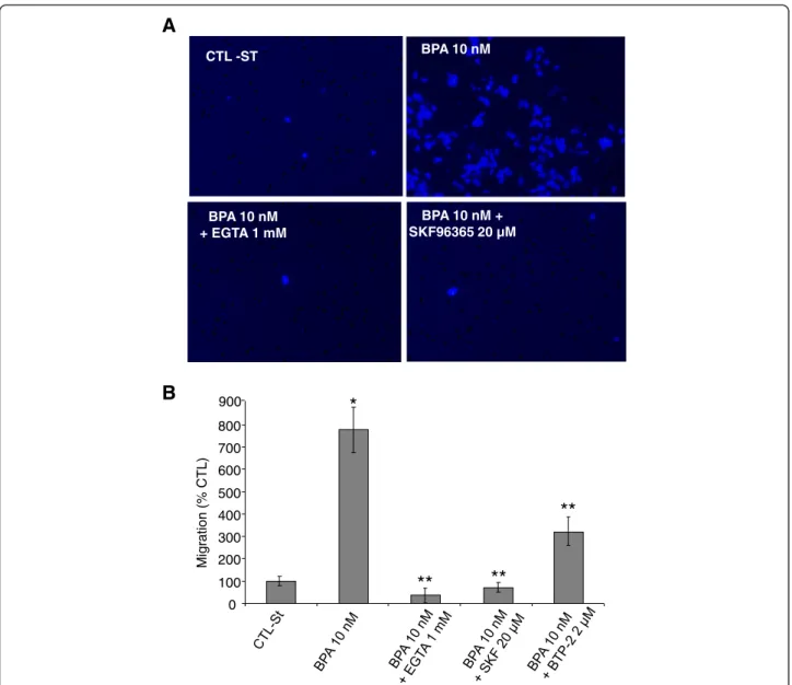
Involvement of calcium entry in BPA-induced cell migration
We showed that Orai1 protein was up-regulated due to BPA treatments of the LNCaP cells and that Orai1 was involved in the amplification of the calcium entry into these cells. To further investigate the correlation be-tween Ca2+ influx, SOCE and cell migration induced by BPA, we studied the effect of blocking Ca2+ influx on LNCaP cell migration. Using a transwell migration assay, we observed that when the cells were incubated in a medium depleted in calcium by the addition of 1 mM EGTA (Calcium chelating agent), thereby reducing the extracellular free calcium to nominally 200 nM, the BPA (10 nM)-induced migration of the LNCaP cells was com-pletely inhibited (Figure 7A and B). In the same manner, in order to study the involvement of SOCE in BPA-in-duced migration, cells were treated with an inhibitor of store-operated Ca2+entry (SKF96365, 20μM) in the pre-sence of BPA (10 nM). As shown in Figures 7A and 7B, SKF96365 blocked all BPA-induced cell migration. When the experiments were performed using BTP2 (2 μM), known to inhibit the calcium channel Orai1 involved in SOCE (Figure 7B), the BPA-induced cell migration was inhibited by 63 ± 11%. These pharmacological inhibitor data show that blocking Ca2+ influx inhibits the BPA-induced migration of LNCaP cells, and that the store-operated Ca2+entry channels including Orai1 are indeed involved in the effects of BPA on cell migration.
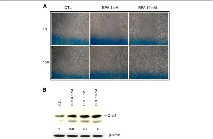
BPA increases the migration of androgen-independent prostate cancer cells
Previous studies reported by Wetherill et al. (2005) sho-wed that low concentrations of BPA induced prolife-ration in AR-dependent cells, but that the environmental
factor failed to affect the growth of AR-negative, andro-gen-independent PCa cell lines (PC-3 and DU-145). In the present study, we examined the effects of BPA on the mi-gration of the androgen-independent PCa cell line PC-3. The cells were incubated for 48 h in CS-RPMI, both com-plemented or not by BPA at different concentrations. The cells were then wounded using a sterile tip and incubated in the corresponding media for an additional 15 h before microscopic examinations of the wound healing of the cells. As shown in Figure 8A, thesein vitro scratch tests of PC-3 cells showed an increase in cell wound closure in re-sponse to BPA, suggesting the induction of cell migration by BPA in the androgen-independent PCa cell line PC-3. In order to study the effects of BPA on Orai1 protein ex-pression, Western blot experiments were performed on total proteins of the PC-3 cells treated for 48 h with BPA at varying concentrations. As shown in Figure 8B, BPA induced a dose-dependent expression of Orai1 in PC-3 cells.
Discussion
It has been suggested that the environmental factor BPA may play an important role in the initiation (Ho et al. 2006) and progression of PCa and in hormonal therapy bypass (Wetherill et al. 2005). At the level of PCa cells, BPA was able to induce androgen-independent tumor cell proliferation and reduced therapeutic efficacy in xenograft models (Wetherill et al. 2006). While these data point to-ward the potential for BPA to assist tumor cells in esca-ping therapy, the molecular mechanisms of this process were not well-known.
This report demonstrates for the first time that the en-vironmentally relevant concentrations of BPA (1–10 nM) induce cell migration by modulating the cell calcium sig-nalling. These data highlight the previously unrecognized action of BPA in the progression of human PCa, thereby providing strong support for the growing recognition of the adverse effects of BPA on human health.
Invasion and metastasis are major events underlying cancer morbidity and mortality (Molloy and Van’t Veer 2008). Because of the widespread metastasis in advanced cancer patients, where a resistance is observed to con-ventional therapies, the mortality rate remains extremely high and warrants new strategies to intervene in the me-tastatic cascade. Thus, an enhanced understanding of the molecular events in the pathogenesis of PCa will offer improved diagnosis, prognosis, therapy and preven-tion measures of the disease, that will ultimately help us to eliminate PCa metastasis. A common regulatory point in several signal transduction pathways is intracellular calcium homeostasis. One approach could be to focus on the intracellular signalling pathways underlying the metastatic process. Several data have clearly shown the involvement of calcium entry in cancer and non-cancer
cell migration (Bisaillon et al. 2010; Li et al. 2011; Schaff et al. 2010; Yang et al. 2009).
In the present work, we present conclusive evidence for the first time that the pre-treatment of human PCa cells with environmentally relevant concentrations of BPA (1–10 nM) induces their migration (Figure 1). By calcium imaging technique, we show that BPA pre-treatment in-duces an amplification of Store-Operated Calcium Entry (SOCE) in LNCaP cells (Figure 2). RT-PCR and Western blot experiments allowed us to identify those ion chan-nel proteins up-regulated by BPA pre-treatments. These
channels include Orai1, a protein known to constitute an important actor in SOCE in various cell systems including human PCa cells (Figure 4). Meanwhile, in our studies, we failed to observe any direct effect of BPA on the rate of basal calcium (Figure 2) whereas in other cell systems, BPA or its derivatives induced a calcium increase. In TM4 Sertoli cells, a direct application of a derivative compound of BPA, Tetrabromobisphenol A (TBBPA), a commonly used brominated flame retardant (BFR), induced an in-crease in the basal free calcium rate originating from internal stores (Ogunbayo et al. 2008). In pituitary tumor
CTL -ST BPA 10 nM + EGTA 1 mM BPA 10 nM + SKF96365 20 µM 0 100 200 300 400 500 600 700 800 Mig ration (% CT L)
A
B
*
**
**
**
BPA 10 nM 900Figure 7 BPA-induced SOCE is involved in prostate cancer cells migration. Cell migration was measured via the Transwell chamber assay. (A) Following the 48 h 10 nM BPA treatment for, LNCaP cells were trypsinized and equal numbers of live cells were plated into inserts and
allowed to migrate for 24h in the presence or absence of BPA 10 nM, with or without the presence of a Ca2+chelator EGTA (1 mM) or the SOCE
inhibitors (SKF, 20μM; BTP-2, 2 μM). Cells in the insert (non-migrated cells) were then eliminated by scraping and migrated cells were fixed and
their nuclei were stained by Hoechst 33342 dye (Blue) and examined under fluorescence microscopy. At least ten different fields per condition were examined and the cells counted. Experiments were performed in duplicate and repeated at least twice. (B) Relative migration (%) ± SE (Bar) is shown for the control condition (CTL-ST), where the cells were incubated in CS-RPMI alone, and for BPA (10 nM) in the presence or absence of the EGTA or SOCE inhibitors. *, P < 0.001 relative to CTL condition; ** P < 0.001 BPA 10 nM versus BPA 10 nM + inhibitors.
cells (GH3/B6/F10 rat Somatomammotropes), Kochukov et al. (2009) showed that BPA at a 1 nM concentration induced a great increase in Ca2+oscillation frequency, the activation of MAPK pathways (ERK1/2) and subsequently a PRL release (Kochukov et al. 2009). Similar results were reported by Bulayeva et al. (2005) and Wozniak et al. (2005) where the authors demonstrated that the BPA-induced Ca2+ influx was strictly dependent on membrane Estrogen Receptor (mER-α) and mediated by L-type voltage-gated Ca2+ channels in pancreatic β cells (Bulayeva et al. 2005; Wozniak et al. 2005). The LNCaP cells used in our present work do not express L-type voltage-gated Ca2+channels (non-excitable cells). This is probably the reason why a direct application of BPA on LNCaP and LNCaP C4.2 cells failed to induce a direct calcium response.
In the present work, when the human PCa cells were pre-treated with BPA (1–10 nM), an increase in SOCE
and a remodelling of ion channel expression was obser-ved. The alterations of the ion channel expression could be mediated by a stimulation of a signal transduction pathways leading to the activation of nuclear transcrip-tion factors. Published data suggest several transductranscrip-tion pathways activated by BPA. In PCa cells, BPA has been shown to be an agonist for mutant androgen receptor (AR-T877A) expressed in recurrent PCa (Wetherill et al. 2002, 2005, 2006), and in the LNCaP cell line used in our studies. According to the authors, BPA induces cell proliferation in cells expressing the mutated AR. The clinical ramifications of BPA activating tumor-derived mutant ARs and inducing androgen-independent tumor cell proliferation may be substantial, as BPA can reduce therapeutic efficacy in xenograft models (Wetherill et al. 2006). In our experiments, the DHT induced the expres-sion of Orai1 (Figure 4C) and BPA appears to mimic these effects on the canonical AR ligand (DHT). However, all
1h 15h BPA 1 nM CTL BPA 10 nM
A
B
CT L BP A 0.1 nM BP A 1 nM BP A 10 nM Orai1 β-actin 2.8 3.9 1 4Figure 8 BPA induces the cell migration of androgen-independent prostate cancer cells. (A) Cell migration was studied via the in vitro wound-healing assay. The PC-3 cells grown to confluent monolayers were incubated for 48 h in CS-RPMI, complemented or not by BPA at different concentrations. The cells were then wounded using a sterile tip and incubated in the corresponding media for an additional 15 h before the microscopic examinations of the wound-healing of the cells. Wound-healing was examined 1 and 15 hours after scratching, in at least 3 different previously identified (Blue line) fields per scratch and representative figures for each condition are presented here. (B) Western blot analysis of whole cell lysate proteins of Orai1, in PC-3 cells treated with BPA at different concentrations. The figure is a representative experiment
showing the variation of Orai1 protein under BPA treatments.β-actin was used as a sample loading control. The fold variation of the Orai1
protein in different samples normalized toβ-actin expression is shown under the panel. Results shown are representative of at least three
the effects of BPA could not be mediated by the activation of AR. In a recent work, Hess-Wilson et al. (2007) showed clearly that BPA and DHT elicited distinct transcrip-tional signatures in PCa cells expressing the BPA-res-ponsive mutant AR-T877A, even if some common genes were activated by both DHT and BPA in LNCaP cells (Hess-Wilson et al. 2007). These observations could ex-plain the cell migration and Orai1 expression induced by BPA in androgen-independent human PCa cells PC-3 (Figure 8), where the AR is absent. BPA could thus vate other signal transduction pathways than the AR acti-vation, to induce the effects observed in our studies on androgen-insensitive PCa cells. The study of the involve-ment of other transduction pathways than those involving the AR receptor (growth factor signalling pathways,. . .) in the effects of BPA in dependent and androgen-independent cells needs further extensive investigations in the future. In this context, BPA was reported to induce the phosphorylation of extracellular signal–regulated kin-ase (ERK), c-Jun N-terminal kinase (JNK), and nuclear translocation of the nuclear factor (NF)-κB, in mouse hippocampal HT-22 cells (Lee et al. 2008). Interestingly, functional NF-κB-binding sites in promoter regions of STIM1 and Orai1 have been identified and the expression of the Orai1 calcium channel was reported to be po-sitively modulated by NF-κB (Eylenstein et al. 2012). Subsequently, the store-operated Ca2+ entry was simi-larly increased by overexpression of p65/p50 or p65/ p52, and decreased by treatment with the NF-κB inhibitor, Wogonin. BPA could thus interfere with the growth fac-tors' signal transduction to activate the PI3K/AkT pathway and thereby induce the activation of the NF-kB transcrip-tion factor, leading in turn to an up-regulatranscrip-tion of the ex-pression of ion channels including Orai1. In this context, authors have shown that the activation of mER-α in-duces the activation of the PI3K/Akt signalling cas-cade (Simoncini et al. 2003; Stirone et al. 2005) and BPA is shown to activate mER-α (Bouskine et al. 2009; Quesada et al. 2002). The activation of the mER-α in androgen-dependent (LNCaP) and androgen-inandrogen-dependent (PC-3) PCa cells may thus induce the PI3K/Akt signalling cascade which leads to the activation of the NF-kB transcription factor and Orai1 gene expression. Several works have also demonstrated the stimulation of the PKA/CREB pathways by nanomolar concentrations of BPA through the activa-tion of mER-α (Bouskine et al. 2009; Quesada et al. 2002). However, the involvement of this pathway in the expres-sion of ion channels needs further investigation. As shown in Figure 5, BPA pre-treatment induced an increase in BKCa and IKCa1 Ca2+-activated potassium channel
ex-pression in LNCaP cells. We previously showed that the IKCa1 Ca2+-activated potassium channels are
invol-ved in SOCE in LNCaP and PC-3 human PCa cells (Lallet-Daher et al. 2009). These potassium channels
could constitute a functional complex with Orai1 protein to promote calcium entry and cell migration. A study is in progress in our lab to show the functional up-regulation and involvement of these Ca2+-activated potassium chan-nels (IKCa1and BKCa) in cell migration in the BPA-treated
LNCaP and PC-3 cancer cells.
Receptor-mediated activation of phospholipases C by the factors present in the serum leads to IP3-mediated
depletion of Ca2+ from the ER, which in turn stimu-lates Ca2+ influx through the plasma membrane involv-ing Orai1/STIM1 complex formation (Varnai et al. 2009). Recent works have elegantly demonstrated that STIM1, Orai1, and SOCE play critical roles in the migration of a number of cell types in cancer and non-cancer cells (Bisaillon et al. 2010; Li et al. 2011). These observations support our data where BPA, by up-regulating the ion channels expression increases the SOCE developed by PCa cells in response to factors present in serum.
Our data show clearly that the BPA-induced cell mi-gration is dependent on the calcium entry and the use of pharmacological tools suggests the involvement of SOCE channels in the effects of BPA on cell migration (Figure 7). Increase in cytoplasmic calcium induced by BPA may have several types of impacts which trigger cell migration, including the induction of the up-regulation of their gene and protein expression, secretion and the activation of the enzymes such as metalloproteinases (MMP2, MMP9), which are involved in cell migration. The MMP proteins are clearly shown to be dependent on calcium for their expression (calcium/Calcineurin/ NFAT, . . .), their processing and their activity (Collier et al. 1988; Mukhopadhyay et al. 2004; Stetler-Stevenson et al. 1989). These observations suggest that the increase in calcium entry induced by BPA pre-treatment could pro-mote all these processes leading to cell migration.
For the first time, we also demonstrate the expression of Orai1 proteins in human PCa tissues (Figure 4F). As shown, a strong immuno-staining of Orai1 protein was found in epithelial cells of the acini and also in the stromal cells. Thus, stromal cells could also be influen-ced by BPA impregnation. Given the importance of the epithelium-stroma (reactive stroma) in the progression of cancer, the potential effects of BPA on calcium signal-ling and on the secretion of growth factors by these cells need further investigation.
Conclusions
BPA is consistently detected in almost all individuals in developed nations (Welshons et al. 2006), suggesting that humans are continuously exposed to BPA. In ad-dition, the rapid metabolic clearance of BPA and its de-tectable levels in human blood and urine suggest that the intake of BPA may be higher than indicated by di-verse studies and that long-term daily intake may lead to
its bioaccumulation, leading to adverse effects on human health and on cancer progression.
These observations suggest that the BPA concentra-tions used in the present study are attainable in humans. The present data provide novel insights into the way in which the molecular mechanisms involved in the effects of environmental factors can promote the progression of the cancer in an androgen-independent manner. Our work also highlights the urgency of taking preventive measures and suggests some potential therapeutic op-portunities of targeting the ion channels involved in SOCE (Orai1) in order to prevent the PCa cell growth and metastasis.
Methods
Chemicals and antibodies
Bisphenol A (BPA) was obtained from Sigma-Aldrich and dissolved in DMSO. Antibodies raised against hu-man ion channel proteins were obtained from commer-cial sources as follows: Rabbit anti-Orai1 (ProSci Inc.), Rabbit anti-STIM1 (ProSci Inc.), Rabbit anti-TRPC1 (Alomone Labs), Rabbit anti-TRPV6 (Santa Cruz nology) and Rabbit anti-β-actin (Santa Cruz Biotech-nology) and horseradish peroxidase-conjugated secondary antibodies (Santa Cruz Biotechnology).
Cell culture
LNCaP, LNCaP-C4.2 and PC-3 PCa cell lines, obtained from the American Type Culture Collection (ATCC, Manassas, VA, USA), were cultured in RPMI 1640 and serum as described by Gackière et al. (Gackiere et al. 2006). For BPA experiments, the cells were treated with Phenol red-free RPMI 1640 containing charcoal-stripped Foetal Calf Serum (FCS) (CS-RPMI). In order to avoid the interference between BPA added for our studies and those leached from RPMI 1640 commercialized in po-lycarbonate bottles, the medium was prepared in glass bottles using RPMI 1640 powder commercialized by SIGMA (L’Isle d’Abeau, France) in ultrapure water and then filtered on 0.2μm filters (Thermo Scientific Nalgene, Fontenay-sous-Bois France).
RT–PCR analysis of mRNA expression
Total RNA isolation and RT-PCR experiments were performed as described earlier (Roudbaraki et al. 1999). The PCR primers (Orai1: 5’-CTTCTTCGACCTCGTC CTCCT-3’ and 5’-CGTAAGGCCAAAGCATGGAA-3’; β-actin : CAGAGCAAGAGAGGCATCCT-3’ and 5’-GTTGAAGGTCTCAAACATGATC-3’ used in this stu-dy were designed on the basis of established GenBank sequences and synthesized by Invitrogen (Carlsbad, CA, USA). The amplified PCR products were of 406 and 212 bp respectively.
siRNA transfections
For siRNA experiments, equal numbers of cells from the same culture were seeded, transfected overnight with 20 nM of control siRNA (targeting Luciferase mRNA) (Eurogentec, Belgium), or raised against Orai1 mRNA (siOrai1) (50-UGAGCAACGUGCACAAUCU (dTdT)-30), using Hyperfect transfection reagent (Qiagen Inc., Courtaboeuf, France) in CS-RPMI containing 10% SVF, according to the manufacturer's instructions. Medium was changed after 24 h and cells were incubated for a further 48h with or without BPA, before performing calcium im-aging experiments. We previously showed the efficiency of the siOrai1 used in the present study, in down-regulating the expression of the Orai1 protein in LNCaP cells (Flourakis et al. 2010).
Orai1 immunofluorescence studies
The protein expression studies of the ion channels in PCa cells were determined by indirect immunofluore-cence analysis performed on acetone-fixed cells. Cells grown on glass cloverslips were incubated with PBS con-taining 0.2% BSA, 0.1% TritonX-100 and 5% donkey serum, for 30 min at room temperature, in order to block the non-specific bindings and to permeabilize the cells. They were then incubated overnight at 4°C with PBS/5% non-immunized serum containing a 1:50e dilu-tion of the primary affinity-purified rabbit anti-Orai1 polyclonal antibody. Cells were then washed with PBS and were incubated with the secondary Alexa fluor 488-labeled anti-rabbit IgG (A-21206; Molecular Probes; dilution 1:2000e) diluted in PBS for 1 h at room temperature. After rinsing three times in PBS, the slides were mounted with Mowiol and the distribution of the labelled proteins was analysed by confocal immunofluo-resccence microscopy (Zeiss LSM 510; acquisition parame-ters: objective 40x/1.3; thickness of confocal slide, 1μm).
For the immunofluorescence studies of Orai1 protein in human PCa, tissues were obtained from consenting patients following local ethical considerations. The tis-sues were diagnosed as cancerous or not by anatomo-pathological examinations. Tissues were from patients prior to any anticancer therapy (chemotherapy, radio-therapy) and were obtained following an office proce-dure, frozen in liquid nitrogen-cooled isopentane and kept in “Tissue-TekW” at −80°C before 10 μm cryo-sections were carried out at −20°C with a cryostat and mounted on glass slides for immunofluorescence studies. All experiments involving patient tissues were carried out under approval number “CP 01/33”, issued by the “Comité Consultatif de Protection des Personnes dans la Recherche. The immunofluorescence experiments for the detection of Orai1 on 7μm cryosections was carried out following the same procedure as for the PCa cell
lines, using anti-Orai 1 antibody and analysed by con-focal microscopy.
Western blot assay
Cells cultured at 80% confluence were harvested and total proteins extracted. 40 micrograms of each sample were analysed by SDS-PAGE on 10% acrylamide and processed for western-blotting using antibody as descri-bed by (Vanoverberghe et al. 2004) using BKca (Alomone, 1:500e), TRPV6 (Alomone, 1:500e), TRPC1 (Alomone, 1:500e), Orai1 (ProSci 1:500e), STIM1 (ProSci, 1:500e), IKCa1 (Santa Cruz, 1:200e). Western blotting was perfor-med with an ECL chemiluminescence kit (Millipore). Quantitative evaluation of protein expression was perfor-med using ImageJ software.
[Ca2+]imeasurements
Cells were grown on glass coverslips for [Ca2+]iimaging
experiments and, before each experiment, the cells were loaded with Fura-2, by adding 2μM Fura-2 AM (Fura-2 Acetoxymethyl esther) (Calbiochem, Meudon, France) to the culture medium for 45 min at 37°C. The cells cells were then washed three times in HBSS (Hanks Balanced Salt Solution; 142 mM NaCl, 5.6 mM KCl, 1 mM MgCl2,
2 mM CaCl2, 340 μM Na2PO4, 440 μM KH2PO4, 10
mM Hepes, 5.6 mM glucose and buffered to pH 7.4). When a Ca2+-free medium was required, CaCl2 was
omitted and replaced by equimolar MgCl2. The
fluores-cent intensity of Fura-2 in each cell was monitored and recorded at 340 and 380 nm. To represent the variation in the intracellular free calcium concentration, either the fluorescence intensity ratio represented by F340/F380 was used as an indicator of changes in cytosolic Ca2+ concentrations, or a calibration was used to represent such variations in nM. All measurements shown are ave-rages of 35–45 cells from a minimum of four experi-ments on different cell cultures.
Cell migration assays
Cell migration assays were performed in duplicate in modified Boyden chambers. These assays consisted in counting cells migrating through a porous membrane with 8μm pores (BD Biosciences, Oxford Science Park, Oxford, UK). After trypsinisation, cells in suspension (1 × 10⁵) were loaded into the upper chamber in phenol-red RPMI without FCS. The lower chamber contained RPMI and 10% charcoal-stripped (CS)-FCS (CS-RPMI). The upper and lower chambers contained the same con-centration of BPA when tested. After 16 h to 24 h at 37°C in a 5% CO2incubator, cells that had attached but
not migrated were scraped from the upper surface, the membranes were fixed in 70% methanol at −20°C and the migrated cells were stained for nuclei with Hoechst 33342 dye (1μg/mL) (blue fluorescent) and evaluated by
counting cell nuclei in 10 randomly chosen fields under fluorescence microscopy. The results are presented as a percentage of control (CTL), where cells were incuba-ted in CS-RPMI culture medium alone. Alternatively, the Wound Healing Assay was also used to study PCa cell migration. The cells were seeded in a 12-well plate (15 × 104). After the cells formed a confluent mono-layer, scratches were performed using a 100 μl tip. The wells were washed with PBS followed by the addition of BPA at different concentrations in CS-RPMI. The closure of scratch was analyzed under the microscope and images were captured 1 and 15 or 24 h after incubation in the presence or absence of BPA.
Statistical analysis
Plots were produced using Origin 5.0 (Microcal Soft-ware, Inc., Northampton, MA). Results are expressed as mean ± S.E. Statistical analysis was performed using unpaired t tests or ANOVA tests followed by either Dunnett (for multiple controlversus test comparisons), or Student-Newman-Keuls post-tests (for multiple compa-risons). The Student’s t-test was used for statistical com-parison of the differences and p < 0.05 was considered significant.
Competing interests
The authors declare that they have no competing interests.
Authors’ contributions
MR and SD initiated and designed the project. SD, MW, PM and PD performed cell culture, calcium imaging, RT-PCR, western and immunofluorescence experiments and analyzed data. PG, BM, and J-LB assured the anatomopathological examinations and prostate tissue processing before immunofluorescence studies. CS supervised the immunofluorescence studies and analysis by confocal microscopy. MR, SD, PM and NP wrote the manuscript. MR directed the research. All the authors discussed and commented on the manuscript. All authors read and approved the final manuscript.
Acknowledgements
We would like to thank E Richard (BICel– IFR 147) for the technical
assistance in image analysis by confocal microscopy, M Masurelle for assistance in the preparation of the manuscript and H Selliez for reading and language corrections of the manuscript.
Funding
This work was supported by grants from the Région Nord Pas-de-Calais,
INSERM, the Ministère de l’Education Nationale, de l’Enseignement Supérieur
et de la Recherche, La Ligue Nationale Contre le Cancer. S. Derouiche was supported by the Région Nord Pas-de-Calais and Association pour la Recherche sur les Tumeurs de la Prostate (ARTP). The funders had no role in study design, data collection and analysis, decision to publish, or preparation of the manuscript.
Author details
1Inserm, U-1003, Equipe labellisée par la Ligue Nationale contre le cancer,
Villeneuve d’Ascq, France.2Laboratory of Excellence, Ion Channels Science
and Therapeutics; Université Lille I Sciences et Technologies, Villeneuve
d’Ascq, France.3Laboratoire d’Anatomie et de Cytologie Pathologique du
groupement hospitalier de l’Institut Catholique de Lille, Faculté Libre de
Médecine, Lille, France.4Service d’Urologie de l’hôpital St-Philibert, Lomme,
Received: 19 October 2012 Accepted: 4 February 2013 Published: 15 February 2013
References
Bisaillon JM, Motiani RK, Gonzalez-Cobos JC, Potier M, Halligan KE, Alzawahra WF, Barroso M, Singer HA, Jourd'heuil D, Trebak M (2010) Essential role for STIM1/ Orai1-mediated calcium influx in PDGF-induced smooth muscle migration.
Am J Physiol Cell Physiol 298(5):C993–C1005
Bouskine A, Nebout M, Brucker-Davis F, Benahmed M, Fenichel P (2009) Low doses of bisphenol A promote human seminoma cell proliferation by activating PKA and PKG via a membrane G-protein-coupled estrogen
receptor. Environ Health Perspect 117(7):1053–1058
Bulayeva NN, Wozniak AL, Lash LL, Watson CS (2005) Mechanisms of membrane estrogen receptor-alpha-mediated rapid stimulation of Ca2+ levels and prolactin release in a pituitary cell line. Am J Physiol Endocrinol Metab
288(2):E388–E397
Calafat AM, Kuklenyik Z, Reidy JA, Caudill SP, Ekong J, Needham LL (2005) Urinary concentrations of bisphenol A and 4-nonylphenol in a human reference
population. Environ Health Perspect 113(4):391–395
Collier IE, Wilhelm SM, Eisen AZ, Marmer BL, Grant GA, Seltzer JL, Kronberger A, He CS, Bauer EA, Goldberg GI (1988) H-ras oncogene-transformed human bronchial epithelial cells (TBE-1) secrete a single metalloprotease capable of
degrading basement membrane collagen. J Biol Chem 263(14):6579–6587
Courjaret R, Machaca K (2012) STIM and Orai in cellular proliferation and division.
Front Biosci 4:331–341, Elite Ed
Eltit JM, Feng W, Lopez JR, Padilla IT, Pessah IN, Molinski TF, Fruen BR, Allen PD, Perez CF (2010) Ablation of skeletal muscle triadin impairs FKBP12/RyR1 channel interactions essential for maintaining resting cytoplasmic Ca2+.
J Biol Chem 285(49):38453–38462
Eylenstein A, Schmidt S, Gu S, Yang W, Schmid E, Schmidt EM, Alesutan I, Szteyn K, Regel I, Shumilina E, Lang F (2012) Transcription factor NF-kappaB regulates expression of pore-forming Ca2+ channel unit, Orai1, and its activator, STIM1, to control Ca2+ entry and affect cellular functions. J Biol
Chem 287(4):2719–2730
Flourakis M, Lehen'kyi V, Beck B, Raphael M, Vandenberghe M, Abeele FV, Roudbaraki M, Lepage G, Mauroy B, Romanin C, Shuba Y, Skryma R, Prevarskaya N (2010) Orai1 contributes to the establishment of an apoptosis-resistant phenotype in prostate cancer cells. Cell Death Dis 1:e75
Gackiere F, Bidaux G, Lory P, Prevarskaya N, Mariot P (2006) A role for voltage gated T-type calcium channels in mediating "capacitative" calcium entry?
Cell Calcium 39(4):357–366
Gronberg H (2003) Prostate cancer epidemiology. Lancet 361(9360):859–864
Gwack Y, Srikanth S, Feske S, Cruz-Guilloty F, Oh-hora M, Neems DS, Hogan PG, Rao A (2007) Biochemical and functional characterization of Orai proteins.
J Biol Chem 282(22):16232–16243
Henley DV, Korach KS (2006) Endocrine-disrupting chemicals use distinct mechanisms of action to modulate endocrine system function.
Endocrinology 147(6 Suppl):S25–S32
Hess-Wilson JK, Webb SL, Daly HK, Leung YK, Boldison J, Comstock CE, Sartor MA, Ho SM, Knudsen KE (2007) Unique bisphenol A transcriptome in prostate cancer: novel effects on ERbeta expression that correspond to androgen
receptor mutation status. Environ Health Perspect 115(11):1646–1653
Ho SM, Tang WY, Belmonte de Frausto J, Prins GS (2006) Developmental exposure to estradiol and bisphenol A increases susceptibility to prostate carcinogenesis and epigenetically regulates phosphodiesterase type 4
variant 4. Cancer Res 66(11):5624–5632
Huff J, Boyd J, Barrett JC (1996) Hormonal carcinogenesis and environmental
influences: background and overview. Prog Clin Biol Res 394:3–23
Jemal A, Siegel R, Ward E, Hao Y, Xu J, Thun MJ (2009) Cancer statistics, 2009.
CA Cancer J Clin 59(4):225–249
Kochukov MY, Jeng YJ, Watson CS (2009) Alkylphenol xenoestrogens with varying carbon chain lengths differentially and potently activate signaling and functional responses in GH3/B6/F10 somatomammotropes. Environ
Health Perspect 117(5):723–730
Komuro H, Rakic P (1993) Modulation of neuronal migration by NMDA receptors.
Science 260(5104):95–97
Lallet-Daher H, Roudbaraki M, Bavencoffe A, Mariot P, Gackiere F, Bidaux G, Urbain R, Gosset P, Delcourt P, Fleurisse L, Slomianny C, Dewailly E, Mauroy B, Bonnal JL, Skryma R, Prevarskaya N (2009) Intermediate-conductance
Ca2 +−activated K + channels (IKCa1) regulate human prostate cancer
cell proliferation through a close control of calcium entry. Oncogene
28(15):1792–1806
Lang F, Foller M, Lang KS, Lang PA, Ritter M, Gulbins E, Vereninov A, Huber SM (2005) Ion channels in cell proliferation and apoptotic cell death. J Membr
Biol 205(3):147–157
LaPensee EW, LaPensee CR, Fox S, Schwemberger S, Afton S, Ben-Jonathan N (2010) Bisphenol A and estradiol are equipotent in antagonizing
cisplatin-induced cytotoxicity in breast cancer cells. Cancer Lett 290(2):167–173
Lee S, Suk K, Kim IK, Jang IS, Park JW, Johnson VJ, Kwon TK, Choi BJ, Kim SH (2008) Signaling pathways of bisphenol A-induced apoptosis in hippocampal neuronal cells: role of calcium-induced reactive oxygen species, mitogen-activated protein kinases, and nuclear factor-kappaB. J Neurosci Res
86(13):2932–2942
Li J, Cubbon RM, Wilson LA, Amer MS, McKeown L, Hou B, Majeed Y, Tumova S, Seymour VA, Taylor H, Stacey M, O'Regan D, Foster R, Porter KE, Kearney MT, Beech DJ (2011) Orai1 and CRAC channel dependence of VEGF-activated Ca2
+ entry and endothelial tube formation. Circ Res 108(10):1190–1198
Marks PW, Maxfield FR (1990) Transient increases in cytosolic free calcium appear to be required for the migration of adherent human neutrophils. J Cell Biol
110(1):43–52
Molloy T, Veer LJ V't (2008) Recent advances in metastasis research. Curr Opin
Genet Dev 18(1):35–41
Mukhopadhyay S, Munshi HG, Kambhampati S, Sassano A, Platanias LC, Stack MS (2004) Calcium-induced matrix metalloproteinase 9 gene expression is differentially regulated by ERK1/2 and p38 MAPK in oral keratinocytes and
oral squamous cell carcinoma. J Biol Chem 279(32):33139–33146
Nishiyama M, Hoshino A, Tsai L, Henley JR, Goshima Y, Tessier-Lavigne M, Poo MM, Hong K (2003) Cyclic AMP/GMP-dependent modulation of Ca2+ channels sets the polarity of nerve growth-cone turning. Nature
423(6943):990–995
Ogunbayo OA, Lai PF, Connolly TJ, Michelangeli F (2008) Tetrabromobisphenol A (TBBPA), induces cell death in TM4 Sertoli cells by modulating Ca2+ transport proteins and causing dysregulation of Ca2+ homeostasis. Toxicol In
Vitro 22(4):943–952
Prevarskaya N, Skryma R, Shuba Y (2011) Calcium in tumour metastasis: new roles
for known actors. Nat Rev Cancer 11(8):609–618
Pupo M, Pisano A, Lappano R, Santolla MF, De Francesco EM, Abonante S, Rosano C, Maggiolini M (2012) Bisphenol A Induces Gene Expression Changes and Proliferative Effects through GPER in Breast Cancer Cells and
Cancer-Associated Fibroblasts. Environ Health Perspect 120(8):1177–1182
Quesada I, Fuentes E, Viso-Leon MC, Soria B, Ripoll C, Nadal A (2002) Low doses of the endocrine disruptor bisphenol-A and the native hormone
17beta-estradiol rapidly activate transcription factor CREB. FASEB J 16(12):1671–1673
Roudbaraki M, Lorsignol A, Langouche L, Callewaert G, Vankelecom H, Denef C (1999) Target cells of gamma3-melanocyte-stimulating hormone detected through intracellular Ca2+ responses in immature rat pituitary constitute a fraction of all main pituitary cell types, but mostly express multiple hormone phenotypes at the messenger ribonucleic acid level. Refractoriness to melanocortin-3 receptor blockade in the lacto-somatotroph lineage.
Endocrinology 140(10):4874–4885
Saidak Z, Boudot C, Abdoune R, Petit L, Brazier M, Mentaverri R, Kamel S (2009) Extracellular calcium promotes the migration of breast cancer cells through the
activation of the calcium sensing receptor. Exp Cell Res 315(12):2072–2080
Schaff UY, Dixit N, Procyk E, Yamayoshi I, Tse T, Simon SI (2010) Orai1 regulates intracellular calcium, arrest, and shape polarization during neutrophil
recruitment in shear flow. Blood 115(3):657–666
Simoncini T, Rabkin E, Liao JK (2003) Molecular basis of cell membrane estrogen receptor interaction with phosphatidylinositol 3-kinase in endothelial cells.
Arterioscler Thromb Vasc Biol 23(2):198–203
Stetler-Stevenson WG, Krutzsch HC, Liotta LA (1989) Tissue inhibitor of metalloproteinase (TIMP-2), A new member of the metalloproteinase
inhibitor family. J Biol Chem 264(29):17374–17378
Stirone C, Boroujerdi A, Duckles SP, Krause DN (2005) Estrogen receptor activation of phosphoinositide-3 kinase, akt, and nitric oxide signaling in cerebral blood vessels: rapid and long-term effects. Mol Pharmacol
67(1):105–113
Vanden Abeele F, Roudbaraki M, Shuba Y, Skryma R, Prevarskaya N (2003a) Store-operated Ca2+ current in prostate cancer epithelial cells. Role of
endogenous Ca2+ transporter type 1. J Biol Chem 278(17):15381–15389
Vanden Abeele F, Shuba Y, Roudbaraki M, Lemonnier L, Vanoverberghe K, Mariot P, Skryma R, Prevarskaya N (2003b) Store-operated Ca2+ channels in prostate
cancer epithelial cells: function, regulation, and role in carcinogenesis. Cell
Calcium 33(5–6):357–373
Vanoverberghe K, Vanden Abeele F, Mariot P, Lepage G, Roudbaraki M, Bonnal JL, Mauroy B, Shuba Y, Skryma R, Prevarskaya N (2004) Ca2+ homeostasis and apoptotic resistance of neuroendocrine-differentiated prostate cancer cells.
Cell Death Differ 11(3):321–330
Varnai P, Hunyady L, Balla T (2009) STIM and Orai: the long-awaited constituents
of store-operated calcium entry. Trends Pharmacol Sci 30(3):118–128
Welshons WV, Thayer KA, Judy BM, Taylor JA, Curran EM, Vom Saal FS (2003) Large effects from small exposures. I. Mechanisms for endocrine-disrupting
chemicals with estrogenic activity. Environ Health Perspect 111(8):994–1006
Welshons WV, Nagel SC, vom Saal FS (2006) Large effects from small exposures, III. Endocrine mechanisms mediating effects of bisphenol A at levels of
human exposure. Endocrinology 147(6 Suppl):S56–S69
Wetherill YB, Petre CE, Monk KR, Puga A, Knudsen KE (2002) The xenoestrogen bisphenol A induces inappropriate androgen receptor activation and
mitogenesis in prostatic adenocarcinoma cells. Mol Cancer Ther 1(7):515–524
Wetherill YB, Fisher NL, Staubach A, Danielsen M, de Vere White RW, Knudsen KE (2005) Xenoestrogen action in prostate cancer: pleiotropic effects dependent
on androgen receptor status. Cancer Res 65(1):54–65
Wetherill YB, Hess-Wilson JK, Comstock CE, Shah SA, Buncher CR, Sallans L, Limbach PA, Schwemberger S, Babcock GF, Knudsen KE (2006) Bisphenol A facilitates bypass of androgen ablation therapy in prostate cancer. Mol
Cancer Ther 5(12):3181–3190
Wozniak AL, Bulayeva NN, Watson CS (2005) Xenoestrogens at picomolar to nanomolar concentrations trigger membrane estrogen receptor-alpha -mediated Ca2+ fluxes and prolactin release in GH3/B6 pituitary tumor cells.
Environ Health Perspect 113(4):431–439
Yang S, Huang XY (2005) Ca2+ influx through L-type Ca2+ channels controls the trailing tail contraction in growth factor-induced fibroblast cell migration.
J Biol Chem 280(29):27130–27137
Yang S, Zhang JJ, Huang XY (2009) Orai1 and STIM1 are critical for breast tumor
cell migration and metastasis. Cancer Cell 15(2):124–134
doi:10.1186/2193-1801-2-54
Cite this article as: Derouiche et al.: Bisphenol A stimulates human prostate cancer cell migration via remodelling of calcium signalling. SpringerPlus 2013 2:54.
Submit your manuscript to a
journal and benefi t from:
7 Convenient online submission 7 Rigorous peer review
7 Immediate publication on acceptance 7 Open access: articles freely available online 7 High visibility within the fi eld
7 Retaining the copyright to your article