HAL Id: hal-01383389
https://hal.sorbonne-universite.fr/hal-01383389
Submitted on 18 Oct 2016HAL is a multi-disciplinary open access archive for the deposit and dissemination of sci-entific research documents, whether they are pub-lished or not. The documents may come from teaching and research institutions in France or abroad, or from public or private research centers.
L’archive ouverte pluridisciplinaire HAL, est destinée au dépôt et à la diffusion de documents scientifiques de niveau recherche, publiés ou non, émanant des établissements d’enseignement et de recherche français ou étrangers, des laboratoires publics ou privés.
Biomarkers of vascular dysfunction and cognitive decline
in patients with Alzheimer’s disease: no evidence for
association in elderly subjects
Alice Breining, Jean-Sébastien Silvestre, Bénédicte Dieudonné, José Vilar,
Véronique Faucounau, Marc Verny, Christian Néri, Chantal M. Boulanger,
Jacques Boddaert
To cite this version:
Alice Breining, Jean-Sébastien Silvestre, Bénédicte Dieudonné, José Vilar, Véronique Faucounau, et al.. Biomarkers of vascular dysfunction and cognitive decline in patients with Alzheimer’s disease: no evidence for association in elderly subjects. Aging Clinical and Experimental Research, Springer Verlag, 2016, pp.1-9. �10.1007/s40520-016-0535-4�. �hal-01383389�
1
Biomarkers of vascular dysfunction and cognitive decline in
patients with Alzheimer’s disease: no evidence for association in
elderly subjects
Alice Breining1,2, MD, Jean-Sébastien Silvestre3, Bénédicte Dieudonné1, José Vilar3, Véronique Faucounau1, MD, Marc Verny1,2, MD,PhD, Christian Néri2, Chantal M. Boulanger3, Jacques Boddaert1,2, MD,PhD
(1) APHP, DHU FAST, Groupe Hospitalier Pitié Salpêtrière - Charles Foix, Centre de Gériatrie, F-75013, Paris, France
(2) Sorbonne Universités, UPMC Univ Paris 06, CNRS, UMR 8256 B2A , F-75005, Paris, France
(3) INSERM, U970, Paris Cardiovascular Research Center—PARCC; and Université Paris Descartes, Sorbonne Paris Cité, UMR-S970, Paris, France
Corresponding author :
Dr Alice Breining Centre de Gériatrie
Groupe Hospitalier Pitié-Salpêtrière 47-83, Bd de l'Hôpital
75651 Paris Cedex 13 France
tel: 33 1 42 16 41 17 ; fax: 33 1 42 16 03 25 e-mail: alice.breining@psl.aphp.fr
2
ABSTRACT
Background: Several studies have suggested that vascular dysfunction plays an important role in Alzheimer’s disease.
Aims: We hypothesized that significant differences might be observed in the levels of blood endothelial biomarkers across elderly population of subjects with dementia.
Methods: We analyzed, in a prospective monocentric study, 3 different endothelial
biomarkers, endothelial microparticles (EMP), endothelial progenitor cells (EPC) and
circulating endothelial cells (CEC) in 132 older patients who underwent a full evaluation of a
memory complaint.
Results: There was no difference in specific EMP, EPC or CEC levels between demented or
non demented patients, nor considering cognitive decline.
Discussion: Blood endothelial biomarkers may be too sensitive and it is likely that the
multimorbidity observed in our patients may lead to opposite and confounding effects on
endothelial biomarkers levels.
Conclusion: Unlike younger AD patients, our results suggest that endothelial biomarkers are
not valuable for the diagnosis of dementia in elderly patients.
Key words : endothelial microparticles, circulating endothelial cells, endothelial progenitors
3
INTRODUCTION
Several studies have suggested that vascular dysfunction plays important roles in Alzheimer’s
disease (AD). Current evidence suggests a decreased vascular density in ageing, leukoaraiosis
and AD. Importantly, cerebrovascular dysfunction may precede cognitive dysfunction and
neurodegeneration [1]. A role for cerebrovascular dysfunction has also been suggested by
epidemiological studies and associations between AD and atherosclerosis [2] as well as
between AD and cerebrovascular disease [3, 4]. Additionally, the majority of
community-dwelling older persons with dementia often have multiple brain pathologies, underscored by
AD and vascular diseases [5]. Finally, people who suffer from both diseases, AD and
infarctions, have a more severe cognitive impairment than those suffering from only one
disease [6, 7], suggesting that neurodegeneration and cerebrovascular disease may be tightly
associated in AD.
Endothelial microparticles (EMP), endothelial progenitor cells (EPC) and circulating
endothelial cells (CEC) have emerged as useful markers to evaluate cardiovascular event risk
and pathology prognosis. Microparticles (MP) are submicron membrane vesicles expressing a
panel of oxidized phospholipids and proteins specific of the cells from which they originate.
Increase of MP levels in plasma, particularly those of endothelial origin, reflects cellular
injury and have been proposed as surrogate marker of vascular dysfunction [8, 9]. CEC are
positive for CD 146 and their detection in a healthy adult is a rare event. In vitro, CEC inhibit
EPC proliferation and migration, thus impacting on the regenerative capacity of vessels [10].
High levels of CEC are associated with the development of various vascular diseases [11, 12].
EPC can derive from bone marrow and participate to the maintenance of the endothelium by
replacing dysfunctional endothelial cells or releasing pro-angiogenic growth factors.
Interestingly, the number and function of EPC inversely correlate with the risk of
4 Although EMP, EPC and CEC measures have clinical potential to assess vascular
dysfunction, a limited number of studies have analysed the diagnostic and prognostic value of
these candidate biomarkers in patients with cognitive impairment. An association between
EPC and the Mini-Mental State Examination score has been suggested in AD but not in non
AD neurodegenerative disease [14, 15]. Some circulating EMP subpopulations might also be
associated with cognitive decline in AD patients [16]. Here, we evaluated the diagnostic and
prognosis value of endothelial dysfunction biomarkers in 132 community dwelling older
persons who present a memory complaint. To fully assess the putative vascular dysfunction in
these patient populations, we analyzed, at the same time, EMP, EPC and CEC levels.
PATIENTS AND METHODS Setting
This observational study was performed during an 8-month period from November 1, 2010
through June 30, 2011 and was designed to analyze endothelial biomarkers in patients with
memory complaint. This study took place in a Memory Center of the Geriatric Department of
Hospital Pitié-Salpètrière, Paris, France. This study has been approved by the ethical
committee (CPP Pitié-Salpêtrière, Ile-de-France VI, Paris, France).
Patients
During the period study, community dwelling older patients consulting for a memory
complaint were prospectively included. No blood sample was collected specifically for the
study. Patients were excluded if they presented with cancer or any acute medical condition
that could influence endothelial biomarkers, such as acute coronary syndrome, acute ischemic
stroke, and infection. A full evaluation was made for each patient, including medical history,
5 cerebral imaging. If needed, they also had a lumbar punction and/or cerebral scintigraphy.
Cardiovascular history was established from medical records.
Diagnosis of cognitive impairment was determined by a multidisciplinary staff and patients
were classified into three groups: mild cognitive impairment (MCI) group, dementia group
and control group without MCI or dementia (see supplementary methodology). A rapid
cognitive decline (RCD) was defined as a decrease of the Mini-Mental State Examination
(MMSE) score of 6 points per year [17].
Patients with dementia were followed at least one year after cognitive evaluation. Surviving
patients and their family or caregivers were seen for a physical examination with a MMS
score according to our routine procedure of follow-up. Missing patients were tracked through
health care providers, particularly general practitioners, or any acquaintances identified.
Diagnostic criteria for dementia
Dementia and MCI were diagnosed according to the Diagnostic and Statistical Manual of
Mental Disorders, 4th Edition (American Psychiatric Association). AD was diagnosed
according to the National Institute of Neurological and Communicative Disorders and Stroke
and the Alzheimer's Disease and Related Disorders Association criteria [18], dementia with
Lewy Body (DLB) according to the McKeith et al criteria [19], fronto temporal dementia
according to the Lund-Manchester criteria [20], vascular dementia according to the National
Institute of Neurological Disorders and Stroke and Association Internationale pour la
Recherche et l'Enseignement en Neurosciences criteria [21].
Endothelial microparticles isolation and analysis
As previously described by Amabile et al., MP were quantified in Platelet-free plasma which
was obtained by successive centrifugation (500g for 15 min followed by 10,000g for 5 min, at
room temperature) of 4 ml of citrated blood. Investigators blinded to subject status
6 EMP were analyzed: CD62e (E-selectin)-positive EMP, CD144 (VE-cadherin)-positive EMP,
and whole pool of MP using calcium-dependent-annexin V labelling. We did not need to
exclude specific platelet phenotype as CD144 and CD62E are not expressed by platelets.
Circulating MP levels were expressed as events per mL (ev/mL).
Assays for circulating endothelial cells
The CEC were counted in whole blood using immunomagnetic separation. Ferrous beads
bound to an anti-CD146 monoclonal antibody were mixed with venous blood in a
head-over-head mixer for 30 min at 4°C. The unbound cells were washed away with buffer. Cells were
labelled with orange acridine and counted in a glass counting chamber under a fluorescent
microscope. CEC levels were expressed as CEC count/ml. Healthy controls display around 0–
10 CEC/ml.
Endothelial progenitors cells
EPC were also analysed in whole blood sample. Mononuclear cells were isolated with the use
of a Ficoll density gradient according to standard protocols and were suspended in 500 μl of a
phosphate-buffered saline. Immunofluorescent cell staining was performed with the use of the
fluorescent conjugated antibody CD34–fluorescein isothiocyanate, KDR (kinase insert
domain receptor)-allophycocyanin, and CD133–phycoerythrin. Cell fluorescence was
measured immediately after staining, and data were analyzed with the use of FlowJo software
(LSRII, Becton Dikinson). Units of all measured components are absolute cell counts
obtained after the measurement of 100,000 events. We measured the number of CD34+ cells,
the number of CD34+ CD133+ KDR cells, which represents a subpopulation of immature
EPC and the number of CD34+ CD133+ CD45- cells to exclude cells with inflammatory
phenotypes.
7 For each patient, endothelial dysfunction biomarkers (EMP, EPC and CEC) were measured
(see supplementary methodology). Results were compared according to the result of the
cognitive and geriatric evaluation, between patients with dementia, patients with MCI, or
patients without any dementia or MCI. Moreover, results were compared according to the
type of dementia, considering neurodegenerative, vascular and mixt origin, i.e.
neurodegenerative and vascular origin. The prognostic value of endothelial biomarkers was
studied with respect to one year mortality. For patients with dementia alive at one year,
prognostic value of biomarkers was analyzed according to the existence of RCD cognitive
decline or not.
Statistics
The statistical analysis was performed with the statistical software Xlstat. Qualitative
variables are expressed as number and percentage, and the quantitative variables as median
and inter quartile rate (IQR). Kruskal-Wallis one-way analysis of variance on ranks and Mann
Whitney Test were used for group comparison for quantitative variables. 2 test was performed for comparisons between groups for categorical variables. A multiple regression
analysis (ANCOVA) was performed to identify factors that were independently associated
with MP counts. The alpha error was set at 0.05.
RESULTS
Among the 373 patients admitted for memory complaint during the study period, 132 patients
had a blood sample drawn at hospital, including 82 patients with dementia, 12 with MCI and
38 controls. Among the 82 patients with dementia, 48 individuals (59%) were diagnosed
having AD (Figure). Among the 38 control patients, 20 individuals (53%) had depression
symptoms. Main characteristics are presented in Table 1. Mean age of patients was 83 ± 6
8 score (p = 0.0002) compared to MCI and control patients. There were no differences for type
and number of cardiovascular risk factors, and associated comorbidities between groups
(Table 1). All patients had EMP measurements. Due to technical issues, only 86 patients had
CEC measurements and 85 patients EPC measurements. All biomarkers were detected in
patients, and we found no significant difference between control patients, patients with MCI and with dementia (table 2). There was no difference between control patients, patients with neurodegenerative disease and patients with vascular and mixed dementia (table 2). All these results were not modified after adjusting for cardiovascular risk factors and comorbidities. In
a multiple regression analysis, we observed a correlation between hypertension and high
levels of EMP (p < 0.01), but no correlation was found between EMP and other risk factors.
Long term outcome
One-year cognitive follow-up was studied. Ten out of 59 living patients with dementia and cognitive follow up had a RCD and were compared to the 49 patients without RCD. There was no significant difference between groups for cardiovascular risk factors and associated comorbidities.
Patients with RCD had a lower MMSE at one year (p=0.008) (Table 3). Patients with RCD
had lower annexin V- MP levels compared to patients not having RCD (268/ml 187-401 versus 530/ml 246-1212, p=0.04). This result was not confirmed with specific EMP. Biomarkers for EPC and CEC did not show any significant towards association (Table 3).
DISCUSSION
To our knowledge, this is the first study analysing the diagnostic and prognostic value of three
different and complementary biomarkers of endothelial dysfunction in older patients with a
memory complaint. Altogether, our results suggest that endothelial biomarkers are not valuable neither for the diagnosis of dementia in older patients, whatever the underlying mechanism, nor to
9
predict cognitive decline in older patients with dementia.
Those three biomarkers were chosen because they are associated with pathways that regulate
endothelial function. Since alteration of vascular homeostasis has been involved in the
development of AD, or appears as a key process in neurodegenerative dementia, we
hypothesized that significant differences might be observed in the levels of endothelial
biomarkers across heterogeneous populations of subjects with dementia. We did not observe
any difference between patients with dementia or not, nor between patients with
neurodegenerative and vascular or mixed dementias. But we obtain suggestive evidence for
differences within the group of demented patients according to their rate of cognitive decline:
comparison of biomarkers of endothelial dysfunction between patients with RCD or not
revealed a significant difference of Annexin V-positive MPs (Table 3). Since Annexin V
labels MP are irrespective of their cellular origin, these results suggest that total MP levels
may be associated with the rate of cognitive decline in patients with dementia. But this
difference was not observed when specific EMP markers were used.
Previous data showed evidence for an increase of certain circulating EMP phenotypes
(CD31+ CD42- and CD62e+ CD42-) correlated with a cognitive decline of AD patients [16].
In this study Xue et al. correlated EMP counts and AD cognitive decline in a multivariate
regression analysis including clinical parameters correlated with EMP counts (i.e. age and cholesterol levels). But they didn’t investigate correlation between EMP counts and
hypertension, nor diabetes and stroke. Yet it is now well demonstrated that hypertension,
diabetes and stroke increases EMP counts [8].
Recent studies reported that patients with AD have reduced EPC levels and functions [14, 15,
22], suggesting that an abnormal capacity to regenerate endothelium is associated with AD.
10 dementia. But Kong et al. assessed that patients with AD and vascular dementia had lower
CD34+CD133+ EPCs counts than controls. Lee et al. found that AD patients have reduced
EPC but for instance, Lee et al. cultured and counted EPC from peripheral blood samples [14,
22] whereas in our study, EPC were directly measured in whole blood sample.
Our results do not confirm Kong et al. and Lee et al. results. However, mean age of patients
enrolled in our study was higher than those from Lee et al. and Kong et al. (83 ± 7 years old
versus 69.1 ± 8.3 years old and 71.4 ± 2.3). Hence, we cannot exclude that differences in
endothelial dysfunction markers in older demented patients could have be influenced by age
and numerous comorbidities observed in these patient population.
Indeed, atherosclerosis, artery coronary disease, severe hypertension, metabolic syndrome,
diabetes, smoking, chronic renal failure, inflammatory diseases and cancer [8] are known to
raise EMP count. Aging, acute coronary syndromes, heart failure are known to decrease EPC
level [23]. Likewise, acute coronary syndromes, heart failure, inflammatory diseases and
cancer raise CEC level in peripheral blood [24]. In our study, patients had on average 2.9
cardiovascular risk factor and other comorbidities that could influence endothelial biomarker’s count. Even though we have excluded patients with acute disease and adjusted
our results with cardio-vascular risk factors, it is likely that the multimorbidity observed in
our patients may lead to opposite and confounding effects on endothelial biomarkers levels.
Our results do not suggest a correlation between vascular function and AD. Many reasons can
explain this result. First, initiation of AD is not a vascular event. Hypoxia resulting from the vascular insufficiency could promote β amyloid accumulation and the attendant deleterious
effects on the brain. Conversely, amyloid precursor protein could induce vascular damage and
worsen vascular insufficiency, which would also lead to brain dysfunction [25]. Synergic or
11 A second hypothesis is that vascular dysfunction is substantial at the early stages of the
disease but afterward other mechanism may have taken over. In this view, patients included in
the study and diagnosed as AD patients are no longer at subclinical stages of the disease.
Initiation of AD may not be a vascular event or vascular dysfunction may be substantial at the
early stages of the disease but afterward other mechanism may have taken over. In this view,
patients included in the study and diagnosed as AD patients are no longer at subclinical stages
of the disease.
The present study has several limitations. First all patients with a memory complaint had an
exhaustive biology but only 1/3 had a blood sample taken at hospital and endothelial
biomarkers measured. The number of patients included in the study is though limited and
measurements of the three endothelial biomarkers are not always available for all the patients
for technical reasons. Finally, the definition of RCD is based on patients with AD [17], and
this definition may not be relevant for all types of dementia.
CONCLUSION
Unlike datas on younger AD patients, our results suggest that endothelial biomarkers are not
valuable for the diagnosis of dementia in older patients, whatever the underlying mechanism.
Vascular endothelial biomarkers may be too sensitive and not specific enough in older population.
This observation, together with previously reported trends, calls for additional studies of the
12
Acknowledgments: The authors thank the INSERM U970 team of the Paris Cardiovascular
Research Center for their support.
Declaration of Sources of Funding: none
Conflict of Interests: The authors declare that there is no conflict of interests regarding the
publication of this paper.
13
REFERENCES
1. Brown WR, Thore CR. Review: cerebral microvascular pathology in ageing and neurodegeneration. Neuropathol Appl Neurobiol. 2011;37(1):56-74.
2. Roher AE, Esh C, Rahman A, Kokjohn TA, Beach TG. Atherosclerosis of cerebral arteries in Alzheimer disease. Stroke. 2004;35(1):2623-7.
3. Gorelick PB. Risk factors for vascular dementia and Alzheimer disease. Stroke. 2004;35(1):2620-2.
4. De la Torre JC. Is Alzheimer’s disease a neurodegenerative or a vascular disorder? Data, dogma, and dialectics. Lancet Neurol. 2004;3(3):184-90.
5. Schneider JA, Arvanitakis Z, Bang W, Bennett DA. Mixed brain pathologies account for most dementia cases in community-dwelling older persons. Neurology. 2007;69(24):2197-204. 6. Riekse RG, Leverenz JB, McCormick W, Bowen JD, Teri L, Nochlin D, et al. Effect of vascular
lesions on cognition in Alzheimer’s disease: a community-based study. J Am Geriatr Soc. 2004;52(9):1442-8.
7. Rockwood K, Davis H, MacKnight C, Vandorpe R, Gauthier S, Guzman A, et al. The
Consortium to Investigate Vascular Impairment of Cognition: methods and first findings. Can J Neurol Sci. 2003;30(3):237-43.
8. Boulanger C-M, Leroyer A-S, Amabile N, Tedgui A. Circulating endothelial microparticles: a new marker of vascular injury. Ann Cardiol Angeiol (Paris). 2008;57(3):149-54.
9. Nozaki T, Sugiyama S, Koga H, Sugamura K, Ohba K, Matsuzawa Y, et al. Significance of a multiple biomarkers strategy including endothelial dysfunction to improve risk
stratification for cardiovascular events in patients at high risk for coronary heart disease. J Am Coll Cardiol. 2009;54(7):601-8.
10. Holmén C, Elsheikh E, Stenvinkel P, Qureshi AR, Pettersson E, Jalkanen S, et al. Circulating inflammatory endothelial cells contribute to endothelial progenitor cell dysfunction in patients with vasculitis and kidney involvement. J Am Soc Nephrol. 2005;16(10):3110-20.
14
11. Werner N, Kosiol S, Schiegl T, Ahlers P, Walenta K, Link A, et al. Circulating endothelial progenitor cells and cardiovascular outcomes. N Engl J Med. 2005;353(10):999-1007. 12. Hill JM, Zalos G, Halcox JPJ, Schenke WH, Waclawiw MA, Quyyumi AA, et al. Circulating
endothelial progenitor cells, vascular function, and cardiovascular risk. N Engl J Med. 2003;348(7):593-600.
13. Vasa M, Fichtlscherer S, Aicher A, Adler K, Urbich C, Martin H, et al. Number and migratory activity of circulating endothelial progenitor cells inversely correlate with risk factors for coronary artery disease. Circ Res. 2001;89(1):E1-7.
14. Lee S-T, Chu K, Jung K-H, Park H-K, Kim D-H, Bahn J-J, et al. Reduced circulating angiogenic cells in Alzheimer disease. Neurology. 2009;72(21):1858-63.
15. Kong X, Zhang Y, Liu L, Sun N, Zhang M, Zhang J. Endothelial progenitor cells with Alzheimer’s disease. Chin Med J. 2011;124(6):901-6.
16. Xue S, Cai X, Li W, Zhang Z, Dong W, Hui G. Elevated plasma endothelial microparticles in Alzheimer’s disease. Dement Geriatr Cogn Disord. 2012;34(3-4):174-80.
17. Schmidt C, Wolff M, Weitz M, Bartlau T, Korth C, Zerr I. Rapidly progressive Alzheimer disease. Arch Neurol. 2011;68(9):1124-30.
18. McKhann G, Drachman D, Folstein M, Katzman R, Price D, Stadlan EM. Clinical diagnosis of Alzheimer’s disease: report of the NINCDS-ADRDA Work Group under the auspices of Department of Health and Human Services Task Force on Alzheimer’s Disease. Neurology. 1984;34(7):939-44.
19. McKeith IG, Galasko D, Kosaka K, Perry EK, Dickson DW, Hansen LA, et al. Consensus guidelines for the clinical and pathologic diagnosis of dementia with Lewy bodies (DLB): report of the consortium on DLB international workshop. Neurology. 1996;47(5):1113-24. 20. Clinical and neuropathological criteria for frontotemporal dementia. The Lund and
15
21. Román GC, Tatemichi TK, Erkinjuntti T, Cummings JL, Masdeu JC, Garcia JH, et al. Vascular dementia: diagnostic criteria for research studies. Report of the NINDS-AIREN
International Workshop. Neurology. 1993;43(2):250-60.
22. Lee S-T, Chu K, Jung K-H, Jeon D, Bahn J-J, Kim J-H, et al. Dysfunctional characteristics of circulating angiogenic cells in Alzheimer’s disease. J Alzheimers Dis. 2010;19(4):1231-40. 23. Lee PSS, Poh KK. Endothelial progenitor cells in cardiovascular diseases. World J Stem
Cells. 2014;6(3):355-66.
24. Erdbruegger U, Haubitz M, Woywodt A. Circulating endothelial cells: a novel marker of endothelial damage. Clin Chim Acta. 2006;373(1-2):17-26.
25. Iadecola C. The overlap between neurodegenerative and vascular factors in the pathogenesis of dementia. Acta Neuropathol. 2010;120(3):287-96.
16
TABLES
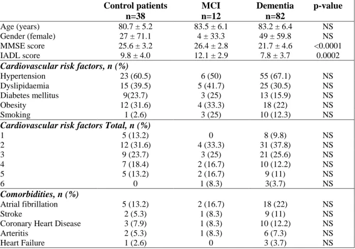
Table 1. Baseline characteristics of patients. Control patients n=38 MCI n=12 Dementia n=82 p-value Age (years) 80.7 ± 5.2 83.5 ± 6.1 83.2 ± 6.4 NS Gender (female) 27 ± 71.1 4 ± 33.3 49 ± 59.8 NS MMSE score 25.6 ± 3.2 26.4 ± 2.8 21.7 ± 4.6 <0.0001 IADL score 9.8 ± 4.0 12.1 ± 2.9 7.8 ± 3.7 0.0002
Cardiovascular risk factors, n (%)
Hypertension 23 (60.5) 6 (50) 55 (67.1) NS
Dyslipidaemia 15 (39.5) 5 (41.7) 25 (30.5) NS
Diabetes mellitus 9(23.7) 3 (25) 13 (15.9) NS
Obesity 12 (31.6) 4 (33.3) 18 (22) NS
Smoking 1 (2.6) 3 (25) 10 (12.3) NS
Cardiovascular risk factors Total, n (%)
1 5 (13.2) 0 8 (9.8) NS 2 12 (31.6) 4 (33.3) 31 (37.8) NS 3 9 (23.7) 3 (25) 21 (25.6) NS 4 7 (18.4) 2 (16.7) 10 (12.2) NS 5 5 (13.2) 2 (16.7) 9 (11) NS 6 0 1 (8.3) 3(3.7) NS Comorbidities, n (%) Atrial fibrillation 5 (13.2) 2 (16.7) 18 (22) NS Stroke 2 (5.3) 1 (8.3) 9 (11) NS
Coronary Heart Disease 3 (7.9) 1 (8.3) 10 (12.2) NS
Arteritis 2 (5.3) 1 (8.3) 6 (7.3) NS
Heart Failure 1 (2.6) 0 3 (3.7) NS
Results were expressed as mean ± sd, number of patients (percentages). MMSE: Mini-mental State Examination; IADL: InstruMini-mental Activities of Daily Living; MCI: Mild Cognitive Impairment.
17
Table 2: Comparison of biomarkers of endothelial dysfunction between control patients, patients with MCI, dementia and according to the type of dementia.
Results are expressed as median values and ranges. Circulating MP levels were expressed as events per mL, CEC levels as CEC count/ml and EPC levels as absolute cell counts obtained after the measurement of 100,000 events. EMP: Endothelial Microparticles, CEC: circulating endothelial cells, EPC: endothelial progenitors cells, MCI: Mild Cognitive Impairment.
Control MCI Dementia All Neurodegenerative Dementia Vascular Dementia Microparticules (n=38) (n=12) (n=82) (n=63) (n=19) NS AnnexinV+ MP 416 [149-1020] 386 [170-1093] 549.7 [248-1110] 531 [233-1063] 688 [380-1255] NS CD144+ EMP 64 [46-143] 92 [37-123] 84.9 [39-174] 81 [39-151] 100 [39-210] NS CD62E+ EMP 78 [39-106] 63 [37-109] 92 [54-118] 94 [56-117] 92 [39-166] NS CEC (n=25) (n=11) (n=50) (n=40) (n=10) 6 [2-72] 6 [2-16] 13 [2-44] 15 [4.8-47.0] 3 [0.5-18] NS EPC (n=25) (n=10) (n=49) (n=39) (n=10) CD34+ 0.07 [0.06-0.08] 0.11 [0.07-0.17] 0.07 [0.05-0.10] 0.07 [0.05-0.10] 0.09 [0.05-0.13] NS CD34+ CD133+ VEGF+ 0.3 [0.0-0.7] 1.2 [0-8.5] 0.4 [0.0-1.4] 0.5 [0.06-1.4] 0.2 [0.03-1.1] NS CD34+ CD133+ CD45- 57 [52-61] 51 [41-61] 57 [46-64] 56.6 [43.9-64.9] 58.2 [53.0-63.9] NS
18
Table 3. Characteristics of patients and comparison of three biomarkers of endothelial dysfunction according to cognitive decline during follow-up.
Results are expressed as median values and ranges. Circulating MP levels were expressed as events per mL, CEC levels as CEC count/ml and EPC levels as absolute cell counts obtained after the measurement of 100,000 events. EMP: Endothelial Microparticles, CEC: circulating endothelial cells, EPC: endothelial progenitors cells, MCI: Mild Cognitive Impairment, RCD: rapid cognitive decline. MMSE: Mini-mental State Examination, IADL: Instrumental Activities of Daily Living.
RCD (n=10)
No RCD (n=49)
p-value Age (years), mean ± sd 81,3 ± 5,3 81,4 ± 5,2 NS
Gender (female), n (%) 8 (80%) 28 (57%) NS
MMSE score, mean ± sd 24 ± 3.2 21.5 ± 6 NS
MMS+1 score, mean ± sd 16.4 ± 4 20.1 ± 5.3 0.008
IADL mean ±sd 8 ± 4.5 8.8 ± 3.3 NS
Cardiovascular risk factors, n (%)
Hypertension 6 (60%) 32 (65%) NS
Dyslipidaemia 3 (30%) 16 (33%) NS
Diabetes mellitus 2 (20%) 10 (20%) NS
Obesity 2 (20%) 16 (32%) NS
Smoking 1 (10%) 4 (8%) NS
Cardiovascular risk factors Total, n (%)
1 2 3 4 5 6 1 (10%) 5 (50%) 2 (20%) 1 (10%) 1(10%) 0 3 (6%) 18 (37%) 13 (27%) 8 (16%) 4 (18%) 3 (6%) NS NS NS NS NS NS Comorbidities, n (%) ACFA 1 (10%) 10 (20%) NS Stroke 0 5 (10%) NS Coronary 3 (30%) 6 (12%) NS AOMI 0 2 (4%) NS Heart failure 0 2 (4%) NS Microparticles Annexin V+ MP CD144+ EMP CD62E+ EMP (n=10) 286 [187-401] 46 [6.6-105] 91 [48-107] (n=49) 530 [246-1212] 97 [56-179] 90 [54-116] 0.04 NS NS CEC (n=9) 37 [10-101] (n=29) 12 [2-32] NS EPC CD34+ CD34+ CD133+ VEGF+ CD34+ CD133+ CD45- (n=9) 0.07 [0.05-0.09] 0.1 [0-1.7] 55 [48-61] (n=29) 0.07 [0.06-0.09] 0.48 [0.0-0.1] 59 [44-63] NS NS NS
19 FIGURE
Figure. Flow chart of community dwelling older persons who present a memory complaint 373 patients with a memory complaint 132 patients with a blood sample 38 control patients 82 patients with dementia 61 neurodegenerative dementia: - 48 Alzheimer disease - 9 dementia with Lewy Body - 2 corticobasal Neurodegeneration - 1 Supranuclear Paralysis - 1 Parkinbson Disease 19 vascular or mixed dementia (9+11) 2 dementia of unknowwn origin 12 patients with MCI