HAL Id: hal-01727158
https://hal.archives-ouvertes.fr/hal-01727158
Submitted on 8 Mar 2018
HAL is a multi-disciplinary open access
archive for the deposit and dissemination of
sci-entific research documents, whether they are
pub-lished or not. The documents may come from
teaching and research institutions in France or
abroad, or from public or private research centers.
L’archive ouverte pluridisciplinaire HAL, est
destinée au dépôt et à la diffusion de documents
scientifiques de niveau recherche, publiés ou non,
émanant des établissements d’enseignement et de
recherche français ou étrangers, des laboratoires
publics ou privés.
Mickael Desvaux, Thomas Candela, Pascale Serror
To cite this version:
Mickael Desvaux, Thomas Candela, Pascale Serror. Surfaceome and Proteosurfaceome in Parietal
Monoderm Bacteria: Focus on Protein Cell-Surface Display. Frontiers in Microbiology, Frontiers
Media, 2018, 9, �10.3389/fmicb.2018.00100�. �hal-01727158�
doi: 10.3389/fmicb.2018.00100
Edited by: Awdhesh Kalia, University of Texas MD Anderson Cancer Center, United States Reviewed by: Konstantin V. Korotkov, University of Kentucky, United States Chenggang Wu, McGovern Medical School, United States *Correspondence: Mickaël Desvaux mickael.desvaux@inra.fr
Specialty section: This article was submitted to Infectious Diseases, a section of the journal Frontiers in Microbiology Received: 29 June 2017 Accepted: 16 January 2018 Published: 14 February 2018 Citation: Desvaux M, Candela T and Serror P (2018) Surfaceome and Proteosurfaceome in Parietal Monoderm Bacteria: Focus on Protein Cell-Surface Display. Front. Microbiol. 9:100. doi: 10.3389/fmicb.2018.00100
Surfaceome and Proteosurfaceome
in Parietal Monoderm Bacteria:
Focus on Protein Cell-Surface
Display
Mickaël Desvaux
1* , Thomas Candela
2and Pascale Serror
31Université Clermont Auvergne, INRA, UMR454 MEDiS, Clermont-Ferrand, France,2EA4043 Unité Bactéries Pathogènes et
Santé, Châtenay-Malabry, France,3Micalis Institute, INRA, AgroParisTech, Université Paris-Saclay, Jouy-en-Josas, France
The cell envelope of parietal monoderm bacteria (archetypal Gram-positive bacteria)
is formed of a cytoplasmic membrane (CM) and a cell wall (CW). While the CM is
composed of phospholipids, the CW is composed at least of peptidoglycan (PG)
covalently linked to other biopolymers, such as teichoic acids, polysaccharides, and/or
polyglutamate. Considering the CW is a porous structure with low selective permeability
contrary to the CM, the bacterial cell surface hugs the molecular figure of the
CW components as a well of the external side of the CM. While the surfaceome
corresponds to the totality of the molecules found at the bacterial cell surface,
the proteinaceous complement of the surfaceome is the proteosurfaceome. Once
translocated across the CM, secreted proteins can either be released in the extracellular
milieu or exposed at the cell surface by associating to the CM or the CW. Following
the gene ontology (GO) for cellular components, cell-surface proteins at the CM can
either be integral (GO: 0031226), i.e., the integral membrane proteins, or anchored
to the membrane (GO: 0046658), i.e., the lipoproteins. At the CW (GO: 0009275),
cell-surface proteins can be covalently bound, i.e., the LPXTG-proteins, or bound
through weak interactions to the PG or wall polysaccharides, i.e., the cell wall binding
proteins. Besides monopolypeptides, some proteins can associate to each other to
form supramolecular protein structures of high molecular weight, namely the S-layer,
pili, flagella, and cellulosomes. After reviewing the cell envelope components and the
different molecular mechanisms involved in protein attachment to the cell envelope,
perspectives in investigating the proteosurfaceome in parietal monoderm bacteria are
further discussed.
Keywords: Gram-positive bacteria, cell-surface protein, surface proteome, subcellular localization, pili/fimbriae/curli, lipoproteins, LPXTG sortase-dependent proteins, membrane proteins
INTRODUCTION
As the interface of the cell with its surrounding, the bacterial cell surface plays a crucial role
in all types of interactions. In the first instance, the diversity of the bacterial cell envelope is
generally viewed as dichotomic, on the one hand, the Gram-positive bacteria, and on the other
hand, the Gram-negative bacteria (
Desvaux et al., 2004, 2009
). This difference is based on the result
of the Gram staining method originally developed by the
Danish pharmacologist and physician Hans Christian Joachim
Gram (
Gram, 1884
) and still routinely used worldwide to
differentiate bacteria (
Beveridge, 2001
). With the development
of microscopic techniques, it first appeared the difference in
staining was the result of profound divergence in structural
organisation of the bacterial cell envelope, where
Gram-positive bacteria have a thick cell wall (CW) sitting atop of
a cytoplasmic membrane (CM) (
Silhavy et al., 2010
). Later
on, molecular analyses further revealed that Gram-positive
bacteria corresponded to a phylogenetically coherent group
within the domain Bacteria and belonged to only two phyla,
namely the low G+C% Gram-positive bacteria of the phylum
Firmicutes and the high G+C% Gram-positive bacteria of
the phylum Actinobacteria (
Woese, 1987
;
Woese et al., 1990
).
Over the years, though, it appears this terminology presents
some ambiguity when considering the diversity of the domain
Bacteria (
Desvaux et al., 2009
). Considering the term
“Gram-positive bacteria,” it can refer to three distinct, and sometimes
incompatible elements, i.e., a Gram staining result, a cell envelope
architecture and/or a taxonomic group. For instance, bacteria
of the class Mollicutes, comprising the genus
Mycoplasma,
cannot retain the Gram stain because they naturally lack
a CW although the low G+C% content of their genomes
and other molecular markers resemble those of Gram-positive
bacteria of the phylum Firmicutes (
Razin et al., 1998
). Species
of the genus Mycobacterium possess a peculiar cell envelope
with a mycomembrane preventing Gram staining and thus
require alternative staining methods called acid-fast (
Somoskovi
et al., 2001
) but nonetheless belong to the high G+C%
Gram-positive bacteria of the phylum Actinobacteria (
Draper, 1998
).
In some deep branches of the phylum Firmicutes, some bacteria
clearly exhibit Gram-negative cell envelope for which a new
class was proposed, i.e., the Negativicutes (
Marchandin et al.,
2010
).
Inspired by the research work of
Gupta
(
1998a,b
,
2000
),
the description of the bacterial cell envelope respective to the
number of biological membranes appeared much more definite
and was first reintroduced in the field of bacterial protein
secretion (
Desvaux et al., 2009
). While monoderm bacteria refer
to species exhibiting only one biological membrane, namely
the CM, diderm bacteria correspond to species exhibiting two
biological membranes, i.e., an inner membrane and an outer
membrane. Monoderm bacteria can be further discriminated
into (i) simple monoderm, lacking a CW (e.g., bacteria from
the genus
Mycoplasma), and (ii) parietal monoderm, exhibiting
a CW (archetypal Gram-positive bacteria) (
Sutcliffe, 2010
;
Gupta, 2011
). As such, parietal monoderm bacteria include
most Firmicutes, e.g., from the class Bacilli and Clostridia,
but of course exclude the class Mollicutes and Negativicutes
as well as the Actinobacteria exhibiting a mycolate outer
membrane.
The CW of parietal monoderm bacteria is a complex structure
composed at least of peptidoglycan (PG) covalently linked
to other biopolymers, such as teichoic acids, polysaccharides,
polyglutamate, or proteins (
Shockman and Barrett, 1983
;
Figure 1
). While constituting the outermost layer of the
cell envelope of parietal monoderm bacteria, the CW is not
impermeable but on the contrary a porous and penetrable
structure. As such, cell envelope proteins are in contact with the
external environment without ever having a domain protruding
out the confines of the CW. Like for the fractal dimension
of the protein surface (
Richards, 1977
;
Banerji and Navare,
2013
), the nature and definition of the bacterial cell surface
strictly depends on the molecule considered, e.g., a water
molecule or a globular protein, which can enter in contact,
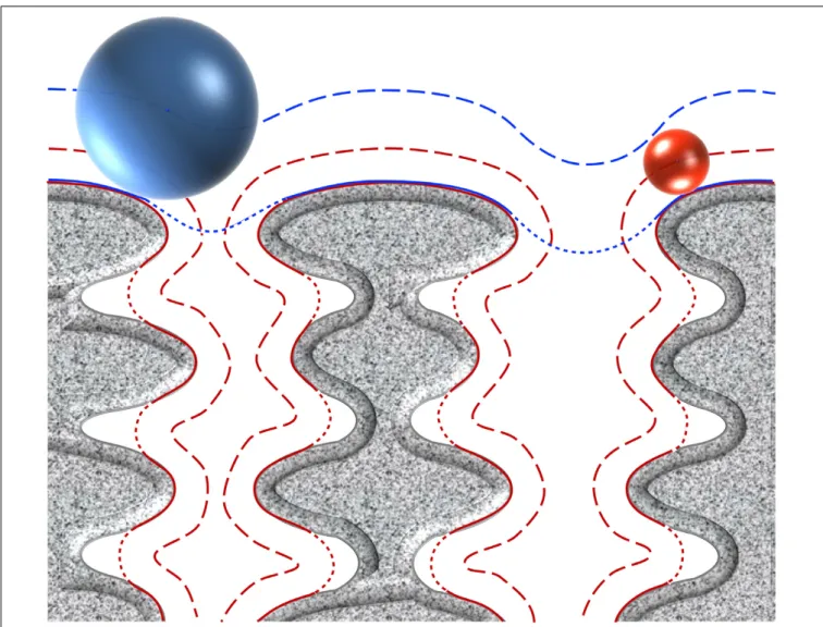
access, diffuse or penetrate differently the CW (Figure 2).
To be exposed at the cell surface of parietal monoderm
bacteria, proteins need to be first secreted across the CM.
Several secretion systems allow protein translocation in parietal
monoderm bacteria (
Tjalsma et al., 2004
;
Desvaux et al., 2005
;
Desvaux and Hébraud, 2006
;
Sibbald et al., 2006
;
Chagnot
et al., 2013
), namely (i) the Sec (secretion), (ii) the Tat
(twin-arginine translocation), (iii) ABC protein exporter, (iv) the
FPE (fimbrilin-protein exporter), (v) the FEA (flagella export
apparatus), and (vi) the ESX (ESAT-6 system), also called
Wss (WXG100 secretion system). Of note, the status of the
holins (hole forming) as protein secretion systems
per se
remain controversial (
Desvaux, 2012
). Proteins secreted via the
Sec translocon generally possess a targeting signal called the
signal peptide (SP) of type I (SP I), which is composed of
three non-conserved domains, namely the n-domain (positively
charged and at the N-terminus), the h-domain (a-helical
hydrophobic core region), and the c-domain (cleavage site
processed by a membrane-bound signal peptidase) (
Fekkes
and Driessen, 1999
). While proteins secreted via Sec, Tat
ABC exporter and FPE possess N-terminal SPs with some
specificities, the signal targeting proteins to the FEA or ESX
remain elusive. Besides transport across the CM, the transport
and maturation of secreted proteins across the CW can be
regulated by different mechanisms, such as the proteolytic
maturation of secreted proenzymes, the requirement of divalent
cations for activation or the post-translocational intervention
of peptidyl-prolyl isomerase chaperones (
Forster and Marquis,
2012
).
To explicitly describe the subcellular localization of proteins,
the gene ontology (GO) respective to the cellular component
is extremely useful (
Ashburner et al., 2000
;
Chagnot et al.,
2013
). Indeed, secreted proteins can have different fate; they
are either (i) associated to the CM (GO: 0005886), (ii)
anchored to the CW (GO: 0009275), (iii) released in the
extracellular milieu (GO: 0005576), the so-called exoproteins
(extracellular proteins), or even (iv) injected into a prokaryotic
or eukaryotic host cell. At the CM, proteins can be either
integral (GO: 0031226), i.e., the IMPs (integral membrane
proteins), or anchored to the membrane (GO: 0046658), i.e.,
the lipoproteins. At the CW, proteins can be covalently bound,
i.e., the LPXTG-proteins, or bound through weak interactions,
i.e., the CW binding proteins. It is worth stressing that all
these extracytoplasmic proteins located at the cell envelope,
wherever at the CM or the CW, can be considered as surface
exposed. Besides monopolypeptides, some organelles can also
be present and result from the assembly of protein subunits to
form supramolecular structures, such as the well-known pili and
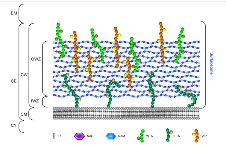
FIGURE 1 | The surfaceome of parietal monoderm bacteria with respect of the organisation and composition of the cell envelope. The cell envelope (CE) of parietal monoderm bacteria is composed of a biological membrane acting as selective permeable barrier, i.e., the cytoplasmic membrane (CM) and a cell wall (CW) providing some resistance to mechanical stresses (e.g., internal turgor pressure) but also somehow acting as a philtre. While the CM is composed of phospholipids (PLs), the CW can be further subdivided into the inner wall zone (IWZ) and the outer wall zone (OWZ). The OWZ constitutes the main CW fabric. It is composed of
N-acetylglucosamine (NAG) and N-acetylmuramic acid (NAM), both constituting the peptidoglycan (PG) with which wall teichoic acids (WTAs), and wall
polysaccharides (WPs) are anchored. Lipoteichoic acids (LTAs) are anchored to the CM and protrude from the CM. As revealed by electron microscopy studies and contrary to the OWZ, the IWZ is a thinner zone of low density most certainly devoid of most cross-linked polymeric CW network, except LTAs and some proteins, e.g., lipoproteins (Matias and Beveridge, 2005, 2006); because this zone is not strictly bounded by two biological membranes like in diderm bacteria, the IWZ resembles but cannot be considered as a periplasm sensu stricto, i.e., it presents some analogies but no homology (Buist et al., 2008;Chagnot et al., 2013). In addition to the proteins present both at the CM and CW and that are not depicted here for clarity (see text and Figures 4–6), these different macromolecular molecules exposed on the external side of the CM constitute the surfaceome in parietal monoderm bacteria. CY, cytoplasm; EM, extracellular milieu.
flagella, but also the S-layer or cellulosome in some bacterial
species.
Following the etymological meaning of the Greek suffix
“-ome” (
Lederberg and McCray, 2001
), the totality of the
molecules found at the bacterial cell surface corresponds to the
surfaceome. Because of the spongy structure of the CW, it is
misleading to restrict the surface of parietal monoderm bacteria
to molecules strictly displayed at the outermost molecular layer
of the CW. Instead, the cell surface of a parietal monoderm
bacterium fits tightly to the molecular outline of the CW
components and to the external side of the CM (Figure 2);
as a biological membrane, the CM has a selective permeability
contrary to the CW. The CW is not a rigid shell but constitutes
a matrix, forming an elastic polyelectrolyte gel (
Doyle and
Marquis, 1994
;
Neuhaus and Baddiley, 2003
), which would then
acts like a sieve during the dynamic transit of solutes. The
proteosurfaceome is the proteinaceous subset of the surfaceome
found at the CW and totally or partially exposed on the external
side of the CM.
THE SURFACEOME OF PARIETAL
MONODERM BACTERIA
The cell envelope of parietal monoderm bacteria is composed
of a CM and a CW, which can be divided into the inner wall
zone (IWZ) and outer wall zone (OWZ) (
Merchante et al., 1995
;
Matias and Beveridge, 2005
;
Zuber et al., 2006
; Figure 1). The CW
surrounding the CM is made of lipoteichoic acids (LTAs) and
a thick layer of PG, decorated with wall teichoic acids (WTAs),
wall polysaccharides (WPs), or/and polyglutamate. The CW
also accommodates some proteins, including monopolypeptides
and cell-surface supramolecular protein structures, namely
pili, flagella, cellulosome, S-layer. Altogether these different
FIGURE 2 | Concepts of molecular surface, contact surface, accessible surface, and reentrant surface to define the bacterial cell surface in parietal monoderm bacteria. Taking molecules of different sizes, their penetration in the cell envelope differs. The blue sphere represents a molecule of high molecular weight unable to penetrate the CW fabric (depicted in grey), whereas the red sphere represents a smaller molecule diffusing through. Depending on the molecules considered, the definition of the bacterial cell surface will also differ. The continuous lines represent the contact surface that is the molecular surface that actually comes in direct contact with the surface of the molecule considered. The dashed lines represent the accessible surface that is the continuous sheet referring to the centre of the molecule considered. The dotted lines correspond to the reentrant surface that is the interior-facing part of the molecule considered when it cannot come in direct contact with the molecular surface of the cell envelope. The definition of bacterial cell surface of parietal monoderm bacteria is thus very different when referring to the molecular surface of the cell envelope or the contact, accessible and reentrant surfaces with respect of the size of the molecule under consideration.
macromolecular molecules and associated molecules constitute
the surfaceome. This part focuses on the components of the cell
envelope, excluding the proteinaceous compounds discussed in
the subsequent part. Cell envelope proteins actually interact with
some of these components for anchoring via different molecular
mechanisms.
Composition and Organisation of the
Cytoplasmic Membrane
The phospholipid bilayer of the membrane parietal monoderm
bacteria is ∼90 Å thick and is composed of 10–40% lipids,
40–75% proteins, and 0.2–20% carbohydrates (
Salton, 1967
;
Ghosh and Carroll, 1968
;
Bodman and Welker, 1969
;
Duda
et al., 2006
). Although membrane phospholipids vary from
one species to another, the most commonly found in the
CM are glycerophospholipids including phosphatidylglycerol,
diphosphatidylglycerol (cardiolipin), and to some extend
phosphatidylethanolamine and their amino acylated forms
(
Fischer et al., 1978
;
Roy, 2009
;
Malanovic and Lohner, 2016
).
Phospholipids vary also by their two fatty acid moieties, which
impact on membrane fluidity (
Mishra et al., 2012
;
Custer
et al., 2014
;
Diomande et al., 2015
;
Malanovic and Lohner,
2016
). Polyisoprenoid lipids are other important regulators of
membrane fluidity. They constitute, together with cardiolipins
and bacterial flotillins acting as scaffolding proteins, nanoscale
functional membrane microdomains, which seem essential to
the proper functioning of signal transduction cascades and
protein transport in
Bacillus subtilis and Staphylococcus aureus
cells (
Lopez and Kolter, 2010
;
Bramkamp and Lopez, 2015
).
By analogy with eukaryotic membranes, these microdomains
are also referred to as lipid rafts. Consistently, membrane
proteins or associated complexes constitute discrete focal sites
in the CM and CW (
Campo et al., 2004
;
Rosch et al., 2007
;
Lopez and Kolter, 2010
;
Kandaswamy et al., 2013
). Biological
significance of functional membrane microdomains could be
to serve as platforms that control the assembly of membrane
and CW proteins and multiprotein complexes involved in
numerous cellular processes, such as cell division, protein
trafficking, genetic transfer, or signal transduction (
Lopez and
Kolter, 2010
;
Schneider et al., 2015
). Subcellular localization
and spatiotemporal distribution of CM and CW proteins or
supramolecular protein complexes are often intimately linked
to their function and vary with the environmental conditions
(
Bierne and Dramsi, 2012
;
Mitra et al., 2016
).
Composition and Organisation of the
Cell Wall
The OWZ constitutes the main part of the CW. It is 15–30 nm
thick and comprises the PG and WTA polymers (
Navarre and
Schneewind, 1999
;
Vollmer et al., 2008
). The PG is made of
N-acetylglucosamine (NAG) and N-acetylmuramic acid (NAM)
forming disaccharide glycan chains of various lengths that are
cross-linked by peptides. PG composition depends on bacteria
(
Schleifer and Kandler, 1972
). The glycan chain is uniform,
whereas the peptide moiety and the cross-links are variable. The
two major PGs in parietal monoderm bacteria have a
meso-diaminopimelic acid (A2pm) or a lysin at the third position of the
peptide. At this position, the cross-link occurs directly or through
a penta-glycine bond, respectively. In
B. subtilis, it is estimated
that the glycan chain length is 1300 disaccharides in average, and
that approximately 20% of the peptide chains are cross-linked
(
Ward, 1973
;
Atrih et al., 1998
;
Hayhurst et al., 2008
). These
glycan chains form helices of ∼50 nm width, and it was proposed
that these cable-like structures coil around the narrow axis of
the bacterium and are cross-linked by peptides (
Hayhurst et al.,
2008
). The glycan chains of ovococcal bacteria, e.g.,
Streptococcus
sp., are formed of more than 100 disaccharide units in average,
whereas the glycan chains of cocci, e.g.,
Staphylococcus sp., are
relatively short with 5–10 disaccharide units in average (
Wheeler
et al., 2011
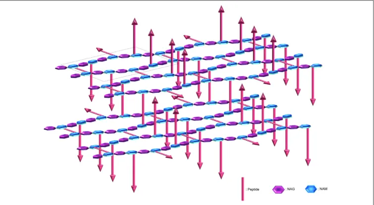
). The average effective mesh size in PG, i.e., the
tessera, is estimated at 2.2 nm (
Koch, 1990
;
Demchick and Koch,
1996
; Figure 3). In other words, hydrophilic molecules of about
25 kDa (but also probably up to 50 kDa) can freely pass through
a structured CW meshwork. Along with this, the CW network
is actually not perfect, e.g., pseudo-tessera, and numerous PG
defects cause increase in the porosity (
Pink et al., 2000
;
Turner
et al., 2013
;
Kim et al., 2015
). Of note, though, the critical hole size
in the CW beyond which lysis occurs, is estimated in the range of
15–24 nm (
Mitchell et al., 2013
). The OWZ of parietal monoderm
bacteria is a very dynamic structure, as bacterial growth requires
FIGURE 3 | Peptidoglycan organisation at the cell wall. The peptidoglycan is composed of N-acetylglucosamine (NAG) and N-acetylmuramic acid (NAM) linked by β-1,4 bonds, where the NAM are further crosslinked via octapeptides either at the same plane or with the upper or lower layer (arrows represent peptides protruding up or down). The peptidoglycan is tiled with hexagonal tesserae, which constitute the structural unit of the CW fabric (one basic unit constituting a tessera is displayed inside the dotted frame). Two layers of tesserae are here schematically represented to highlight the network form by the peptide crosslinking. Of note, defects due to abnormal tesserae with more edges and larger area can also occur and resulting in the increase in porosity.
constant remodelling of the CW, which has a turnover rate of 50%
per generation (
Koch and Doyle, 1985
). Remodelling is mediated
by CW-anchored autolysins that are active on the outermost layer
of the PG (
Jolliffe et al., 1981
).
LTAs and WTAs are zwitterionic polymers anchored to the
CM and CW, respectively. They are major polyanionic teichoic
acids of the envelope of parietal monoderm bacteria. LTAs are
localised in the IWZ at the interface of the CM and the CW
(
Neuhaus and Baddiley, 2003
;
Reichmann and Grundling, 2011
;
Schneewind and Missiakas, 2012
;
Percy and Grundling, 2014
).
The most common LTA structure found in Firmicutes and,
referred as type I LTAs, consists in a polyglycerol phosphate
polymer linked to a glycolipid anchor, often a
diglucosyl-diacylglycerol (Glc2-DAG), anchored to the CM. Type II, III,
IV, and V LTAs have more complex repeating units that contain
glycosyl residues, e.g., in
Streptococcus pneumoniae, type IV LTA
is decorated with phosphocholine.
WTAs are covalently attached by the PG disaccharide unit
via a phosphodiester linkage to NAM (
Neuhaus and Baddiley,
2003
;
Brown et al., 2013
). Although the structures of WTAs
vary considerably between species, the most common ones are
composed of glycerol-phosphate or ribitol-phosphate repeats.
LTAs and WTAs are often modified with sugar moieties and
D-alanine esters, which introduce positive charges to neutralise
the negatively charged phosphates in the polymer backbone
(
Wooldridge and Williams, 1993
;
Xia et al., 2010
;
Schneewind
and Missiakas, 2012
;
Percy and Grundling, 2014
;
Carvalho et al.,
2015
). In addition to their diversity between and within species,
the degree of
D-alanylation of teichoic acids is fine tuned in
changing environments and thus likely influences the protein
repertoire displayed at the CW. The zwitterionic WTA polymers
potentially contribute to the sequestration of divalent cations
within the OWZ, including Ca
2+, Mg
2+, and Fe
2+(
Beveridge
and Murray, 1980
), and might thus influence the regulation
of protein transport across the CW (
Forster and Marquis,
2012
).
WPs have various compositions, e.g., teichuronic acids in
Bacillus (
Ward, 1981
) or highly diverse heteropolysaccharides
in
Lactococcus (
Yasuda et al., 2011
;
Vinogradov et al., 2013
;
Ainsworth et al., 2014
), which complexity and diversity can be
even greater than expected as revealed by the ever increasing
genome data regularly made available. The last and most external
layer of the CW may be composed of a capsule, generally
composed of WPs (
Jones, 2005
;
Yother, 2011
). Although the
WP capsule structures are well documented, the anchoring was
recently proposed to be at the
β-
D-N-acetylglucosamine of the
PG via a direct glycosidic bond (
Larson and Yother, 2017
). In
some cases, the capsule is composed of polyglutamate, e.g., in
Bacillus anthracis (
McLean et al., 1992
;
Candela and Fouet, 2006
).
Poly-
γ-
D-glutamate anchoring was reported to be covalent at the
PG (
Candela and Fouet, 2005
;
Candela et al., 2005
). However,
the exact anchoring mechanism is still controversial and may be
either on the A2pm or on the PG glucosamine (
Richter et al.,
2009
;
Candela et al., 2014
).
Overall, the CW of parietal monoderm bacteria is a
complex structure that protects them from mechanical and
osmotic lysis, and serves as a scaffold for anchoring proteins,
glycopolymers, and cations that perform various functions
(
Navarre and Schneewind, 1999
;
Weidenmaier and Peschel,
2008
). While WPs or WTAs can be essential for bacterial
growth (
Oh et al., 2017
), WTAs have been shown to be
dispensable in some other bacterial species (
Chapot-Chartier and
Kulakauskas, 2014
;
Mistou et al., 2016
). However, wall rhamnose
polysaccharides (RhaWPs) can be a functional counterpart of
WTAs, as suggested in
Streptococcus agalactiae and Streptococcus
pyogenes (
Caliot et al., 2012
;
van Sorge et al., 2014
), where
they appear to be covalently linked to PG NAM (
Deng et al.,
2000
).
CELL-SURFACE PROTEINS LOCALISED
AT THE CYTOPLASMIC MEMBRANE
(GO: 0005737)
Cell-surface proteins specifically localised at the CM can either
be integral to the CM (GO:0031226) or anchored to the CM
(GO: 0046658). Besides, some proteins can interact by weak
interactions with components of the CM surface and be extrinsic
to the CM (GO:0019897).
Proteins Integral to the Cytoplasmic
Membrane (GO: 0031226): The IMPs
As
a
common
theme,
all
IMPs
exhibit
hydrophobic
transmembrane
α-helical domains (TMDs) enabling anchoring
of the protein to the membrane (
White and von Heijne, 2004
).
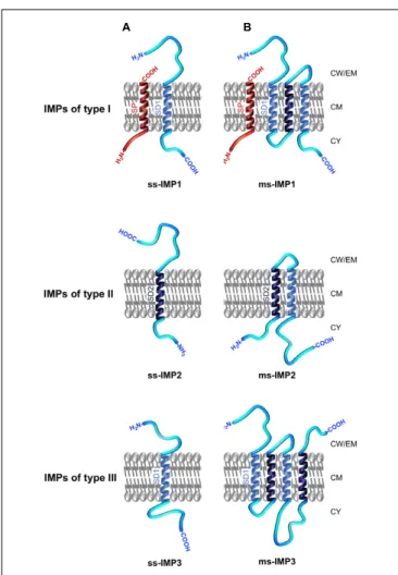
IMPs can be broadly discriminated between single-spanning
IMPs (ss-IMPs) exhibiting a single TMD and
multispanning-IMPs (ms-multispanning-IMPs) with more than one TMD (Figure 4;
Goder
and Spiess, 2001
;
Higy et al., 2004
). Whereas most IMPs
are not synthesised with a cleavable N-terminal SP, some
IMPs are (
Facey and Kuhn, 2004
). For the latters and after
cleavage of the hydrophobic transmembrane
α-helical SP
by a signal peptidase (SPase), the ss-IMPs remain anchored
to CM thanks to an additional hydrophobic TMD, i.e., the
stop-transfer sequence also called signal domain of type I
(SD1), which exhibits a N
out–C
intopology; as such, these
IMPs refer to the type I (IMP1; Figure 4). Type II
ss-IMPs (ss-IMP2) have a signal-anchor sequence also called
signal domain of type II (SD2), with a N
in–C
outtopology,
which actually corresponds to an uncleavable SP. Type III
ss-IMPs (ss-IMP3) have reverse signal-anchor sequence, i.e.,
a SD1 (TMD with a N
out–C
intopology); in the literature,
they are sometimes described as ss-IMP1 without SP since
the reverse signal-anchor sequence is a SD1. Of note, while
the translocation mechanism of both type I and type II IMPs
is in line with our current knowledge about the Sec/YidC
translocation, i.e., involving an N-terminal SP (whenever
cleavable or uncleavable) targeting the protein to CM, the
mechanism for the translocation of type III IMPs in the absence
of a SP remain unclear. In ms-IMPs, the type I (N
out–C
inTMD
topology) and type II (N
in–C
outTMD topology) signals alternate
along the protein sequence. Based on topology of the most
N-terminal TMD enabling anchoring of the ms-IMP to the
FIGURE 4 | Topology and nomenclature of IMPs. IMPs are primarily categorised into (A) single-spanning IMPs (ss-IMPs) and (B) multi-spanning IMPs (ms-IMPs). Indeed, IMPs are anchored to the CM via hydrophobic transmembraneα-helical peptide domains (TMDs); when a TMD has a Nout–Cintopology, it is called a signal domain of type I (SD1; depicted in light
blue), whereas a TMD with Nin–Couttopology is called a signal domain of type
II (SD2; depicted dark blue) (White and von Heijne, 2004). In ss-IMPs, only one TMD is present, whereas at least two TMDs are present in ms-IMPs. Whenever ss-IMPs or ms-IMPs, they are further subcategorised into three types. A ss-IMP of type I (ss-IMP1) possesses a cleavable N-terminal signal peptide (SP; depicted in red) and are actually anchored to the CM by a SD1 (TMD with a Cin–Nouttopology). A ss-IMP of type II (ss-IMP2) is anchored to
the CM by a SD2 (TMD with a Nin–Couttopology). Like for a ss-IMP1, a
ss-IMP of type III (ss-IMP3) is anchored to the CM by a SD1 but it did not originally exhibit a SP. For ms-IMPs, the classification is similar and based on the most N-terminal TMD anchoring the ms-IMP to the CM. As such, a ms-IMP of type I (ms-IMP1) has a cleavable SP followed by a SD1. A ms-IMP of type II (ms-IMP2) has a SD2 as the most N-terminal TMD. A ms-IMP of type III (ms-IMP3) has a SD1 as the most N-terminal TMD (and no cleavable SP). Of note, the TMD of a cleavable SP actually corresponds to a SD2; as such, a SD2 in IMPs of type II can be referred as an uncleavable SP. In ms-IMPs, a SD1 necessarily alternates with a SD2 along the polypeptide chain, and vice versa. Except for the TMDs, other regions of the IMPs can be in contact with the IWZ but also the OWZ or the extracellular milieu.
CM, the three types mentioned here above can be discriminated
(Figure 4).
IMP biogenesis in lipopolysaccharidic-diderm bacteria
(archetypal Gram-negative bacteria) involves an integrase known
as YidC (
Scotti et al., 2000
). Up to two paralogues of the integrase
YidC have been uncovered in parietal monoderm bacteria,
namely SpoIIIJ and YqjG (
Tjalsma et al., 2000
;
van Wely et al.,
2001
). While both SpoIIIJ and YqjG are involved IMP biogenesis
and are essential for cell viability (
Murakami et al., 2002
;
Tjalsma
et al., 2003
), SpoIIIJ is required for sporulation in
B. subtilis
but not YqjG (
Errington et al., 1992
;
Murakami et al., 2002
).
Lately, these proteins have been renamed YidC1 and YidC2,
respectively, in parietal monoderm bacteria (
Funes et al., 2009
;
Wang and Dalbey, 2011
;
Palmer et al., 2012
). In
E. coli, YidC is
associated to the Sec translocase enabling insertion of all IMPs
to the CM in a SRP (signal-recognition particle) dependent
mechanism (
Scotti et al., 2000
;
Fröderberg et al., 2003
;
Ziehe
et al., 2017
). In this species, the YidC pathway is quite versatile
since integration of IMPs to the CM can be SecA-, SecB-,
and/or Sec-independent (
Samuelson et al., 2000
;
Beck et al.,
2001
;
Yen et al., 2002
;
Fröderberg et al., 2003
;
White and von
Heijne, 2004
). Moreover, flotillin-like proteins could contribute
to the insertion of IMPs (
Dempwolff et al., 2016
). So far, these
aspects have been poorly investigated in parietal monoderm
bacteria.
Cell-Surface Proteins Anchored to the
Cytoplasmic Membrane (GO: 0046658):
The Lipoproteins
In parietal monoderm bacteria, lipoproteins are synthesised
as pre-prolipoproteins that are exported by the Sec pathway
and exposed on the outer face of the CM (
Hutchings et al.,
2009
; Figure 5). The pre-prolipoproteins exhibit a SP of
type II (SP II) that is harbouring a conserved lipobox motif
at the cleavage site (
Sutcliffe and Harrington, 2002
). The
consensus sequence for the lipobox is [LVI]
−3-[ASTVI]
−2-[GAS]
−1-[C]
+1(
Sutcliffe and Harrington, 2002
;
Babu et al.,
2006
). Once translocated across the CM, the lipoprotein
maturation pathway in parietal monoderm bacteria is a
two-step process. First, the lipobox motif is recognised by a
prolipoprotein diacylglyceryl transferase (Lgt), which transfers
of a diacylglyceryl moiety from a glycerophospholipid onto
the thiol group of the conserved cysteine, giving rise to the
prolipoprotein. Then, the SP II of the prolipoprotein is cleaved
off by a lipoprotein signal peptidase (Lsp), generating a mature
lipoprotein. The lipoprotein is consequently covalently bound
to the acyl moiety of two fatty acids from the diacylglyceride
by a cysteine residue at position 1 of the N-terminal end
(
Lai et al., 1980
). Besides this classical form of lipid-modified
cysteine for lipoprotein anchoring to the CM, intensive mass
spectrometry analyses revealed three novel forms of mature
lipoproteins in parietal monoderm bacteria (
Nakayama et al.,
2012
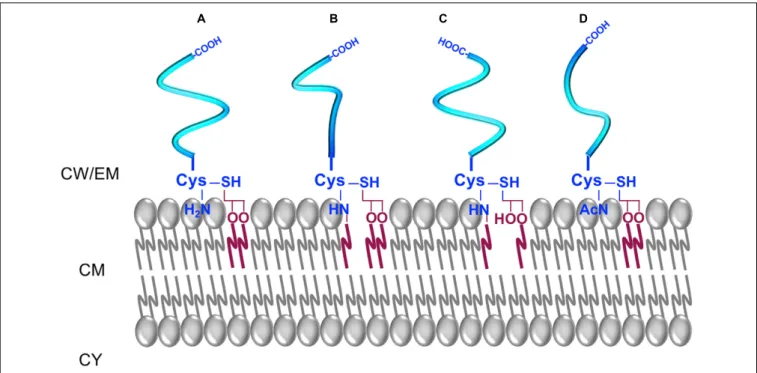
; Figure 5). The N-acylated triacyl form of lipoproteins
containing
N-acyl-S-diacyl-glyceryl-cysteine
was
identified
in
S. aureus and S. epidermidis (
Kurokawa et al., 2009
;
Asanuma et al., 2011
). The
N-acetyl form of lipoproteins
identified in different Bacillaceae contains
N-acetyl-S-diacyl-glyceryl-cysteine (
Kurokawa et al., 2012b
). The lyso-form
of lipoproteins containing an
N-acyl-S-monoacyl-glyceryl-cysteine was identified in
Bacillus cereus, Enterococcus
FIGURE 5 | The different forms of lipoproteins in parietal monoderm bacteria. (A) A diacyl-lipoprotein contains an N-acyl-S-diacylated cysteine residue. (B) A N-acylated-triacyl-lipoprotein contains an N-acyl-S-triacylated cysteine residue. (C) A lyso-lipoprotein contains an N-acyl-S-monoacyl-glyceryl-cysteine. (D) A N-acetyl-form contains a N-acetyl-S-diacyl-glyceryl-cysteine.
faecalis, Lactobacillus bulgaricus, and Streptococcus sanguinis
(
Asanuma et al., 2011
). It further appeared that environmental
conditions influenced the ratio between diacyl and triacyl
forms of lipoproteins in
S. aureus, with an accumulation
of the diacyl lipoprotein form at high temperatures and
high salt concentrations (
Kurokawa et al., 2012a
). Together,
these recent findings are suggestive of uncharacterised
non-canonical pathways for differential lipoprotein lipidation in
parietal monoderm bacteria, analogous to the N-acylation of the
lipidated cysteine by the apolipoprotein
N-acyltransferase (Lnt)
in lipopolysaccharidic-diderm bacteria. Actually, the lipoprotein
intramolecular transacylase (Lit) involved in N-lyso-form
biosynthesis was recently identified in
E. faecalis and B. cereus
(
Armbruster and Meredith, 2017
). If N-acylation is likely to
involve acyltransferases adapted to specific phospholipids as
acyl-donor substrates, novel enzymes and maybe pathways
are to be discovered to explain how these alternative
N-acetyl
lipoprotein forms are biosynthesised in parietal monoderm
bacteria.
CELL-SURFACE PROTEINS LOCALISED
AT THE CELL WALL (GO: 0009275)
The first surface associated proteins were described because of
their activities on the bacterial CW. Most of them were autolysins
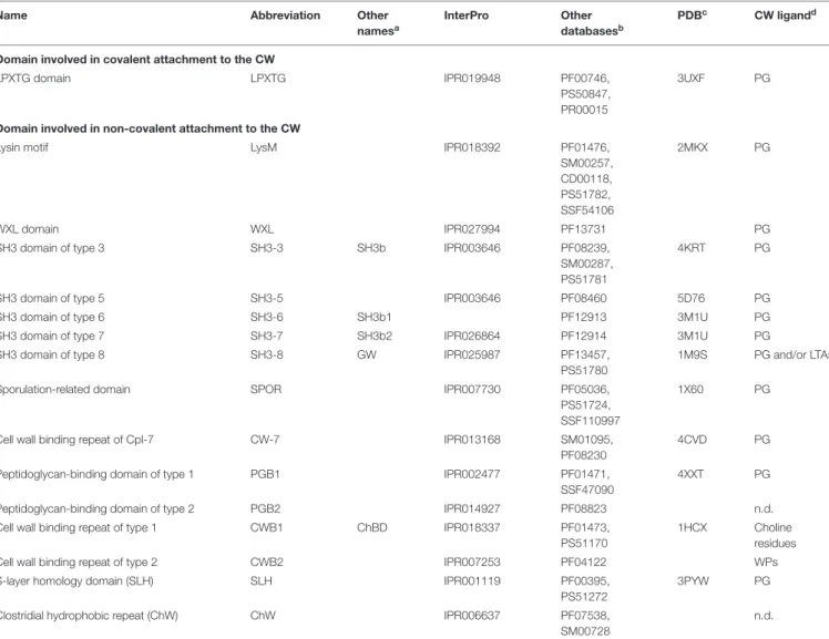
or proteases. PG-binding domains were thereafter observed
thanks to the sequencing data and bioinformatic analyses. Indeed,
amino acid repetitions involved in the surface binding were
highlighted. Most of the characterised and conserved domains
are registered and classified by bioinformatic resources, especially
InterPro (IPR;
Zdobnov and Apweiler, 2001
;
Finn et al., 2017
)
regrouping several databases for motif signatures, such as Pfam
(
Soohammer et al., 1997
;
Finn et al., 2016
), Prosite (
Hulo et al.,
2006
;
Sigrist et al., 2013
), or SMART (
Schultz et al., 1998
;
Letunic et al., 2015
) (Table 1). Of note, the use of underscore
(“_”), as given in the name of domains in databases, must
be abstained by reminding the readers this sign is primarily
designed for bioinformatics purpose when a space cannot be
used due to command line constraints but are meant to be
replaced by a space (“ ”) or a dash (“-”) in textbook. These
binding domains allow protein subcellular location at the CW
and are therefore often crucial for their activity on the surface
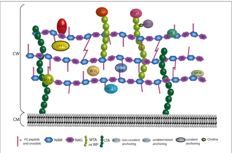
structure and organisation (Figure 6). They can be divided into
three main classes: domains that are (i) covalently attached
to the PG, (ii) non-covalently bound to the PG, and (iii)
non-covalently bound to WPs (Figure 6). Besides, the CW
components targeted by some domains remain uncertain. These
proteins are generally secreted by the Sec translocon and possess
a SP I.
Cell-Surface Proteins Covalently Bound
to the Peptidoglycan: The
LPXTG-Proteins
Covalent binding of LPXTG-proteins to the CW has been the
subject of intensive studies and is certainly one of the best
characterised molecular mechanisms for protein anchoring
to the PG (
Fischetti et al., 1990
;
Schneewind et al., 1992
).
Here, we review the major mechanism of anchoring. In
TABLE 1 | Domains involved in protein attachment to the cell wall in parietal monoderm bacteria.
Name Abbreviation Other
namesa
InterPro Other databasesb
PDBc CW ligandd
Domain involved in covalent attachment to the CW
LPXTG domain LPXTG IPR019948 PF00746, PS50847, PR00015
3UXF PG
Domain involved in non-covalent attachment to the CW
Lysin motif LysM IPR018392 PF01476,
SM00257, CD00118, PS51782, SSF54106 2MKX PG WXL domain WXL IPR027994 PF13731 PG
SH3 domain of type 3 SH3-3 SH3b IPR003646 PF08239, SM00287, PS51781
4KRT PG
SH3 domain of type 5 SH3-5 IPR003646 PF08460 5D76 PG SH3 domain of type 6 SH3-6 SH3b1 PF12913 3M1U PG SH3 domain of type 7 SH3-7 SH3b2 IPR026864 PF12914 3M1U PG SH3 domain of type 8 SH3-8 GW IPR025987 PF13457,
PS51780
1M9S PG and/or LTAs
Sporulation-related domain SPOR IPR007730 PF05036, PS51724, SSF110997
1X60 PG
Cell wall binding repeat of Cpl-7 CW-7 IPR013168 SM01095, PF08230
4CVD PG
Peptidoglycan-binding domain of type 1 PGB1 IPR002477 PF01471, SSF47090
4XXT PG
Peptidoglycan-binding domain of type 2 PGB2 IPR014927 PF08823 n.d. Cell wall binding repeat of type 1 CWB1 ChBD IPR018337 PF01473,
PS51170
1HCX Choline residues Cell wall binding repeat of type 2 CWB2 IPR007253 PF04122 WPs S-layer homology domain (SLH) SLH IPR001119 PF00395,
PS51272
3PYW PG
Clostridial hydrophobic repeat (ChW) ChW IPR006637 PF07538, SM00728
n.d.
aOther names found in the literature to name the respective domain. Name and abbreviation given in the two first columns are preferred and must be favoured. SH3b,
sarcome homology 3 domain of bacterial type; ChBD, choline-binding domain.
bSignatures from InterPro member databases used to construct the entry, namely from Pfam (PF), SMART (SM), Conserved Domain Database (CD), Prosite (PS), Prints
(PR), SuperFamily (SF).
cAccession number to the resolved structure in PDB (protein data bank).
dCW, cell wall; PG, peptidoglycan; LTAs, lipoteichoic acids; WTAs, wall teichoic acids; WPs, wall polysaccharides; n.d., not determined. Choline residues are found in
WTAs and LTAs.
parietal monoderm bacteria, a range of proteins called LPXTG
(IPR019948) is covalently linked to the PG by enzymes
named sortases. Among LPXTG-proteins are found colonising
factors, toxins and proteases. In parietal monoderms, the
LPXTG motif was identified in both classes of Actinobacteria
and Firmicutes, especially in the orders of Coriobacteriales,
Streptomycetales,
Propionibacteriales,
Bifidobacteriales,
Micrococcales, and Corynebacteriales for the former, and
the orders of Erysipelotrichales, Clostridiales, Lactobacillales,
Bacillales, and Tissierellales for the latter. This is a C-terminal
motif composed of the LPXTG sequence where X represents
any amino acids, followed by a hydrophobic domain and
a short positively charged tail. Several variations around
this motif were reported, e.g., NP(Q/K)TN, but the overall
motifs remain homologous and are included for profile
search (
Boekhorst et al., 2005
). In any case, the motif is
recognised by sortases that are classified into six classes
from A to F (
Dramsi and Bierne, 2017
;
Siegel et al., 2017
).
Sortase A anchors a wide range of LPXTG-proteins, whereas
sortase B recognises the NP(Q/K)TN related motif. Sortase
C allows the pilus assembly (see below), whereas sortases
D, E, and F have been much less characterised. Sortases
anchor the LPXTG-proteins on the nascent PG through their
transpeptidase activity, by cleaving between T and G (or N) and
transferring the protein on the PG. Depending on the PG nature,
molecular binding can occur at the pentaglycine crossbridge
(
Marraffini and Schneewind, 2005
) or at the A2pm (
Budzik et al.,
2008
).
FIGURE 6 | Anchoring localization of protein domains interacting with the CW. The localization of the CW proteins depends on their domains. Domains are interacting covalently or not at the bacterial CW through interaction with surface structures that are the PG, the WTAs, the WPs or the LTAs. LPXTG proteins are covalently attached to the A2pm or K residue of the PG. Proteins harbouring a LysM, SH3 of type 5 (SH3-5), SH3 of type 6 (SH3-6), SPOR, or CW-7 domain interact non-covalently with the PG. WXL interacts with PG but the precise anchoring region is undetermined. Proteins possessing a CWB2 or SLH domain are localised at the WTAs or WPs extremities, whereas proteins harbouring a CWB1 domain interact with the WTAs through a choline. For SH3 of type 8 (SH3-8), the CW target remains controversial.