HAL Id: hal-01868522
https://hal.archives-ouvertes.fr/hal-01868522
Submitted on 5 Sep 2018
HAL is a multi-disciplinary open access
archive for the deposit and dissemination of sci-entific research documents, whether they are pub-lished or not. The documents may come from teaching and research institutions in France or abroad, or from public or private research centers.
L’archive ouverte pluridisciplinaire HAL, est destinée au dépôt et à la diffusion de documents scientifiques de niveau recherche, publiés ou non, émanant des établissements d’enseignement et de recherche français ou étrangers, des laboratoires publics ou privés.
High Resolution pQCT micro-architectural parameters
to predict bone failure in the case of a forward fall
Martin Revel, Francois Duboeuf, François Bermond, Jean-Paul Roux, David
Mitton, Hélène Follet
To cite this version:
Martin Revel, Francois Duboeuf, François Bermond, Jean-Paul Roux, David Mitton, et al.. High Resolution pQCT micro-architectural parameters to predict bone failure in the case of a forward fall. 8th World Congress of Biomechanics, Jul 2018, DUBLIN, France. 2 p. �hal-01868522�
High Resolution pQCT micro-architectural
parameters to predict bone failure in the case of a
forward fall
M. Revel
1,2, F. Duboeuf
1, F. Bermond², J-P. Roux
1, D. Mitton², H. Follet
1 1Univ Lyon, Université Claude Bernard Lyon 1, INSERM, Lyos UMR1033, Lyon, France ²Univ Lyon, Université Claude Bernard Lyon 1, IFSTTAR, LBMC UMR_T9406, Lyon, France
Keywords: HR-pQCT, osteoporosis, micro-architecture, fall, fracture, human radius.
Introduction
The gold standard for the diagnosis of osteoporosis is the measurement of areal bone mineral density by Dual X-ray absorptiometry. But the assessment of the bone fracture risk is still not sufficiently discriminant. Indeed, half of the patient considered non-osteoporotic will fracture [1]. In this context, the objective of this study is to evaluate whether bone micro-architecture of distal radius assessed by high-resolution peripheral quantitative computed tomography (HR-pQCT) manage to discriminate fractured from non-fractured bones obtained in a previous ex vivo experimental study reproducing a forward fall under dynamic loading conditions [2].
Method
Thirty radii from elderly donors (79 ± 12 y.o., 15 males, 15 females) were scanned intact using a HR-pQCT device (XtremeCT, Scanco Medical AG, Switzerland) before being loaded at 2 m.s-1 to mimic impact that corresponds to a fall, leading to two groups: fractured and non-fractured. Clinical standard evaluation was performed on the images in order to assess bone micro-architecture. Statistical non-parametric Spearman’s test and Wilcoxon’s rank-sum test were performed using R© software (The R foundation for statistical Computing, Austria) and p-values under 0.05 were considered as significant.
Results
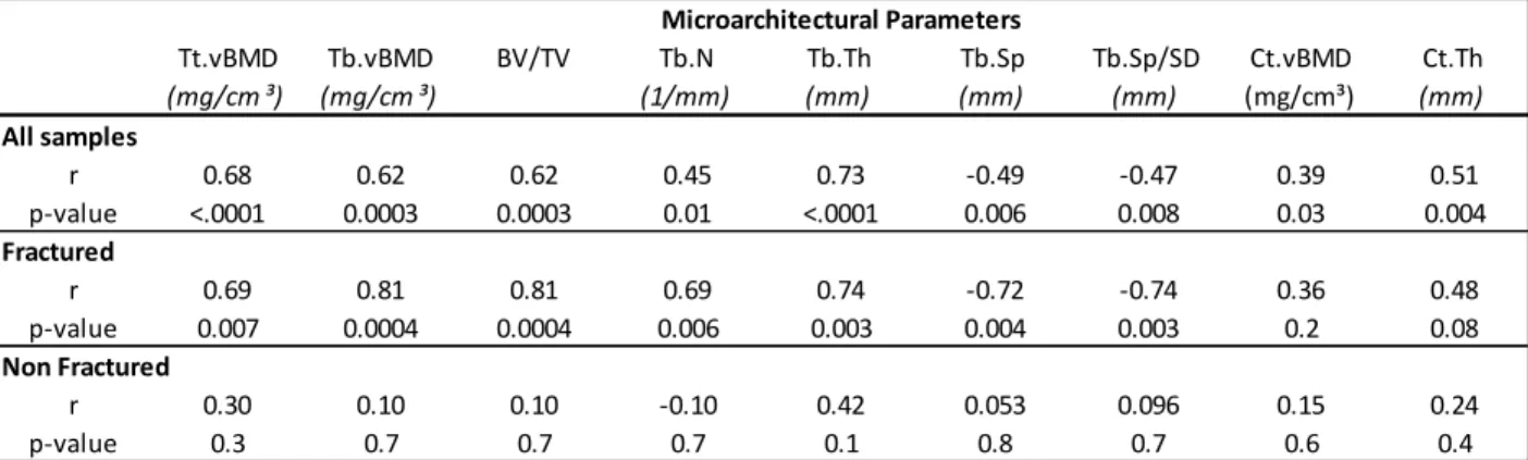
On the entire population, all micro-architectural parameters were significantly correlated to the maximal load. But important differences appeared when analyzing in two groups. Regarding the fractured group, the Spearman’s correlation coefficient ranged between 0.69 to 0.81 (Tables 1) for the trabecular parameters whereas the cortical ones were found as not significant. As to non-fractured group, no link was found with any cortical or trabecular parameters (Table 1). Furthermore, the fractured and non-fractured group showed in the second test a significant difference for: all trabecular parameters (p-values < 0.02), the cortical thickness (p-value < 0.05) and the experimental load (p-value < 0.001) but the cortical vBMD still remain non-significant (p-value = 0.11).
Discussion
The aim of this study was to assess whether micro-architectural parameters obtained by HR-pQCT could discriminate fractured and non-fractured bones obtained experimentally. The correlations showed that trabecular parameters assessed by HR-pQCT explained the bone strength for the fractured group. Furthermore, we found that trabecular parameters, BV/TV and experimental load showed a significant difference between the two groups (p-values < 0.02) contrary to the cortical parameters. These results are in accordance with an epidemiologic study where the authors conclude that the trabecular micro-architecture in the radius is a good predictor of incident fracture in postmenopausal women [3]. Finite element analysis will be performed to enhance our understanding of bone fracture during a fall.
References
[1] E. S. Siris et al., (2004) Arch.Intern.Med., 164(10) p1108-1112 [2] E. Zapata et al., (2017) J.Biomech., 63 p174-178
[3] E. Sornay-Rendu et al, (2017) JBMR, 32(6) p1243-1251 Tt.vBMD (mg/cm ³) Tb.vBMD (mg/cm ³) BV/TV Tb.N (1/mm) Tb.Th (mm) Tb.Sp (mm) Tb.Sp/SD (mm) Ct.vBMD (mg/cm³) Ct.Th (mm) All samples r 0.68 0.62 0.62 0.45 0.73 -0.49 -0.47 0.39 0.51 p-value <.0001 0.0003 0.0003 0.01 <.0001 0.006 0.008 0.03 0.004 Fractured r 0.69 0.81 0.81 0.69 0.74 -0.72 -0.74 0.36 0.48 p-value 0.007 0.0004 0.0004 0.006 0.003 0.004 0.003 0.2 0.08 Non Fractured r 0.30 0.10 0.10 -0.10 0.42 0.053 0.096 0.15 0.24 p-value 0.3 0.7 0.7 0.7 0.1 0.8 0.7 0.6 0.4 Microarchitectural Parameters
Table 1 : Spearman correlation (r) between the experimental load and the micro-architectural parameters. Tt: Total, Tb: Trabecular, Ct: Cortical, vBMD: Volumetric Bone Mineral Density, N: number, Th: Thickness,