HAL Id: hal-03154463
https://hal.archives-ouvertes.fr/hal-03154463
Submitted on 1 Mar 2021
HAL is a multi-disciplinary open access
archive for the deposit and dissemination of
sci-entific research documents, whether they are
pub-lished or not. The documents may come from
teaching and research institutions in France or
abroad, or from public or private research centers.
L’archive ouverte pluridisciplinaire HAL, est
destinée au dépôt et à la diffusion de documents
scientifiques de niveau recherche, publiés ou non,
émanant des établissements d’enseignement et de
recherche français ou étrangers, des laboratoires
publics ou privés.
Distributed under a Creative Commons Attribution| 4.0 International License
Giuffra, Sonia Lacroix-Lamandé, Agnès Wiedemann
To cite this version:
Martin Beaumont, Fany Blanc, Claire Cherbuy, Giorgia Egidy, Elisabetta Giuffra, et al.. Intestinal
organoids in farm animals. Veterinary Research, BioMed Central, 2021, 52 (1), 15 p.
�10.1186/s13567-021-00909-x�. �hal-03154463�
REVIEW
Intestinal organoids in farm animals
Martin Beaumont
1*†, Fany Blanc
2†, Claire Cherbuy
3†, Giorgia Egidy
2†, Elisabetta Giuffra
2†,
Sonia Lacroix‑Lamandé
4†and Agnès Wiedemann
4,5†Abstract
In livestock species, the monolayer of epithelial cells covering the digestive mucosa plays an essential role for nutri‑ tion and gut barrier function. However, research on farm animal intestinal epithelium has been hampered by the lack of appropriate in vitro models. Over the past decade, methods to culture livestock intestinal organoids have been developed in pig, bovine, rabbit, horse, sheep and chicken. Gut organoids from farm animals are obtained by seeding tissue‑derived intestinal epithelial stem cells in a 3‑dimensional culture environment reproducing in vitro the stem cell niche. These organoids can be generated rapidly within days and are formed by a monolayer of polarized epithe‑ lial cells containing the diverse differentiated epithelial progeny, recapitulating the original structure and function of the native epithelium. The phenotype of intestinal organoids is stable in long‑term culture and reflects characteristics of the digestive segment of origin. Farm animal intestinal organoids can be amplified in vitro, cryopreserved and used for multiple experiments, allowing an efficient reduction of the use of live animals for experimentation. Most of the studies using livestock intestinal organoids were used to investigate host‑microbe interactions at the epithe‑ lial surface, mainly focused on enteric infections with viruses, bacteria or parasites. Numerous other applications of farm animal intestinal organoids include studies on nutrient absorption, genome editing and bioactive compounds screening relevant for agricultural, veterinary and biomedical sciences. Further improvements of the methods used to culture intestinal organoids from farm animals are required to replicate more closely the intestinal tissue complexity, including the presence of non‑epithelial cell types and of the gut microbiota. Harmonization of the methods used to culture livestock intestinal organoids will also be required to increase the reproducibility of the results obtained in these models. In this review, we summarize the methods used to generate and cryopreserve intestinal organoids in farm animals, present their phenotypes and discuss current and future applications of this innovative culture system of the digestive epithelium.
Keywords: Enteroids, Epithelium, Gut, Monolayer, Culture, Polarity, Pig, Bovine, Chicken, Horse, Rabbit
© The Author(s) 2021. This article is licensed under a Creative Commons Attribution 4.0 Inter‑
national License, which permits use, sharing, adaptation, distribution and reproduction in any medium or format, as long as you give appro‑ priate credit to the original author(s) and the source, provide a link to the Creative Commons licence, and indicate if changes were made. The images or other third party material in this article are included in the article’s Creative Commons licence, unless indicated otherwise in a credit line to the material. If material is not included in the article’s Creative Commons licence and your intended use is not permitted by statutory regulation or exceeds the permitted use, you will need to obtain permission directly from the copyright holder. To view a copy of this licence, visit http://creat iveco mmons .org/licen ses/by/4.0/. The Creative Commons Public Domain Dedication waiver (http://creat iveco mmons .org/publi cdoma in/zero/1.0/) applies to the data made available in this article, unless otherwise stated in a credit line to the data.
Open Access
*Correspondence: martin.beaumont@inrae.fr
†All authors equally contributed to this work and are listed in alphabetical order
1 GenPhySE, Université de Toulouse, INRAE, ENVT, Castanet‑Tolosan 31326, France
Full list of author information is available at the end of the article
Table of Contents 1 Introduction
2 Methods to generate and cryopreserve livestock intes‑ tinal organoids
2.1 Methods to grow intestinal organoids 2.2 Organoid cryopreservation and biobanking
3 Phenotype of livestock intestinal organoids 3.1 Insights from transcriptome analysis 3.2 Epithelial polarization
3.3 Barrier function 3.4 Nutrient absorption
4 Host‑microbe interactions in livestock intestinal organoids
4.1 Enteric infections modeling 4.2 Microbiota‑epithelium interactions
5 Future directions of research with livestock intestinal organoids
1 Introduction
The intestinal epithelium is formed by a monolayer of cells located at the mucosal surface. Intestinal epithe‑ lial cells (IEC) contribute to food digestion and nutrient absorption while acting as a physical and immunologi‑ cal barrier against harmful luminal components (micro‑
organisms, toxins, food antigens) [1]. This dual function
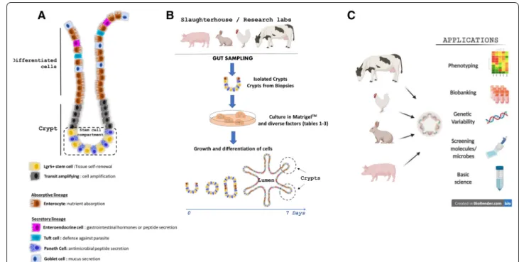
of the intestinal epithelium is performed by the coordi‑ nated action of differentiated epithelial cells specialized for nutrient absorption (enterocytes), hormone secre‑ tion (enteroendocrine cells), antimicrobial peptide secre‑ tion (Paneth cells), anti‑parasite immunity (tuft cells) or
mucus secretion (goblet cells) (Figure 1A). All these dif‑
ferentiated IEC types derive from intestinal epithelial stem cells (IESC) located at the base of epithelial crypts
[2]. Paneth cells remain at the bottom of crypts whereas
the other IEC types differentiate while they migrate towards the lumen. Epithelial cell type distribution also differs along the length of the intestine, which reflects the
functional specialization of digestive segments [3]. The
small intestine is divided into 3 segments: duodenum, jejunum and ileum. The duodenum receives the food chyme, bile juice and pancreatic secretions to complete the chemical digestion. The jejunum is the main site for
nutrient absorption while the ileum absorbs residual nutrients, vitamins and conjugated bile acids. The large intestine is composed of the caecum and the colon, which main functions are to absorb water, electrolytes and microbial fermentation products.
The study of the intestinal epithelium in livestock spe‑ cies has major implications for agricultural (e.g. improve‑ ment of feed efficiency), veterinary (e.g. resistance to enteric pathogens) or biomedical (e.g. large animal mod‑ els of human diseases) research. However, models com‑ monly used to study livestock epithelium present major limitations. In vitro immortalized intestinal epithelial cell lines (e.g. porcine IPEC‑J2) lack cellular heterogeneity, do not fully reproduce tissue functionality and present
genomic abnormalities [4, 5] or have not been established
for some species (e.g. chicken, bovine). Ex vivo culture of intestinal explants or primary isolated IECs recapitulate key features of the in vivo tissue but they are not suitable for long‑term experiments due to limited viability (24–
48 h as reviewed by Randall et al. [6]). The recent devel‑
opment of livestock intestinal organoids solved most of these limitations.
Intestinal organoids are self‑organized 3D structures composed of a monolayer of polarized IEC surrounding
a hollow lumen [7] (Figure 1B). This innovative model
recapitulates in vitro the multicellular composition of the References
Figure 1 Models of intestinal organoids in farm animals. A Diverse cell lineages constitute the digestive epithelium. Paneth and stem cells
are localized at the bottom of the crypts, while enterocytes, goblet cells, enteroendocrine cells and tuft cells migrate towards the lumen upon differentiation. B Schematic illustration of livestock intestinal organoid generation from epithelial crypts isolated from fresh intestinal tissue or from frozen biopsies. C Schematic representation of farm animal intestinal organoid applications in basic and applied science.
intestinal epithelium, its architecture with crypt‑domains and its main roles such as nutrient absorption and bar‑ rier function. “Enteroids” and “colonoids” refer to intes‑ tinal organoids derived from the small intestine or from
the colon, respectively [8]. Importantly, intestinal orga‑
noids retain in vitro phenotypic features of their diges‑
tive segment of origin [3]. Intestinal organoid culture was
first developed in mouse and humans after identification of the signaling pathways involved in the maintenance
and proliferation of Lgr5+ IESC [9]. Intestinal organoid
culture is based on the reproduction of the IESC niche in vitro: Wnt pathway activation, epidermal growth fac‑ tor (EGF) signaling stimulation and bone morphogenic
protein (BMP) pathway inhibition [2]. IESC are seeded
in a gel of extracellular matrix proteins (e.g. Matrigel™)
that provides a structural scaffold for 3D growth and
promotes cell survival (Figure 1B). Human and mouse
intestinal organoids can be derived from intestinal tissue‑ derived IESC or from induced pluripotent stem (iPS) or
embryonic stem (ES) cells [10]. Organoids obtained from
tissue‑derived IESC are composed only of epithelial cells. In contrast, both epithelial and mesenchymal cells (e.g. myofibroblasts) are present in iPS and ES‑derived intes‑ tinal organoids, which however remain in an immature,
fetal‑like state [10].
Herein, we first review the main methods used to gen‑ erate and cryopreserve intestinal organoids from farm animals. Then, we present the main characteristics of these innovative intestinal epithelium culture systems. Finally, we detail current and future applications of live‑
stock intestinal organoids (Figure 1C).
2 Methods to generate and cryopreserve livestock intestinal organoids
2.1 Methods to grow intestinal organoids
Despite recent progress for the development of domes‑
tic animal iPS and ES cells [11, 12], to our knowledge, all
intestinal organoid models from livestock species have been developed with tissue‑derived‑IESC. Methods orig‑ inally developed to culture mouse and human intestinal
organoids from tissue‑derived IESC [13, 14] were recently
adapted to numerous farm animal species including pig, rabbit, cow, sheep, horse and chicken (summarized in
Table 1 for pig, Table 2 for ruminant and herbivorous
species and Table 3 for chicken) (Figure 2). Farm ani‑
mal intestinal organoids were successfully obtained from several digestive segments (duodenum, jejunum, ileum, caecum and colon). The first step of livestock intestinal organoid culture is to isolate epithelial crypts that con‑ tain IESC. Most of the studies used a dissociation buffer containing ethylenediaminetetraacetic acid (EDTA) and dithiothreitol (DTT). There are great variations in EDTA concentration (0.8–30 mM), time and temperature of
incubation according to the studies, species and digestive
segments (Tables 1, 2 and 3). Some studies supplemented
the crypt isolation buffer with Y27632, a ROCK inhibi‑ tor that prevents epithelial cell death. Isolated crypts
are then seeded in Matrigel™ and the growth medium is
added. Alternatively, a hanging drop culture system with‑
out Matrigel™ embedding has been reported for embry‑
onic chicken intestinal organoids [15].
The composition of livestock intestinal organoid growth medium has been directly adapted from human
and mouse protocols (see Tables 1, 2 and 3 for refer‑
ences). Although recombinant growth factors are most often not commercially available for farm animals, human or mouse orthologues can be used due to high evolution‑
ary conservation of their amino acid sequences [14, 16].
Wnt signaling activation is generally induced by mouse or human recombinant Wnt3a and R‑spondin, either used purified or in conditioned media from engineered cell lines. Some studies also used CHIR99021, an inhibi‑ tor of glycogen synthase kinase 3 to further activate the Wnt pathway. BMP pathway signaling is usually inhib‑ ited by human or mouse recombinant Noggin, either used purified or in conditioned media. For instance, the conditioned medium from L‑WRN cells (mouse L cell
line secreting Wnt3a, R‑spondin and Noggin, ATCC®
CRL‑3276™) was successfully used to culture intestinal
organoids from several species [14, 16] (Figure 2). The
BMP inhibitor LDN193189 was used instead of Nog‑ gin in the growth medium of rabbit caecum organoids
[16]. TGF‑β receptor inhibitors (LY2157299, A8301,
SB43542) and the p38 MAPK inhibitor SB202190 were used in several studies to promote epithelial proliferation
and inhibit differentiation [17]. Epithelial proliferation is
stimulated by human or murine EGF in several studies, while other methods used fetal bovine serum (FBS) that is a potent source of growth factors, though undefined. When organoid culture medium does not contain FBS, the cell culture supplements N2 and B27 are used to pro‑ vide vitamins or hormones. Most studies supplemented the growth medium with the ROCK inhibitor Y27632 to prevent isolated epithelial cell death but it can be omitted
after 2–3 days of culture when organoids are formed [18].
Continuous use of Y27632 was also reported to prevent the formation of tight junctions in pig intestinal organoid cells, when cultured in 2D monolayers (described below)
[19]. Other common constituents of livestock intestinal
organoid growth media include HEPES (buffer), N‑ace‑ tylcysteine (antioxidant), GlutaMAX (stable glutamine), nicotinamide and antibiotics/antifungals. As an alter‑ native to custom‑made culture media, pig and bovine
intestinal organoids can be cultured in IntestiCult™, a
proprietary defined medium available from STEMCELL
Table 1 P ig in testinal or ganoids CM: c onditioned medium, NA C: N-ac et yl c yst eine , L -WRN: L c
ell line eng
ineer ed t o secr et e W
nt3a, R-spondin 3 and Nogg
in, r m: r ec ombinan t mouse , r h: r ec ombinan t human, FBS: f etal bo
vine serum, EGF
: epider mal gr owth fac tor . A ntibiotics and an
tifungals added in the g
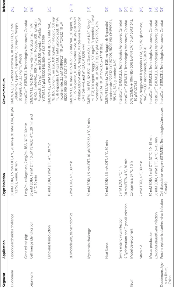
ro wth medium ar e not pr esen ted . Seg men t A pplica tion Cr ypt isola tion G ro wth medium Ref er enc e Duodenum Lipopolysacchar ides challenge 30 mM ED TA, 1.5 mM D TT , 4 °C, 20 min + 30 mM ED TA, 10 µM Y27632, war m, 10 min
DMEM, N2, B27 without vitamin A, 10
mM HEPES, 2 mM L‑ glutamine , 1 μg/mL R ‑spondin1, 100 ng/mL Nogg in, 50 ng/mL EGF , 2.5 ng/mL W nt ‑3a [ 60 ] G ene edit ed pigs 1 mg/mL collagenase , 2 mg/mL BSA, 37 °C, 30 min Int estiC ult ™ (STEMCELL Technolog ies , V ancouv er , C anada) [ 37 ] Jejunnum
Cell lineage identification
30 mM ED TA, 1 mM D TT , 10 µM Y27632, 4 °C, 20 min and 37 °C 10 min DMEM/F12, N ‑2, B
‑27 minus vitamin A, Glutamax, 1
mM HEPES, 1 mM NA C, 100 ng/mL r m Nogg in, 500 ng/mL r h R‑ spondin, 50 ng/mL r m EGF , 100 ng/mL r h W nt3a, 10 µM Y‑ 27632, 10 µM SB202190, 500 nM L Y2157299 [ 39 ] Lentivirus transduc tion 10 mM ED TA, 1 mM D TT , 4 °C, 30 min DMEM/F ‑12, 2 mM glutaM A X, 10 mM HEPES, 1 mM NA C, N2, B27, 50 ng/mL r m EGF , 100 ng/mL r m Nogg in, 500 ng/ mL r h R ‑spondin 1, W nt3a CM, 1 mM valpr oic acid , 10 mM nicotinamide , 2.5 μM CHIR99021, 10 μM Y27632, 10 μM SB202190, 500 nM L Y2157299 [ 25 ] 2D monola yers , transcr ipt omics 2 mM ED TA, 4 °C, 30 min DMEM/F12, 10 mM HEPES, B ‑27, 1.25 mM NA C, 50 ng/mL r h EGF , 15 nM gastr in, 10 mM nicotinamide , 10 μM p38 M APK inhibit or , 600 nM A83 ‑01, Nogg in CM (15% v/v), R ‑spondin CM (15% v/v), and W nt3A CM (30% v/v) [ 5 , 19 ] M ycot oxin challenge 30 mM ED TA, 1.5 mM D TT , 10 µM Y27632, 4 °C, 30 min DMEM, 10% FBS, N2, B27, 1X GlutaM A X, 1 mM NA C, 50 ng/ mL EGF , 100 ng/mL Nogg in, 500 ng/mL R ‑spondin1, 10 mM nicotinamide , 10 µM SB202190, 0.5 µM L Y2157299, 45% W nt3a CM, 10 µM Y27632 (2–3 first da ys) [ 18 ] Heat Str ess 30 mM ED TA, 1.5 mM D TT , 4 °C, 30 min DMEM/F12, W nt3a CM, r m EGF , r m Nogg in, r h R ‑spondin1, nicotinamide , Y27632, L Y2157299, SB202190, CHIR99021, FBS, N2, B27, glutamine , NA C [ 26 ] Swine ent er ic virus inf ec tion 5 mM ED TA, 4 °C, 1 h Int estiC ult ™ (STEMCELL Technolog ies , V ancouv er , C anada) [ 34 ] S. T yphimur ium and T. gondii inf ec tion 0.8 mM ED TA, 4 °C, 30 min Int estiC ult ™ (STEMCELL Technolog ies , V ancouv er , C anada) [ 20 ] Ileum M odel de velopment Collagenase , 37 °C, 1.5 h 50% (DMEM + 10% FBS), 50% L ‑WRN CM, 10 µM SB431542, 10 µM Y27632 [ 14 ] Vitamin A 2 mM ED TA, 4 °C, 30 min W nt3a, Nogg in and R ‑spondin 1 CM, B27, N2, glutamine , NA C, r m EGF , nicotinamide , SB202190 [ 45 ] M ucus pr oduc tion 30 mM ED TA, 1 mM D TT , 37 °C, 10–15 min Int estiC ult ™ (STEMCELL Technolog ies , V ancouv er , C anada) [ 43 ] Lawsonia intr ac ellularis inf ec tion 30 mM ED TA, 1 mM D TT , 37 °C, 5–15 min Int estiC ult ™ (STEMCELL Technolog ies , V ancouv er , C anada) [ 36 ] Duodenum, J eju ‑ num, I leum, Colon Por
cine epidemic diar
rhea virus inf
ec tion Cell dissociation r eagent (STEMCELL Technolog ies ,V ancouv er , Canada) Int estiC ult ™ (STEMCELL Technolog ies , V ancouv er , C anada) [ 21 ]
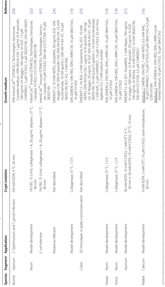
Table 2 Ruminan t and her biv or ous sp ecies (b ovine , sheep , horse and r abbit) in testinal or ganoids CM: c onditioned medium, NA C: N-ac et yl c yst eine , L -WRN: L c
ell line eng
ineer ed t o secr et e W
nt3a, R-spondin 3 and Nogg
in, r m: r ec ombinan t mouse , r h: r ec ombinan t human, FBS: f etal bo
vine serum, EGF
: epider mal gr owth fac tor . A ntibiotics and an
tifungals added in the g
ro wth medium ar e not pr esen ted . Species Seg men t A pplica tion Cr ypt isola tion G ro wth medium Ref er enc e Bo vine Jejunum S. T yphimur ium and T. gondii inf ec tion 0.8 mM ED TA, 4 °C, 30 min 50% I nt estiC ult ™ (STEMCELL T echnolog ies , V ancouv er , Canada) medium, 50% W nt3a CM, 1 μg/mL r h R ‑spondin, 100 ng/mL r m Nogg in, 100 ng/mL r m EGF , 1.5 μM CHIR99021, 5 μM Y27632, 5 μM SB202190, 250 nM A8301 [ 20 ] Ileum M odel de velopment 1% FBS, 75 U/mL collagenase 1 ‑A, 20 µg/mL dispase I, 37 °C, 40 min Int estiC ult ™ (mouse) (STEMCELL Technolog ies , V ancouv er , Canada), Y27632, L Y2157299, SB202190 [ 22 ] E. c oli inf ec tion 1% FBS, 75 U/mL collagenase 1 ‑A, 20 µg/mL dispase I, 37 °C, 40 min Int estiC ult ™ (mouse) (STEMCELL Technolog ies , V ancou ‑ ver , C anada), 10 µM Y27632, 500 nM L Y2157299, 10 µM SB202190 [ 47 ] Rota virus inf ec tion Not descr ibed DMEM/F12, 10 mM HEPES, GlutaM A X, 50 ng/mL EGF , 10% Nogg in CM, 20% R ‑spondin CM, 50% W nt3a CM, 10 mM nicotinamide , 10 nM gastr in I, 500 nM A ‑83–01, 10 μM SB202190, B27, N2, 1 mM NA C [ 24 ] M odel de velopment Collagenase , 37 °C, 1.5 h 50% (DMEM + 10% FBS), 50% L ‑WRN CM, 10 µM SB431542, 10 µM Y27632 [ 14 ] Colon 2D monola yer , in ‑plat e cr yoconser vation Not descr ibed DMEM/F12, 1% BSA, 2 mM Glutamine , N2, B27, 10 mM HEPES, 50% W nt3a ‑CM, 1 µg/mL r h R ‑spondin, 100 ng/ mL r h Nogg in, 50 ng/mL r h EGF , 500 nM A ‑83–01, 10 μM SB202190, 10 nM [L eu]15 ‑gastr in ‑1, 10 mM nicotinamide , 1 mM NA C, 5 μM CHIR99021, 10 μM Y‑ 27623, 50 nm P ros ‑ taglandin E2, 2 mM sodium acetat e [ 23 ] Sheep Ileum M odel de velopment Collagenase , 37 °C, 1.5 h 50% (DMEM + 10% FBS), 50% L ‑WRN CM, 10 µM SB431542, 10 µM Y27632 [ 14 ] Horse Ileum M odel de velopment Collagenase , 37 °C, 1.5 h 50% (DMEM + 10% FBS), 50% L ‑WRN CM, 10 µM SB431542, 10 µM Y27632 [ 14 ] Jejunum M odel de velopment 30 mM ED TA, 10 mM Y27632, 1 mM D TT , 4 °C, 30 min + 30 mM ED TA, 10 mM Y27632, 37 °C, 10 min DMEM/F12, N ‑2, B27, GlutaM A X, 1 mM Hepes , 100 ng/mL hr nogg in, 500 ng/mL r h R ‑spondin, 50 ng/mL hr EGF , 100 ng/mL, hr W nt3a, 10 mM Y27632, 10 mM SB202190, 500 nM L Y215799, 2,5 µM CHIR99021 [ 27 ] Rabbit Caecum M odel de velopment 9 mM ED TA, 3 mM D TT , 10 µM Y27632, r oom t emperatur e, 30 min M edium 1 : DMEM, 10% FBS, 1 mM HEPES, 0.5 mM NA C, 10 µM CHIR99021, 10 µM Y27632,10 µM SB431542, 0.2 µM LDN193189 M edium 2: 50% (DMEM + 10% FBS), 50% L ‑WRN condi ‑ tioned medium, 10 µM Y27632, 10 µM SB431542 [ 16 ]
2). This commercial growth medium has been optimized for mouse and human organoids but not for other spe‑
cies. Indeed, supplementation of mouse IntestiCult™
organoid growth medium with additional growth fac‑
tor was required to culture bovine organoids [20, 22]
(Table 2). After initial growth, intestinal organoid differ‑
entiation can be enhanced by reducing the concentration of the IESC niche factors. For instance, rabbit caecum organoids differentiation was obtained by reducing the concentration of L‑WRN conditioned medium from 50
to 5% for 2 days [16]. Similarly, bovine enteroid differen‑
tiation was triggered by withdrawal of Wnt3a and other
niche factors [23, 24].
Intestinal organoid culture medium is replaced every 2–3 days. After 5–10 days of culture, intestinal organoids are dissociated by mechanical and/or enzymatic meth‑ ods and are subcultured with a 1:3 to 1:8 dilution ratio
in fresh Matrigel™. A study showed that pig enteroids
cultured at 39 °C (body temperature of pigs) expressed
lower levels of the IESC marker Lgr5 +, when compared
to organoids grown at 37 °C [25]. This result suggests
that the temperature of cell culture incubators should be adapted to each species. Another study in pig enteroids showed that the organoid forming efficiency was higher when jejunum crypts were cultured at 37 °C, when com‑ pared to 41 °C, an experimental condition used to mimic
heat stress [26]. However, potentially lower stability of
human and mouse recombinant growth factors at tem‑ peratures above 37 °C might contribute to the effects of temperature on organoid phenotype.
2.2 Organoid cryopreservation and biobanking
Farm animal intestinal organoids can be successfully recovered after cryopreservation in freezing medium (containing FBS, dimethyl sulfoxide and in some stud‑
ies Y27632) and long‑term storage in liquid nitrogen [14,
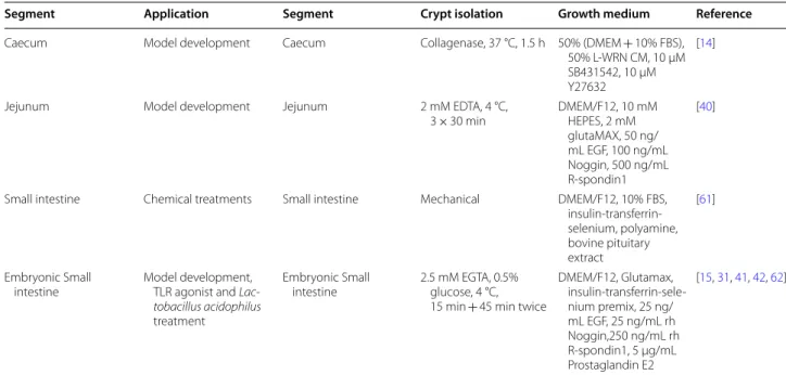
Table 3 Chicken intestinal organoids
CM: conditioned medium, L-WRN: L cell line engineered to secrete Wnt3a, R-spondin 3 and Noggin, rh: recombinant human, FBS: fetal bovine serum, EGF: epidermal growth factor. Antibiotics and antifungals added in the growth medium are not presented.
Segment Application Segment Crypt isolation Growth medium Reference
Caecum Model development Caecum Collagenase, 37 °C, 1.5 h 50% (DMEM + 10% FBS), 50% L‑WRN CM, 10 µM SB431542, 10 µM Y27632
[14]
Jejunum Model development Jejunum 2 mM EDTA, 4 °C,
3 × 30 min DMEM/F12, 10 mM HEPES, 2 mM glutaMAX, 50 ng/ mL EGF, 100 ng/mL Noggin, 500 ng/mL R‑spondin1
[40]
Small intestine Chemical treatments Small intestine Mechanical DMEM/F12, 10% FBS, insulin‑transferrin‑ selenium, polyamine, bovine pituitary extract [61] Embryonic Small
intestine Model development, TLR agonist and
Lac-tobacillus acidophilus
treatment
Embryonic Small
intestine 2.5 mM EGTA, 0.5% glucose, 4 °C, 15 min + 45 min twice
DMEM/F12, Glutamax, insulin‑transferrin‑sele‑ nium premix, 25 ng/ mL EGF, 25 ng/mL rh Noggin,250 ng/mL rh R‑spondin1, 5 µg/mL Prostaglandin E2 [15, 31, 41, 42, 62]
Figure 2 Morphology of intestinal organoids from farm animals. Brightfield images of intestinal organoids grown in Matrigel™ with 50%
L‑WRN conditioned media. Organoids from pig, cow, horse and sheep were obtained from the terminal ileum. Organoids from rabbit and chicken were obtained from the caecum. Scale bars: 200 µM. Images were adapted from previous publications distributed under the terms of Creative Commons Licenses: Powell and Behnke (pig, cow, horse, sheep and chicken organoids) [14] and from Mussard et al. (rabbit organoids) [16].
16, 20, 25]. High‑throughput applications (e.g. bioac‑ tive molecules screening) might benefit from the recent development of an in‑plate cryopreservation technique
for bovine colonic organoids [23]. However, since slaugh‑
terhouses are often located away from the cell culture laboratory, the isolation and culture of intestinal epithe‑ lial crypts from fresh tissues is not always feasible for farm animals. Methods have been developed to directly cryopreserve either isolated epithelial crypts or intestinal tissues retaining the ability to generate organoids after thawing. These methods allow sampling of a large num‑ ber of animals at once, which would not be compatible with immediate labor‑intensive organoid culture. Cryo‑ preserved epithelial crypts isolated from pig and equine jejunum were successfully used after thawing for the cul‑
ture of organoids [25, 27]. Intestinal tissue fragments can
also be cryopreserved and used to culture organoids as
shown for human biopsies [28]. The principle is to freeze
small pieces of intestinal tissue before the crypt isolation step. Once stored in liquid nitrogen, these specimens can be shipped frozen, stored and later thawed to isolate crypts and generate cultures of intestinal organoids, even if a delay in organoid formation is observed. Organoids can thus be generated from biobanked tissues following the best lab schedule. In addition, in our hands, cryo‑ preservation of tissues has guaranteed organoid culture generation free from bacterial and fungal contaminations compared to freshly generated organoids. We have suc‑ cessfully produced pig intestinal organoids after isolation
of epithelial crypts from small pieces (< 1 mm2) of sev‑
eral digestive segments cryopreserved in FBS (90%) and dimethyl sulfoxide (10%) (Blanc et al., unpublished data)
(Figure 3, Additional file 1).
3 Phenotype of livestock intestinal organoids
3.1 Insights from transcriptome analysis
The objective of intestinal organoid cultures is to repli‑ cate in vitro as closely as possible the phenotype of the intestinal epithelium observed in vivo. Transcriptome analysis revealed that pig jejunum organoids resembled more pig jejunum tissue than the pig IPEC‑J2 cell line
[5]. For instance, genes coding for enterohormones or
mucins were similarly expressed in jejunum tissue and organoids while they were not expressed in IPEC‑J2, probably because of the lack of cellular diversity in this
cell line [5]. Gene expression patterns also largely over‑
lapped between bovine epithelial crypts and enteroids
[22]. Thus, livestock intestinal organoids appear to effi‑
ciently reproduce epithelial transcriptome in vitro. The ability of intestinal organoids to retain digestive‑segment specific epithelial phenotype is an important feature pre‑
viously observed in mice [3]. Pig organoids from jejunum
expressed higher levels of genes involved in digestion
and nutrient transport than organoids derived from the ileum, while the opposite was observed for immune‑
related genes [5]. These digestive‑segment specific gene
expression profiles in pig organoids are consistent with known roles of the jejunum for nutrient absorption and of the ileum for host‑microbiota interaction. Another study in pig revealed specific transcription patterns in organoids derived from duodenum, jejunum and colon; although differentially expressed genes were not detailed
[29]. Stability of gene expression in organoids across
extended period of time and multiple passage is neces‑ sary to ensure reproducibility of the results obtained. Transcriptome analysis of pig jejunum organoid lines revealed stable gene expression profile after a long period
of culture in vitro (12 weeks/17 passages) [5]. Bovine
enteroid transcriptome also showed an overall stabil‑
ity across 5 passages [22]. Finally, inter‑individual dif‑
ferences persisted after multiple passages of organoids
obtained from two piglets [5]. This observation suggests
that experiments on intestinal organoids should be per‑ formed in parallel with several organoid lines obtained from different animals in order to cover inter‑individ‑ ual variability. A study showed that the age‑dependent expression of genes used as markers of intestinal matu‑ ration (e.g. lactase and sucrase‑isomaltase) were not reproduced in jejunum enteroids generated from piglets at several developmental stages (embryonic, suckling and
post‑weaning) [30]. These results suggest that enteroids
Figure 3 Morphological features of porcine gut organoids obtained from frozen tissues. Organoids are derived from: A
duodenum (7 days, passage 3), B jejunum (7 days, passage 1), C ileum (7 days, passage 3) and D colon (8 days, passage 1). Observation by phase contrast microscopy. Bars: 200 µm. Images are representative of organoids obtained from 4 pigs for each digestive segment.
do not retain the epithelial phenotype associated with the age of the animal used for crypt isolation. More stud‑ ies are needed to confirm this result with other digestive segments, other markers of developmental stage (e.g. related to innate immunity) and by testing different cul‑ ture conditions. Future experiments using single‑cell RNA sequencing will also be helpful to further character‑ ize each epithelial cell type present in livestock intestinal organoids.
3.2 Epithelial polarization
IEC are highly polarized and interactions with luminal components (nutrients, microorganisms or toxins) occur at their apical side. The localization of apical surface markers (actin or villin) or visualization of microvilli by transmission electronic microscopy at the luminal side indicates that epithelial cells are also polarized in vitro in intestinal organoids from pig, cow, chicken and rab‑
bit [16, 20, 22, 25, 31] (Figures 4A, F and 5A). Although
physiologically relevant, this 3D organization leads to a basolateral exposure of experimental treatments added in the culture medium. Thus, the apical side of organoid epithelial cells is not directly accessible. The microinjec‑ tion technique was used to inject micro‑organisms into
organoid lumen [32]. This procedure is labor‑intensive
and to our knowledge, no study using micro‑injection in livestock intestinal organoids has been published yet. Moreover, it requires a hollow structure in the organoid (lumen) that is often reduced in differentiated organoids. We have successfully used this technique with pig colon
organoids (Cherbuy et al., unpublished data) (Figure 6).
However, concerns have been raised regarding repro‑ ducibility of this technique due to the variable volume injected in each organoid and unintended leak into the medium. A recent study showed that human enteroid cell polarity can be reversed by removal of the extracel‑
lular matrix [33]. The method developed by Co et al. was
applied to reverse epithelial polarity in pig jejunum enter‑
oids in suspension cultures [34]. However, in this study,
the polarity failed to be reversed in ~20% organoids after 3 days, which indicates phenotypic variability in these culture conditions. We also have observed that the trans‑
fer of pig colon organoids from Matrigel™ to suspension
culture for 24 h induces epithelial polarity reversal in
some but not all organoids (Figure 5B, additional file 1)
(Beaumont et al., unpublished data). This polarity inver‑ sion was associated with the loss of the organoid hol‑ low structure (lumen), as observed previously in human
enteroids [33]. In our hands, we observed a rapid viability
loss of organoids cultured in suspension (< 3 days) which
Figure 4 Characterization of intestinal organoids from farm animals. A Pig colon organoids stained for Villin (orange). Scale bar: 20 µm. B Monolayer of rabbit caecum organoid cells stained for actin (red). Scale bar: 100 µm. C Pig colon organoid stained for E‑cadherin (red), and
proliferating cell nuclear antigen (PCNA, green). The arrow indicates a proliferative zone in the organoid bud. Scale bar: 100 µm. D Pig colon organoid cells were seeded in Transwell inserts at several densities. Transepithelial electrical resistance (TEER) of pig organoid cell monolayers was measured 3 days post‑seeding. Kruskal–Wallis test indicated a significant effect of cell density on TEER. E Pig colon organoid stained for mucins (Periodic Acid‑Schiff staining). Scale bar: 20 µm. F Characterization of chicken intestinal organoid by transmission electron microscopy. The polarized organization of the cells, the brush border and the intracellular dense vesicles containing packaged mucins (marked with asterisk) were morphologically distinguishable. L: lumen. Scale bar: 2 µm. G Pig colon organoid stained for chromogranin A (CgA, orange). Scale bar: 20 µm. A, B, C and G: DNA (nuclei) is stained in blue.
restricts the use of this culture condition to short‑term
experimental treatments. Alternatively, incubation of human intestinal organoids with an inflammatory cock‑ tail composed of tumor necrosis factor‑alpha, interleu‑ kin‑6 and interleukin‑1 induced the inversion of organoid
polarity [35]. This technique has not been applied yet to
reverse epithelial polarity in farm animal organoids. The culture of 2D monolayers of organoid epithelial cells is another method to facilitate the access to the api‑ cal side and it has been applied to pig, rabbit and bovine
intestinal organoids [16, 19, 21, 23, 36–38] (Figure 4B).
To our knowledge, this model has not been developed in chicken and other farm animals yet. The principle is to seed dissociated intestinal organoid cells in Tran‑ swells inserts or 96‑well plates previously incubated with
diluted Matrigel™ (0.5–2.5% v/v) or collagen. This coating
procedure produces a thin layer of extracellular matrix, which allows the attachment and growth of organoid epi‑ thelial cells in 2D but not in 3D. The culture of pig, rabbit and bovine intestinal organoid cells in Transwell inserts produced a tight monolayer and the increased transepi‑ thelial electrical resistance (TEER) overtime indicated
the gradual formation of an intact epithelial barrier [19,
23]. In this 2D setting, the apical side of organoid epithe‑
lial cells faces up and is thus accessible to experimental
treatments [16, 19, 37]. Epithelial differentiation can be
induced in 2D monolayer by removing niche factors (as described above) or by using an air liquid interface, as
shown for pig enteroids [37]. In the latter case, pig orga‑
noid cell monolayers were cultured on Transwells for two days in proliferation medium before establishment of
Figure 5 Reversal of epithelial polarity in piglet colon organoid. After 7 days of culture in Matrigel™ (A), piglet colon organoids were cultured
in suspension for 24 h (B). Organoids were observed by confocal laser scanning microscopy to visualize horizontal (xy, A1 and B1) and vertical (xz, A2 and B2) sections. Phalloidin staining (red) shows actin and DAPI staining (blue) shows nuclei. Arrows indicate the apical side of epithelial cells.
L Lumen.
Figure 6 Microinjection in a pig colon organoid to access the luminal side: initial experiments using a non-toxic food dye. The
micro‑injection was monitored under a microscope and successively shows the insertion of the needle into the organoid (A and B), the injection of the dye into the pig organoid (B and C) and the removal of the needle (D). Organoid size: 200 µm.
the air liquid interface on the third day by removal of the medium in the upper (apical) compartment combined with the switch to differentiation medium lacking Wnt in
the lower (basal) compartment [37]. The main limitation
of monolayers of epithelial cells is the loss of the complex 3D organization of intestinal organoids such as buddings that reproduces in vitro crypt domains.
3.3 Barrier function
A crucial function of the intestinal epithelium is to form a physical and immunological barrier to prevent the entry in the organism of harmful luminal components. The constant renewal of IEC is an important mecha‑ nism to maintain epithelial integrity. Stem/progeni‑ tor cells and proliferation markers (LGR5, SOX9, KI67, PCNA) are expressed in pig, rabbit, horse, chicken and cow organoids, which indicates that epithelial prolif‑
eration is maintained in vitro [14–16, 19, 21–23, 25, 27,
39–41] (Figure 4C). Epithelial permeability is controlled
by tight and adherens junction proteins (TJP1, OCLN, CDH1) that are expressed in pig, rabbit and bovine intes‑
tinal organoids [5, 16, 22, 23, 42]. TEER measurement
and analysis of apical‑to‑basal transport of fluorescent probes indicated that 2D monolayer of pig, rabbit and bovine organoid cells is an efficient model to study para‑
cellular epithelial permeability [16, 19, 23] (Figure 4D,
Additional file 1). Mucin 2, the main gel‑forming mucin
secreted by goblet cells, is expressed by intestinal orga‑
noids from pig, rabbit, horse and cows [16, 19–23, 25,
27, 43] (Figure 4E and F). Expression of antimicrobial
peptides or Paneth cell markers (REG3G, LYZ) was also
detected in pig, horse, cow and rabbit organoids [16, 19,
22, 25, 39]. Overall, the main cellular and molecular com‑
ponents required to study the epithelial barrier function are reproduced in vitro in livestock intestinal organoids. For instance, experiments in pig intestinal organoids showed that the food contaminant mycotoxin deoxyni‑
valenol impairs epithelial renewal [18]. Another study in
pig enteroids showed that in vitro heat stress (42 °C ver‑ sus 37 °C) reduced the expression of proliferation mark‑
ers and tight junction proteins in pig enteroids [26].
3.4 Nutrient absorption
Besides its role of barrier, the intestinal epithelium also contributes to food digestion, nutrient absorption and hormonal regulation. Markers of absorptive enterocytes (ALPI, KRT20), digestive enzymes (sucrase‑isomaltase), ion/nutrient transporters (MCT1, SGLT1, NHE3) are expressed with the appropriate cellular localization in
pig, rabbit, chicken and cow organoids [15, 16, 19–21, 37,
39, 41]. Moreover, the characteristic microvilli of mature
enterocytes were observed in pig and chicken enteroids
[19, 40] (Figure 4F). Markers of enteroendocrine cells
(PYY, CHGA) that are involved in digestive hormone secretion are also expressed in pig, rabbit and bovine
organoids [16, 19–21, 23, 25, 27, 39] (Figure 4G). Thus,
farm animal intestinal organoids have a great potential to study nutrient transport and gut hormone production, as it has already been performed in mouse or human
enteroids [33, 44]. Experiments in 2D monolayers of pig
organoid cells demonstrated that nutrients (amino acids, vitamins and choline) were transported from the api‑ cal to the basolateral side of epithelial cells, indicating
functional nutrient absorption [5]. Intestinal organoids
can also be used to test the effects of nutrients on epi‑ thelial homeostasis. For instance, the treatment of piglet organoids with dietary vitamin A reduced buddings and markers for differentiated cells while it increased stem
cell markers [45]. In another study, treatment of pig orga‑
noids with glutamate increased epithelial proliferation
[46].
4 Host‑microbe interactions in livestock intestinal organoids
4.1 Enteric infections modeling
Intestinal 3D and 2D organoid models can be used to characterize the effects of enteric pathogens on the epi‑ thelium, providing valuable insights into host–microbe interactions. Indeed, the different steps of the infection can be modelled. For example, the mechanisms of entry (on the basolateral or apical side), intracellular replication and propagation but also the exit of pathogens can be dis‑ sected with the help of organoids. More particularly, the role of specific cell types in these processes or the impact of the infection on the intestinal epithelium development and cell differentiation was impossible to study in vitro prior to the development of organoids. From now on, the inflammatory responses induced by pathogens may be reproduced in vitro thanks to the organoid model. For example, studies using intestinal organoids infected with pathogens may give insights into the effects of microbial antigens on the function of Paneth cells, leading to the release of antimicrobial factors into the gut lumen.
Livestock intestinal organoids were successfully infected by a variety of enteric pathogens. Pig intestinal organoids were infected by several swine coronaviruses (porcine epidemic diarrhea virus (PEDV), transmissible gastroenteritis virus (TGEV) and porcine deltacoronavi‑
rus (PDCoV)) [21, 29, 34, 38], by the bacteria Salmonella
Typhimurium [20] and Lawsonia intracellularis [36]
and by the protozoan parasite Toxoplasma gondii [20].
Bovine intestinal organoids were infected by Salmonella Typhimurium, Toxoplasma gondii and by group A rotavi‑
ruses [20, 24]. To study the infection process, it is essen‑
tial to replicate the physiological route of entry of enteric pathogen. Apical‑out organoids and 2D organoid cell
monolayers can be used to study enteric pathogens that penetrate epithelial cells through the apical side, as per‑ formed for the study of swine coronaviruses or Lawsonia
intracellularis in pig intestinal organoids [21, 29, 34, 36,
38]. Alternatively, the apical surface of organoid epithelial
cells can be exposed to luminal components when orga‑ noids are mechanically disrupted during the subculture process, as performed for Toxoplasma gondii or
Salmo-nella Typhimurium infection in pig and bovine intesti‑
nal organoids [20]. However, both apical and basolateral
sides of organoid cells are exposed in this setting, which might not be physiologically relevant.
The multicellular composition of intestinal organoids enables the identification of epithelial cell types targeted by enteric pathogens. For instance, double immunostain‑ ing experiments in pig enteroids revealed that PEDV and PDCoV infected mainly enterocytes, stem cells and
goblet cells [21, 29, 38]. Intestinal organoids can also be
used to unravel the digestive‑segment tropism of enteric pathogens. Indeed, intestinal organoids retain in vitro the phenotype of their digestive segment of origin, as
discussed above for pig enteroids [5, 29]. The suscepti‑
bility to PEDV and PDCoV infection was higher in pig small intestine organoids when compared to colon orga‑
noids, reflecting in vivo observations [21, 29]. In the case
of PDCoV, the jejunum tropism was associated with a higher expression of the PDCoV entry receptor amin‑ opeptidase N in jejunum enteroids, when compared to
organoids derived from other digestive segments [29].
Intestinal organoids can also be used to study epithelial innate immune responses triggered by enteric patho‑ gens. For instance, swine coronaviruses (PEDV, PDCoV and TGEV) regulated the gene expression of type‑I inter‑
ferons and inflammatory cytokines [21, 29, 38]. Finally,
intestinal organoids are suitable to study the effects of toxins produced by enteric pathogens. For instance, the treatment of bovine ileal enteroids with Shiga toxin‑con‑ taining supernatant from Escherichia coli reduced orga‑
noid growth [47].
4.2 Microbiota-epithelium interactions
In vivo, the gut epithelium is in constant contact with a vast array of commensal microorganisms, collectively called the gut microbiota. Intestinal epithelial cells play a major role in maintaining the balance between the tolerance of commensal microbes and defense against
them [1]. In turn, the gut microbiota regulates the main
functions of the intestinal epithelium (i.e. digestion and nutrient absorption, barrier function). In recent stud‑ ies, intestinal organoids have been used as accurate in vitro models to further decipher the complex inter‑ play between individual resident microorganisms and the epithelium. For instance, studies using murine intestinal
organoids revealed a link between gut microbiota and epithelial regeneration. The underlying mechanisms
involve a constituent of bacterial cell walls [48] and com‑
mensal bacteria‑derived short‑chain fatty acids [49]. In
another study, mouse ileal organoids were exposed to two specific commensal gut bacteria Akkermansia
mucin-iphila and Faecalibacterium prausnitzii or to bacterial
metabolites [50]. This study revealed a modulation in the
expression of genes involved in host lipid metabolism and epigenetic activation/silencing of gene transcription
[50]. Besides monoculture of bacteria with organoids, a
complex human gut microbiota was cultivated for 4 days
after microinjection in colon organoids [51].
Only few studies in livestock intestinal organoids have started exploring microbiota‑epithelium interac‑ tions. The probiotic strain Lactobacillus acidophilus and the TLR2 ligand Pam3CSK4 promoted the growth of
chicken embryo intestinal epithelial organoids [31]. A
recent study has investigated the impact of gut microbi‑
ota‑derived metabolites on rabbit cecum organoids [52].
Sterile supernatant of caecal contents prepared from suckling rabbits ingesting or not solid foods were incu‑ bated with rabbit caecal organoids. Data shows that the changes in the luminal environment (i.e. metabolites) at the suckling‑to‑weaning transition regulate gene expres‑ sion in epithelial cells, and can contribute to the matu‑
ration of the gut barrier [52]. Future developments are
needed to colonize livestock intestinal organoids with monoculture of commensal bacteria or with a complex microbiota. The culture of intestinal organoids from germ‑free farm animals would also highlight the role of the gut microbiota on epithelial physiology. In the com‑ ing years, intestinal organoids will be helpful to under‑ stand the role of the gut microbiota in the regulation of key digestive functions such as nutrient absorption and resistance to enteric infections in farm animals.
5 Future directions of research with livestock intestinal organoids
Since organoids develop from stem cells, they provide an ideal source of material for genome editing strategies, producing transgenic cells ready to clonally expand and differentiate. In genotype‑to‑phenotype research in farm animals, organoids make powerful systems for testing candidate causal mutations in intestinal epithelial cells
[53]. Genome editing in livestock intestinal organoids
also has a great potential to explore molecular mecha‑ nisms, such as the identification of receptors involved in enteric pathogen invasion. Pig intestinal organoids have
been successfully transduced with lentivirus [25]. Orga‑
noids are also amenable to gene editing by CRISPR‑Cas9 technologies but, to our knowledge, it has not been used in farm animal intestinal organoids yet. Alternatively,
enteroids obtained from genetically edited animals can be used to study intestinal epithelial biology. For instance, enteroids were produced from pigs genetically edited to express a mutation in the MYO5B gene, which is involved
in microvillus inclusion disease in humans [37]. MYO5B
mutant pig enteroids displayed a poorly developed brush border and an abnormal localization of several trans‑ porters, these epithelial phenotypes being also observed
in vivo in mutant pigs and in human patients [37]. Over‑
all, farm animal intestinal organoids represent a powerful in vitro tool to study the impact of genome modifications on epithelial physiology.
Livestock intestinal organoids derived from tissue‑ IESC are composed only of epithelial cells. This feature is a clear advantage to study direct effects of experimen‑ tal treatments on epithelial cells. However, the complex‑ ity of the intestinal mucosa is not reproduced in these organoids obtained from tissue‑derived IESC. Immune, neuronal, mesenchyme and vascular cells are lack‑ ing. Oxygen gradients, mechanical forces (peristalsis motions and shear stress) and the microbiota are also not reproduced in current models of farm animal intestinal organoids despite all these factors are key regulators of intestinal physiology. In order to increase the complexity of livestock intestinal organoids, efforts should be made to develop: (i) co‑culture models (e.g. epithelial organoids and immune cells), (ii) iPS/ES‑derived organoid culture containing mesenchymal cells, (iii) 3D‑bioprinting of epi‑ thelial and non‑epithelial cells and (iv) microfluidic intes‑ tine‑on‑a‑chip models. The latter devices were initially
developed with cell lines such as Caco‑2 [54] and recently
with gut organoid [55, 56]. In these models, organoid
dissociated cells are cultured on a porous extracellular matrix‑coated membrane within a microfluidic device under flow. This allows multi‑lineage differentiation and the formation of epithelium architecture similar to that of intestinal organoids but with normal cell–cell and cell‑ lumen interfaces and mimicking the complex physical and biochemical microenvironment. To our knowledge, intestine‑on‑a‑chip technology has not been applied to farm animals.
Experiments performed in livestock intestinal orga‑ noids can provide ground data for less‑ and/or better‑ defined experiments and validations of hypotheses in vivo. This has a strong ethical benefit for reducing the number of animals used in vivo (3R rule: reduce, refine and replace). The creation of biobanks for farm animal intestinal organoids available as open‑resources to the research community would also greatly contribute to the reduction of experiments in live animals. As a pre‑ requisite for the creation of shared‑biobanks, harmo‑ nization of protocols used for the culture of livestock intestinal organoid (e.g. culture media composition) and
the proposal of guidelines to ensure the reproducibility of models across laboratories would be required. Indeed, methods to culture intestinal organoids from livestock
animals are clearly not unified yet (Tables 1, 2 and 3). So
far, the protocols used to culture farm animal intestinal organoids have been directly inspired from those devel‑ oped for mice or humans. It may be judicious to take into account the specificities related to the intestinal physiol‑ ogy of each of the animal species studied (e.g. in relation with the presence or absence of Paneth cells). It is also crucial to fully characterize the status of animals from which IESC are isolated to produced intestinal organoids (e.g. developmental stage, diet, gut microbiota, infectious status) since IESC imprinting in vivo might impact long
term epithelial phenotype in organoids in vitro [57, 58].
However, human studies showed that traces of epigenetic regulation in intestinal organoids (related for example to effects of microbiota or chronic disease condition) were progressively erased during cell culture passaging for
amplification [59]. Study of epigenetic regulations in live‑
stock intestinal organoids is another important field that has not been explored yet.
In many areas, organoids are replacing all traditional in vitro models. In livestock, the development of this biological tool is all the more important as it repre‑ sents the only cell models available for certain species. It is important to note that intestinal organoid models have not been developed for all farm animals yet. For instance, the development of fish intestinal organoids could clearly benefit the fish farming sector. The fields of potential application of intestinal organoids in livestock
are multiple (Figure 1C) ranging from studies of pheno‑
typing, genome editing, screening of molecules/microbes or basic science in relation to production traits such as resilience, feed efficiency, and susceptibility/resistance to disease. Most of these applications rely on the possibility of high throughput screening. It is therefore necessary to look for the optimal conditions to carry out these tests based on organoids, for example in terms of homogene‑ ity between culture wells, sufficient cell production (e.g. large number are required for monolayer cultures) and cost‑effective solutions.
6 Conclusions
In this review, we described the current state of the art of the emerging field of intestinal organoids obtained from tissue‑derived IESC in livestock species. Culture methods are now available to generate intestinal organoids from the main farm animals (pig, bovine, horse, sheep, rabbit and chicken). Like their mouse and human counterparts, farm animal intestinal organoids retain characteristics of their digestive segment of origin, contain all epithelial cell types and recapitulate the main epithelial functions.
Most of the studies with farm animal intestinal organoids aimed to model enteric infections but numerous other applications are foreseen in veterinary, agricultural and biomedical sciences. Further developments of livestock intestinal organoids are still required to facilitate the access to the lumen, to increase cellular complexity and to reproduce essential features of the gut environment, including the presence of the microbiota. Creation of shared biobanks of cryopreserved farm animal intestinal organoids and harmonization of culture conditions will be needed to increase accessibility and reproducibility of these innovative in vitro culture systems of the intes‑ tinal epithelium. Finally, the development of livestock intestinal organoids represents a great opportunity to reduce efficiently the number of animals used for in vivo experiments.
Supplementary Information
The online version contains supplementary material available at https ://doi. org/10.1186/s1356 7‑021‑00909 ‑x.
Additional file 1: Additional material and methods.
Abbreviations
BMP: Bone morphogenic protein; 2D: 2‑Dimensional; 3D: 3‑Dimensional; DMSO: Dimethylsulfoxyde; DTT: Dithiothreitol; EDTA: Ethylenediaminetet‑ raacetic acid; EGF: Epidermal growth factor; ES: Embryonic stem cell; FBS: Fetal bovine serum; IEC: Intestinal epithelial cell; IESC: Intestinal epithelial stem cell; iPS: Induced pluripotent stem; MAPK: Mitogen‑activated protein kinase; PDCoV: Porcine deltacoronavirus; PEDV: Porcine epidemic diarrhea virus; TEER: Transepithelial electrical resistance; TGEV: Transmissible gastroenteritis virus; TGF‑β: Transforming Growth Factor‑Beta; TNF‑α: Tumor necrosis factor‑alpha. Acknowledgements
MB thanks E. Mussard and J. Viémon‑Desplanque (GenPhySE, INRAE, Toulouse, France) for their contribution to the rabbit organoid cell monolayer culture and polarity reversal in pig organoids (Figures 4B and 5). CC would like to thank A. Bruneau, N. Daniel and V. Brochat (Micalis and Breed Labs, respec‑ tively, INRAE, France) for the micro‑injection experiments and C. Le Poupon for the 3D staining in pig intestinal organoids (Figures 4C and 6). EG, FB and GE thank J. Rivière and M. Vilotte (@BRIDGe facility, GABI, INRAE, AgroParisTech, Paris‑Saclay University) for histology and virtual slide scanner (Figure 4). SL‑L and AW thank A. Gagneux and O. Bernardi for their contribution to the chicken organoid culture and J. Burlaud‑Gaillard (Plateforme IBiSA de Microscopie Electronique, Université de Tours et CHRU de Tours) for transmission electron microscopy analysis (Figure 4F).
Authors’ contributions
All authors wrote the manuscript and approved the final version. Funding
MB is a recipient of grants from INRAE PHASE division (“Minigut” project) and Institut Carnot France Futur Elevage (“Organopig” project). SL‑L and AW received fundings from Fédération de recherche en infectiologie de la Région Centre Val de Loire (FeRi) and the Région Centre within the “ANIMALT” project granted by the Délégation Régionale à la Recherche et à la Technologie du Centre (FEDER) and by the Région Centre. EG, FB and GE received a grant from the Animal Genetics Branch of INRAE (“SNOOPY” project). CC received fund‑ ings from Institut Carnot Qualiment (“Model_gut” project).
Competing interests
The authors declare that they have no competing interests.
Author details
1 GenPhySE, Université de Toulouse, INRAE, ENVT, Castanet‑Tolosan 31326, France. 2 GABI, INRAE, AgroParisTech, Université Paris‐Saclay, Jouy‐en‐ Josas 78350, France. 3 Micalis, INRAE, AgroParisTech, Université Paris‑Saclay, Jouy‑en‑Josas 78350, France. 4 ISP, INRAE, Université de Tours, Nouzilly 37380, France. 5 IRSD ‑ Institut de Recherche en Santé Digestive, Université de Tou‑ louse, INSERM, INRAE, ENVT, UPS, Toulouse, France.
Received: 27 November 2020 Accepted: 4 February 2021
References
1. Peterson LW, Artis D (2014) Intestinal epithelial cells: regulators of barrier function and immune homeostasis. Nat Rev Immunol 14:141–153. https ://doi.org/10.1038/nri36 08
2. Gehart H, Clevers H (2019) Tales from the crypt: new insights into intestinal stem cells. Nat Rev Gastroenterol Hepatol 16:19–34. https ://doi. org/10.1038/s4157 5‑018‑0081‑y
3. Middendorp S, Schneeberger K, Wiegerinck CL, Mokry M, Akkerman RD, van Wijngaarden S, Clevers H, Nieuwenhuis EE (2014) Adult stem cells in the small intestine are intrinsically programmed with their location‑ specific function. Stem Cells 32:1083–1091. https ://doi.org/10.1002/ stem.1655
4. Vergauwen H (2015) The IPEC‑J2 Cell Line. In: Verhoeckx K, Cotter P, Lopez‑Exposito I et al. (eds) The Impact of Food Bioactives on Health: in vitro and ex vivo models. Cham (CH), pp 125–134. doi:https ://doi. org/10.1007/978‑3‑319‑16104 ‑4_12
5. van der Hee B, Madsen O, Vervoort J, Smidt H, Wells JM (2020) Congru‑ ence of transcription programs in adult stem cell‑derived jejunum organoids and original tissue during long‑term culture. Front Cell Dev Biol 8:375. https ://doi.org/10.3389/fcell .2020.00375
6. Randall KJ, Turton J, Foster JR (2011) Explant culture of gastrointestinal tissue: a review of methods and applications. Cell Biol Toxicol 27:267–284. https ://doi.org/10.1007/s1056 5‑011‑9187‑5
7. Sato T, Vries RG, Snippert HJ, van de Wetering M, Barker N, Stange DE, van Es JH, Abo A, Kujala P, Peters PJ, Clevers H (2009) Single Lgr5 stem cells build crypt‑villus structures in vitro without a mesenchymal niche. Nature 459:262–265. https ://doi.org/10.1038/natur e0793 5
8. M Stelzner M Helmrath JC Dunn SJ Henning CW Houchen C Kuo J Lynch L Li ST Magness MG Martin MH Wong J Yu Consortium NIHISC 2012 A nomenclature for intestinal in vitro cultures Am J Physiol Gastrointest Liver Physiol 302 G1359 1363 https ://doi.org/10.1152/ajpgi .00493 .2011 9. Sato T, Stange DE, Ferrante M, Vries RG, Van Es JH, Van den Brink S, Van
Houdt WJ, Pronk A, Van Gorp J, Siersema PD, Clevers H (2011) Long‑term expansion of epithelial organoids from human colon, adenoma, adeno‑ carcinoma, and Barrett’s epithelium. Gastroenterology 141:1762–1772. https ://doi.org/10.1053/j.gastr o.2011.07.050
10. In JG, Foulke‑Abel J, Estes MK, Zachos NC, Kovbasnjuk O, Donowitz M (2016) Human mini‑guts: new insights into intestinal physiology and host‑pathogen interactions. Nat Rev Gastroenterol Hepatol 13:633–642. https ://doi.org/10.1038/nrgas tro.2016.142
11. Gao X, Nowak‑Imialek M, Chen X, Chen D, Herrmann D, Ruan D, Chen ACH, Eckersley‑Maslin MA, Ahmad S, Lee YL, Kobayashi T, Ryan D, Zhong J, Zhu J, Wu J, Lan G, Petkov S, Yang J, Antunes L, Campos LS, Fu B, Wang S, Yong Y, Wang X, Xue SG, Ge L, Liu Z, Huang Y, Nie T, Li P, Wu D, Pei D, Zhang Y, Lu L, Yang F, Kimber SJ, Reik W, Zou X, Shang Z, Lai L, Surani A, Tam PPL, Ahmed A, Yeung WSB, Teichmann SA, Niemann H, Liu P (2019) Establishment of porcine and human expanded potential stem cells. Nat Cell Biol 21:687–699. https ://doi.org/10.1038/s4155 6‑019‑0333‑2 12. Bogliotti YS, Wu J, Vilarino M, Okamura D, Soto DA, Zhong C, Sakurai M,
Sampaio RV, Suzuki K, Izpisua Belmonte JC, Ross PJ (2018) Efficient deriva‑ tion of stable primed pluripotent embryonic stem cells from bovine blas‑ tocysts. Proc Natl Acad Sci U S A 115:2090–2095. https ://doi.org/10.1073/ pnas.17161 61115
13. Sato T, Clevers H (2013) Growing self‑organizing mini‑guts from a single intestinal stem cell: mechanism and applications. Science 340:1190–1194. https ://doi.org/10.1126/scien ce.12348 52
14. Powell RH, Behnke MS (2017) WRN conditioned media is sufficient for in vitro propagation of intestinal organoids from large farm and small
companion animals. Biol Open 6:698–705. https ://doi.org/10.1242/ bio.02171 7
15. Panek M, Grabacka M, Pierzchalska M (2018) The formation of intestinal organoids in a hanging drop culture. Cytotechnology 70:1085–1095. https ://doi.org/10.1007/s1061 6‑018‑0194‑8
16. Mussard E, Pouzet C, Helies V, Pascal G, Fourre S, Cherbuy C, Rubio A, Vergnolle N, Combes S, Beaumont M (2020) Culture of rabbit caecum organoids by reconstituting the intestinal stem cell niche in vitro with pharmacological inhibitors or L‑WRN conditioned medium. Stem Cell Res 48:101980. https ://doi.org/10.1016/j.scr.2020.10198 0
17. Holmberg FE, Seidelin JB, Yin X, Mead BE, Tong Z, Li Y, Karp JM, Nielsen OH (2017) Culturing human intestinal stem cells for regenerative applica‑ tions in the treatment of inflammatory bowel disease. EMBO Mol Med 9:558–570. https ://doi.org/10.15252 /emmm.20160 7260
18. Li XG, Zhu M, Chen MX, Fan HB, Fu HL, Zhou JY, Zhai ZY, Gao CQ, Yan HC, Wang XQ (2019) Acute exposure to deoxynivalenol inhibits porcine enteroid activity via suppression of the Wnt/beta‑catenin pathway. Toxicol Lett 305:19–31. https ://doi.org/10.1016/j.toxle t.2019.01.008 19. van der Hee B, Loonen LMP, Taverne N, Taverne‑Thiele JJ, Smidt H, Wells
JM (2018) Optimized procedures for generating an enhanced, near physi‑ ological 2D culture system from porcine intestinal organoids. Stem Cell Res 28:165–171. https ://doi.org/10.1016/j.scr.2018.02.013
20. Derricott H, Luu L, Fong WY, Hartley CS, Johnston LJ, Armstrong SD, Randle N, Duckworth CA, Campbell BJ, Wastling JM, Coombes JL (2019) Developing a 3D intestinal epithelium model for livestock species. Cell Tissue Res 375:409–424. https ://doi.org/10.1007/s0044 1‑018‑2924‑9 21. Li L, Fu F, Guo S, Wang H, He X, Xue M, Yin L, Feng L, Liu P (2019) Porcine
intestinal enteroids: a new model for studying enteric coronavirus porcine epidemic diarrhea virus infection and the host innate response. J Virol 93:e01682‑e1718. https ://doi.org/10.1128/JVI.01682 ‑18
22. Hamilton CA, Young R, Jayaraman S, Sehgal A, Paxton E, Thomson S, Katzer F, Hope J, Innes E, Morrison LJ, Mabbott NA (2018) Development of in vitro enteroids derived from bovine small intestinal crypts. Vet Res 49:54. https ://doi.org/10.1186/s1356 7‑018‑0547‑5
23. Topfer E, Pasotti A, Telopoulou A, Italiani P, Boraschi D, Ewart MA, Wilde C (2019) Bovine colon organoids: from 3D bioprinting to cryopreserved multi‑well screening platforms. Toxicol In Vitro 61:104606. https ://doi. org/10.1016/j.tiv.2019.10460 6
24. Alfajaro MM, Kim JY, Barbe L, Cho EH, Park JG, Soliman M, Baek YB, Kang MI, Kim SH, Kim GJ, Park SI, Pendu JL, Cho KO (2019) Dual recognition of sialic acid and alphaGal epitopes by the VP8* domains of the bovine rotavirus G6P[5] WC3 and of its mono‑reassortant G4P[5] RotaTeq vaccine strains. J Virol 93:e00941‑e1019. https ://doi.org/10.1128/JVI.00941 ‑19 25. Khalil HA, Lei NY, Brinkley G, Scott A, Wang J, Kar UK, Jabaji ZB, Lewis M, Martin MG, Dunn JC, Stelzner MG (2016) A novel culture system for adult porcine intestinal crypts. Cell Tissue Res 365:123–134. https ://doi. org/10.1007/s0044 1‑016‑2367‑0
26. Zhou JY, Huang DG, Zhu M, Gao CQ, Yan HC, Li XG, Wang XQ (2020) Wnt/ beta‑catenin‑mediated heat exposure inhibits intestinal epithelial cell proliferation and stem cell expansion through endoplasmic reticulum stress. J Cell Physiol 235:5613–5627. https ://doi.org/10.1002/jcp.29492 27. Stewart AS, Freund JM, Gonzalez LM (2018) Advanced three‑dimensional
culture of equine intestinal epithelial stem cells. Equine Vet J 50:241–248. https ://doi.org/10.1111/evj.12734
28. Tsai YH, Czerwinski M, Wu A, Dame MK, Attili D, Hill E, Colacino JA, Nowacki LM, Shroyer NF, Higgins PDR, Kao JY, Spence JR (2018) A method for cryogenic preservation of human biopsy specimens and subsequent organoid culture. Cell Mol Gastroenterol Hepatol 6:218‑222.e7. https :// doi.org/10.1016/j.jcmgh .2018.04.008
29. Yin L, Chen J, Li L, Guo S, Xue M, Zhang J, Liu X, Feng L, Liu P (2020) Aminopeptidase N expression, not interferon responses, determines the intestinal segmental tropism of porcine deltacoronavirus. J Virol 94:e00480‑e520. https ://doi.org/10.1128/JVI.00480 ‑20
30. Mohammad MA, Didelija IC, Stoll B, Burrin DG, Marini JC (2020) Modeling age‑dependent developmental changes in the expression of genes involved in citrulline synthesis using pig enteroids. Physiol Rep 8:e14565. https ://doi.org/10.14814 /phy2.14565
31. Pierzchalska M, Panek M, Czyrnek M, Gielicz A, Mickowska B, Grabacka M (2017) Probiotic Lactobacillus acidophilus bacteria or synthetic TLR2 ago‑ nist boost the growth of chicken embryo intestinal organoids in cultures
comprising epithelial cells and myofibroblasts. Comp Immunol Microbiol Infect Dis 53:7–18. https ://doi.org/10.1016/j.cimid .2017.06.002
32. Heo I, Dutta D, Schaefer DA, Iakobachvili N, Artegiani B, Sachs N, Boonekamp KE, Bowden G, Hendrickx APA, Willems RJL, Peters PJ, Riggs MW, O’Connor R, Clevers H (2018) Modelling Cryptosporidium infection in human small intestinal and lung organoids. Nat Microbiol 3:814–823. https ://doi.org/10.1038/s4156 4‑018‑0177‑8
33. Co JY, Margalef‑Catala M, Li X, Mah AT, Kuo CJ, Monack DM, Amieva MR (2019) Controlling epithelial polarity: a human enteroid model for host‑pathogen interactions. Cell Rep 26(2509–2520):e2504. https ://doi. org/10.1016/j.celre p.2019.01.108
34. Li Y, Yang N, Chen J, Huang X, Zhang N, Yang S, Liu G, Liu G (2020) Next‑ generation porcine intestinal organoids: an apical‑out organoid model for swine enteric virus infection and immune response investigations. J Virol 94:e01006‑e1020. https ://doi.org/10.1128/JVI.01006 ‑20
35. d’Aldebert E, Quaranta M, Sebert M, Bonnet D, Kirzin S, Portier G, Duffas JP, Chabot S, Lluel P, Allart S, Ferrand A, Alric L, Racaud‑Sultan C, Mas E, Deraison C, Vergnolle N (2020) Characterization of human colon organoids from inflammatory bowel disease patients. Front Cell Dev Biol 8:363. https ://doi.org/10.3389/fcell .2020.00363
36. Resende TP, Medida RL, Vannucci FA, Saqui‑Salces M, Gebhart C (2020) Evaluation of swine enteroids as in vitro models for Lawsonia intracellula‑ ris infection. J Anim Sci 98: skaa011. doi:https ://doi.org/10.1093/jas/skaa0 11
37. Engevik AC, Coutts AW, Kaji I, Rodriguez P, Ongaratto F, Saqui‑Salces M, Medida RL, Meyer AR, Kolobova E, Engevik MA, Williams JA, Shub MD, Carlson DF, Melkamu T, Goldenring JR (2020) Editing myosin VB gene to create porcine model of microvillus inclusion disease, with microvillus‑ lined inclusions and alterations in sodium transporters. Gastroenterology 158(2236–2249):e2239. https ://doi.org/10.1053/j.gastr o.2020.02.034 38. Luo H, Zheng J, Chen Y, Wang T, Zhang Z, Shan Y, Xu J, Yue M, Fang W, Li X
(2020) Utility evaluation of porcine enteroids as PDCoV infection model in vitro. Front Microbiol 11:821. https ://doi.org/10.3389/fmicb .2020.00821 39. Gonzalez LM, Williamson I, Piedrahita JA, Blikslager AT, Magness ST (2013) Cell lineage identification and stem cell culture in a porcine model for the study of intestinal epithelial regeneration. PLoS One 8:e66465. https ://doi. org/10.1371/journ al.pone.00664 65
40. Li J, Li J Jr, Zhang SY, Li RX, Lin X, Mi YL, Zhang CQ (2018) Culture and char‑ acterization of chicken small intestinal crypts. Poult Sci 97:1536–1543. https ://doi.org/10.3382/ps/pey01 0
41. Pierzchalska M, Grabacka M, Michalik M, Zyla K, Pierzchalski P (2012) Prostaglandin E2 supports growth of chicken embryo intestinal organoids in Matrigel matrix. Biotechniques 52:307–315. https ://doi. org/10.2144/00001 13851
42. Pierzchalska M, Grabacka M (2016) The potential role of some phy‑ tochemicals in recognition of mitochondrial damage‑associated molecular patterns. Mitochondrion 30:24–34. https ://doi.org/10.1016/j. mito.2016.06.001
43. Ferrandis Vila M, Trudeau MP, Hung YT, Zeng Z, Urriola PE, Shurson GC, Saqui‑Salces M (2018) Dietary fiber sources and non‑starch polysaccha‑ ride‑degrading enzymes modify mucin expression and the immune pro‑ file of the swine ileum. PLoS One 13:e0207196. https ://doi.org/10.1371/ journ al.pone.02071 96
44. Zietek T, Rath E, Haller D, Daniel H (2015) Intestinal organoids for assess‑ ing nutrient transport, sensing and incretin secretion. Sci Rep 5:16831. https ://doi.org/10.1038/srep1 6831
45. Wang Z, Li J, Wang Y, Wang L, Yin Y, Yin L, Yang H, Yin Y (2020) Dietary vitamin A affects growth performance, intestinal development, and func‑ tions in weaned piglets by affecting intestinal stem cells. J Anim Sci. https ://doi.org/10.1093/jas/skaa0 20
46. Zhu M, Qin YC, Gao CQ, Yan HC, Wang XQ (2020) l‑Glutamate drives porcine intestinal epithelial renewal by increasing stem cell activity via upregulation of the EGFR‑ERK‑mTORC1 pathway. Food Funct 11:2714– 2724. https ://doi.org/10.1039/c9fo0 3065d
47. Fitzgerald SF, Beckett AE, Palarea‑Albaladejo J, McAteer S, Shaaban S, Morgan J, Ahmad NI, Young R, Mabbott NA, Morrison L, Bono JL, Gally DL, McNeilly TN (2019) Shiga toxin sub‑type 2a increases the efficiency of
Escherichia coli O157 transmission between animals and restricts epithe‑
lial regeneration in bovine enteroids. PLoS Pathog 15:e1008003. https :// doi.org/10.1371/journ al.ppat.10080 03