HAL Id: hal-01864307
https://hal-amu.archives-ouvertes.fr/hal-01864307
Submitted on 29 Aug 2018HAL is a multi-disciplinary open access archive for the deposit and dissemination of sci-entific research documents, whether they are pub-lished or not. The documents may come from teaching and research institutions in France or abroad, or from public or private research centers.
L’archive ouverte pluridisciplinaire HAL, est destinée au dépôt et à la diffusion de documents scientifiques de niveau recherche, publiés ou non, émanant des établissements d’enseignement et de recherche français ou étrangers, des laboratoires publics ou privés.
Lipid catabolism in microalgae
Fantao Kong, Ismael Torres Romero, Jaruswan Warakanont, Yonghua
Li-Beisson
To cite this version:
Fantao Kong, Ismael Torres Romero, Jaruswan Warakanont, Yonghua Li-Beisson. Lipid catabolism in microalgae. New Phytologist, Wiley, 2018, 218 (4), pp.1340 - 1348. �10.1111/nph.15047�. �hal-01864307�
Lipid catabolism in microalgae
Kong F1, Romero I.T1, Warakanont J1,2 , Li‐Beisson Y1
1 Commissariat à l'Energie Atomique et aux Energies Alternatives, CNRS, Aix Marseille Université, UMR7265, Institut de Biosciences et Biotechnologies Aix Marseille, Cadarache, France
2 Department of Botany, Faculty of Science, Kasetsart University, Chatuchak, Bangkok, Thailand
Author for correspondence: Yonghua Li‐Beisson Tel: +33 4 42 25 28 97 Email: [email protected]
Summary
Lipid degradation processes are important in microalgae because survival and growth of microalgal cells under fluctuating environmental conditions require permanent remodeling or turnover of membrane lipids as well as rapid mobilization of storage lipids. Lipid catabolism comprises two major spatially and temporarily separated steps, namely lipolysis, which releases fatty acids and head groups and is catalyzed by lipases at membranes or lipid droplets, and degradation of fatty acids to acetyl‐CoA, which occurs in peroxisomes through the β‐oxidation pathway in green microalgae, and can sometimes occur in mitochondria in some other algal species. Here we review the current knowledge on the enzymes and regulatory proteins involved in lipolysis and peroxisomal β‐oxidation and highlight gaps in our understanding of lipid degradation pathways in microalgae. Metabolic use of acetyl‐CoA products via glyoxylate cycle and gluconeogenesis is also reviewed. We then present the implication of various cellular processes such as vesicle trafficking, cell cycle and autophagy on lipid turnover. Finally, physiological roles and the manipulation of lipid catabolism for biotechnological applications in microalgae are discussed.
Introduction
Microalgae are a group of unicellular photosynthetic eukaryotic organisms that are found in various aquatic habitats ranging from cold seas to desert microbiotic crusts, freshwaters and soils. They represent a polyphyletic group because they are derived from several heterotrophic eukaryotic lineages (Keeling, 2010). However, microalgae have in common the presence of one or several plastids, which host the photosynthetic apparatus and account for a major part of the microalgal membranes. Depending on the microalgal lineage, plastids are derived from engulfment of a cyanobacterium or another alga and they are thus bounded by two membranes
(glaucophytes, green algae and red algae), or three (euglenophytes and many dinoflagellates) or four membranes (diatoms and all other algae). From this complex evolutionary history, it is expected that a wide variety of lipid compositions, lipid metabolism and lipid trafficking pathways might have evolved within algal diversity.
In the past 10 yr, interest in lipid‐based biofuels and ω‐3 fatty acids (FAs) has triggered intensive research on algal lipid metabolism, which has been mostly performed in a few algal models, namely the soil‐living green microalga Chlamydomonas reinhardtii, the marine diatom Phaeodactylum tricornutum and two species of the brown algae‐related genus Nannochloropsis. This research has resulted in a wealth of new data in microalgae on FA and membrane lipid synthesis, as well as storage lipid accumulation (Harwood & Guschina, 2009; Li‐Beisson et al., 2015; Du & Benning, 2016). It has also led to the realization that lipid catabolism plays an important role in microalgae. Indeed, faced with rapid changes in temperature, light and/or nutrient conditions, microalgal cells must rapidly remodel or degrade membrane and storage lipids from plastids, lipid droplets and other organelles to ensure survival and growth. Here, we focus on recent advances in the characterization of microalgal enzymes and regulatory proteins involved in the lipolysis of FA‐based lipids (i.e. acyl‐lipids) and the β‐ oxidation of FAs, with an emphasis on Chlamydomonas reinhardtii(hereafter referred to as Chlamydomonas).
Lipolysis
A schematic overview of lipolysis in a subcellular context and the subsequent FA degradation processes that occur in algal peroxisomes are shown in Fig. 1. The function of lipolysis is multifold, including providing carbon and energy for growth, remodeling membrane lipid composition, or generating signaling molecules. Here we do not review membrane lipid remodeling or lipid signaling per se, but focus on the breakdown of bulk membrane or storage lipids and the degradation of FAs to two‐carbon compounds.
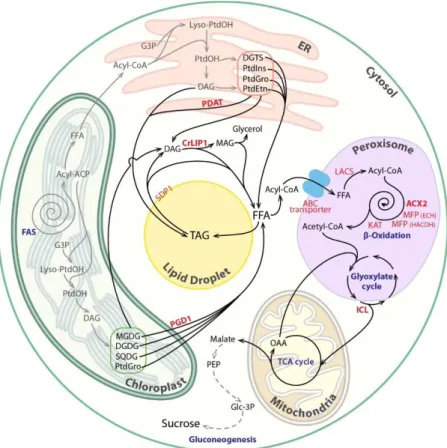
Figure 1 Pathways and processes involved in lipid catabolism in microalgae. Fatty acids (FAs) are made in the chloroplast; one part of them is being used to make plastidial membranes, and the other part is exported to the endoplasmic reticulum (ER) where they are assembled to glycerol to make the extraplastidial membranes. In response to developmental signals or adverse conditions, cells accumulate some of these acyl‐chains as triacylglycerols (TAGs) in lipid droplets. Lipid turnover starts from lipolysis of structural or storage lipids through action of various lipases. The released free FAs enter peroxisomes for complete degradation. FA β‐ oxidation operates as a spiral reaction consisting of four repeating enzymatic steps. Acetyl‐ Coenzyme A (CoA), the final product of FA degradation, is then used to make a four‐carbon compound by the glyoxylate cycle partly operating in the peroxisomes. Succinates produced are used to fuel the tricarboxylic acid cycle in the mitochondria and subsequently to produce sucrose for growth through the gluconeogenesis pathway in the cytoplasm. It is worth mentioning here that this pathway is drawn mostly based on what we know in the model alga Chlamydomonas, and in some other algal species, FA β‐oxidation is known to occur in mitochondria. Known enzymes are written in red and bold. Biosynthetic steps are shown as gray lines, whereas degradative steps are in black. ABC, ATP binding cassette; ACP, acyl‐ carrier protein; ACX, acyl‐CoA oxidase; DAG, diacylglycerol; DGDG, digalactosyldiacylglycerol; DGTS, diacylglyceryl N,N,N‐trimethylhomoserine; FAS, fatty acid synthase; FFA, free fatty acid; G3P, glycerol‐3‐phosphate; KAT, ketoacyl‐CoA thiolase; ICL,
isocitrate lyase; LACS, long‐chain acyl‐CoA synthetase; CrLIP1, Chlamydomonas lipase 1; MAG, monoacylglycerol; MFP, multifunctional protein; MGDG, monogalactosyldiacylglycerol; OAA, oxaloacetate; PDAT, phospholipid:diacylglycerol acyltransferase; PDG1, plastid galactoglycerolipid degradation 1; PEP, phosphoenoylpyruvate; PtdGro, phosphatidylglycerol; PtdEtn, phosphatidylethanolamine; PtdIns, phosphatidylinositol; PtdOH, phosphatidic acid; SDP1, SUGAR‐DEPENDENT1; SQDG, sulfoquinovosyldiacylglycerol; TCA, tricarboxylic acid.
In the laboratory, lipid degradation is mostly studied via manipulation of nitrogen (N) content in the culture medium: N depletion triggers accumulation of triacylglycerols (TAGs) in the lipid droplets (LDs), which is partly reliant on de novo FA synthesis and partly on recycling of membrane lipids (Siaut et al., 2011). By contrast, N resupply causes massive degradation of the TAGs and resynthesis of membrane lipids. N depletion followed by N resupply is thus a convenient way to study enzymes and reactions of membrane and storage lipid catabolism and anabolism.
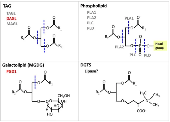
Lipolysis starts with the cleavage of FAs or the head groups from the glycerol backbone in glycerolipids. Depending on substrate preference, lipases can be divided into galactolipases, phospholipases, TAG lipases, diacylglycerol (DAG) lipases, and monoacylglycerol (MAG) lipases. All these lipases release free fatty acids and are thus carboxylic ester hydrolases. By contrast, phospholipases C and phospholipases D do not release acyl chains from glycerolipids but break the phosphodiester bond in the phosphorylated head group present at the sn‐3 position of glycerol, and thus phospholipase C and D belong to the phosphoric diester hydrolase category, which also includes nucleases. Major lipase classes and the sites of cleavage for specific lipases are summarized in Fig. 2.
Figure 2 Schematic drawing of representative acyl lipid structures indicating sites of cleavage by lipases. Sites of lipase action are drawn as dashed lines in blue. Classes of lipases with characterized members are shown in red, whereas uncharacterized ones are in gray. Phospholipases can again be divided into several subclasses: phospholipase A1 and A2 (PLA1 and PLA2) remove specifically the acyl chain at the positions sn‐1 and sn‐2, respectively. Lysophospholipases release the fatty acid of a lysophospholipid. Phospholipase C removes the phosphorylated head group from glycerol to release diacylglycerol (DAG) while phospholipase D cleaves off the head group from the phosphate to produce a phosphatidic acid. DGTS, diacylglyceryl N,N,N‐trimethylhomoserine; R, represents any acyl group; MGDG, monogalactosyldiacylglycerol; PGD1, Plastid Galactoglycerolipid Degradation 1; MAGL, monoacylglycerol lipase; DAGL, diacylglycerol lipase; TAGL, triacylglycerol lipase; PLC, phospholipase C; PLD, phospholipase D.
TAG breakdown
Owing to its economic importance, TAG lipases have been intensively studied, which resulted in the discovery of major TAG lipase in Arabidopsis thaliana (SDP1, the sugar‐dependent 1) and Saccharomyces cerevisae (TGL3P and TGL4P). These TAG lipases are located to LDs, the major site of neutral lipid storage (Athenstaedt & Daum, 2005; Eastmond, 2006). A TAG lipase (TGL1) has also been identified in the diatom Phaeodactylum tricornutum (Barka et al., 2016). TGL1 showed high sequence similarity to Arabidopsis SDP1. In vitro, the recombinant TGL1 possessed TAG lipase activity. Artificial microRNA knockdown lines had almost twice the
amount of TAGs compared with the wild‐type (WT), without compromising growth. A SDP1 homolog from the oleaginous alga Lobosphaera incisa (LiSDP1) was also studied (Siegler et al., 2017). LiSDP1 shares 44% identity to Arabidopsis SDP1, and contains a patatin domain harboring a putative catalytic dyad, which includes a serine located within a GXSXG motif. Interestingly the LiSDP1, when expressed heterologously in the tobacco pollen tube system, was not located to the LDs. Moreover, when expressed in the Arabidopsismutant sdp1, only partial restoration of the seedling growth phenotype of sdp1 was observed. Collectively, this study highlighted potential differences in the functions of SDP1 in an evolutionary context. Eight putative TAG lipases from Chlamydomonas showing reduction in transcription during N starvation compared with the N‐sufficient condition were tested using a function complementation approach (Li et al., 2012b). Among the eight candidate lipases (CrLIP1‐8), five were cloned as full‐length cDNAs and expressed in the yeast TAG lipase mutant (tgl3Δtgl4Δ), respectively. Only CrLIP1 was able to complement tgl3Δtgl4Δ. This result highlights the difficulty in assigning functions to putative lipases. Artificial microRNA silencing of CrLIP1 resulted in slower TAG degradation during N recovery. However, the in vitro lipase assay on recombinant CrLIP1 showed that the protein was not able to degrade TAG but rather DAG and polar lipids. Overall, CrLIP1 is proposed to be a DAG lipase with broad substrate specificity, therefore acting downstream of the TAG lipase in oil breakdown. In addition, CrLIP1 is highly expressed during optimal growth where TAG synthesis is low; therefore it might also play a key role in degrading DAGs resulting from removal of head groups in some membrane lipids.
Thaps3_264297, a homolog of the human Comparative Gene Identification‐58 (CGI‐58), was recently identified as involved in lipid catabolism in the diatom Thalassiosira pseudonana(Trentacoste et al., 2013). CGI‐58 belongs to the family 5 of α/β‐hydrolase fold proteins. It is a soluble enzyme that associates with LDs in certain conditions, and its absence results in overaccumulation of TAGs in plants and mammals (James et al., 2010). Trentacoste et al. (2013) reported that Thaps3_264297 exhibits TAG lipase, phospholipase, and acyltransferase activities in in vitro assays, but in a later study, CGI‐58 was concluded as only acting as a co‐activator of TAG lipase (McMahon et al., 2014). Targeted knockdown of this enzyme increased lipid yields by two‐ to fourfold during exponential growth and under silicon starvation in Thalassiosira. These suggested a role for Thaps3_264297 in lipid homeostasis. A putative ortholog of CGI‐58 is encoded in many other sequenced algal genomes, including the one of Chlamydomonas (Merchant et al., 2007), but whether and how it is involved in lipid metabolism in these algae remain to be elucidated.
Recently, the function of a highly abundant putative TAG lipase (LiLBP36) of the LDs isolated from the green alga Lobosphaera incisa was investigated (Siegler et al., 2017). An LD location was further confirmed via in vivo expression in tobacco pollen tube. LiLBP36 was identified as a class 3 lipase, and contains a putative catalytic triad of serine, aspartate and histidine that is found in many lipases, with the serine residue being part of a conserved GXSXG motif. The structure of LiLBP36 matches very closely a group of known fungal‐ and yeast‐secreted lipases that act on MAG, DAG and TAG (Siegler et al., 2017). Moreover, the transcription of LiLBP36 is up‐regulated by N starvation, and down‐regulated upon N resupply. However, only moderate functional restoration of the Arabidopsis sdp1 mutant phenotype was observed, and evidence for lipase activity of this protein remains to be determined.
Membrane lipid breakdown
Plastid Galactoglycerolipid Degradation 1 (PGD1) is the only lytic enzyme that has been demonstrated to be involved in membrane lipid turnover in Chlamydomonas (Li et al., 2012a). PGD1 catalyzes the hydrolysis of acyl chains at the sn‐1 position of the glycerol backbone of the plastidial galactolipid monogalacosyldiacylglycerol (MGDG). The null mutant contained 50% fewer TAGs with a reduced amount of oleic acid (18 : 1Δ9) during N starvation. FA fluxes from plastidial lipids to TAGs are also reduced in pgd1. Although the steady‐state concentrations of membrane lipids of pgd1 were not significantly different from those of WT, the pulse‐chase labeling assay and in vitro lipase assay with recombinant protein suggested that MGDG is a substrate of PGD1. In in vitro assay, PGD1 hydrolyzed only the newly synthesized MGDG containing 18 : 1Δ9. Taken together, these results indicate that PGD1 acts as a lipase removing low desaturated acyl chains from MGDG, which are then used for TAG synthesis under N deprivation. Such a route has not yet been reported for land plants, but as putative orthologs of PGD1 are present in plant and other algal genomes, the transfer of acyl‐chains from MGDG to TAG might be operating in plants and a wider group of algae.
No enzyme responsible for degradation of other major membrane lipids (i.e. diacylglycerylN,N,N‐trimethylhomoserine, sulfoquinovosyldiacylglycerol, phosphatidylglycerol, phosphatidylethanolamine, and digalactosyldiacylglycerol) has been identified in microalgae. Nevertheless, the phospholipid:diacylglycerol acyltransferase (PDAT) of Chlamydomonas has been shown, in addition to acyltransferase activity, to exhibit lipase activity toward a wide range of lipids (TAG, phospholipids, galactolipids and cholesteryl esters) (Yoon et al., 2012). PDAT contains an α/β‐hydrolase fold domain, where the conserved lipase motif (GXSXG) is present. PDAT is known to synthesize TAG by transferring acyl moiety from phospholipids to DAG in yeasts and higher plant cells (Dahlqvist et al., 2000). This
transacylation pathway mediated by PDAT has been also shown to contribute to TAG synthesis in Chlamydomonas, because the pdat1 mutant accumulates 25% less TAG compared with the WT (Boyle et al., 2012). It is therefore believed that PDAT is involved in both membrane lipid turnover and TAG synthesis in microalgae, similar to its role in higher plants (Zhang et al., 2009).
Fatty acid degradation
Fatty acid degradation is an oxidative process that results in production of C2 acetyl units that
are linked to coenzyme A (acetyl‐CoA). FA degradation is known to occur in peroxisomes of land plants and yeast, and in both peroxisomes and mitochondria in mammalian cells. In microalgae both mitochondrial and/or peroxisomal FA breakdown have been reported depending on algal lineage (Stabenau, 1984; Stabenau et al., 1984a,b, 1988; Winkler et al., 1988). Differences in the compartmentation of FA β‐oxidation enzymes have been suggested as a consequence of different phylogenetic development. Owing to the limited literature on microalgal β‐oxidation, we focus our discussion here only on green microalgae, where peroxisomal FA β‐oxidation has recently been demonstrated through study of aChlamydomonas mutant knocked‐out for an acyl‐CoA oxidase (Kong et al., 2017).
Microalgal peroxisomes
Peroxisomes (also called microbodies) are globular organelles (0.1–1 μm) surrounded by a single membrane and are present in all eukaryotic cells. Peroxisomes are defined as organelles that carry out oxidative reactions producing hydrogen peroxide (H2O2), which is subsequently
converted to water by catalase. Therefore, catalase is often considered a marker for peroxisomes. Peroxisomes have been reported to occur in various algal species, such as Mougeotia, Spirogyra, Dunaliella, Etemosphaera, Chlorogonium elongatum (Silverberg & Sawa, 1974; Silverberg, 1975; Stabenau et al., 1984b, 1993). In Chlamydomonas, the first trackable report on peroxisomes was published in 1974 (Stabenau, 1974). This paper studied the distribution of microbody enzymes on sucrose gradient. Not until 2009 has another paper focusing on peroxisomes from Chlamydomonas appeared (Shinozaki et al., 2009), where the authors reported the functional presence of a peroxisomal targeting sequence (PTS) in Chlamydomonas (Shinozaki et al., 2009). They further showed that the Chlamydomonas PTS can target GFP to peroxisomes in the green alga Closterium ehrenbergii. This study provided additional evidence that the protein import machinery between the two algal species is conserved. Peroxisomes of Chlamydomonas were visualized only as recently as 5 yr ago under both confocal microscopy and transmission electron
microscopy (Hayashi & Shinozaki, 2012). The same group further showed that the number and size of peroxisomes in a cell can be manipulated via changing carbon source; for example, the number of peroxisomes in Chlamydomonas can be boosted up to three times when cultured with acetate (Hayashi et al., 2015). This suggests dynamic turnover of the algal peroxisomes and their active participation to metabolism.
Acyl delivery to peroxisomes
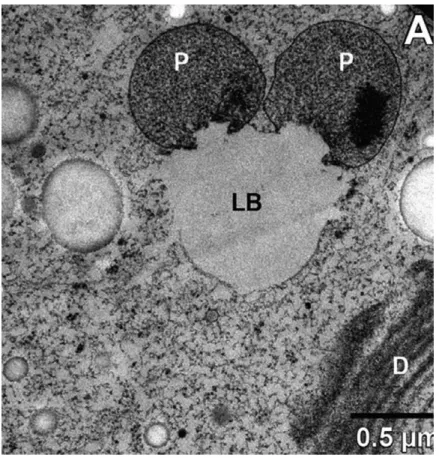
Degradation of lipids by lipases involves trafficking of FA released from all organelles to peroxisomes.Whether this occurs by transport processes used for lipid synthesis or by specific means is unknown. Studies in mammals and plants have suggested that TAG stored in LDs serves as an intermediate in the transport of otherwise toxic free FAs released from membrane lipid breakdown (Listenberger et al., 2003; Fan et al., 2014). Physical interactions between LDs and peroxisomes have been observed in plants and also in Chlamydomonasand other algal species (Hayashi et al., 2015; Schwarz et al., 2017) (Fig. 3). It remains to be determined how LDs move in the cell to approach peroxisomes, or vice versa.
Figure 3 Transmission electron micrograph (TEM) of a section through an algal cell showing the close proximity of lipid droplets and peroxisomes. The freshwater alga Micrasterias denticulata(Streptophyta) was kept in the dark for 9 wk before being fixed and imaged under TEM (Schwarz et al., 2017). LB, lipid body; P, peroxisome; D, dictyosome.
Once FAs get close to the peroxisome, studies in Arabidopsis show that they are transported across the single membrane by the ATP‐binding cassette transporter (ABCD1, also known as PXA1/CTS/PED3 (Zolman et al., 2001). ABCD1 has been shown to possess, in addition to the transporter activity, a thioesterase activity (De Marcos Lousa et al., 2013). This means that FA has to be activated to its CoA ester before being transported by ABCD1 across the peroxisomal membrane. After entering peroxisome, FAs are activated to their CoA esters via the action of two peroxisomal long‐chain acyl‐CoA synthetases (LACS6/7) (Fulda et al., 2004). A physical interaction between the ABCD1 and LACS6/7 has been demonstrated (De Marcos Lousa et al., 2013). Chlamydomonas encodes one putative ABCD1 and three LCS (=LACS) proteins (Li‐Beisson et al., 2015), among which two are associated with LDs in Chlamydomonas (Nguyen et al., 2011). Only the LD‐associated LCS2 has been shown to participate in TAG synthesis during LD formation (Li et al., 2016). The function of the putative peroxisomal isoform remains to be validated.
Reactions and enzymes of fatty acid β‐oxidation
Fatty acid β‐oxidation is the principal pathway implicated in degradation of FAs in the peroxisomes of land plants (Poirier et al., 2006; Graham, 2008). The core reactions of FA β‐ oxidation require a cyclic reaction of four enzymatic steps: oxidation, hydration, dehydrogenation and thiolytic cleavage of an acyl‐CoA, and are catalyzed by three enzymes: acyl‐CoA oxidase (ACX), multifunctional protein and 3‐ketoacyl‐CoA thiolase. The end product of these reactions is acetyl‐CoA (C2). Genes homologous to known Arabidopsis FA β‐
oxidation enzymes are encoded in the genome of Chlamydomonas (Merchant et al., 2007), and their IDs and number of homologs are described in Li‐Beisson et al. (2015). Except for ACX2, none of the other enzymes of FA β‐oxidation has been characterized.
We have recently isolated and characterized a knockout mutant for ACX2. The acx2 mutant is impaired in oil remobilization upon N resupply (Kong et al., 2017). Oil remobilization is severely (60–80%) but not completely blocked in acx2, suggesting functional redundancy with the other four ACXs (Li‐Beisson et al., 2015). Indeed, overlapping substrate specificities between different ACX isoenzymes have been reported for Arabidopsis where no phenotype can be observed with any of the single acx mutants (Eastmond et al., 2000). Furthermore, except for ACX2, all four other Chlamydomonas ACXs contain a putative PTS (either PTS1 or PTS2) sequence (Kong et al., 2017), suggesting potential functions in peroxisome metabolism. In in vitro assays, the recombinant Chlamydomonas ACX2 catalyzes the conversion of acyl‐ CoA to trans‐2‐enoyl‐CoA, requiring FAD as a co‐factor and producing H2O2. Thus, it
tagging to mCherry, ACX2 was localized to peroxisomes in Chlamydomonas. Therefore, this study demonstrated for the first time that Chlamydomonasuses a peroxisomal pathway for FA degradation, and that H2O2‐producing activities had already evolved in green microalgae.
In addition to acetyl‐CoAs, FA β‐oxidation produces H2O2 and NADH. In higher plants, the
highly oxidative H2O2 is usually decomposed to water by the peroxisome‐resident catalase,
whereas there is still debate about the subcellular localization of catalases in Chlamydomonas (Kato et al., 1997). Nevertheless, homologs of ascorbate peroxidases are encoded in the genome of Chlamydomonas (Merchant et al., 2007), and these enzymes could be used by cells to quench H2O2. Continued operation of FA β‐oxidation requires a steady
supply of oxidized NAD+. This can occur either through oxidation of NADH to NAD+ by a peroxisomal malate dehydrogenase (MDH; Pracharoenwattana et al., 2007) or through import of NAD+ from the cytosol to the peroxisome with the aid of a homolog of the NAD+carrier protein (PXN) (Bernhardt et al., 2012). Both proteins have been demonstrated to play a role in FA respiration in land plants, and their homologs occur in microalgae but experimental demonstration for their function is lacking.
In parallel to the core activities of the β‐oxidation spiral which are mainly responsible for oxidation of saturated straight chain fatty acids, microalgae are rich in unsaturated fatty acids, and which can make up to 70% of their membranes in Chlamydomonas (Nguyen et al., 2013). The β‐oxidation of unsaturated fatty acids are proposed to go through two alternative pathways in land plants (Li‐Beisson et al., 2013). The best characterized protein of these auxiliary pathways is the enoyl‐CoA isomerase (ECI) (Goepfert et al., 2008), and a protein encoded by the locus Cre03.g190850 showing weak homology to the plant ECI can be identified in Chlamydomonas but its function remains to be validated.
Metabolic fate of acetyl‐CoA produced from FA β‐oxidation
Acetyl units (C2), generated by the β‐oxidation of FAs, can usually be utilized by cells after
being synthesized to 4‐carbon compounds (i.e. succinate) through the glyoxylate cycle. Subsequently, succinate enters the tricarboxylic acid cycle in mitochondria, releasing malate which can be converted to hexose and sucrose in the cytosol via gluconeogenesis. Coordinate regulation of transcription and enzyme activities of the three pathways (i.e. FA β‐oxidation, glyoxylate cycle, and gluconeogenesis) at the onset of oilseed germination have been demonstrated, suggesting global regulatory mechanisms (Rylott et al., 2001). However, the identity of the signal(s) or regulatory proteins that induces all these genes is not known. Chlamydomonas encodes all known enzymes of the glyoxylate cycle, that is, citrate
synthase (CYS), isocitrate lyase (ICL), malate synthase (MS), aconitase (AOS) and MDH (Merchant et al., 2007), which have shown up‐regulation in the dark, similar to core enzymes of FA β‐oxidation (Zones et al., 2015). Either N‐terminal or C‐terminal fusion to fluorescence proteins revealed that all of these (CYS, MS, AOS, MDH), except for ICL which is cytosolic, are localized in the peroxisomes (Lauersen et al., 2016). The localization of the glyoxylate enzymes in Chlamydomonas differs from that of plants, where ICL is peroxisomal. The icl mutant cannot grow in the dark and grows at a slower rate when cultivated mixotrophically in the light (Plancke et al., 2014), demonstrating that the glyoxylate cycle is important for acetate metabolism in Chlamydomonas. Moreover, the icl mutant overaccumulated both total FAs and neutral lipids when cultivated mixotrophically. Based on quantitative proteomic data, the authors consider that the observed changes in lipid could be the result of a slowdown in the β‐oxidation of FAs and is less likely to be a result of increased de novo FA synthesis. The availability of the icl mutant provides an exciting opportunity to address the question of whether, in addition to the glyoxylate cycle, there are alternative routes to transport acetyl units from the peroxisome to mitochondria in microalgae.
Lipid degradation in relation to other cellular processes
Cellular lipid content and composition vary according to developmental stage, phase of cell cycle, and in response to nutrient availability, as well as changes in environmental conditions (e.g. light, temperature). This suggests tight and complex control of lipid synthesis and degradation. We review here the processes shown to be implicated in lipid catabolism in microalgae.
Vesicle trafficking
Brefeldin A (BFA), is a known inhibitor of ADP‐ribosylation factor 1 that promotes the formation of COPI‐coated vesicles (important for peroxisome vesiculation) and is thought to inhibit the delivery of TAG lipase to LDs in mammalian systems (Farese & Walther, 2009). Studies in Arabidopsis seeds have recently shown that mutants defective in the retromer function are impaired in the delivery of SDP1 lipase from peroxisome to LDs through peroxisome extensions, thereby perturbing FA degradation and seed germination (Thazar‐ Poulot et al., 2015). Two independent studies have observed an increase in cellular TAG content in Chlamydomonas upon addition of BFA (Kato et al., 2013; Kim et al., 2013). Kato et al. (2013) showed that the increased TAG accumulation is a result of inhibition of lipid degradation. Progression of FA β‐oxidation in pre‐mature peroxisomes was shown to be delayed. This was further supported by FA feeding assays, as, in the presence of BFA,
fluorescent FAs are not degraded. Indeed, proteins implicated in vesicle trafficking are present in the LD proteome of Chlamydomonas (Moellering & Benning, 2010; Nguyen et al., 2011). Taken together, these studies highlight the participation of vesicle trafficking in lipid catabolism in microalgae.
Cell cycle
Lipids of Chlamydomonas are observed to show specific alterations in relation to the phase in the cell cycle; and three unique patterns can be seen (Juppner et al., 2017). TAGs have shown a bimodal accumulation, with degradation occurring in the middle of the day, as well as at the beginning of the night. This suggests fine‐tuning of lipolysis to cell cycle progression. Indeed, a direct link between cell cycle and TAG degradation has been demonstrated in S. cerevisiae, where the major TAG lipase (TGL4) is phosphorylated and activated by cyclin‐dependent kinase 1 (Cdk1/Cdc28) (Kurat et al., 2009). Whether such a system occurs in microalgae remains to be tested. Upon N starvation, algal cells enter a quiescent state and accumulate TAGs. Upon N resupply, cells degrade TAGs and exit quiescence thereby restarting growth. Tsai et al. (2014) showed that exiting quiescence is linked to TAG remobilization, because the cht7 (compromised in hydrolysis of TAG) mutant, which is defective in a gene required for exiting quiescence, is severely impaired in TAG degradation. CHT7 encodes a CXC‐domain containing DNA‐binding protein and is proposed as a transcription factor regulating quiescence and proliferation. This study provides evidence that TAG remobilization is tuned to cell division and growth in algae.
Autophagy‐mediated lipid turnover
In addition to lipase‐mediated lipid turnover, autophagy was also reported to be involved in lipid degradation in mammals, plant kingdoms and fungi (Singh et al., 2009; Kurusu et al., 2014; van Zutphen et al., 2014). In yeast cells, LDs were observed to be taken up by vacuoles/lysosomes and subsequently degraded (van Zutphen et al., 2014). Inhibition of autophagy in a mouse model increased TAG content, thereby providing evidence that autophagy plays a role in lipid turnover (Singh et al., 2009). Formation of autophagosomes has been observed in N‐starved Chlamydomonas cells, and could be averted upon acetate boost, thereby explaining the additional obesity in acetate‐boosted cells (Goodenough et al., 2014). This provides the first evidence that averting autophagy can increase oil content in microalgae. It is also reported that in the green alga Auxenochlorella protothecoides, LDs were observed to be directly sequestered by the vacuole during the transition from heterotrophy to autotrophy when a large amount of TAGs were degraded (Zhao et al., 2014). The authors suggest that LD
autophagy observed in A. protothecoides resembles more a microautophagy‐like mechanism rather than a classical autophagy, as during this process no autophagosome is observed. By contrast, LD degradation during carbon starvation in the green alga Micrasterias denticulata (Streptophyta) involves a classical autophagy pathway (Schwarz et al., 2017). Finally, inhibition of autophagy in Chlorella zofingiensis by the autophagy inhibitor 3‐ methyladenine during N starvation resulted in cells accumulating 15–20% less TAG than control cells (Zhang et al., 2018). The authors considered that the reduced TAG accumulation during N starvation upon inhibition of autophagy could be the result of inhibition of lipolysis of membrane lipids, which contributes to TAG amount in addition to de novo synthesis. Detailed studies of the type of autophagy (lipophagy, pexophagy, nonselective autophagy…) associated with each condition should help to clarify the complex interrelationship between autophagy and lipid metabolism.
Physiological functions and biotechnological applications of lipid catabolism
Microalgae are exposed to constant changes in their habitat, light, temperature, and nutrient availability; therefore the need to remodel their membranes to adapt to these changes is crucial for survival. This explains the high expression of many genes encoding lipolytic enzymes and enzymes of fatty acid β‐oxidation under stress (Miller et al., 2010). In rapidly growing cells, lipid degradation plays also a ‘housekeeping’ role in removing harmful free FAs which can originate from acylated‐protein and lipid degradation. During the N recovery, lipid catabolism is important to degrade TAG storage to supply cells with carbon precursors for synthesis of structural membranes. Moreover, FA β‐oxidation is found to be important in regulating the amount of FAs during day/night cycles, and is essential for FA utilization upon carbon starvation (Kong et al., 2017). In addition, lipid turnover has also been found to contribute to resource allocation during aging or senescence in land plants (Troncoso‐Ponce et al., 2013). Whether such a function holds true in algal cells remains to be determined.
In addition to their importance in cell metabolism and physiology, lipid catabolisms are also very useful targets for biotechnological applications: discovery of new lipases can provide the industry with novel enzymes; shutting down lipid catabolism has been shown to increase cellular oil content (Trentacoste et al., 2013; Tsai et al., 2014; Kong et al., 2017); and mutants defective in FA β‐oxidation, which prevents futile cycling, can serve as background strains for genetic engineering strategies aiming to produce exotic or unusual FAs which are often otherwise degraded.
Remaining questions
Although considerable progress has been made towards our understanding of lipid catabolism in microalgae, many unknowns remain. For example, the identity of a true TAG lipase remains to be identified in Chlamydomonas. The function of the major lipid droplet protein is still not clear. Does it possess enzymatic activities, similar to that of oleosin (Parthibane et al., 2012)? As lipases are water‐soluble, while their substrates are mostly insoluble in water, lipolysis is an interfacial process. What are the processes or enzymes involved in opening up the protein coat of LDs, therefore allowing the access to lipase to the TAG core? Are lipolysis and FA β‐ oxidation regulated by a common signal or transcription factor? In addition to acetyl‐CoAs, FA β‐oxidation produces H2O2 and NADH. How these products are metabolized or transported into
or out of the algal peroxisomes remains unknown. Furthermore, in addition to FA catabolism, do algal peroxisomes also play roles in the metabolism of substrates other than lipids, as is the case in higher plants? Answering these and many other questions should help to expand our basic knowledge on lipid catabolism and aid in the design of engineering strategies in the creation of high TAG strains for biofuel, nutrition and green chemistry applications.
Acknowledgements
The research in the authors’ laboratory is funded by French ANR funding. I.T.R. acknowledges the CEA for an international PhD studentship, and J.W. thanks the Junior Research Fellowship Program from the French Embassy in Bangkok (Thailand) for funding a 6‐month scholarship.
References
Athenstaedt K, Daum G. 2005. Tgl4p and Tgl5p, two triacylglycerol lipases of the yeast Saccharomyces cerevisiae are localized to lipid particles. Journal of Biological Chemistry 280: 37301–37309.
Barka F, Angstenberger M, Ahrendt T, Lorenzen W, Bode HB, Buchel C. 2016. Identification of a triacylglycerol lipase in the diatom Phaeodactylum tricornutum. Biochimica et Biophysica Acta 1861: 239–248.
Bernhardt K, Wilkinson S, Weber AP, Linka N. 2012. A peroxisomal carrier delivers NAD+ and contributes to optimal fatty acid degradation during storage oil mobilization. Plant Journal 69: 1–13.
Boyle NR, Page MD, Liu B, Blaby IK, Casero D, Kropat J, Cokus SJ, Hong‐Hermesdorf A, Shaw J, Karpowicz SJ et al. 2012. Three acyltransferases and nitrogen‐responsive regulator are implicated in nitrogen starvation‐induced triacylglycerol accumulation in Chlamydomonas. Journal of Biological Chemistry 287: 15811–15825.
Dahlqvist A, Stahl U, Lenman M, Banas A, Lee M, Sandager L, Ronne H, Stymne H. 2000. Phospholipid: diacylglycerol acyltransferase: an enzyme that catalyzes the acyl‐CoA‐ independent formation of triacylglycerol in yeast and plants. Proceedings of the National Academy of Sciences, USA 97: 6487–6492.
De Marcos Lousa C, van Roermund CWT, Postis VLG, Dietrich D, Kerr ID, Wanders RJA, Baldwin SA, Baker A, Theodoulou FL. 2013. Intrinsic acyl‐CoA thioesterase activity of a peroxisomal ATP binding cassette transporter is required for transport and metabolism of fatty acids. Proceedings of the National Academy of Sciences, USA 110: 1279–1284.
Du ZY, Benning C. 2016. Triacylglycerol accumulation in photosynthetic cells in plants and algae. SubCellular Biochemistry 86: 179–205.
Eastmond PJ. 2006. SUGAR‐DEPENDENT1 encodes a patatin domain triacylglycerol lipase that initiates storage oil breakdown in germinating Arabidopsis seeds. Plant Cell 18: 665–675. Eastmond PJ, Hooks M, Graham IA. 2000. The Arabidopsis acyl‐CoA oxidase gene family. Biochemical Society Transactions 28: 755–757.
Fan J, Yan C, Roston R, Shanklin J, Xu C. 2014. Arabidopsis Lipins, PDAT1 Acyltransferase, and SDP1 triacylglycerol lipase synergistically direct fatty acids toward β‐oxidation, thereby maintaining membrane lipid homeostasis. Plant Cell 26: 4119–4134.
Farese RV Jr, Walther TC. 2009. Lipid droplets finally get a little R‐E‐S‐P‐E‐C‐T. Cell 139: 855–860.
Fulda M, Schnurr J, Abbadi A, Heinz E, Browse J. 2004. Peroxisomal acyl‐CoA synthetase activity is essential for seedling development in Arabidopsis thaliana. Plant Cell 16: 394–405. Goepfert S, Vidoudez C, Tellgren‐Roth C, Delessert S, Hiltunen JK, Poirier Y. 2008. Peroxisomal Δ3, Δ2‐enoyl CoA isomerases and evolution of cytosolic paralogues in embryophytes. Plant Journal 56: 728–742.
Goodenough U, Blaby I, Casero D, Gallaher SD, Goodson C, Johnson S, Lee J‐H, Merchant SS, Pellegrini M, Roth R et al. 2014. The path to triacylglyceride obesity in the sta6 strain of Chlamydomonas reinhardtii. Eukaryotic Cell 13: 591–613.
Graham IA. 2008. Seed storage oil mobilization. Annual Review of Plant Biology 59: 115–142. Harwood JL, Guschina IA. 2009. The versatility of algae and their lipid metabolism. Biochimie 91: 679–684.
Hayashi Y, Sato N, Shinozaki A, Watanabe M. 2015. Increase in peroxisome number and the gene expression of putative glyoxysomal enzymes in Chlamydomonas cells supplemented with acetate. Journal of Plant Research 128: 177–185.
Hayashi Y, Shinozaki A. 2012. Visualization of microbodies in Chlamydomonas reinhardtii. Journal of Plant Research 125: 579–586.
James CN, Horn PJ, Case CR, Gidda SK, Zhang DY, Mullen RT, Dyer JM, Anderson RGW, Chapman KD. 2010. Disruption of the Arabidopsis CGI‐58 homologue produces Chanarin– Dorfman‐like lipid droplet accumulation in plants. Proceedings of the National Academy of Sciences, USA 107: 17833–17838.
Juppner J, Mubeen U, Leisse A, Caldana C, Brust H, Steup M, Herrmann M, Steinhauser D, Giavalisco P. 2017. Dynamics of lipids and metabolites during the cell cycle of Chlamydomonas reinhardtii. Plant Journal 92: 331–343.
Kato N, Dong T, Bailey M, Lum T, Ingram D. 2013. Triacylglycerol mobilization is suppressed by brefeldin A in Chlamydomonas reinhardtii. Plant and Cell Physiology 54: 1585–1599. Kato J, Yamahara T, Tanaka K, Takio S, Satoh T. 1997. Characterization of catalase from green algae Chlamydomonas reinhardtii. Journal of Plant Physiology 151: 262–268.
Keeling PJ. 2010. The endosymbiotic origin, diversification and fate of plastids. Philosophical Transactions of the Royal Society B: Biological Sciences 365: 729–748.
Kim S, Kim H, Ko D, Yamaoka Y, Otsuru M, Kawai‐Yamada M, Ishikawa T, Oh H‐M, Nishida I, Li‐Beisson Y et al. 2013. Rapid induction of lipid droplets in Chlamydomonas reinhardtii and Chlorella vulgaris by brefeldin A. PLoS ONE 8: e81978.
Kong F, Liang Y, Legeret B, Beyly‐Adriano A, Blangy S, Haslam RP, Napier JA, Beisson F, Peltier G, Li‐Beisson Y. 2017. Chlamydomonas carries out fatty acid beta‐oxidation in ancestral peroxisomes using a bona fide acyl‐CoA oxidase. Plant Journal 90: 358–371.
Kurat CF, Wolinski H, Petschnigg J, Kaluarachchi S, Andrews B, Natter K, Kohlwein SD. 2009. Cdk1/Cdc28‐dependent activation of the major triacylglycerol lipase Tgl4 in yeast links lipolysis to cell‐cycle progression. Molecular Cell 33: 53–63.
Kurusu T, Koyano T, Hanamata S, Kubo T, Noguchi Y, Yagi C, Nagata N, Yamamoto T, Ohnishi T, Okazaki Y et al. 2014. OsATG7 is required for autophagy‐dependent lipid metabolism in rice postmeiotic anther development. Autophagy 10: 878–888.
Lauersen KJ, Willamme R, Coosemans N, Joris M, Kruse O, Remacle C. 2016. Peroxisomal microbodies are at the crossroads of acetate assimilation in the green microalga Chlamydomonas reinhardtii. Algal Research 16: 266–274.
Li XB, Benning C, Kuo MH. 2012b. Rapid triacylglycerol turnover in Chlamydomonas reinhardtii requires a lipase with broad substrate specificity. Eukaryotic Cell 11: 1451–1462. Li X, Moellering ER, Liu B, Johnny C, Fedewa M, Sears BB, Kuo M‐H, Benning C. 2012a. A galactoglycerolipid lipase is required for triacylglycerol accumulation and survival following nitrogen deprivation in Chlamydomonas reinhardtii. Plant Cell 24: 4670–4686.
Li X, Zhang R, Patena W, Gang SS, Blum SR, Ivanova N, Yue R, Robertson JM, Lefebvre PA, Fitz‐Gibbon ST et al. 2016. An indexed, mapped mutant library enables reverse genetics studies of biological processes in Chlamydomonas reinhardtii. Plant Cell 28: 367–387.
Li‐Beisson Y, Beisson F, Riekhof W. 2015. Metabolism of acyl‐lipids in Chlamydomonas reinhardtii. Plant Journal 82: 504–522.
Li‐Beisson Y, Shorrosh B, Beisson F, Andersson MX, Arondel V, Bates PD, Baud S, Bird D, DeBono A, Durrett TP et al. 2013. Acyl‐lipid metabolism. Arabidopsis Book 8: 1–65.
Listenberger LL, Han XL, Lewis SE, Cases S, Farese RV, Ory DS, Schaffer JE. 2003. Triglyceride accumulation protects against fatty acid‐induced lipotoxicity. Proceedings of the National Academy of Sciences, USA 100: 3077–3082.
McMahon D, Dinh A, Kurz D, Shah D, Han G‐S, Carman GM, Brasaemle DL. 2014. Comparative gene identification 58/α/β hydrolase domain 5 lacks lysophosphatidic acid acyltransferase activity. Journal of Lipid Research 55: 1750–1761.
Merchant SS, Prochnik SE, Vallon O, Harris EH, Karpowicz SJ, Witman GB, Terry A, Salamov A, Fritz‐Laylin LK, Marechal‐Drouard L et al. 2007. The Chlamydomonas genome reveals the evolution of key animal and plant functions. Science 318: 245–250.
Miller R, Wu GX, Deshpande RR, Vieler A, Gartner K, Li XB, Moellering ER, Zauner S, Cornish AJ, Liu BS et al. 2010. Changes in transcript abundance in Chlamydomonas reinhardtii following nitrogen deprivation predict diversion of metabolism. Plant Physiology 154: 1737– 1752.
Moellering ER, Benning C. 2010. RNA interference silencing of a major lipid droplet protein affects lipid droplet size in Chlamydomonas reinhardtii. Eukaryotic Cell 9: 97–106.
Nguyen HM, Baudet M, Cuiné S, Adriano J‐M, Barthe D, Billon E, Bruley C, Beisson F, Peltier G, Ferro M et al. 2011. Proteomic profiling of oil bodies isolated from the unicellular green microalga Chlamydomonas reinhardtii: with focus on proteins involved in lipid metabolism. Proteomics 11: 4266–4273.
Nguyen HM, Cuiné S, Beyly‐Adriano A, Légeret B, Billon E, Auroy P, Beisson F, Peltier G, Li‐Beisson Y. 2013. The green microalga Chlamydomonas reinhardtii has a single ω‐3 fatty acid desaturase that localizes to the chloroplast and impacts both plastidic and extraplastidic membrane lipids. Plant Physiology 163: 914–928.
Parthibane V, Rajakumari S, Venkateshwari V, Iyappan R, Rajasekharan R. 2012. Oleosin is bifunctional enzyme that has both monoacylglycerol acyltransferase and phospholipase activities. Journal of Biological Chemistry 287: 1946–1954.
Plancke C, Vigeolas H, Hohner R, Roberty S, Emonds‐Alt B, Larosa V, Willamme R, Duby F, Onga Dhali D, Thonart P et al. 2014. Lack of isocitrate lyase in Chlamydomonas leads to changes in carbon metabolism and in the response to oxidative stress under mixotrophic growth. Plant Journal 77: 404–417.
Poirier Y, Antonenkov VD, Glumoff T, Hiltunen JK. 2006. Peroxisomal beta‐oxidation – a metabolic pathway with multiple functions. Biochimica Et Biophysica Acta–Molecular Cell Research 1763: 1413–1426.
Pracharoenwattana I, Cornah JE, Smith SM. 2007. Arabidopsis peroxisomal malate dehydrogenase functions in β‐oxidation but not in the glyoxylate cycle. Plant Journal 50: 381– 390.
Rylott EL, Hooks MA, Graham IA. 2001. Co‐ordinate regulation of genes involved in storage lipid mobilization in Arabidopsis thaliana. Biochemical Society Transactions 29: 283–287. Schwarz V, Andosch A, Geretschlager A, Affenzeller M, Lutz‐Meindl U. 2017. Carbon starvation induces lipid degradation via autophagy in the model alga Micrasterias. Journal of Plant Physiology 208: 115–127.
Shinozaki A, Sato N, Hayashi Y. 2009. Peroxisomal targeting signals in green algae. Protoplasma 235: 57–66.
Siaut M, Cuiné S, Cagnon C, Fessler B, Nguyen M, Carrier P. 2011. Oil accumulation in the model green alga Chlamydomonas reinhardtii: characterization, variability between common laboratory strains and relationship with starch reserves. BMC Biotechnology 11: 7–22.
Siegler H, Valerius O, Ischebeck T, Popko J, Tourasse NJ, Vallon O, Khozin‐Goldberg I, Braus GH, Feussner I. 2017. Analysis of the lipid body proteome of the oleaginous alga Lobosphaera incisa. BMC Plant Biology 17: 98–105.
Silverberg BA. 1975. 3,3‐diaminobenzidine (DAB) ultrastructural cytochemistry of
Silverberg BA, Sawa T. 1974. Cytochemical localization of oxidase activities with diaminobenizidine in the green alga Chlamydomonas dysosmos. Protoplasma 81: 177–188. Singh R, Kaushik S, Wang YJ, Xiang YQ, Novak I, Komatsu M, Tanaka K, Cuervo AM, Czaja MJ. 2009. Autophagy regulates lipid metabolism. Nature 458: U1131–U1164.
Stabenau H. 1974. Verteilung von microbody‐enzymen aus Chlamydomonas in dichtegradienten. Planta 118: 35–42.
Stabenau H 1984. Microbodies in different algae. In: Wiessner W, Robinson DG, Starr RC eds. Compartments in algal cells and their interaction. Berlin/Heidelberg, Germany: Springer Berlin Heidelberg, 183–190.
Stabenau H, Winkler U, Saftel W. 1984a. β‐oxidation in algal peroxisomes of the leaf and unspecialized type. Plant Physiology 75: 79.
Stabenau H, Winkler U, Saftel W. 1984b. Enzymes of β‐oxidation in different types of algal microbodies. Plant Physiology 75: 531–533.
Stabenau H, Winkler U, Saftel W. 1988. Compartmentation of enzymes of the β‐oxidation pathway in different types of algae. Biological Chemistry Hoppe‐Seyler 369: 19.
Stabenau H, Winkler U, Säftel W. 1993. Localization of glycolate dehydrogenase in two species of Dunaliella. Planta 191: 362–364.
Thazar‐Poulot N, Miquel M, Fobis‐Loisy I, Gaude T. 2015. Peroxisome extensions deliver the Arabidopsis SDP1 lipase to oil bodies. Proceedings of the National Academy of Sciences, USA 112: 4158–4163.
Trentacoste EM, Shrestha RP, Smith SR, Glé C, Hartmann AC, Hildebrand M, Gerwick WH. 2013. Metabolic engineering of lipid catabolism increases microalgal lipid accumulation without compromising growth. Proceedings of the National Academy of Sciences, USA 110: 19748–19753.
Troncoso‐Ponce MA, Cao X, Yang Z, Ohlrogge JB. 2013. Lipid turnover during senescence. Plant Science 205–206: 13–19.
Tsai C‐H, Warakanont J, Takeuchi T, Sears BB, Moellering ER, Benning C. 2014. The protein Compromised Hydrolysis of Triacylglycerols 7 (CHT7) acts as a repressor of cellular quiescence in Chlamydomonas. Proceedings of the National Academy of Sciences, USA 111: 15833–15838.
Winkler U, Saftel W, Stabenau H. 1988. Beta‐oxidation of fatty acids in algae – localization of thiolase and acyl‐CoA oxidizing enzymes in 3 different organisms. Planta 175: 91–98.
Yoon K, Han D, Li Y, Sommerfeld M, Hu Q. 2012. Phospholipid: diacylglycerol acyltransferase is a multifunctional enzyme involved in membrane lipid turnover and degradation while synthesizing triacylglycerol in the unicellular green microalga Chlamydomonas reinhardtii. Plant Cell 24: 3708–3724.
Zhang M, Fan J, Taylor DC, Ohlrogge JB. 2009. DGAT1 and PDAT1 acyltransferases have overlapping functions in Arabidopsis triacylglycerol biosynthesis and are essential for normal pollen and seed development. Plant Cell 21: 3885–3901.
Zhang Z, Sun D, Cheng K‐W, Chen F. 2018. Inhibition of autophagy modulates astaxanthin and total fatty acid biosynthesis in Chlorella zofingiensis under nitrogen starvation. Bioresource Technology 247(Suppl C): 610–615.
Zhao L, Dai J, Wu Q. 2014. Autophagy‐like processes are involved in lipid droplet degradation in Auxenochlorella protothecoides during the heterotrophy‐autotrophy transition. Frontiers in Plant Science 5: 400.
Zolman BK, Silva ID, Bartel B. 2001. The Arabidopsis pxa1 mutant is defective in an ATP‐ binding cassette transporter‐like protein required for peroxisomal fatty acid β‐oxidation. Plant Physiology 127: 1266–1278.
Zones JM, Blaby IK, Merchant SS, Umen JG. 2015. High‐resolution profiling of a synchronized diurnal transcriptome from Chlamydomonas reinhardtii reveals continuous cell and metabolic differentiation. Plant Cell 27: 2743–2769.
van Zutphen T, Todde V, de Boer R, Kreim M, Hofbauer HF, Wolinski H, Veenhuis M, van der Klei IJ, Kohlwein SD. 2014. Lipid droplet autophagy in the yeast Saccharomyces cerevisiae. Molecular Biology of the Cell 25: 290–301.