Chemotaxis during Adherent-Invasive Escherichia coli Infection
Nicolas Dreux,a,bMaria del Mar Cendra,cSébastien Massier,a,bArlette Darfeuille-Michaud,a,b,d†Nicolas Barnich,a,b,dEduard Torrentsc
Clermont Université, “M2iSH Microbe intestin inflammation et Susceptibilité de l’Hôte,” UMR 1071 Inserm/Université d’Auvergne,a
and Unité Sous Contrat Institut National de la Recherche Agronomique 2018,b
Clermont-Ferrand, France; Institute for Bioengineering of Catalonia (IBEC), Bacterial infections and antimicrobial therapies group, Barcelona, Spainc
; Institut Universitaire de Technologie, Génie Biologique, Aubière, Franced
A critical step in the life cycle of all organisms is the duplication of the genetic material during cell division. Ribonucleotide
re-ductases (RNRs) are essential enzymes for this step because they control the de novo production of the deoxyribonucleotides
required for DNA synthesis and repair. Enterobacteriaceae have three functional classes of RNRs (Ia, Ib, and III), which are
tran-scribed from separate operons and encoded by the genes nrdAB, nrdHIEF, and nrdDG, respectively. Here, we investigated the
role of RNRs in the virulence of adherent-invasive Escherichia coli (AIEC) isolated from Crohn’s disease (CD) patients.
Interest-ingly, the LF82 strain of AIEC harbors four different RNRs (two class Ia, one class Ib, and one class III). Although the E. coli RNR
enzymes have been extensively characterized both biochemically and enzymatically, little is known about their roles during
bac-terial infection. We found that RNR expression was modified in AIEC LF82 bacteria during cell infection, suggesting that RNRs
play an important role in AIEC virulence. Knockout of the nrdR and nrdD genes, which encode a transcriptional regulator of
RNRs and class III anaerobic RNR, respectively, decreased AIEC LF82’s ability to colonize the gut mucosa of transgenic mice that
express human CEACAM6 (carcinoembryonic antigen-related cell adhesion molecule 6). Microarray experiments demonstrated
that NrdR plays an indirect role in AIEC virulence by interfering with bacterial motility and chemotaxis. Thus, the development
of drugs targeting RNR classes, in particular NrdR and NrdD, could be a promising new strategy to control gut colonization by
AIEC bacteria in CD patients.
R
ibonucleotide reductase (RNR) is an essential enzyme in all
living organisms. It catalyzes the reduction of
ribonucle-otides (nucleoside triphosphates [NTPs]) to their
correspond-ing 2=-deoxyribonucleotides (deoxynucleoside triphosphates
[dNTPs]) and therefore plays an essential role in DNA
synthe-sis and repair. Three RNR classes (classes I, II, and III) exist;
these classes exhibit different primary structures, subunit
co-factor requirements, and quaternary three-dimensional (3D)
structures, but they all are allosterically regulated and share
similar catalytic mechanisms (
1
,
2
). Class I RNRs are
oxygen-dependent enzymes that occur in eubacteria, eukaryotes, and
some viruses. This class comprises two main subgroups (Ia and
Ib). Class Ia RNRs are encoded by an operon containing nrdA
and nrdB genes. These genes encode the NrdA subunit, which is
catalytically and allosterically regulated, and the NrdB subunit,
which possesses radical-generating activity. Class Ib RNRs are
encoded by an operon containing the nrdH, nrdI, nrdE, and
nrdF genes, which encode the corresponding specific redoxin
NrdH, the activating subunit NrdI, the catalytic subunit NrdE,
and the radical-generating subunit NrdF. Class III RNRs are
present in facultative anaerobic and strict anaerobic
microor-ganisms and use S-adenosylmethionine and iron-sulfur
clus-ters in the NrdG accessory protein to create a stable glycyl
radical. nrdD and nrdG form an operon and encode the
cata-lytic NrdD subunit and the activating protein NrdG,
respec-tively. This system works only under strict anaerobic
condi-tions because oxygen is toxic to these enzymes (
1
,
2
). Notably,
the distribution of the different RNR classes is difficult to
elu-cidate, as the different bacterial phylogenetic groups do not
display any common RNR combinations. One possible RNR
distribution appears to correlate with the bacterial growth
con-ditions. We identified bacteria that encode one RNR class,
whereas others encode more than one RNR class, and many
RNR combinations exist in nature (
3
,
4
).
Escherichia coli and all Enterobacteriaceae encode three RNR
classes: classes Ia, Ib, and III (
5
,
6
). In E. coli, class Ia RNRs are
active during aerobic growth, class III RNRs are active under
an-aerobic growth conditions, and class Ib RNRs are active under
some special circumstances, such as growth in an iron-deficient
medium or biofilm formation (
6
,
7
). However, the mechanisms
that govern different RNR activities in pathogenic E. coli are not
fully understood. An additional protein, termed NrdR, was
de-Received 21 October 2014 Returned for modification 5 November 2014 Accepted 9 January 2015
Accepted manuscript posted online 20 January 2015
Citation Dreux N, Cendra MDM, Massier S, Darfeuille-Michaud A, Barnich N, Torrents E. 2015. Ribonucleotide reductase NrdR as a novel regulator for motility and chemotaxis during adherent-invasive Escherichia coli infection. Infect Immun 83:1305–1317.doi:10.1128/IAI.02772-14.
Editor: B. A. McCormick
Address correspondence to Nicolas Barnich, nicolas.barnich@udamail.fr, or Eduard Torrents, etorrents@ibecbarcelona.eu.
† Deceased.
N.B. and E.T. contributed equally to this article as senior authors. N.D. and M.D.M.C. contributed equally to this article.
Supplemental material for this article may be found athttp://dx.doi.org/10.1128 /IAI.02772-14.
Copyright © 2015, American Society for Microbiology. All Rights Reserved.
scribed as a novel transcriptional regulator capable of modulating
the expression of all RNR present in one organism (
8
). NrdR
pro-teins are composed of 140 to 200 amino acids, with two
differen-tiated domains: a zinc ribbon DNA-binding domain and an
ATP-cone domain with the capacity to bind nucleotides. NrdR changes
its conformation and binds to its cognate DNA recognition
se-quences to repress RNR gene expression depending on which
nu-cleotide it is bound to (
9
).
A role for E. coli has been demonstrated in Crohn’s disease
(CD), an inflammatory bowel disease that occurs in individuals
with a genetic predisposition to environmental or infectious
trig-gers that cause an abnormal immune response (
10–13
). Most E.
coli strains isolated from the ileal mucosa of CD patients are able to
adhere to and invade intestinal epithelial cells (
14–16
) and belong
to the pathogenic group of adherent invasive E. coli (AIEC) (
17
).
AIEC is highly associated with the ileal mucosa in CD patients
(
14–16
,
18–23
). CD-associated AIEC cells adhere to the brush
border of primary ileal enterocytes isolated from CD patients but
not to enterocytes isolated from controls without inflammatory
bowel disease. This adhesion is involved in the recognition
be-tween the variant FimH adhesin motifs located on the top of type
1 pili expressed on the AIEC bacterial surface and the
carcinoem-bryonic antigen-related cell-adhesion molecule 6 (CEACAM6)
abnormally expressed in the ileal epithelial cells of CD patients
(
24
,
25
). AIEC strain LF82 colonizes and induces strong gut
in-flammation in CEABAC10 transgenic mice, which express human
CEACAMs and mimic CD susceptibility (
25
,
26
). In the present
study, we gained insight into the roles of the different RNR classes
during the course of infection with AIEC LF82 using intestinal
cells and an in vivo model of infection. Interestingly, we
demon-strated that the transcriptional regulator NrdR governs the
viru-lence of CD-associated E. coli by regulating AIEC LF82 motility
and chemotaxis.
MATERIALS AND METHODS
Bacterial strains, plasmids, and growth conditions. Adherent invasive
Escherichia coli (AIEC) was used throughout this study and for construct-ing gene knockouts. The bacteria and plasmids used are listed inTable 1. The bacterial cells were routinely grown in LB medium at 37°C supple-mented with 50g ampicillin ml⫺1, 50g kanamycin ml⫺1, 30g chlor-amphenicol ml⫺1, or 30g X-Gal (5-bromo-4-chloro-3-indolyl--D -ga-lactopyranoside) ml⫺1. Bacterial growth was measured as optical density at 550 nm (OD550).
DNA manipulations. Restriction endonucleases, T4 DNA ligase,
al-kaline phosphatase, DNA polymerase (Klenow fragment), and DNA-modifying enzymes were purchased from Fermentas and used according to the manufacturer’s instructions. Plasmid DNA was isolated using the QIAprep spin Miniprep kit (Qiagen) as described by the manufacturer.
PCR amplifications were performed using high-fidelity polymerase (Fermentas). PCR master mix (2⫻) was used for screening assays accord-ing to the manufacturer’s specifications (Fermentas) with the primers described inTable 2. Other molecular assays and manipulations were performed according to standard procedures (27).
Construction of E. coli LF82 nrd mutants and complementation plasmids. Isogenic mutants were generated using PCR products, as
described by Datsenko and Wanner (28) and modified by Chaveroche et al. (29). Briefly, we replaced a chromosomal sequence with a select-able antibiotic resistance gene (kanamycin or chloramphenicol) gen-erated by PCR. For the nrd mutants, this PCR product was gengen-erated using primers with 50-nucleotide (nt) extensions that are homologous to regions adjacent to the target gene and the E. coli template strain harboring the kanamycin resistance gene on the pKD4 plasmid. In paral-lel, the E. coli AIEC strain LF82 was transformed with the pKD46 plasmid, which encodes Red proteins from phage synthesized under the control of anL-arabinose-inducible promoter. This plasmid was maintained in
bacteria at 30°C with 25g/ml chloramphenicol and was eliminated at 37°C. Strain LF82/pKD46 was grown at 30°C with 1 mML-arabinose to
induce Red expression. When the OD620reached 0.6, the bacterial culture was incubated for 20 min at 42°C to eliminate the plasmid. The bacteria were washed three times with 10% glycerol and then electroporated
TABLE 1 Plasmids and strains used in this study
Plasmid or
strain Descriptiona Source
Plasmids
pGEM-T easy A/T cloning vector (Ampr) Promega
pJET1.2 Positive selection cloning vector (Ampr) Fermentas
pBAD18 Arabinose-inducible expression vector (Ampr) 47
pETS174 Chromosomal nrdAB operon cloned into EcoRI and XbaI sites of pBAD18 (Ampr) This work
pETS175 Plasmid nrdAB operon cloned into EcoRI and XbaI sites of pBAD18 (Ampr) This work
pETS179 nrdR (with native promoter) cloned into ApaI-SacI sites of pBBR1mcs5 (Gmr) This work
pETS188 nrdDG coding region cloned into EcoRI-XbaI sites of pJN105 (Gmr) This work
pBBR1mcs5 Broad-host-range plasmid vector (pBBR) (Gmr) 48
pJN105 araBAD promoter broad-host-range vector (Gmr) 49
pKD4 Source of kanamycin-resistant cassette 28
pKD46 Encode red protein from phage underL-arabinose promoter control 28
E. coli strains
DH5␣ recA1 endA1 hsdR17 supE44 thi-1 relA1⌬(lacZYA-argF)U169 deoR 80⌬lacZM15 Laboratory stock
LF82 Wild-type adherent-invasive E. coli LF82 36
LF82⌬nrdR E. coli LF82⌬nrdR::kan (Kanr) This work
LF82⌬nrdE E. coli LF82⌬nrdE::kan (Kanr) This work
LF82⌬nrdD E. coli LF82⌬nrdD::kan (Kanr) This work
LF82⌬fliC E. coli LF82⌬fliC::kan (Kanr) 43
E101 thr1 leuB6 fhuA21 lacY1 glnV44 rfbC1 nrdA(Ts) thyA6 rpsL67 thi1 deoC1 deoB37 50
a
with the PCR products. Isogenic mutants were selected on LB agar con-taining 50g/ml kanamycin. Gene replacement by the kanamycin resis-tance cassette in mutants was confirmed by PCR (primers are listed in
Table 2).
Complementation plasmids (pETS179 and pETS188) were con-structed by cloning the nrdR gene (970 bp) with its promoter region and the nrdDG (class III RNR; 3,298-bp) coding region into plasmids pBBR1MCS-5 and pJN105, respectively, using the primer pairs LF82 R up/LF82 R low and LF82 DG up/LF82 DG low, respectively (Table 2). Sequencing and restriction analysis confirmed the correct cloning and orientation of the inserts.
Motility and swim plate chemotaxis assays. Bacterial motility was
evaluated on soft LB agar (Difco). Four different chemoattractants (glu-cose, maltose,L-serine, andL-fucose) and one chemorepellent (L-valine)
(Sigma) were added to 0.25% LB agar plates to a final concentration of 2 mM. The LF82 and LF82⌬nrdR overnight cultures were adjusted to an OD550of 1.0, and 5l of the cultures was spotted onto the plate surface. The motility plates were incubated for 12 h at 30°C, after which the diam-eter of the bacterial movement was measured. Three independent exper-iments were performed, and significant differences in the motility diam-eter were ddiam-etermined and plotted.
The swim plates for chemotaxis assays were prepared as described previously (30). The chemoattractants were added at a final concentration of 2M forL-serine, 1M for thymine, and 100 M for maltose. Similar to the procedure used for the motility plates, the overnight cultures of the LF82 and the LF82⌬nrdR strains were adjusted to an OD550of 1.0 and spotted (5l) onto the plate surfaces. The plates were incubated at 30°C, and the chemotaxis expansion halos were measured at 4 h, 8 h, 12 h, and
TABLE 2 Primers used in this study
Name Sequence (5=¡3=) Application
LF82 R up AATGCGGGTAAAGGGTCATT Cloning
LF82 R lw CGCGCCATGTAATACTCGT Cloning
LF82up-DGpBAD AAAGAATTCATGACACCGCATGTGAA Cloning
LF82lw-DGpBAD AAATCTAGAGCAACGCCTATCACCAGA Cloning
M13 dir GTTTTCCCAGTCACGAC Verification-cloning
M13 rev CAGGAAACAGCTATGAC Verification-cloning
EcE-up CCGATGAACGCATTCGCAA cDNA amplification
EcE-lw AAAGTGCACAGGAGACGCAG Reverse transcription-cDNA amplification
EcGAP-up GGCCAGGACATCGTTTCCA cDNA amplification
EcGAP-lw GATGATGTTCTGGGAAGCGC Reverse transcription-cDNA amplification
LF82 fliS RT up GCAAGGCAAAGGCGTCTCTTTGTC cDNA amplification
LF82 fliS RT lw GACATCGTTGCGTAAATTGGCTTGC Reverse transcription-cDNA amplification
LF82 motA RT up CAATACCAAACGCCGGAAGTGAGTC cDNA amplification
LF82 motA RT lw GATAGCGTCATGCTTGAATTTATCGTCG Reverse transcription-cDNA amplification
LF82 cheR RT up GCTCCTGACGCACGCGTAC cDNA amplification
LF82 cheR RT lw CAGCATAGCGATGACGCTGGC Reverse transcription-cDNA amplification
LF82 kdgK RT up CGAAGCCGCCGCCAAATTCTGG cDNA amplification
LF82 kdgK RT lw GTCTCTTCTTTGCTGGCCCACAG Reverse transcription-cDNA amplification
LF82 tsr RT up GCTGGCGGAGCTGATACAACTG cDNA amplification
LF82 tsr RT lw GCCTACCAGAATCCACATCGCC Reverse transcription-cDNA amplification
LF82 fliC RT up CGGCAAATACCGCCTGATACGTC cDNA amplification
LF82 fliC RT lw GACTGCATCAGTCACGATGGGG Reverse transcription-cDNA amplification
ABc-pBADup AAGAATTCGGAGTGAAAGCATGAATCAGAATCTGCTG pBAD cloning
ABc-pBADlow AATCTAGAGGGCCATTCAGAGCTGGAAG pBAD cloning
ABp-pBADup AAGAATTCGGAGTGTACATGATAAGCATCGTAAAACGTAAC pBAD cloning
ABp-pBADlow AATCTAGATCGGTTTACAGGTCAGCAAAC pBAD cloning
MInrdR(F) TCAGTTCAGGGTAAAATAGATTTCTGTTAACCACCTGG
TCAGGACGCCGTGTAGGCTGGAGCTGCTTCG
Construction of isogenic nrdR mutant
MInrdR(R) CGTTGCGCCAGCTTTAGCGCCCGCGCCATGTAATACTC
GTCCTGCACGGCCATATGAATATCCTCCTTAG
Construction of isogenic nrdR mutant
MInrdAc(F) CTCACTACAGGTAGTCTGCATGAAGCTATTGAAAAACA
GGTACGACATACGTAGGCTGGAGCTGCTTCG
Construction of isogenic nrdAc mutant
MInrdAc(R) TGGACCCGTAGGCCGGGTAGGGCGCTCACGCCGCATCC
GGCATCACAATACATATGAATATCCTCCTTAG
Construction of isogenic nrdAc mutant
MInrdAp(F) TCTATTAACATGGGCCACTATATTTAGTGGCCCTCTTT
ATTTGGTGATACGTAGGCTGGAGCTGCTTCG
Construction of isogenic nrdAp mutant
MInrdAp(R) AAACAAGGCTTACAAATCAAAGAGAAGGTGGGGATATC
CCCCACCATATGCATATGAATATCCTCCTTAG
Construction of isogenic nrdAp mutant
MInrdD(F) GCCTTTCCCAATTTCTGTGGATAACCTGTTCTTAAAAA
TATGGAGCGATCGTAGGCTGGAGCTGCTTCG
Construction of isogenic nrdD mutant
MInrdD(R) GGATAAGGCGTTTACGCCGCATCCGGCATTTGCTTAAC
ACAGAGTGAAGACATATGAATATCCTCCTTAG
Construction of isogenic nrdD mutant
MInrdE(F) GTAACCGAATTTTGGCAACGACAACCGCAGAACGCCTG
ACGCAGGAAACGGTAGGCTGGAGCTGCTTCG
Construction of isogenic nrdE mutant
MInrdE(R) ATCTTGTTCCAGTTGATGGCGCTGATACGTGAGAGTTT
CATAGATATTCCCATATGAATATCCTCCTTAG
24 h of incubation. For statistical validation, three independent experi-ments were performed.
RNA isolation. RNA was isolated from three independent E. coli
cul-tures of the wild-type LF82 strain and the LF82⌬nrdR strain, which were grown aerobically at 37°C to an OD550of 0.5 using the RNeasy kit (Qia-gen). The cultures were treated with the RNAprotect bacteria reagent (Qiagen) to stabilize and preserve the RNA before total RNA extraction and concentration, as described by the manufacturer. The RNA’s integ-rity, quality, and quantity were assessed using a Bioanalyzer (Bio-Rad) and a NanoDrop spectrophotometer (ND-1000; NanoDrop).
RT-PCR and real-time PCR. One microgram of total RNA was mixed
with 1 pmol of each reverse primer (listed inTable 2) and 2l of 10 mM dNTPs, and the mixture was brought to a volume of 12l with sterile distilled water. The mixture was heated to 65°C and iced for 1 min. Four microliters of 5⫻ First-Strand buffer, 1 l of 0.1 M dithiothreitol (DTT), 1l RNaseOUT (Invitrogen), 1 l of SuperScript reverse transcriptase (RT; 200 units/l; Invitrogen), and 1 l of DEPC (diethyl pyrocarbon-ate)-treated water (Invitrogen) were added to the mix. The PCR was per-formed according to the manufacturer’s recommendations. One micro-liter of each reverse transcriptase reaction was used as a template for amplifying cDNA using a 2⫻ PCR master mix (Fermentas) according to the manufacturer’s instructions. The primers used for cDNA amplifica-tion are listed inTable 2.
For quantification of gene expression by real-time PCR, the RNAs were reverse transcribed and amplified using specific primers to nrdR, nrdAp, nrdAc, nrdD, nrdE, or 16S rRNA (Table 2). Results are expressed as the number of mRNA-encoding nrd genes for 107copies of mRNA-en-coding 16S. The amplification of a single expected PCR product was con-firmed by electrophoresis on a 2% agarose gel. Real-time PCR was per-formed using an Eppendorf Realplex, and the RNA levels were quantified using the RNA master SYBR green I (Roche Diagnostic) with 0.25 mg of total RNA.
Microarray studies and data analysis. For the nrdR microarray study,
we used the Affymetrix GeneChip E. coli Genome 2.0 Array (Affymetrix). The microarray samples were analyzed to determine the nrdR expression profile in strain LF82. The data were normalized and compared to calcu-late the significance of the expression log ratios and standard deviations for open reading frames (ORFs) that exhibited at least a 2-fold change and P values of⬍0.05. Genes with expression ratios in the ⌬nrdR strain that changed more than 2-fold were considered up- or downregulated com-pared with the wild-type strain.
Ribonucleotide reductase complementation assay. The entire
chro-mosomally carried E. coli LF82 wild-type nrdAB (nrdABc) and plasmid nrdAB genes (nrdABp) were amplified from genomic DNA by PCR using the primer combinations ABc-pBADup/ABc-pBADlow and ABp-pBADup/ABp-pBADlow, respectively. The respective primers were de-signed to generate an EcoRI and XbaI restriction site at the start and end of the resulting amplified fragment, respectively. All amplified fragments were cloned into the pGEM-T Easy vector as described by the manufac-turer (Promega). The EcoRI/XbaI-digested DNA fragments ABc (3,606 bp) and ABp (3,573 bp) were cloned into the pBAD18 vector, thus gen-erating pETS174 and pETS175, respectively. Both plasmids were se-quenced in both directions and transformed into E. coli strain E101. This recipient strain contains conditional lethal mutations in nrdA(Ts), which result in a temperature-sensitive phenotype. As a result, E101 can grow at 42°C only if complementary NrdAB activity is supplied in trans (31). The complementation assay was previously used to verify the activity of atyp-ical RNRs (31–33).
Complementation was determined by plating serial dilutions of liquid cultures on LB agar plates supplemented with thymine (50g/ml) and either 0.2%L-arabinose or 0.2%D-glucose to induce or repress the Para
promoter, respectively, at both 30 and 42°C for 24 to 48 h. As a positive control, the LF82 nrdAB chromosomal copy cloned into the pBAD18 vector (plasmid pETS174) was used, and the pBAD18 vector was used as a negative control; both were transformed into strain E101.
Cell culture and infections. T-84 cells were purchased from Flow
Lab-oratories, Inc., McLean, VA. The cultured cells were maintained in 5% CO2at 37°C in Dulbecco’s modified Eagle medium (DMEM)-Ham’s F12 medium (Seromed, Biochrom KG, Berlin, Germany) supplemented with 10% (vol/vol) fetal calf serum (FCS; Seromed), 1% nonessential amino acids (Life Technologies, Cergy-Pontoise, France), 1%L-glutamine (Life
Technologies), 200 U of penicillin, 50 mg of streptomycin and 0.25 mg of amphotericin B per liter, and 1% minimal essential medium (EMEM) vitamin mix X-100 (Life Technologies). Monolayers were seeded in 24-well tissue culture plates (Polylabo, Strasbourg, France) with 4⫻ 105 cells/well and incubated for 48 h. The monolayers were then infected with 1 ml of the cell culture medium without antibiotics and with heat-inacti-vated fetal bovine serum at a multiplicity of infection (MOI) of 10 bacteria per epithelial cell. After a 3-h incubation period at 37°C, the monolayers were washed five times in phosphate-buffered saline (PBS; pH 7.2). The epithelial cells were then lysed with 1% Triton X-100 (Sigma Chemical Company, St. Louis, MO) in deionized water. The samples were diluted and plated onto Mueller-Hinton agar plates to determine the number of CFU corresponding to the total number of cell-associated bacteria (ad-herent and intracellular bacteria). The bacterial invasion was measured using the gentamicin protection assay (17). After a 3-h incubation period at 37°C, the monolayers were washed three times in PBS (pH 7.2). To determine the number of intracellular bacteria, fresh cell culture medium containing 100 mg/ml gentamicin (Sigma) was added for 1 h to kill extra-cellular bacteria. The monolayers were then lysed with 1% Triton X-100, and the intracellular bacteria were quantified as described above. When needed, the infected monolayers were centrifuged for 10 min at 1,000⫻ g before the 3-h infection period.
Animal model of infection. Twelve-week-old FVB/N CEABAC10
transgenic male mice (body weight, 26 to 28 g) that expressed human CEACAM6 were first pretreated orally with the broad-spectrum antibi-otic streptomycin (5 mg per mouse administered intragastrically) to dis-rupt the normal resident bacterial flora in the intestinal tract (34) and then orally challenged with 109bacteria 24 h later. The animals received an extremely low dose of 0.25% (wt/vol) dextran sulfate sodium (DSS; mo-lecular mass, 36,000 to 50,000 Da; MP Biomedicals) in their drinking water starting 3 days before infection to increase the bacteria’s accessibility to the surface of the epithelial layer.
One, 2, 3, and 6 days after bacterial infection, fresh fecal pellets (100 to 200 mg) were collected from individual mice and resuspended in PBS. After serial dilution, the bacteria were enumerated by plating them on LB agar medium containing ampicillin (50 mg/l) and erythromycin (20 mg/l) to isolate AIEC LF82 bacteria and isogenic mutants and then in-cubating them at 37°C overnight.
Bacterial interactions were also studied using mouse colonic loops, as previously described (35). The mice were starved for 12 h before surgery, with water available ad libitum. They were anesthetized, and their intes-tines were exteriorized through a midline incision. Two colonic segments (approximately 1 cm) were ligated and inoculated with approximately 5 ⫻ 107bacteria. After 4 h, the mice were anesthetized with isoflurane and then euthanized by cervical dislocation. Colonic loops were removed, and the associated bacteria were seeded onto agar plates containing the appro-priate antibiotics.
Transmission electron microscopy. The bacteria were grown
over-night at 37°C without shaking in LB broth, placed for 1 min on carbon-Formvar copper grids (Electron Microscopy Sciences, Hatfield, England), and negatively stained for 1 min with phosphotungstic acid, pH 6.0. The grids were examined with a Hitachi H-7650 transmission electron micro-scope.
Statistical analysis. Values are expressed as the means⫾ standard
errors of the means (SEM) or medians. Data were compared using Stu-dent’s t test analysis or nonparametric one-way analysis of variance Mann-Whitney test when appropriate. Statistical analyses were per-formed using the GraphPad Prism 4.00 (GraphPad Software, San Diego, CA, USA) software package for PC. Single comparisons were performed
with the unpaired Mann-Whitney test. Correlation analyses were per-formed using a nonparametric correlation Spearman test. P values of ⬍0.05 were considered statistically significant.
Ethics statement. This study was performed in strict accordance with
the recommendations in the Guide for the Care and Use of Laboratory Animals of the University of Clermont-Ferrand, Clermont-Ferrand, France. The animal protocol was approved by the Committee for Re-search and Ethical Issues, Auvergne department (CEMEA Auvergne), ac-cording to international directive 86/609/CEE (n°CE16-09).
Microarray data accession number. Microarray data are available in
the ArrayExpress database (www.ebi.ac.uk/arrayexpress) under accession numberE-MTAB-2288.
RESULTS
AIEC LF82 strain harbors four RNR classes in the genome. Like
all Enterobacteriaceae, the AIEC strain LF82 encodes three classes
of RNRs (classes Ia, Ib, and III). Interestingly, this E. coli strain also
harbors the pLF82 plasmid, with an additional operon encoding
class Ia proteins (here denoted nrdABp). The positions of the
dif-ferent ribonucleotide reductase genes on the LF82 chromosome
and plasmid are shown in
Fig. 1
. An alignment comparison
be-tween E. coli LF82 chromosomal and plasmid NrdAB proteins (see
Fig. S1 in the supplemental material) revealed only 59% identity
and 75% similarity. In contrast, a comparison between the LF82
chromosomal NrdAB proteins and chromosomal NrdABs from
other Enterobacteriaceae revealed 97% identity and 98%
similar-ity. Only extremely important amino acid residues located in the
active site and those with allosteric specificity in the active site are
present in the E. coli LF82 NrdAp. The residues that are important
for iron ligand binding, R1 interaction, and hydrophobic pocket
integrity in the NrdB proteins are also conserved in the E. coli LF82
NrdBp. Concerning class Ib and III RNRs, the proteins encoded
by the chromosome exhibited 99 to 100% identity/similarity to
other similar RNR classes in Enterobacteriaceae.
Plasmid-encoded NrdAB proteins have been detected only in
the Salmonella enterica serovar Typhi strain CT18 and several
Yer-sinia pestis biovars and in no other members of the E. coli species
that have been sequenced to date. These sequences are located on
several plasmids (pLF82, pHCM2, and pMT1) that share a
plas-mid structure, sequence, and genes (
3
,
4
,
36
). Phylogenetic
recon-structions demonstrated that the plasmid-encoded NrdABp
dras-tically differs from the chromosomal copy (NrdABc) (
Fig. 2
). The
latter is much closer to other enterobacterial and
gammaproteo-bacterial sequences. This suggests that plasmid copies evolve at a
different rate from that of their corresponding chromosomal
cop-ies and that the acquisition of such copcop-ies could occur via
hori-zontal gene transfer.
To assess whether the pLF82-encoded NrdA and NrdB were
functional, an enzymatic complementation assay was
per-formed. The complete nrdAB sequences from the chromosome
and plasmid copy were cloned into an arabinose-inducible
pBAD18 vector, as described in Materials and Methods. As
expected, the positive-control pETS174 complemented the
nrdA(Ts) deficiency, which indicates that these proteins are
fully functional and active (
Table 3
). The absence of bacterial
growth using the pETS175 construction at 42°C in arabinose
LB plates revealed an absence of complementation of the
tem-perature-sensitive phenotype. This indicates that the plasmid
FIG 1 Four different RNR classes in E. coli strain LF82. The localizations of the different ribonucleotide reductase classes and the transcriptional factor NrdR areillustrated in the representation of the adherent-invasive E. coli genome and plasmid. The genetic organizations of the different RNR classes are also presented, and their positions and lengths are drawn to scale.
class Ia RNR (nrdABp) is not enzymatically functional under
the conditions tested in our experiment.
Expression of RNR genes during intestinal cell infection. To
explore if the different RNR proteins have a role in promoting the
adherence of AIEC bacteria to intestinal epithelial cells, we
mea-sured the levels of nrdR, nrdAp, nrdAc, nrdD, and nrdE mRNA in
AIEC LF82 bacteria using real-time PCR. This measurement was
performed after different infection periods using T-84 intestinal
epithelial cells or after bacterial growth in DMEM during similar
periods (
Fig. 3
). The levels of nrdR mRNA increased during the
bacterium-cell interaction, whereas the level of nrdR mRNA
sig-nificantly decreased when the bacteria were grown in DMEM
dur-ing a similar period. nrdAp was not expressed durdur-ing bacterial
growth in DMEM, but its expression increased after contact with
T-84 cells. Finally, we observed decreased nrdAc, nrdD, and nrdE
expression when the bacteria were grown in DMEM, whereas
sim-ilar mRNA levels for these three genes were detected during the
bacterium-cell interaction. These results indicate that
ribonucle-otide reductases may play crucial roles during the infection of
intestinal epithelial cells by AIEC bacteria.
nrdR is required for AIEC adhesion to intestinal epithelial
T-84 cells and the ileal mucosa. To evaluate the roles of the
dif-ferent RNRs in promoting the adherence of the AIEC strain LF82
to T-84 cells, we constructed insertion mutants with interruptions
in several nrd genes (nrdAp, nrdR, nrdD, nrdE). We could not
create a chromosomal nrdAB mutant since class Ia RNR mutants
are nonviable under laboratory growing conditions (
5
,
6
).
The mutants lacking nrd (nrdR, nrdD, and nrdE mutants)
dis-played growth curves in DMEM growth medium that were similar
to those of the wild-type LF82 strain (
Fig. 4A
) over an 8-h period.
Interestingly, under anaerobic conditions, the nrdD mutants grew
more slowly than the wild-type strain and other nrd mutants did
(
Fig. 4B
), indicating that this ribonucleotide reductase class is
im-portant under anaerobiosis. To correlate loss of function of the
nrdD to anaerobic growth, this mutation was complemented with
the corresponding wild-type nrdDG genes (pETS188), restoring
the wild-type phenotype (
Fig. 4B
). The wild-type strain
trans-formed with pJN105 did not show any change in the anaerobic
growth curve (data not shown).
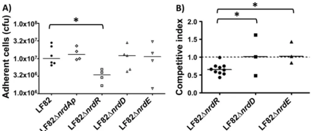
A significant decrease in adhesion was observed only for the LF82
⌬nrdR mutant (
Fig. 5A
). This mutant’s decreased ability to adhere
was type 1 pilus independent, as the LF82
⌬nrdR mutant still
ex-pressed functional type 1 pili on the bacterial surface, as determined
by the ability of bacteria to aggregate yeast cells via binding to
D-mannose residues located on the yeast surface (data not shown). The
role of nrdR gene in adhesion ability of AIEC LF82 bacteria was
con-firmed by a transcomplementation assay restoring the wild-type
phe-notype (see Fig. S3 in the supplemental material). The bacterial
inter-action of LF82, LF82
⌬nrdR, LF82 ⌬nrdD, and LF82 ⌬nrdE was
investigated using ex vivo mouse ileal loop assays. The mean
compet-FIG 2 Plasmid NrdAB genes are acquired by horizontal gene transfer. Unrooted phylogenetic tree of representative class Ia (NrdA) catalytic RNR subunit. TheNeighborNet phylogenetic network was generated from a representative collection of the 26 closest BLAST neighbors in RNRdb to the class Ia catalytic subunit (NrdA) from E. coli LF82 and S. enterica CT18 that were encoded chromosomally and by the plasmid. Yersinia pestis strain 91001, Salmonella enterica strain CT18, and E. coli strain LF82 are highlighted in black when the class Ia RNR copy is encoded chromosomally and in light gray when encoded by a plasmid. All bootstrap values are higher than 950.
TABLE 3 Low enzymatic activity of plasmid NrdAB proteins in a heterologous complementation assay
Plasmid Activity (CFU/ml)a Arabinose (0.2%) Glucose (0.2%) 30°C 42°C 30°C 42°C E101(pETS174) 3.1⫻ 109⫾ 1.2 ⫻ 107 2.9⫻ 109⫾ 3.2 ⫻ 107 2.9⫻ 109⫾ 4.2 ⫻ 106 ⬍10 E101(pETS175) 5.2⫻ 109⫾ 7.2 ⫻ 106 ⬍10 3.1⫻ 109⫾ 1.2 ⫻ 107 ⬍10 E101(pBAD18) 2.1⫻ 109⫾ 1.2 ⫻ 106 ⬍10 3.1⫻ 109⫾ 1.2 ⫻ 107 ⬍10
itive index for LF82
⌬nrdD and ⌬nrdE isogenic mutants compared to
wild-type LF82 bacteria was close to 1. In contrast, for LF82
⌬nrdR,
the mean competitive index was 0.65
⫾ 0.15 (
Fig. 5B
). These indexes
suggest that nrdR is important for the ileal mucosal interaction and
that the loss of NrdR expression reduced LF82’s ability to interact
with the ileal mucosa.
Importance of nrdD and nrdR to AIEC persistence in the gut
of CEABAC10 mice. To investigate the role of Nrd proteins in gut
colonization by AIEC LF82, CEABAC10 transgenic mice expressing
human CEACAM molecules (CEACAM3, CEACAM5, CEACAM6,
and CEACAM7) were challenged with LF82, LF82
⌬nrdR, LF82
⌬nrdD, or LF82 ⌬nrdE bacteria. The quantification of bacteria in
stool samples at day 2 postinfection revealed an 80.0-fold decrease
for LF82
⌬nrdR (5.0 ⫻ 10
7CFU/g of feces) and an 87.8-fold
de-crease for LF82
⌬nrdD (3.6 ⫻ 10
7CFU/g of feces) compared with
wild-type AIEC LF82 (3.0
⫻ 10
8CFU/g of feces) (
Fig. 6
). The
significantly lower concentration of LF82
⌬nrdR and LF82 ⌬nrdD
mutants in the stool was confirmed at day 3 postinfection (4.0
⫻
10
6for LF82
⌬nrdR; 3.9 ⫻ 10
6for LF82
⌬nrdR versus 5.7 ⫻ 10
7CFU/g of feces, respectively; P
⫽ 0.01 and P ⫽ 0.05) and at day 6
postinfection (6.7
⫻ 10
4for LF82
⌬nrdR; 2.5 ⫻ 10
6for LF82
⌬nrdR versus 1.5 ⫻ 10
6CFU/g of feces, respectively; P
⫽ 0.01 and
P
⫽ 0.05). No significant difference in bacterial persistence in the
gut of CEABAC10 mice was observed for LF82
⌬nrdE compared
with wild-type LF82 (
Fig. 6
).
These results suggest that the deletion of nrdR and nrdD genes
in the AIEC LF82 genome dramatically decreased the ability of
bacteria to persist in the gut of CEABAC10 mice.
Interference of nrdR regulon in AIEC virulence. We
demon-strated that NrdR is involved in LF82 adhesion and persistence.
Thus, we pursued an investigation of the mechanisms leading to
these phenotypes. Total RNA from aerobic LF82 and LF82
⌬nrdR
cultures grown to mid-exponential phase was extracted to obtain
an nrdR expression profile in the AIEC LF82 strain. The
microar-ray data from this transcriptomic analysis demonstrated that 33
genes were differentially expressed between the LF82
⌬nrdR
mu-tant and a wild-type strain. Of those genes, only 7 were
upregu-lated in the absence of nrdR, whereas 26 genes were
downregu-FIG 3 Expression of the different nrd genes depends on cell interaction. mRNA levels of the nrdR, nrdAp, nrdAc, nrdD, and nrdE genes in associated bacteria(after a 3-h, 8-h, or 24-h infection period) relative to those of AIEC LF82 bacteria grown during similar periods in MEM cell culture medium were compared using quantitative RT-PCR. As controls, 16S rRNA levels were measured. Results are expressed as the number of mRNA-encoding nrd genes for 107copies of
mRNA-encoding 16S rRNA. Only experiments that demonstrated the same levels of 16S rRNA for each sample were taken into account. The data represent the means from at least three separate experiments, and the bars represent the SEM.
lated in the LF82
⌬nrdR mutant (
Table 4
). As we expected from
previous studies (
8
), the operon for the class Ib RNR expression,
nrdHIEF, was induced in the nrdR mutant strain. This RNR is
encoded by genes that were identified along with two genes
in-volved in cell division, dicB and ydfD, among the few upregulated
genes obtained in the LF82
⌬nrdR microarray.
Interestingly, 8 fully transcriptional units corresponding to 18
genes (ycgR, fliDST, aer, tsr, motAB, cheAW, tar, tap, cheRBYZ,
fliC, and yhjH) from the 26 repressed genes have functions in
bacterial motility and chemotaxis (
Table 4
). Four genes (aer, tsr,
tar, and tap), encoding the chemotaxis receptors known as
meth-yl-accepting chemotaxis proteins (MCPs), and the 6 che loci
(cheA, cheB, cheR, cheZ, cheY, and cheW) responsible for the
che-motactic movements of E. coli were downregulated by an
approx-imately 2-log-fold change (FC) in an LF82
⌬nrdR mutant. The
genes are involved in flagellar biosynthesis (fliDST) and the
flagel-line (fliC) and flagellar (ycgR) functions, and they were
downregu-lated in the LF82
⌬nrdR mutant, demonstrating the possible
par-ticipation of this transcriptional regulator in LF82 motility. The
EAL domain-containing phosphodiesterase YhjH-encoding gene,
yhjH, was also downregulated by 2.26 log FC in the LF82
⌬nrdR
mutant compared with the wild-type strain. The results of the
transcriptomic experiment were confirmed via quantitative PCR
of several of the genes described in
Table 4
(
Fig. 7
).
Regulation of LF82 motility and chemotaxis by NrdR. To
phenotypically corroborate our microarray results, which
indi-cated a possible role of NrdR in motility and chemotaxis, we
eval-uated the behavior of LF82 and LF82
⌬nrdR on soft agar
supple-mented with four different chemoattractants (
D-glucose, maltose,
L-serine, and
L-fucose) and one chemorepellent (
L-valine). After a
12-h incubation period at 30°C for all conditions, the LF82
⌬nrdR
mutant exhibited reduced colony motility compared with its
cor-responding wild-type strain (
Fig. 8A
). The diameters of the
mo-tility halos in the LF82
⌬nrdR were one-half of those of the halos
produced by the wild-type strain (
Fig. 8B
), indicating the
impor-tance of NrdR in E. coli LF82 motility. Experiments to evaluate
FIG 5 Adhesion of LF82⌬nrd mutants using T-84 cells or ileal loops. (A) Cell-associated bacteria were quantified using undifferentiated T-84 cells after a 3-hinfection. (B) Competitive indexes of LF82 with LF82⌬nrdR, LF82 ⌬nrdD, and LF82 ⌬nrdE. Intestinal ileal loops were inoculated with a mixed inoculum comprising equivalent numbers of two bacterial strains. After 5 h, the bacterial presence was compared using competitive index analysis, which provides a sensitive measurement of the relative degree of attenuation. *, P⬍ 0.05.
FIG 6 Bacterial persistence in the gut of CEABAC10 mice. Quantification of LF82, LF82⌬nrdR, LF82 ⌬nrdE, and LF82 ⌬nrdD bacteria in the feces of CEABAC10 mice that received 0.25% DSS in their drinking water after oral infection with 109bacteria at day 0. *, P⬍ 0.05; **, P ⬍ 0.01.
chemotaxis using specific soft-agar plates supplemented with
ser-ine, maltose, and thymine yielded results similar to those of the
motility assays, with reduced chemotactic expansion of the LF82
⌬nrdR mutant compared with the wild-type strain (see Fig. S2 in
the supplemental material).
Electron microscopy indicated reduced expression of flagella at
the surface of the LF82
⌬nrdR mutant bacteria compared with the
wild-type strain (
Fig. 8C
). LF82
⌬nrdD, ⌬nrdE, and ⌬nrdABp
mu-tants did not display any alterations in flagellum production
com-pared with the wild-type strain (data not shown). In addition,
Western blot analysis indicated reduced FliC expression in the
LF82
⌬nrdR mutant compared with the wild-type strain,
reinforc-ing our previous results of reduced motility and demonstratreinforc-ing a
reduced FliC protein level (
Fig. 8D
). In addition,
complementa-tion of LF82
⌬nrdR mutant with a cloned nrdR gene restored the
motility phenotype of the
⌬nrdR mutant to a level similar to the
wild-type level (
Fig. 8E
and
F
), confirming the effect of NrdR in
the regulation of LF82 motility.
DISCUSSION
Ribonucleotide reductases operate under a range of
environmen-tal conditions, which suggests that although the propensity to
syn-thesize deoxyribonucleotides is an essential function, the type of
RNR present will have an impact on the microorganism’s ability
to adapt to different environments. In addition, when more than
one RNR class is encoded by the bacterial genome, it is crucial to
understand which RNR is important during the adhesion,
coloni-zation, and persistence of pathogenic bacteria. Adherent-invasive
E. coli (AIEC), especially the reference strain LF82, is of particular
interest because AIEC virulence evolution involves the selection of
amino acid mutations in common bacterial proteins, such as the
TABLE 4 NrdR-dependent expression of genes identified by transcriptomic analysisGene Operon arrangement
Log fold
changea Gene product
nrdH nrdHIEF 2.29 Glutaredoxin-like protein
nrdI 2.32 Ribonucleotide reductase stimulatory protein: flavodoxin
nrdE 2.28 Ribonucleotide-diphosphate reductase subunit alpha
nrdF 1.92 Ribonucleotide-diphosphate reductase subunit beta
dicB dicB ydfDE insD intQ 1.15 Cell division inhibition protein
ydfD 1.44 Hypothetical protein
yieI yieIJ 1.13 Putative inner membrane protein
ycgR ycgR ⫺1.08 Flagellar function protein
fliD fliDST ⫺1.21 Flagellar filament capping protein
fliS ⫺1.34 Flagellar biosynthesis protein FliS
fliT ⫺1.39 Flagellar biosynthesis protein FliT
aer aer ⫺1.47 Aerotaxis receptor
fliC fliC ⫺1.56 Flagelline
motA motAB cheAW ⫺1.63 Flagellar motor protein; proton conduct
motB ⫺1.75 Flagellar motor protein; motor rotation
cheA ⫺1.82 Chemotaxis protein; chemotactic sensory histidine kinase
cheW ⫺2.06 Purine-binding chemotaxis protein
tar tar tap cheRBYZ ⫺2.26 Methyl-accepting chemotaxis protein II
tap ⫺2.36 Methyl-accepting chemotaxis protein IV
cheR ⫺1.96 Chemotaxis methyltransferase; regulator
cheB ⫺2.01 Chemotaxis-specific methylesterase; chemotaxis regulator
cheY ⫺2.18 Chemotaxis regulator transmitting signal
cheZ ⫺2.01 Chemotaxis protein phosphatase for CheY; chemotaxis regulator
tsr tsr ⫺2.35 Methyl-accepting chemotaxis protein I
yhjH yhjH ⫺2.26 EAL domain-containing protein
ymdA ymdA ⫺1.31 Hypothetical protein
yjcZ yjdAZ ⫺1.88 Hypothetical protein
yhjG yhjG ⫺1.48 Hypothetical protein
kdgK kdgK ⫺2.79 2-Dehydro-3-deoxygluconokinase
aLog fold change was determined by comparing the transcription (ratio of mRNA) of Pseudomonas aeruginosa⌬nrdR to its wild-type strain PAO1 (⌬nrdR mRNA/wild-type
mRNA).
FIG 7 RT-PCR analysis of the transcription levels of the nrdE, tsr, fliC, cheR,
kdgK, motA, and fliS genes from the AIEC LF82 (wt) and LF82⌬nrdR isogenic mutant strains. The gapA gene was used as a control to ensure that equivalent quantities of templates were loaded. Log FC values from the array are shown. Twenty-five PCR cycles were performed in all cases.
FimH protein, and leads to the development of chronic
inflam-matory bowel disease in a genetically susceptible host (
25
). The
AIEC reference strain LF82 is of great interest among the different
E. coli species and Enterobacteriaceae, as it provides an additional
RNR class of plasmid origin. This bacterium harbors four
differ-ent RNR classes, three of which are chromosomally encoded
(classes Ia, Ib, and III) and an additional copy (class Ia) that is
encoded by the pLF82 plasmid. In the present study, we
demon-strated that this extra class Ia RNR copy encoded by pLF82 was
acquired by horizontal gene transfer. Several other examples of
FIG 8 Effect of⌬nrdR on LF82 motility. (A and B) Motility on soft LB agar of wild-type and nrdR mutant LF82 strains after 12 h of incubation at 30°C. The errors bars in panel B represent the standard deviations from three independent experiments. (C) Electron microscopy of AIEC LF82 and the nrdR mutants (magni-fication,⫻10,000). (D) Western blot analysis of FliC expression in wild-type and LF82 ⌬nrdR mutant strains. (E and F) Motility on soft LB agar of complemented ⌬nrdR mutant strains with cloned nrdR gene (pETS179) and the empty vector (pBBR1). The error bars in panel F represent the standard deviations from three independent experiments. Significant differences in motility were determined using a paired Student t test; *, P⬍ 0.01.plasmid- and prophage-encoded RNR proteins have been
previ-ously reported (for an example, see reference
3
), providing clear
evidence of horizontal gene transfer events that play an important
role in the evolution of the RNR distribution for a great variety of
RNR genes from all three domains of life.
However, for suspected horizontal gene transfer cases, we must
assess whether the transferred copy is enzymatically active. In the
AIEC LF82 strain, this is not the case because plasmid NrdAp was
inactive in our experiments. Surprisingly, all-important residues
located at the active and allosteric sites, iron ligand and NrdA, and
the NrdA-NrdB interaction were also conserved in the E. coli LF82
NrdABp. Thus, the loss of E. coli LF82 NrdAB activity may involve
residues that are important for protein folding, activation, and
subunit interaction.
Bacteria need tight gene regulation to maintain the balanced
dNTP pools important for DNA replication. For this reason, tight
transcriptional regulation must be achieved when a bacterium
encodes more than one class of RNR. Therefore, it is important to
understand which is the role of each RNR class during AIEC LF82
virulence processes, such as adhesion to intestinal epithelial cells
and AIEC LF82 persistence in the gut mucosa of a mouse model
mimicking CD susceptibility. An expression analysis of the
differ-ent RNR classes presdiffer-ent when AIEC LF82 was grown in cells
asso-ciated with T-84 cells compared with cells grown in cellular
me-dium revealed important differences. nrdR and nrdD (class III)
mRNA levels increased during growth when AIEC LF82 bacteria
were associated with T-84 cells, whereas the corresponding
mRNA levels decreased significantly when bacteria were grown in
cellular medium during a similar period, suggesting that NrdR
and NrdD protein levels in AIEC LF82 bacteria are crucial for
controlling bacterial virulence.
The importance of each RNR class during infection was
inves-tigated by assessing the bacterium’s ability to attach to T-84 cells
and to the ileal loop and to persist in the gut mucosa of
CE-ABAC10 mice. The adhesion assays demonstrated that the nrdR
mutant exhibited an impaired ability to interact with intestinal
epithelial cells compared with the wild type. The other nrd-lacking
mutants used in this study did not display alterations in adhesion.
Conversely, the results indicate that only the nrdR and nrdD gene
products reduced the ability of AIEC LF82 to persist in the gut and
thus impaired pathogenesis. Several authors have previously
de-scribed the anaerobic environments in the gut of mammals (
37
),
and under these conditions, anaerobic enzymes are
transcription-ally activated. Class III enzymes are enzymatictranscription-ally active only
un-der anaerobic conditions, and their importance has been well
es-tablished (
5
). The nrdD mutant (class III) has a markedly reduced
capacity to infect the gut, which is correlated with the capacity for
DNA replication under anaerobic conditions. Other RNR classes
cannot compensate for nrdD deficiency. A comparison of the
LF82 nrdD promoter region with the E. coli MG1655 nrdD
pro-moter region revealed two FNR boxes (TTGATGTAGCTCAA
and TTGATGCAAAGCAC) located at
⫺211 and ⫺242 bp
up-stream of the translation start point. The almost-identical
posi-tions in the two promoter region strains demonstrate that this
RNR class is transcriptionally activated during anaerobic
metab-olism and is important for the specific dNTP supply in the AIEC
LF82 strain facing anaerobic conditions during gut infection.
We also assessed why the AIEC transcriptional regulator NrdR
is important for AIEC’s ability to adhere to T-84 cells, interact
with intestinal mucosa in the ileal loop model, and persist in the
gut mucosa of mice that mimic CD susceptibility. To answer this
question, we investigated NrdR’s ability to regulate global gene
expression. In our transcriptomic analysis, the genes that were
transcriptionally altered were ones that regulate motility and
che-motaxis. Genes involved in chemotaxis, such as MotA and MotB,
together apply the required force to FliG for flagellar rotation (
38
).
MotA interaction with FliG is essential for switching the flagellar
direction from clockwise (CW) movement and stimulating cell
tumble to the counterclockwise (CCW) state that promotes the
forward movement of the bacteria (
39
). The effect of deletion of
motA and motB, which promotes CCW movement and reduces
bacterial chemotaxis, is well known (
40–42
) and corroborates our
array results indicating reduced chemotaxis for the AIEC LF82
strain (see Fig. S2 in the supplemental material). In addition, the
AIEC LF82
⌬nrdR mutant exhibited reduced motility capacity
compared to the wild-type strain (
Fig. 8
). Flagella of the AIEC
LF82 strain have established roles in the adhesion to and invasion
of intestinal epithelial cells (
43
,
44
). Electronic microscopy
analy-sis of the wild-type and LF82
⌬nrdR isogenic mutant
demon-strated reduced flagellar expression when the nrdR gene was
at-tenuated. This was confirmed by Western blot analysis of the
expression of the major component of the flagella, FliC (
Fig. 8
). It
is well described that flagella contribute to AIEC virulence.
In-deed, the nonmotile aflagellar LF82 mutants (
⌬fliC or ⌬fliA
mu-tants) exhibit a drastic reduction in adherence and invasion ability
(
43
,
44
), and the flagellar sigma factor FliA regulates the adhesion
and invasion of Crohn’s disease-associated Escherichia coli via a
cyclic dimeric GMP-dependent pathway (
44
). In addition,
Crohn’s disease-associated virulent AIEC LF82 bacteria, via
flagel-lar expression, are able to potentiate an inflammatory mucosal
immune response in the dextran sulfate sodium (DSS)-injured
colons of BALBc/J mice via increased expression of Toll-like
re-ceptor 5 (TLR5) and IPAF flagellin rere-ceptors (
45
). In addition, we
reported that CEABAC10 mice infected with LF82
⌬fliC did not
loss body weight compared to mice infected with LF82 bacteria
(see Fig. S4 in the supplemental material). Thus, the decreased
virulence ability of the LF82 mutant lacking nrdR could be
ex-plained in part by impaired flagellar expression. In addition,
flagellin receptor TLR5-deficient mice (T5KO) display elevated
intestinal proinflammatory gene expression and colitis. The
Crohn’s disease-associated E. coli LF82 strain induced chronic
colitis in T5KO, which persisted well after the exogenously
intro-duced bacterial species had been eliminated (
46
).
An extended understanding of the pathogenicity and
physiol-ogy of AIEC could aid in the development of novel drugs active
against these pathogens. In the present study, we investigated the
role of RNRs in the virulence of AIEC isolated from Crohn’s
dis-ease patients and demonstrated that knockout of the nrdR gene,
which encodes a transcriptional regulator of RNR, decreased the
ability of AIEC LF82 to adhere to intestinal epithelial T-84 cells,
interact with the ileal mucosa using the ileal loop model, and
col-onize the gut mucosa of transgenic mice that express human
CEACAM6. NrdR plays a crucial role in AIEC virulence by
inter-fering with bacterial motility and chemotaxis. Thus, the
develop-ment of new drugs targeting RNR classes, particularly NrdR,
could be a promising new strategy for controlling AIEC flagellum
expression and motility and subsequently controlling gut
coloni-zation and AIEC-induced inflammation in CD patients.
ACKNOWLEDGMENTS
This work was supported by the Ministerio de Economia y Competitivi-dad with grants BFU2011-24066, CSD2008-00013, and ERA-NET PathoGenoMics (BIO2008-04362-E) to E.T. This work was also ported by the Generalitat de Catalunya SGR2014-01260. E.T. was sup-ported by the Ramón y Cajal and I3 programs from the Ministerio de Ciencia e Innovación. This study was supported by INSERM (UMR1071) and INRA (USC-2018) and by grants from the Association F. Aupetit (AFA), ANR, in the frame of ERA-NET PathoGenomics (grant no. 0315443).
The funders did not participate in the study design, data collection and analysis, decision to publish, or preparation of the manuscript. REFERENCES
1. Nordlund P, Reichard P. 2006. Ribonucleotide reductases. Annu Rev Biochem 75:681–706. http://dx.doi.org/10.1146/annurev.biochem.75 .103004.142443.
2. Torrents E, Sahlin M, Sjöberg B-M. 2008. The ribonucleotide reductase family— genetics and genomics, p 17–77. In Anderson KK (ed), Ribonu-cleotide reductases. Nova Science Publishers, Hauppauge, NY. 3. Lundin D, Gribaldo S, Torrents E, Sjoberg BM, Poole AM. 2010.
Ribonucleotide reduction— horizontal transfer of a required function spans all three domains. BMC Evol Biol 10:383.http://dx.doi.org/10.1186 /1471-2148-10-383.
4. Lundin D, Torrents E, Poole AM, Sjöberg B-M. 2009. RNRdb, a curated database of the universal enzyme family ribonucleotide reductase, reveals a high level of misannotation in sequences deposited to Genbank. BMC Genomics 10:589.http://dx.doi.org/10.1186/1471-2164-10-589. 5. Garriga X, Eliasson R, Torrents E, Jordan A, Barbe J, Gibert I, Reichard
P. 1996. nrdD and nrdG genes are essential for strict anaerobic growth of
Escherichia coli. Biochem Biophys Res Commun 229:189 –192.http://dx .doi.org/10.1006/bbrc.1996.1778.
6. Cendra M, Juarez A, Torrents E. 2012. Biofilm modifies expression of ribonucleotide reductase genes in Escherichia coli. PLoS One 7:e46350.
http://dx.doi.org/10.1371/journal.pone.0046350.
7. Martin JE, Imlay JA. 2011. The alternative aerobic ribonucleotide reduc-tase of Escherichia coli, NrdEF, is a manganese-dependent enzyme that enables cell replication during periods of iron starvation. Mol Microbiol
80:319 –334.http://dx.doi.org/10.1111/j.1365-2958.2011.07593.x. 8. Torrents E, Grinberg I, Gorovitz-Harris B, Lundstrom H, Borovok I,
Aharonowitz Y, Sjöberg BM, Cohen G. 2007. NrdR controls differential
expression of the Escherichia coli ribonucleotide reductase genes. J Bacte-riol 189:5012–5021.http://dx.doi.org/10.1128/JB.00440-07.
9. Torrents E. 2014. Ribonucleotide reductases: essential enzymes for bac-terial life. Front Cell Infect Microbiol 4:52. http://dx.doi.org/10.3389 /fcimb.2014.00052.
10. Strober W, Fuss I, Mannon P. 2007. The fundamental basis of inflam-matory bowel disease. J Clin Invest 117:514 –521.http://dx.doi.org/10 .1172/JCI30587.
11. Xavier RJ, Podolsky DK. 2007. Unravelling the pathogenesis of inflam-matory bowel disease. Nature 448:427– 434. http://dx.doi.org/10.1038 /nature06005.
12. Kaser A, Zeissig S, Blumberg RS. 2010. Inflammatory bowel disease. Annu Rev Immunol 28:573– 621. http://dx.doi.org/10.1146/annurev-immunol -030409-101225.
13. Chassaing B, Darfeuille-Michaud A. 2011. The commensal microbiota and enteropathogens in the pathogenesis of inflammatory bowel diseases. Gastroenterology 140:1720 –1728.http://dx.doi.org/10.1053/j.gastro.2011.01 .054.
14. Darfeuille-Michaud A, Neut C, Barnich N, Lederman E, Di Martino P,
Desreumaux P, Gambiez L, Joly B, Cortot A, Colombel JF. 1998.
Presence of adherent Escherichia coli strains in ileal mucosa of patients with Crohn’s disease. Gastroenterology 115:1405–1413.http://dx.doi.org /10.1016/S0016-5085(98)70019-8.
15. Martin HM, Campbell BJ, Hart CA, Mpofu C, Nayar M, Singh R,
Englyst H, Williams HF, Rhodes JM. 2004. Enhanced Escherichia coli
adherence and invasion in Crohn’s disease and colon cancer. Gastroenter-ology 127:80 –93.http://dx.doi.org/10.1053/j.gastro.2004.03.054. 16. Swidsinski A, Ladhoff A, Pernthaler A, Swidsinski S, Loening-Baucke V,
Ortner M, Weber J, Hoffmann U, Schreiber S, Dietel M, Lochs H. 2002.
Mucosal flora in inflammatory bowel disease. Gastroenterology 122:44 – 54.http://dx.doi.org/10.1053/gast.2002.30294.
17. Boudeau J, Glasser AL, Masseret E, Joly B, Darfeuille-Michaud A. 1999. Invasive ability of an Escherichia coli strain isolated from the ileal mucosa of a patient with Crohn’s disease. Infect Immun 67:4499 – 4509. 18. Conte MP, Schippa S, Zamboni I, Penta M, Chiarini F, Seganti L,
Osborn J, Falconieri P, Borrelli O, Cucchiara S. 2006. Gut-associated
bacterial microbiota in paediatric patients with inflammatory bowel dis-ease. Gut 55:1760 –1767.http://dx.doi.org/10.1136/gut.2005.078824. 19. Kotlowski R, Bernstein CN, Sepehri S, Krause DO. 2007. High
preva-lence of Escherichia coli belonging to the B2⫹D phylogenetic group in inflammatory bowel disease. Gut 56:669 – 675.http://dx.doi.org/10.1136 /gut.2006.099796.
20. Neut C, Bulois P, Desreumaux P, Membre JM, Lederman E, Gambiez
L, Cortot A, Quandalle P, van Kruiningen H, Colombel JF. 2002.
Changes in the bacterial flora of the neoterminal ileum after ileocolonic resection for Crohn’s disease. Am J Gastroenterol 97:939 –946.http://dx .doi.org/10.1111/j.1572-0241.2002.05613.x.
21. Baumgart M, Dogan B, Rishniw M, Weitzman G, Bosworth B, Yantiss R,
Orsi RH, Wiedmann M, McDonough P, Kim SG, Berg D, Schukken Y, Scherl E, Simpson KW. 2007. Culture independent analysis of ileal mucosa
reveals a selective increase in invasive Escherichia coli of novel phylogeny relative to depletion of Clostridiales in Crohn’s disease involving the ileum. ISME J 1:403– 418.http://dx.doi.org/10.1038/ismej.2007.52.
22. Sasaki M, Sitaraman SV, Babbin BA, Gerner-Smidt P, Ribot EM,
Garrett N, Alpern JA, Akyildiz A, Theiss AL, Nusrat A, Klapproth JM.
2007. Invasive Escherichia coli are a feature of Crohn’s disease. Lab Invest
87:1042–1054.http://dx.doi.org/10.1038/labinvest.3700661.
23. Martinez-Medina M, Aldeguer X, Lopez-Siles M, Gonzalez-Huix F,
Lopez-Oliu C, Dahbi G, Blanco JE, Blanco J, Garcia-Gil LJ, Darfeuille-Michaud A. 2009. Molecular diversity of Escherichia coli in the human
gut: new ecological evidence supporting the role of adherent-invasive E. coli (AIEC) in Crohn’s disease. Inflamm Bowel Dis 15:872– 882.http://dx .doi.org/10.1002/ibd.20860.
24. Barnich N, Carvalho FA, Glasser AL, Darcha C, Jantscheff P, Allez M,
Peeters H, Bommelaer G, Desreumaux P, Colombel JF, Darfeuille-Michaud A. 2007. CEACAM6 acts as a receptor for adherent-invasive E.
coli, supporting ileal mucosa colonization in Crohn disease. J Clin Invest
117:1566 –1574.http://dx.doi.org/10.1172/JCI30504.
25. Dreux N, Denizot J, Martinez-Medina M, Mellmann A, Billig M, Kisiela
D, Chattopadhyay S, Sokurenko E, Neut C, Gower-Rousseau C, Co-lombel JF, Bonnet R, Darfeuille-Michaud A, Barnich N. 2013. Point
mutations in FimH adhesin of Crohn’s disease-associated adherent-invasive Escherichia coli enhance intestinal inflammatory response. PLoS Pathog 9:e1003141.http://dx.doi.org/10.1371/journal.ppat.1003141. 26. Carvalho FA, Barnich N, Sivignon A, Darcha C, Chan CH, Stanners CP,
Darfeuille-Michaud A. 2009. Crohn’s disease adherent-invasive
Esche-richia coli colonize and induce strong gut inflammation in transgenic mice expressing human CEACAM. J Exp Med 206:2179 –2189.http://dx.doi .org/10.1084/jem.20090741.
27. Sambrook J, Fritsch EF, Maniatis T. 1989. Molecular cloning: a labora-tory manual, 2nd ed. Cold Spring Harbor Laboralabora-tory Press, Cold Spring Harbor, NY.
28. Datsenko KA, Wanner BL. 2000. One-step inactivation of chromosomal genes in Escherichia coli K-12 using PCR products. Proc Natl Acad Sci U S A 97:6640 – 6645.http://dx.doi.org/10.1073/pnas.120163297.
29. Chaveroche MK, Ghigo JM, d’Enfert C. 2000. A rapid method for effi-cient gene replacement in the filamentous fungus Aspergillus nidulans. Nucleic Acids Res 28:E97.http://dx.doi.org/10.1093/nar/28.22.e97. 30. Adase CA, Draheim RR, Manson MD. 2012. The residue composition
of the aromatic anchor of the second transmembrane helix determines the signaling properties of the aspartate/maltose chemoreceptor Tar of Escherichia coli. Biochemistry 51:1925–1932. http://dx.doi.org/10.1021 /bi201555x.
31. Roca I, Torrents E, Sahlin M, Gibert I, Sjoberg BM. 2008. NrdI essen-tiality for class Ib ribonucleotide reduction in Streptococcus pyogenes. J Bacteriol 190:4849 – 4858.http://dx.doi.org/10.1128/JB.00185-08. 32. Jordan A, Aragall E, Gibert I, Barbe J. 1996. Promoter identification and
expression analysis of Salmonella typhimurium and Escherichia coli nrdEF operons encoding one of two class I ribonucleotide reductases present in both bacteria. Mol Microbiol 19:777–790. http://dx.doi.org/10.1046/j .1365-2958.1996.424950.x.
mutant forms of Escherichia coli ribonucleotide reductase. J Bacteriol 185: 1167–1173.http://dx.doi.org/10.1128/JB.185.4.1167-1173.2003. 34. Stecher B, Robbiani R, Walker AW, Westendorf AM, Barthel M,
Kre-mer M, Chaffron S, Macpherson AJ, Buer J, Parkhill J, Dougan G, von Mering C, Hardt WD. 2007. Salmonella enterica serovar typhimurium
exploits inflammation to compete with the intestinal microbiota. PLoS Biol 5:2177–2189.http://dx.doi.org/10.1371/journal.pbio.0050244. 35. Chassaing B, Etienne-Mesmin L, Bonnet R, Darfeuille-Michaud A.
2013. Bile salts induce long polar fimbriae expression favouring Crohn’s disease-associated adherent-invasive Escherichia coli interaction with Pey-er’s patches. Environ Microbiol 15:355–371.http://dx.doi.org/10.1111/j .1462-2920.2012.02824.x.
36. Miquel S, Peyretaillade E, Claret L, de Vallee A, Dossat C, Vacherie B,
Zineb EH, Segurens B, Barbe V, Sauvanet P, Neut C, Colombel JF, Medigue C, Mojica FJ, Peyret P, Bonnet R, Darfeuille-Michaud A. 2010.
Complete genome sequence of Crohn’s disease-associated adherent-invasive E. coli strain LF82. PLoS One 5(9):pii:e12714.http://dx.doi.org /10.1371/journal.pone.0012714.
37. Jones SA, Gibson T, Maltby RC, Chowdhury FZ, Stewart V, Cohen PS,
Conway T. 2011. Anaerobic respiration of Escherichia coli in the mouse
intestine. Infect Immun 79:4218 – 4226. http://dx.doi.org/10.1128/IAI .05395-11.
38. Kojima S, Blair DF. 2001. Conformational change in the stator of the bacterial flagellar motor. Biochemistry 40:13041–13050.http://dx.doi.org /10.1021/bi011263o.
39. Berg HC. 2003. The rotary motor of bacterial flagella. Annu Rev Biochem
72:19 –54.http://dx.doi.org/10.1146/annurev.biochem.72.121801.161737. 40. Zhou J, Sharp LL, Tang HL, Lloyd SA, Billings S, Braun TF, Blair DF.
1998. Function of protonatable residues in the flagellar motor of Esche-richia coli: a critical role for Asp 32 of MotB. J Bacteriol 180:2729 –2735. 41. Morimoto YV, Che YS, Minamino T, Namba K. 2010.
Proton-conductivity assay of plugged and unplugged MotA/B proton channel by cytoplasmic pHluorin expressed in Salmonella. FEBS Lett 584:1268 –1272.
http://dx.doi.org/10.1016/j.febslet.2010.02.051.
42. Parkinson JS, Houts SE. 1982. Isolation and behavior of Escherichia coli deletion mutants lacking chemotaxis functions. J Bacteriol 151:106 –113.
43. Barnich N, Boudeau J, Claret L, Darfeuille-Michaud A. 2003. Reg-ulatory and functional co-operation of flagella and type 1 pili in adhe-sive and invaadhe-sive abilities of AIEC strain LF82 isolated from a patient with Crohn’s disease. Mol Microbiol 48:781–794.http://dx.doi.org/10 .1046/j.1365-2958.2003.03468.x.
44. Claret L, Miquel S, Vieille N, Ryjenkov DA, Gomelsky M,
Darfeuille-Michaud A. 2007. The flagellar sigma factor FliA regulates adhesion and
invasion of Crohn disease-associated Escherichia coli via a cyclic dimeric GMP-dependent pathway. J Biol Chem 282:33275–33283.http://dx.doi .org/10.1074/jbc.M702800200.
45. Carvalho FA, Barnich N, Sauvanet P, Darcha C, Gelot A,
Darfeuille-Michaud A. 2008. Crohn’s disease-associated Escherichia coli LF82
aggra-vates colitis in injured mouse colon via signaling by flagellin. Inflamm Bowel Dis 14:1051–1060.http://dx.doi.org/10.1002/ibd.20423. 46. Carvalho FA, Koren O, Goodrich JK, Johansson ME, Nalbantoglu I,
Aitken JD, Su Y, Chassaing B, Walters WA, Gonzalez A, Clemente JC, Cullender TC, Barnich N, Darfeuille-Michaud A, Vijay-Kumar M, Knight R, Ley RE, Gewirtz AT. 2012. Transient inability to manage
proteobacteria promotes chronic gut inflammation in TLR5-deficient mice. Cell Host Microbe 12:139 –152.http://dx.doi.org/10.1016/j.chom .2012.07.004.
47. Guzman LM, Belin D, Carson MJ, Beckwith J. 1995. Tight regulation, modulation, and high-level expression by vectors containing the arabi-nose PBAD promoter. J Bacteriol 177:4121– 4130.
48. Kovach ME, Elzer PH, Hill DS, Robertson GT, Farris MA, Roop RM, II,
Peterson KM. 1995. Four new derivatives of the broad-host-range cloning
vector pBBR1MCS, carrying different antibiotic-resistance cassettes. Gene
166:175–176.http://dx.doi.org/10.1016/0378-1119(95)00584-1. 49. Newman JR, Fuqua C. 1999. Broad-host-range expression vectors that
carry the L-arabinose-inducible Escherichia coli araBAD promoter and the araC regulator. Gene 227:197–203.http://dx.doi.org/10.1016/S0378 -1119(98)00601-5.
50. Wechsler JA, Gross JD. 1971. Escherichia coli mutants temperature-sensitive for DNA synthesis. Mol Gen Genet 113:273–284.http://dx.doi .org/10.1007/BF00339547.