HAL Id: inserm-02911633
https://www.hal.inserm.fr/inserm-02911633
Submitted on 4 Aug 2020
HAL is a multi-disciplinary open access
archive for the deposit and dissemination of sci-entific research documents, whether they are pub-lished or not. The documents may come from teaching and research institutions in France or abroad, or from public or private research centers.
L’archive ouverte pluridisciplinaire HAL, est destinée au dépôt et à la diffusion de documents scientifiques de niveau recherche, publiés ou non, émanant des établissements d’enseignement et de recherche français ou étrangers, des laboratoires publics ou privés.
Non-Endemic area
Federico Gobbi, Dora Buonfrate, Michel Boussinesq, Cédric Chesnais,
Sébastien Pion, Ronaldo Silva, Lucia Moro, Paola Rodari, Francesca
Tamarozzi, Marco Biamonte, et al.
To cite this version:
Federico Gobbi, Dora Buonfrate, Michel Boussinesq, Cédric Chesnais, Sébastien Pion, et al.. Perfor-mance of two serodiagnostic tests for loiasis in a Non-Endemic area. PLoS Neglected Tropical Dis-eases, Public Library of Science, 2020, 14 (5), pp.e0008187. �10.1371/journal.pntd.0008187�. �inserm-02911633�
RESEARCH ARTICLE
Performance of two serodiagnostic tests for
loiasis in a Non-Endemic area
Federico GobbiID1*, Dora Buonfrate1, Michel Boussinesq2, Cedric B. Chesnais2,
Sebastien D. Pion2, Ronaldo Silva1, Lucia Moro1, Paola RodariID1, Francesca Tamarozzi3,
Marco Biamonte4, Zeno Bisoffi1,5
1 IRCCS Sacro Cuore Don Calabria Hospital, Center for Tropical Diseases, Negrar, Verona, Italy, 2 Institut de Recherche pour le De´ veloppement (IRD), UMI 233-INSERM U1175-Montpellier University, Montpellier, France, 3 Department of Infectious Diseases, Foodborne and Neglected Parasitic Diseases Unit, Istituto Superiore di Sanità, Rome, Italy, 4 Drugs & Diagnostics for Tropical Diseases, San Diego, California, United States of America, 5 Infectious Infectious Diseases and Tropical Medicine Section, Diagnostic and Public Health Department, University of Verona, Verona, Italy
*federico.gobbi@sacrocuore.it
Abstract
Loiasis, caused by the filarial nematode Loa loa, is endemic in Central and West Africa where about 10 million people are infected. There is a scarcity of convenient, commercial diagnostics for L. loa. Microscopy requires trained personnel and has low sensitivity, while the serodiagnosis is currently not standardized. Individual case management is also impor-tant in non-endemic countries to treat migrants, expatriates and tourists. We retrospectively compared the performance of a Loa Antibody Rapid Test (RDT) and a commercial ELISA pan-filarial test on 170 patients, 65 with loiasis [8 with eyeworm, 29 with positive microfilare-mia, 28 with neither microfilaremia nor history of eyeworm but eosinophilia and history of Calabar swelling (probable loiasis)], 95 with other common parasitic infections and no previ-ous exposure to L. loa (37 with M. perstans, 1 with Brugia sp., 18 with strongyloidiasis, 20 with schistosomiasis, 5 with hookworm, 4 with Ascaris lumbricoides infection, 10 with hyper-reactive malarial splenomegaly), and 10 uninfected controls. The sensitivity of the RDT and of the ELISA were 93.8% (61/65) and 90.8% (59/65), respectively. For the RDT, most of the cross-reactions were observed in patients with M. perstans: 7/37 (18.9%), followed by 1/10 (10%) with hyper-reactive malarial splenomegaly and 1/20 (5%) with schistosomiasis. None of the 27 subjects infected with intestinal nematodes was found positive at this test. The ELISA is meant to be a pan-filarial assay, and reacted extensively with cases of M. perstans (95%), as expected, and also in 11/18 (61.1%) patients with strongyloidiasis and in 3/5 (60%) with hookworm infection. The RDT and the ELISA are both highly sensitive for the diagnosis of loiasis. The main difference lies in the extent of cross-reactivity with other para-sites. Considering that the RDT is specifically meant for Loa loa infection, and its high sensi-tivity, this test could be a useful tool for the diagnosis of occult loiasis.
a1111111111 a1111111111 a1111111111 a1111111111 a1111111111 OPEN ACCESS
Citation: Gobbi F, Buonfrate D, Boussinesq M,
Chesnais CB, Pion SD, Silva R, et al. (2020) Performance of two serodiagnostic tests for loiasis in a Non-Endemic area. PLoS Negl Trop Dis 14(5): e0008187.https://doi.org/10.1371/journal. pntd.0008187
Editor: Paul J. Brindley, George Washington
University School of Medicine and Health Sciences, UNITED STATES
Received: December 6, 2019 Accepted: March 2, 2020 Published: May 26, 2020
Copyright:© 2020 Gobbi et al. This is an open access article distributed under the terms of the
Creative Commons Attribution License, which permits unrestricted use, distribution, and reproduction in any medium, provided the original author and source are credited.
Data Availability Statement: The study database is
available in Mendeley Data, URL:https://data. mendeley.com/datasets/dg4j6wb6h8/1, doi10. 17632/dg4j6wb6h8.1. All other data is available in the manuscript.
Funding: This work was supported by the Italian
Ministry of Health “Fondi Ricerca Corrente - Linea 3, progetto 8” to IRCCS Sacro Cuore Don Calabria Hospital. The funders had no role in study design,
Author summary
Loa loa is a filarial worm which infects millions of people living in the forested areas of central Africa. The infection is rarely diagnosed outside Africa, but individual case man-agement is also important in non-endemic countries to treat migrants, tourists and expa-triates. Aim of this study was to describe the performance of a Rapid Test and an ELISA pan filarial test for the diagnosis ofLoa loa infection. Both tests demonstrated good per-formance for the detection of cases of loiasis. In case of infection with other parasites, the RDT gave less false positive results.
Introduction
Loiasis, the disease caused by the infection with the filarial nematodeLoa loa, is transmitted through the bite of tabanid flies of the genusChrysops. It is endemic in Central and West Africa where, according to the most recent estimates, about 10 million people are infected [1].
The adult worms reside in the subcutaneous tissues and in the intermuscular layers, while the progeny, microfilariae (mf), circulate in the blood where they are picked up by the vector during a blood meal, allowing the perpetuation of the biological cycle [2].
The two most specific signs of infection are transient oedemas known as “Calabar swell-ings”, and the movement of the adult worm under the conjunctiva (“eye worm”) [2]. These manifestations are major reasons for people seeking medical advice in endemic areas [3]. Loia-sis is still regarded as a benign condition even if a recent a study found an association between highL. loa microfilarial density (MFD) and increased mortality risk [4]. Importantly, severe adverse events (SAEs) after administration of diethylcarbamazine (DEC) and ivermectin (IVM) may occur in individuals with highL. loa MFD [5]. Hence, it is mandatory to rule out this infection before treatment with these drugs, or at least to quantify the number of circulat-ing mf.
From a global health perspective, the World Health Organization (WHO)-driven pro-grammes for the elimination of onchocerciasis rely on mass drug administration (MDA) of IVM. In areas where onchocerciasis is meso or hyperendemic (prevalence of nodules >20%) and loiasis is co-endemic, MDA is possible but a specific surveillance system has to be put in place because of the risk of IVM-induced SAEs. When hypoendemic onchocerciasis coexists with loiasis, a test and treat strategy can be used, so that the whole population is screened to identify those individuals who are at risk of SAEs [6]. Individual case management is also important both in endemic and non-endemic countries to treat migrants, tourists and expatri-ates. For instance, the Department of Infectious/Tropical Diseases (DITM), IRCCS Sacro Cuore Don Calabria Hospital, Negrar, Verona, Italy has diagnosed and treated at least 120 patients with loiasis in the last 30 years [7]. Clinical diagnosis is possible, although difficult: the passage of the adult worm under the conjunctiva is sporadic, and oedemas are also transient and have been reported in infections with another filarial species,Mansonella perstans [8,9]. The identification of circulatingL. loa mf by microscopy confirms the diagnosis of infection. Unfortunately, this method has low sensitivity.L. loa microfilaraemia in the peripheral blood shows a diurnal periodicity, with maximum MFD found between 10 am and 4 pm; therefore the time of sampling influences the sensitivity of microscopy [2]. Further, mf are not present in the blood during the pre-patent period (4–8 months after the infective bite by the vector) and about 40% of the infected individuals present a so-called “occult loiasis”, i.e. they will never show any mf in the peripheral blood, due to a genetic predisposition [10]. There is a scarcity of convenient, commercial diagnostics forL. loa. Microscopy is time-consuming and
data collection and analysis, decision to publish, or preparation of the manuscript.
Competing interests: Dr. Biamonte is CEO of
Drugs & Diagnostics for Tropical Diseases, the company that produces the rapid test described herein. The rapid test was donated to support this research.
requires trained personnel. A cell-phone based microscopy has been described to simplify the measurement of circulating mf and identify individuals at risk of IVM-induced adverse events [11]. Molecular biology techniques such as PCR or LAMP (Loop-mediated Isothermal Ampli-fication) have been developed, but have not entered in clinical routine practice yet [12–15].
The serodiagnosis of loiasis is currently not standardized. In published clinical-based reports, serodiagnosis was carried out using a variety of techniques and both homologous and heterologous antigen sources [7,16–22]. The specificity of the antigenic preparations to detect antibodies againstL. loa has not been widely explored, but a variable level of cross-reactivity is generally observed in individuals with other filarial infections and also in case of infections with other helminthiases such as strongyloidiasis [23]. Sensitivity is also variable, with only a proportion of infected patients, as assessed by presence of circulating mf or PCR, who have detectable antibodies. Using sera from monkeys experimentally infected withL. loa, Klion and colleagues identified an antigen,Ll-SXP-1, which was poorly sensitive (56%) for human loiasis but highly specific (98%) when tested in an ELISA-IgG4 assay using sera from patients infected with other filarial worms and nematodes [24]. Burbelo and colleagues subsequently evaluated this recombinant antigen using an IgG Luciferase Immuno-Precipitation System (LIPS) improving both sensitivity (67%) and specificity (99%) [25]. Finally, Pedram and colleagues developed a recombinantLl-SXP-1 based lateral flow rapid diagnostic test (RDT) with further improved sensitivity (94%), and acceptable specificity (82%-100% depending on the control panel tested) [26]. This test is the firstL. loa-specific available assay. A RDT could be useful both inL. loa endemic areas (for loiasis mapping or for a first-step screening of individuals who should be excluded from MDA with IVM) and in non-endemic settings, where it could speed up and improve diagnosis, even in absence of highly-skilled parasitologists.
The primary objective of this study was to evaluate and compare the performance of the novel lateral flow RDT for loiasis and of a commercial ELISA test for filarial infections in a non-endemic setting, in a mixed population of migrants, tourists and expatriates.
Secondary objectives were to assess the diagnostic concordance between the two tests and
to assess the reproducibilityof the RDT.
Methods
This diagnostic accuracy study was carried out in the laboratory of the DITM. The study pro-tocol received ethical clearance from the Ethics Committee for Clinical Experimentation of Verona and Rovigo on February 13, 2019 (protocol number 8575).
The RDT was applied on archived serum specimens kept frozen at -80˚C at the same institu-tion, from the day of the sample collection till the day of test execution. The results of the ELISA test were already available as part of the routine tests performed at the time of diagnosis.
Participant selection and study groups
The study was carried out using fully anonymized serum samples available at the DITM and collected between 1994 and 2018. Criteria for inclusion were: presence of a signed informed consent for using the biological sample for research purpose; availability of clinical and demo-graphic information relevant for the study (including age, sex, symptoms, country where the infection was likely acquired, eosinophil count). Expatriates are defined as people who stayed in an endemic country for more than six consecutive months. Three different groups of sera were constituted:
• Group I (“Cases”) was composed by patients diagnosed withL. loa infection according to one of the following case definitions:
a. “eyeworm”: patients with negative microfilaraemia but with documented passage of an adultL. loa under the conjunctiva;
b. “mf-positive”: patients with circulatingL. loa mf;
c. “probable loiasis”: patients with neither eyeworm nor microfilaraemia, but presenting eosinophil counts �500/μL and history of Calabar swelling in the 2 months preceding the blood sampling.
• Group II (Non-endemic controls): samples collected from patients born and resident in non-endemic areas for loiasis, with no travel history to endemic countries and presenting to the hospital with other clinical conditions, excluding any parasitic infection; the sera were present in the biobank of the DITM and retrospectively tested with both the ELISA and RDT for this study.
• Group III included samples collected from subjects with no previous exposure toL. loa (i.e. who had never lived in countries where loiasis is endemic: Nigeria, Cameroon, Gabon, Equa-torial Guinea, Congo, Democratic Republic of Congo, Central African Republic, Angola, Uganda, South Sudan, Chad), but infected with other common parasitic infections, which may be of concern for cross-reactivity:
a. patients with circulatingM. perstans or Brugia sp. mf;
b. patients with strongyloidiasis, diagnosed by presence of larvae in stool (by routine microscopy of formol-ether concentrated feces [FECF] or stool culture) and/or high titre (>1:160) positive serology (in-house immunofluorescence antibody test [IFAT]) [27];
c. patients with schistosomiasis, diagnosed by microscopy of FECF or microscopy of fil-tered urines;
d. patients with hookworm infection diagnosed by microscopy of FECF;
e. patients withAscaris lumbricoides infection diagnosed by microscopy of FECF;
f. patients from sub-Saharan Africa with hyper-reactive malarial splenomegaly (anti-malar-ial antibody titre >1:160, IFAT-Biome´rieux);
Test methods
L. loa, M. perstans and Brugia sp. mf were detected with leukoconcentration method process-ing 13 mL of venous blood, accordprocess-ing to the routine procedure followed in our laboratory; in addition, the MFD was assessed by examining Giemsa-stained thick smears, prepared with 100μL of blood.
The Loa Antibody Rapid Test (Drugs & Diagnostics for Tropical Diseases, San Diego, CA, USA) detects human IgG against a 148-aminoacid sequence ofLl-SXP-1, a protein with 51– 53% sequence identity withWuchereria bancrofti and Onchocerca volvulus, the two most clini-cally relevant filarial species [26]. When read with the naked eye, the RDT is geared towards high sensitivity (94%) and medium specificity versus other filariae (82–88%) [26]. An optional companion reader, or an inexpensive visual score card, can be used to set a threshold of test line intensity above which the assay is considered to be positive, as would be the case in an ELISA. Depending on the selected threshold, the balance between sensitivity and specificity can be adjusted towards a less sensitive but more specific assay, e.g. 71% sensitivity and 96– 100% specificity vs. other filariae. The intensity of the test line does not correlate with the
MFD, only with the probability that the infection is due toL. loa rather than another filaria. In this study, the assay was read with the naked eye, in favour of high sensitivity. The RDT was run within 24 hours from thawing of sera. Two lab technicians independently read the results of the RDT at 20 minutes from execution, and reported a qualitative result (positive/negative/ indeterminate) on an electronic sheet. Indeterminate results were reported as such in order to evaluate of the ease of interpretation of the RDT. Laboratory staff involved in the study were blinded to the results of the ELISA (comparator) and of any other lab test previously performed.
The commercial ELISA kit usingA. viteae antigens (Bordier Affinity Products, Crissier, Swit-zerland) was used as per manufacturer instructions [28]. This test is not specific for single filarial species, and detects IgG against various filarial nematodes affecting humans. A Serological Index (SI) is calculated as per manufacturer’s protocol and a SI � 1 is considered positive. Liter-ature reported test’s accuracy is (a) a sensitivity of 95% in patients with filariasis (certain or probable); (b) a specificity of 98% in blood donors, and of 69% in patients with other parasitic infections [28]. This test had been previously evaluated at the DITM, in association with other parameters (eosinophilia and microfilaraemia), for the follow-up of the patients withM. per-stans and L. loa, highlighting a seroconversion within 20 months from first treatment [29]. It was chosen as a comparator as part of the present study because it is routinely used at the DITM and in most French hospitals for the screening of patients with suspected filarial infection. The diagnostic algorithm at the DITM entails examination for the presence of microfilaraemia in patients positive to the ELISA, which enables the identification of the filarial species [29].
The results of both tests (RDT and ELISA) were entered in an electronic database set up for the study and protected by a password. The principal investigator monitored the data entry and was in charge of validation in case of discrepancies.
At the time of writing, the cost of the Bordier ELISA is US $ 10–11 per patient, depending on the sales volumes. The Loa Antibody Rapid Test costs $ 3–4 per patient, depending on the sales volumes. The test can be used without additional equipment, as we have done in this arti-cle. In addition, the manufacturer proposes an optional companion reader ($1,000) to adjust the balance between sensitivity vs specificity. As a cost-effective alternative to the reader, the manufacturer also proposes a scorecard for simple visual scoring. The scorecard is not only cheaper but also easier to ship internationally and does not require maintenance. Herein, we have not experimented with either the reader or the scorecard.
Data analysis
The sample size of this study was determined by the available number of archived specimens. The results were summarized using descriptive statistics. Diagnostic estimated parameters were reported with exact 95% confidence intervals (CI) and statistical significance level was fixed at 5%. Both statistical methods and plots were used to assess concordance between tests or readers (Cohen’s kappa coefficient).
The diagnostic performance of the RDT, according to the two readers, was reported as fre-quencies, from which sensitivity and specificity were derived comparing them to the known diagnosis. As an exploratory analysis, test accuracies were further assessed stratifying the popu-lation by infection type. Data analysis was performed using SAS1software version 9.4.
Results
Description of the study groups
The sera were collected from 170 patients. Sixty-five belonged to Group I (confirmed or proba-ble loiasis), 10 to Group II (no history of travel/residence inL. loa endemic countries and
admitted for other reasons) and 95 to Group III (no history of travel/residence inL. loa endemic countries but infected with other helminths/parasites that can be co-endemic withL. loa). Thirty-eight were females and 132 males. The median age of females was 42 years (inter-quartile range, IQR = 28–57) and that of males was 26 years (IQR = 21–43.5). One hundred nineteen patients (70%) were migrants, 38 (22%) were expatriates and 13 (8%) were tourists. Among the 65 patients of Group I, 8 had a history of eye worm, 29 presentedL. loa mf, and 28 had a “probable loiasis”. Group II included 10 negative controls. The 95 subjects of Group III included 37 withM. perstans microfilaraemia, one with Brugia sp. microfilaraemia, 57 with a different parasitic infection that was tested for possible cross-infection (A. lumbricoides, hook-worm,Schistosoma sp., S. stercoralis or hyper-reactive malarial splenomegaly).
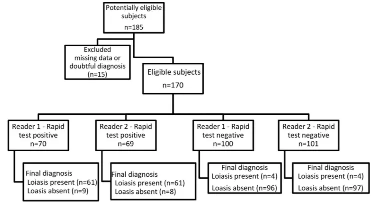
The study flow (of the RDT) in relation to the final diagnosis is reported inFig 1(Flow chart).
The agreement between the two readers was excellent (kappa’s coefficient = 0.99, 95% CI 0.96–1). Only one subject out of 170 had discordant RDT results (indeterminate versus posi-tive). This subject was from Senegal, was co-infected withM. perstans (103 mf/mL) and S. ster-coralis, and had an ELISA SI of 1.22.
Sensitivity and specificity of the RDT and of the ELISA test
Table 1shows the sensitivity of the two tests calculated against Group I, and in each of the sub-groups (patients with microfilaremia, eye worm and probable loiasis). Among the four subjects with a “probable loiasis” who were “missed” by the RDT, two were also negative at the ELISA test, with SI of 0.44 and 0.58. None of these four subjects was a migrant (three were expatriates and one was a tourist). Two were co-infected withS. stercoralis and one with Schistosoma sp., and their eosinophil counts were 1,240, 1,430, 1,830 and 4,770/μL.
Fig 1. The study flow of the RDT in relation to the final diagnosis. https://doi.org/10.1371/journal.pntd.0008187.g001
Table 2shows the specificity of the two tests calculated against Group II and III. For the RDT, most of the cross-reactions were observed in patients withM. perstans microfilaraemia: 7/37 (18.9%), followed by 1/10 (10%) with hyper-reactive malarial splenomegaly and 1/20 (5%) with schistosomiasis. None of the 27 subjects infected with intestinal nematodes was found positive at this test. In 11/18 (61.1%) patients with strongyloidiasis and in 3/5 (60%) with hookworm the ELISA test resulted positive. The ELISA (panfilarial) test was also positive in 35/37 samples from patients withM. perstans infection. No false positive results were observed when testing the 10 sera of patients with no parasitic infections.
According to reader 1, the sensitivity of the RDT was 93.8% (95% CI 85–98.3), the specific-ity 91.4% (95% CI 85–96). According the reader 2, the sensitivspecific-ity was of 93.8% (95% CI 88– 99.7), the specificity 92.4% (95% CI 85.5–96.7).
Discussion
This is the first publication describing the performances of two serological assays forL. loa using the same sample collection to allow for direct comparison between an ELISA test which is not specific for a single filarial species, and the RDT specific for loiasis. The agreement between readers of the RDT was excellent as only one sample had a different result between the two lab technicians. The accuracy of the RDT was very good. In particular, the test was pos-itive for all cases of confirmed loiasis. The sensitivity was lower in case of probable loiasis (85.7%). As patients with eye worm and/or microfilaraemia do not need further tests for con-firmation of loiasis, this RDT would be most useful for clinical decision-making of “patients
Table 2. Specificity of the RDT and ELISA test by infection type.
No. of ELISA positive samples (specificity; 95% CI)
No. of RDT positive samples (specificity; 95% CI) Reader 1
No. of RDT positive samples (specificity; 95% CI) Reader 2
Negative Controls Group II (n = 10) 0 (100%; 69.2–100)
Patients of Group III (n = 95) 51 (46.3%; 36.0–56.9) 9 (90.5%; 82.8–95.6) 8 (91.6%; 84.1–96.3) Patients of group III with hyper-reactive malarial
splenomegaly (n = 10)
0 (100%; 69.2–100) 1a(90.0%; 55.6–99.8)
Patients of group III withMansonella (n = 37) 35 (b) 7 (81.1%; 64.8–92.0) 6 (83.8%; 68.0–93.8)
Patient of group III withBrugia (n = 1) 1 (b) 0 (100%; 2.5–100)
Patients of group III withSchistosoma (n = 20) 1c(95%; 75.2–99.9)
Patients of group III withStrongyloides (n = 18) 11 (38.9%; 17.3-64-3) 0 (100%; 81.5–100) Patients of group III withAncylostoma (n = 5) 3 (40%; 5.3–85.3) 0 (100%; 47.8–100) Patients of group III withAscaris (n = 4) 0 (100%; 39.8–100)
a
correspond to the same patient (a migrant from Togo), not tested for mf, with an ELISA SI at 0.20. b
specificity was not reported as ELISA is a pan-filarial test. c
correspond to the same patient (a migrant from Senegal), not tested for mf, with an ELISA SI at 2.05.
https://doi.org/10.1371/journal.pntd.0008187.t002
Table 1. Sensitivity of the RDT and ELISA test by infection type.
ELISA RDT
No. of positive samples Sensitivity (95% CI) No. of positive samples Sensitivity (95% CI)
Patients with microfilaraemia (n = 29) 27 93.1% (77–99) 29 100% (88.1–100)
Patients with eye worm (n = 8) 6 75.0% (35.0–97.0) 8 100% (63.1–100)
Patients with probable loiasis (n = 28) 26 92.9% (76.5–99.1) 24 85.7% (67.3–96.0) All patients with confirmed and probable loiasis (n = 65) 59 90.8% (81.0–96.5) 61 93.8% (85.0–98.3)
with probable loiasis”. However, the sensitivity for the latter group was not as high as for the other two categories of loiasis. This can be due for instance to the inclusion of false positive cases (in particular two cases with negative RDT and positive ELISA that might have cross-reacted withStrongyloides stercoralis). For this reason, sensitivity and specificity of a new diag-nostic test forL. loa should be ideally assessed using PCR as the reference standard. Actually this is a limitation of our study, but retrospectively it was not possible to carry out PCR due to unavailability of whole blood. Overall, the sensitivity of the RDT was high (93.8%), i.e. very similar to the value (94%) reported by Pedram et al. [26]. The sensitivity of the ELISA was also high (90.8%) and in the same range as the rapid test when taking into account the 95% confi-dence intervals. The denominators for specificity were different for the two tests, in consider-ation of the fact that the Group III patients were not subjected to microfilaremia research, so we cannot exclude the presence ofM. perstans mf. Even after excluding the other filarial infec-tions from the denominator, the ELISA presented several cross-reacinfec-tions (in particular withS. stercoralis) that affected the specificity (77.6%). A smaller proportion of false positive results was observed for the rapid test, which cross-reacted mostly withM. perstans. The original report of the performance of the Loa Antibody Rapid Test was rich inO. volvulus (n = 99), W. bancrofti (n = 49), and S. stercoralis (n = 40) samples, and had fewer cases of M. perstans (n = 16) with reported specificities of 82–88% [26]. Our sample set was different, with no O. volvulus samples and only one W. bancrofti sample, but more sera from people with M. perstans (n = 37) for which the specificity was 81.1%, confirming the specificity range of the original article [26]. In summary, both assays perform as claimed by their respective manufac-turers. The RDT and the ELISA are both highly sensitive. The main difference lies in the extent of cross-reactivity with other parasites; the RDT cross reacts when the control population is composed of subjects infected withM. perstans (18.9%) and does not cross react with S. ster-coralis, while the ELISA reacts extensively with cases of M. perstans (95%), obviously being a pan-filarial test, andS. stercoralis (61%).
The RDT detects IgG and not the antigen, hence it results positive also in case of pastLoa loa infections. This can affect the specificity of the test that, differently to the ELISA, and can-not be recommended for the post-treatment follow-up.
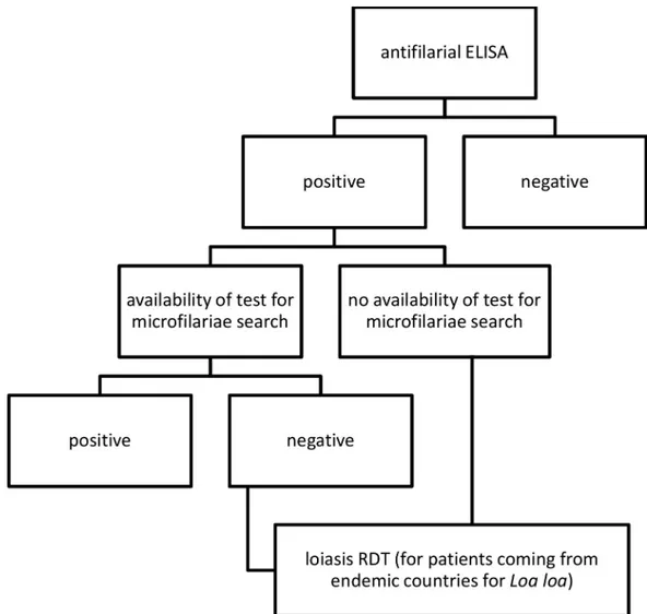
The Loa rapid test is a Research Use Only device intended primarily for epidemiological purposes and has not been approved at this point for individual case management. The RDT appears to be suitable for mappingL. loa prevalence in endemic countries. From a practical stand-point, we found it easy to run, reproducible, and convenient for use at the point of care. However, the RDT has the potential to be useful for a Tropical Diseases Center in a non-endemic country for loiasis, both for centers that assess microfilaraemia and for those that do not (Fig 2).
In case of availability of mf detection, the RDT might be carried out in case of negative microfilaraemia to confirm probable loiasis cases, while in case of unavailability of mf detec-tion, the RDT might be carried out in case of positive ELISA test to differentiate a case of loia-sis from other filarialoia-sis. Our proposed algorithm does not include costs of the tests, as a cost-effectiveness was not among the purposes of this study.
With the shown high sensitivity, this RDT can replace the microscopy in facilities that are not familiar with loiasis, as all microfilaraemic patients were detected by this RDT. However, as the SAE is related to the burden of mf and the RDT band strength does not correlate to MFD in any case it is necessary to assess microscopy before treatment. This aspect is very important considering that the treatment forL. loa is different from that of M. perstans infec-tion. For loiasis the treatment of choice is DEC [30] or IVM + ALB [31] in case of DEC unavailability. Instead, forM. perstans infection the treatment of choice seems to be doxycy-cline [32,33], although the treatment usually preferred in European countries is still
Conclusions
The novel lateral flow RDT has proven to be an accurate and user-friendly tool for the diagno-sis ofL. loa infection. While some cross-reactivity with M. perstans should be taken into account when considering its potential application as a screening tool in endemic areas, on the other hand this new test appears to be promising in the of non-endemic setting, where it could be included in a management algorithm.
Acknowledgments
We acknowledge Monica Degani, Eleonora Rizzi and Stefano Tais for the precious technical support.
Author Contributions
Conceptualization: Federico Gobbi. Data curation: Ronaldo Silva.
Formal analysis: Federico Gobbi, Dora Buonfrate, Ronaldo Silva.
Fig 2. Proposed algorithm for diagnosis of filariasis in non-endemic countries. https://doi.org/10.1371/journal.pntd.0008187.g002
Investigation: Federico Gobbi, Lucia Moro, Paola Rodari.
Methodology: Federico Gobbi, Dora Buonfrate, Michel Boussinesq, Cedric B. Chesnais,
Sebastien D. Pion, Francesca Tamarozzi, Zeno Bisoffi.
Project administration: Federico Gobbi. Supervision: Federico Gobbi, Zeno Bisoffi.
Validation: Federico Gobbi, Dora Buonfrate, Ronaldo Silva. Visualization: Federico Gobbi.
Writing – original draft: Federico Gobbi, Dora Buonfrate.
Writing – review & editing: Michel Boussinesq, Cedric B. Chesnais, Sebastien D. Pion,
Ronaldo Silva, Lucia Moro, Paola Rodari, Francesca Tamarozzi, Marco Biamonte, Zeno Bisoffi.
References
1. Metzger WG, Mordmuller B (2013) Loa loa-does it deserve to be neglected? Lancet Infect Dis. 2. Boussinesq M (2006) Loiasis. Ann Trop Med Parasitol 100: 715–731.
3. Pinder M (1988) Loa loa—a neglected filaria. Parasitol Today 4: 279–284.
4. Chesnais CB, Takougang I, Paguele M, Pion SD, Boussinesq M (2017) Excess mortality associated with loiasis: a retrospective population-based cohort study. Lancet Infect Dis 17: 108–116. 5. Gardon J, Gardon-Wendel N, Demanga N, Kamgno J, Chippaux JP, et al. (1997) Serious reactions
after mass treatment of onchocerciasis with ivermectin in an area endemic for Loa loa infection. Lancet 350: 18–22.
6. Kamgno J, Pion SD, Chesnais CB, Bakalar MH, D’Ambrosio MV, et al. (2017) A Test-and-Not-Treat Strategy for Onchocerciasis in Loa loa-Endemic Areas. N Engl J Med 377: 2044–2052.
7. Gobbi F, Postiglione C, Angheben A, Marocco S, Monteiro G, et al. (2014) Imported loiasis in Italy: an analysis of 100 cases. Travel Med Infect Dis 12: 713–717.
8. Adolph PE, Kagan IG, Mc QR (1962) Diagnosis and treatment of Acanthocheilonema perstans filaria-sis. Am J Trop Med Hyg 11: 76–88.
9. Gobbi F, Beltrame A, Buonfrate D, Staffolani S, Degani M, et al. (2017) Imported Infections with Manso-nella perstans Nematodes, Italy. Emerg Infect Dis 23: 1539–1542.
10. Garcia A, Abel L, Cot M, Richard P, Ranque S, et al. (1999) Genetic epidemiology of host predisposition microfilaraemia in human loiasis. Trop Med Int Health 4: 565–574.
11. Emukah E, Rakers LJ, Kahansim B, Miri ES, Nwoke BEB, et al. (2018) In Southern Nigeria Loa loa Blood Microfilaria Density is Very Low Even in Areas with High Prevalence of Loiasis: Results of a Sur-vey Using the New LoaScope Technology. Am J Trop Med Hyg 99: 116–123.
12. Fink DL, Kamgno J, Nutman TB (2011) Rapid molecular assays for specific detection and quantitation of Loa loa microfilaremia. PLoS Negl Trop Dis 5: e1299.
13. Drame PM, Fink DL, Kamgno J, Herrick JA, Nutman TB (2014) Loop-mediated isothermal amplification for rapid and semiquantitative detection of Loa loa infection. J Clin Microbiol 52: 2071–2077.
14. Toure FS, Bain O, Nerrienet E, Millet P, Wahl G, et al. (1997) Detection of Loa loa-specific DNA in blood from occult-infected individuals. Exp Parasitol 86: 163–170.
15. Toure FS, Mavoungou E, Kassambara L, Williams T, Wahl G, et al. (1998) Human occult loiasis: field evaluation of a nested polymerase chain reaction assay for the detection of occult infection. Trop Med Int Health 3: 505–511.
16. Antinori S, Schifanella L, Million M, Galimberti L, Ferraris L, et al. (2012) Imported Loa loa filariasis: three cases and a review of cases reported in non-endemic countries in the past 25 years. Int J Infect Dis 16: e649–662.
17. Churchill DR, Morris C, Fakoya A, Wright SG, Davidson RN (1996) Clinical and laboratory features of patients with loiasis (Loa loa filariasis) in the U.K. J Infect 33: 103–109.
18. Gantois N, Rapp C, Gautret P, Ficko C, Savini H, et al. (2013) Imported loiasis in France: a retrospective analysis of 47 cases. Travel Med Infect Dis 11: 366–373.
19. Nutman TB, Miller KD, Mulligan M, Ottesen EA (1986) Loa loa infection in temporary residents of endemic regions: recognition of a hyperresponsive syndrome with characteristic clinical manifestations. J Infect Dis 154: 10–18.
20. Saito M, Armstrong M, Boadi S, Lowe P, Chiodini PL, et al. (2015) Clinical Features of Imported Loiasis: A Case Series from the Hospital for Tropical Diseases, London. Am J Trop Med Hyg 93: 607–611. 21. Develoux M, Hennequin C, Le Loup G, Paris L, Magne D, et al. (2017) Imported filariasis in Europe: A
series of 31 cases from Metropolitan France. Eur J Intern Med 37: e37–e39.
22. Cobo F, Cabezas-Fernandez MT, Salas-Coronas J, Cabeza-Barrera MI, Vazquez-Villegas J, et al. (2015) Filariasis in sub-Saharan immigrants attended in a health area of southern Spain: clinical and epidemiological findings. J Immigr Minor Health 17: 306–309.
23. Bisoffi Z, Buonfrate D, Sequi M, Mejia R, Cimino RO, et al. (2014) Diagnostic accuracy of five serologic tests for Strongyloides stercoralis infection. PLoS Negl Trop Dis 8: e2640.
24. Klion AD, Vijaykumar A, Oei T, Martin B, Nutman TB (2003) Serum immunoglobulin G4 antibodies to the recombinant antigen, Ll-SXP-1, are highly specific for Loa loa infection. J Infect Dis 187: 128–133. 25. Burbelo PD, Ramanathan R, Klion AD, Iadarola MJ, Nutman TB (2008) Rapid, novel, specific,
high-throughput assay for diagnosis of Loa loa infection. J Clin Microbiol 46: 2298–2304.
26. Pedram B, Pasquetto V, Drame PM, Ji Y, Gonzalez-Moa MJ, et al. (2017) A novel rapid test for detect-ing antibody responses to Loa loa infections. PLoS Negl Trop Dis 11: e0005741.
27. Boscolo M, Gobbo M, Mantovani W, Degani M, Anselmi M, et al. (2007) Evaluation of an indirect immu-nofluorescence assay for strongyloidiasis as a tool for diagnosis and follow-up. Clin Vaccine Immunol 14: 129–133.
28. Gueglio B, Bordier C, Marjolet M (1995) Mise au point d’un test ELISA pour le diagnostic des filarioses humaines. Bulletin de la Societe´ Franc¸aise de Parasitologie 13: 67–72.
29. Gobbi F, Tamarozzi F, Buonfrate D, Rodari P, Tais S, et al. (2019) Laboratory Parameters after Treat-ment for Loa loa and Mansonella perstans: The Experience of a Single Referral Center for Tropical Dis-eases in a Non-Endemic Area. Am J Trop Med Hyg 100: 914–920.
30. Boussinesq M (2012) Loiasis: new epidemiologic insights and proposed treatment strategy. J Travel Med 19: 140–143.
31. Gobbi F, Buonfrate D, Tamarozzi F, Degani M, Angheben A, et al. (2019) Efficacy of High-Dose Alben-dazole with Ivermectin for Treating Imported Loiasis, Italy. Emerg Infect Dis 25: 1574–1576.
32. Coulibaly YI, Dembele B, Diallo AA, Lipner EM, Doumbia SS, et al. (2009) A randomized trial of doxycy-cline for Mansonella perstans infection. N Engl J Med 361: 1448–1458.
33. Batsa Debrah L, Phillips RO, Pfarr K, Klarmann-Schulz U, Opoku VS, et al. (2019) The Efficacy of Doxy-cycline Treatment on Mansonella perstans Infection: An Open-Label, Randomized Trial in Ghana. Am J Trop Med Hyg 101: 84–92.