HAL Id: hal-01790616
https://hal-amu.archives-ouvertes.fr/hal-01790616
Submitted on 14 May 2018
HAL is a multi-disciplinary open access
archive for the deposit and dissemination of
sci-entific research documents, whether they are
pub-lished or not. The documents may come from
teaching and research institutions in France or
abroad, or from public or private research centers.
L’archive ouverte pluridisciplinaire HAL, est
destinée au dépôt et à la diffusion de documents
scientifiques de niveau recherche, publiés ou non,
émanant des établissements d’enseignement et de
recherche français ou étrangers, des laboratoires
publics ou privés.
Non-pollen palynomorphs notes: 2. Holocene record of
Megalohypha aqua - dulces , its relation to the fossil
form genus Fusiformisporites and association with
lignicolous freshwater fungi
Lyudmila Shumilovskikh, Astrid Ferrer, Frank Schlütz
To cite this version:
Lyudmila Shumilovskikh, Astrid Ferrer, Frank Schlütz. Non-pollen palynomorphs notes: 2. Holocene
record of Megalohypha aqua - dulces , its relation to the fossil form genus Fusiformisporites and
association with lignicolous freshwater fungi. Review of Palaeobotany and Palynology, Elsevier, 2017,
246, pp.167-176. �10.1016/j.revpalbo.2017.07.002�. �hal-01790616�
Review papers
Non-pollen palynomorphs notes: 2. Holocene record of Megalohypha
aqua-dulces, its relation to the fossil form genus Fusiformisporites and
association with lignicolous freshwater fungi
Lyudmila S. Shumilovskikh
a,b,c,⁎
, Astrid Ferrer
d, Frank Schlütz
eaUniversity of Goettingen, Goettingen, Germany b
Tomsk State University, Tomsk, Russia
c
Mediterranean Institute of Marine and Terrestrial Biodiversity and Ecology, Aix-en-Provence, France
d
University of Illinois, Urbana-Champaign, USA
e
Lower Saxony Institute for Historical Coastal Research, Wilhelmshaven, Germany
a b s t r a c t
a r t i c l e i n f o
Article history: Received 4 April 2017
Received in revised form 29 June 2017 Accepted 3 July 2017
Available online 6 July 2017
Thefirst Holocene record of the freshwater ascomycete Megalohypha aqua-dulces from the sediment core Kongor (NE Iran) is presented here. Based on the similarity of the spore morphology with the fossil form genus Fusiformisporites, we establish a link between extant and fossil taxa. Comparative analysis of morphological char-acteristics of fossil spores of Fusiformisporites indicates that several different fungal groups might be included in this form genus. At leastfive species of Fusiformisporites share similar morphology with spores of Megalohypha aqua-dulces: Fusiformisporites annafrancescae, Fusiformisporites crabbii, Fusiformisporites keralensis, Fusiformisporites paucistriatus, and Fusiformisporites pseudocrabbii. Based on Fusiformisporites, the evolution of Megalohypha aqua-dulces can be traced to the late Cretaceous, corresponding with diversification of the flowering plants and pointing to a co-evolution of both groups. Megalohypha aqua-dulces has a tropical to subtropical dis-tribution but also occurs in the semi-arid steppe environments of Kongor together with other freshwater fungal genera such as Xylomyces, Dictyosporium, and Sporoschisma, which spores we describe here. The ecological re-quirements of Megalohypha indicate that its spores can be used for the palaeoecological sign of dead submerged wood as well as of tropical to subtropical conditions.
© 2017 Elsevier B.V. All rights reserved. Keywords: Palaeomycology Xylomyces Dictyosporium Sporoschisma Freshwater fungi Lignicolous fungi Fossil fungi Contents 1. Introduction . . . 168
2. Material and methods . . . 169
3. Results . . . 169
3.1. Spore morphology of Megalohypha aqua-dulces . . . 169
3.2. Records of freshwater fungi in Kongor . . . 169
4. Discussion . . . 173
4.1. Taxonomic relationship between Fusiformisporites and Megalohypha aqua-dulces . . . 173
4.2. Ecology and palaeoecology . . . 173
4.3. Geological evidence for the evolution of aquatic ascomycetes. . . 174
5. Conclusions . . . 175
Acknowledgements . . . 175
References . . . 175
⁎ Corresponding author at: University of Goettingen, Albrecht-von-Haller-Institute for Plant Sciences, Department of Palynology and Climate Dynamics, Wilhelm-Weber-Str. 2a, 37073 Goettingen, Germany.
E-mail address:lshumil@gwdg.de(L.S. Shumilovskikh).
http://dx.doi.org/10.1016/j.revpalbo.2017.07.002
0034-6667/© 2017 Elsevier B.V. All rights reserved.
Contents lists available atScienceDirect
Review of Palaeobotany and Palynology
1. Introduction
Morphologically distinctive fungal spores present in the geologi-cal record provide valuable information about a wide variety of envi-ronmental conditions, including climate, hydrological conditions, fire and erosion history, vegetation type, and organismal interactions (e.g.Elsik, 1976; Sherwood-Pike, 1988; Pirozynski, 1989; Van Geel and Aptroot, 2006; Taylor et al., 2015). The description of fossil fun-gal spores is usually carried out based on fossil material, which rarely provides identification to extant taxa (Elsik, 1976; Jansonius and Kalgutkar, 2000). Studies on the relationship between extant and fossil taxa however deliver important information for geological and mycological research by combining geological records with known fungal ecology. For example, investigation of the monotypic genus Potamomyces indicate that it likely contains several species (Schlütz and Shumilovskikh, 2013; Nuñez Otaño et al., 2016), or in
the case of Caryospora callicarpa led to the discovery of species thought to be extinct (Hawksworth et al., 2016). Further connections of fossil fungal spores to recent species are required for a deeper un-derstanding of the history, evolution, ecology and (palaeo)geogra-phy of fungal taxa.
During palynological investigations of a sediment core from Kongor (NE Iran), covering the last 6000 years (Shumilovskikh et al., 2016), fungal spores of Megalohypha aqua-dulces Ferrer et Shear-er wShear-ere documented (Plate I) and identified using mycological liter-ature (Ferrer et al., 2007). These spores share similar characteristics to additional fossilised spores from geological records described as FusiformisporitesRouse, 1962. Continuing our series of“non-pollen palynomorphs notes” (Schlütz and Shumilovskikh, 2017), in this paper we provide thefirst Holocene record of the extant species Megalohypha aqua-dulces and discuss its relation to the fossil form genus Fusiformisporites.
Plate I. Spores of Megalohypha aqua-dulces (KNG 62) from the sediment core Kongor, NE Iran, showing variation in spore morphology and different preservation grade (1–2: 176 cm core depth, 3: 224 cm, 4: 64 cm, 5–6: 48 cm, 7–8: 96 cm, 9: 72 cm, 10: 80 cm). Photos at 500× magnification with oil immersion.
2. Material and methods
The Kongor core was obtained from the temporary lake Kongor, lo-cated in the Artemisia-steppe of the eastern Gorgan Plain, NE Iran. Sam-ples from the core were treated with standard palynological laboratory procedures and studied for pollen and non-pollen palynomorphs in-cluding microscopic plant, animal and fungal remains (details in
Shumilovskikh et al., 2016). For the purpose of this paper we present an abbreviated version of the palynological diagram with freshwater fungi, arboreal pollen and tree and shrub macroremains (Fig. 1). The de-scription of the Megalohypha aqua-dulces spores (Plate I) and of the spores of other freshwater fungi (Plate III) from the sediment core Kongor follows the scheme ofElsik (1983)with an abbreviation KNG (Kongor) for thefirst described types. Spore measurements were car-ried out on 1000× magnification.
The samples from Panama were collected from freshwater habitats at the Soberania National Park, which support lowland tropical forest. Samples of submerged partially decomposed wood were incubated in the laboratory in plastic boxes containing moistened paper towels at room temperature and examined with a dissecting microscope period-ically over 12 months. Fungi were removed from the substrate and placed in a drop of distilled water on a glass slide. Measurements and photographs of the spores and fruiting bodies were made in material mounted in distilled water (Plate II;Ferrer et al., 2007).
3. Results
3.1. Spore morphology of Megalohypha aqua-dulces
Spores of Megalohypha aqua-dulces from the sediment core Kongor (KNG 62;Plate I): spores are fusiform, dark reddish-brown, dicellate, 47–60 × 21–27 μm (average = 54.4 × 23.4 μm, SD = 4.5 × 1.7 μm, n = 10), inaperturate, slightly constricted at the septum, wall
thickness 1–1.5 μm, up to 3 μm at apices. The axis is straight, 2 sym-metrical cells are separated by a septum 2–3 μm thick. The sculpture is longitudinally striate with 5–7 ridges exposed on each flattened sector. The striate pattern merges to a coarse reticulum at the apices. The morphology corresponds to the original description of spores of the extant fungus Megalohypha aqua-dulces (Ferrer et al., 2007): asco-spores 40–55 × 19–22 μm (mean = 48.3 × 18.8 μm, SD = 2.8 × 0.91 μm, n = 30), ellipsoidal, acutely tapered at apices, brown to dark brown, 1-septate, septum appearing as a dark band, both cells of equal shape and size, rough walled with longitudinal sulcate striations lacking appendages or a gelatinous sheath (Plate II).
In addition, the spores from Kongor resemble spores of the fossil form genus Fusiformisporites Rouse, 1962 with holotype Fusiformisporites crabbiiRouse, 1962(Kalgutkar and Jansonius, 2000): spores are distinctly fusiform in outline. The unit is split into two equal halves by an equatorial wall that appears to be continuous, thus completely dividing the unit. Longitudinal grooves spread out along the wall from either pole like a spindle; some reach the equator, others stop short of it. Only occasionally is a groove continuous across the di-viding wall. The wall is moderately thick, about 3μm. Ornamentation levigate. Size range 20–100 μm. Size of Fusiformisporites crabbii is 45– 52μm (Rouse, 1962).
According toKalgutkar and Jansonius (2000), Fusiformisporites in-cludes forms with less obvious parallel ornamentation elements (stria-tions) (Table 1). The authors place it taxonomically into Fungi Imperfecti, Didimosporae.
3.2. Records of freshwater fungi in Kongor
In the Kongor sediment core, Megalohypha aqua-dulces occurs to-gether with freshwater lignicolous fungi such as Sporoschisma saccardoi-type, Dictyosporium heptasporum, Dictyosporium digitatum, Zopfiella cf. submersa, and Xylomyces chlamydosporus-type (Fig. 1). Ar-boreal pollen varies between 5 and 20% throughout the record. Macroremains of trees and shrubs, including seeds, epidermis and wood, occur in the upper metre of the core.
Below we provide a description of the spores of freshwater fungi from the Kongor site.
Dictyosporium digitatum (KNG 27b;Plate III: 4–6)
Conidia are 58–62 × 28–32 μm in size, reddish-brown, multiseptate, composed of 6–7 parallel arms, closely branched from the terminal cell, flattened in one plane. The wall is smooth, about 1 μm thick. The spore morphology resembles Dictyosporium digitatum Chen, Hwang, Tzean, which is commonly found on submerged dead wood in Australia, Brunei Darussalam, Hong Kong, Seychelles, Taiwan, and Thailand (Goh et al., 1999). In Kongor,five spores were found from the Middle and Late Ho-locene (Fig. 1;Shumilovskikh et al., 2016).
Dictyosporium heptasporum (HdV 1053 byVan Geel et al., 2011;
Plate III: 1–3)
Conidia are 62–68 × 22–26 μm in size, with 7 parallel arms, branched from the terminal cell in form of a cylinder, apices of arms are incurved. The spore morphology resembles Dictyosporium heptasporum with co-nidia broad ellipsoidal, 42–71 × 21–25 μm, branched, composed of ca. 7 rows of cells (Damon, 1952).Van Geel et al. (2011)described type HdV 1053 from Lake Challa in Africa and identified them as Dictyosporium cf. heptasporum (Garov.) Damon. Two species of Dictyosporium have similar size and cylindrical morphology of conidia: D. heptasporum and Dictyosporium cocophilum (Goh et al., 1999). How-ever, the apex of the arms is straight by the latter species. Therefore, we identified our specimen as D. heptasporum. Dictyosporium heptasporum has been observed on dead wood and submerged wood in Australia, Belize, Brunei Darussalam, Cuba, Europe, Hong Kong, India, Ecuador, Mexico, Peru, Taiwan, Tanzania, Thailand, USA (Goh et al., 1999). Subfossilfinds are known from the Late Holocene from Lake Challa (Africa,Van Geel et al., 2011) and the Middle and Late Holocene from Kongor (NE Iran,Shumilovskikh et al., 2016). Spores of another
Fig. 1. Freshwater fungi in the palynological record of the Kongor sediment core. Grey lines show exaggeration line ×10. Circle indicates presence of Morus alba wood.
Plate II. Megalohypha aqua-dulces (1–2) grown in culture (Holotype AF005-2) and (3–6) collected from wood (Holotype AF005-1): 1–2: stalked ascomata, 3: longitudinal section through ascoma, 4: surface of ascospores with coarse reticulum at apices, 5: longitude section through ascospores, 6: surface of ascospores with sulcate striations. Further details inFerrer et al. (2007).
species, Dictyosporium australiense, have been documented from Holo-cene peat sediments from Germany (Shumilovskikh et al., 2015).
Sporoschisma saccardoi-type (UG 1002 byGelorini et al., 2011;
Plate III: 13–15)
Conidia are 45–50 × 15–17 μm in size, composed of 4 to 6 cells with dark central and hyaline short apical cells, subtruncate at both ends; the spore is slightly constricted at septa, with a smooth wall 1–2 μm thick. Inner dark septa 3–4 μm thick, distal septa 1–1.5 μm. Central cells are normally equal but sometimes unequal (comparePlate III: 13 and 14). Some spores were found within the conidiophore, showing production of 5 and 3-septate spores by the same fungus specimen (Plate III: 15).
Gelorini et al. (2011)associated the type UG 1002 with Sporoschisma spp. possibly with Sporoschisma saccardoi. In addition, at least Sporoschisma nigroseptatum has similar morphology (Goh et al., 1997b) and may be another Sporoschisma species, therefore here the Sporoschisma saccardoi-type is erected. Both species have been observed on submerged dead wood from Australia, Brunei Darussalam, Ecuador, Europe, Hong Kong, Indonesia, Malaysia, Peru, South Africa, Taiwan (Goh et al., 1997b). Subfossil sporefinds are known from African mod-ern lake sediments (Gelorini et al., 2011), Holocene sediments from NE Iran (Shumilovskikh et al., 2016) and terrestrial surface samples from Nepal (Shumilovskikh, unpubl.).Prager et al. (2006)affiliated EMA 12 to Sporoschisma or Chalara or hyphae. Morphologically EMA 12 differs from conidia of Sporoschisma saccardoi-type, and it might be a part of hyphae.
Xylomyces chlamydosporus-type (KNG 7;Plate III: 16–17) Conidia are 145–255 × 35–40 μm in size, fusiform, straight or slightly curved, dark-brown with pale end cells. Spores have 6–14 septa, 2–5 μm
thick. Spores are constricted at septa. The wall is 2–4 μm thick with scarce irregular longitudinal ornamentation (rugulate to coarse striate). Based on similar morphology, the type KNG 7 is assigned to hyphomy-cete genus Xylomyces. According to mycobank database (www. mycobank.org), the genus Xylomyces consists of nine species but only four, Xylomyces chlamydosporus Goos, Brooks & Lamore, Xylomyces giganteus Goh, Ho, Hyde & Tsui, Xylomyces rhizophorae Kohlm. & Volkm.-Kohlm. and Xylomyces acerosisporus Oliveira, Malosso & Castañeda, produce large spores over 140μm long with 6–14, 6–26, 11–43(–64) and 7–15 septa, respectively (Goos et al., 1977; Goh et al., 1997a; Kohlmeyer and Volkmann-Kohlmeyer, 1998; Oliveira et al., 2015). Morphologically, the type KNG 7 is the most close to X. chlamydosporus and X. giganteus. For this two and possibly upcoming species with same spore morphology the X. chlamydosporus-type is erected here.
Xylomyces chlamydosporus is the type species of Xylomyces and it wasfirst described from dead, decaying wood submerged in freshwater in southern Rhode Island and Alabama (Goos et al., 1977). The fungus was found during all seasons at water temperature ranging from 1.7 to 23.8 °C; it grows well on laboratory media within temperature range 15 to 30 °C and at salinities of up to 22.15‰ (Goos et al., 1977). Its known distribution is from Brunei Darussalam, Hong Kong, Sey-chelles, and the United States (Goh et al., 1997a). Xylomyces giganteus was described from submerged wood in Australia and is also found in South Africa and United Kingdom (Goh et al., 1997a). Ourfindings re-veal the presence of X. chlamydosporus-type during the Late Holocene in NE Iran.Campbell et al. (2007)refer Xylomyces to the order Jahnulales based on the wide mycelium and molecular evidence, whileSivichai et
Table 1
Morphological characteristics of Megalohypha aqua-dulces and Fusiformisporites species (based onKalgutkar and Jansonius, 2000; Ferrer et al. 2007). Species Length
(μm)
Width (μm)
Wall Septum Striation Apical ends Form
Megalohypha aqua-dulces
Ferrer & Shearer 2007
40–55 19–22 – Dark band Sulcate striations Acutely tapered Fusiform Subfossil M. aqua-dulces
(present study)
47–60 21–27 1–1.5 μm 2–3 μm 5–7 grooves with broader ridges Facetted sectors Fusiform Fusiformisporites
annafrancescae Norris 1997
44–55 19–24 0.25–0.5 μm (1 μm at apices)
1–2 μm 0.25–0.5 μm wide, spaced 0.5–1 μm Facetted sectors Fusiform F. crabbii Rouse 1962 45–52 – 3μm 5 grooves exposed on eachflattened
sector – Fusiform F. duenasii Kalgutkar and Jansonius 2000 28–32 8–10 – – – – – F. elongatus Ramanujam & Rao 1978
35–38.5 8–12 1μm 2.5μm Fine striate with ridges as thick as grooves Blunt ends – F. foedus Salujha, Kindra
& Rehman 1974
43.2–46.4 24.5–27.2 1.2 μm 2–2.5 μm 10 ridges about 1.5μm wide Pointed ends – F. keralensis Ramanujam
& Rao 1978
51–56 32–36 1.5μm, much thicker at each end
4μm Striae numerous, ridges slightly broader than grooves
Ends truncate to broadly arched
Fusiform to rhomboidal F. lineatus Rouse &
Mustard 1997
58–62 23–29 0.75–1.0 μm uniform
– 3–5 in each cell with a uniform width of 0.3–0.9 μm
Polar cap readily detaches or hinges open
Fusiform F. lineolatus Sheffy and
Dilcher 1971
33.8 18.4 – 1μm 6–7 ribs – Fusiform
F. mackenziei Parsons & Norris 1999
31–41 15.5– 24 Thicker at apices Incompl. septate
15 to 20 ribs Rounded apices, slight apical nub
Fusiform F. marii Elsik 1968 21 12 1μm Thicker than
wall
Two ridges in one hemisphere rotated 90° from two ridges in the opposite hemisphere
– Capsular to
ovoid F. microstriatus Hopkins
1969
42–49 – Thick, granular – Fine longitudinal ribs – Oval
F. paucistriatus Rouse & Mustard 1997
39–42 15–22 0.25–0.5 μm – 3–10 striae, very thin, irregular 1.5–2 μm thick at apices – F. pseudocrabbii Elsik 1968 40–45 25 Inner layer 0.5μm, outer 0.5–1.5 μm 2μm; two layers
Broad longitudinal ribs or folds 1.5–2 μm thick at apices Fusiform F. rugosus Sheffy and
Dilcher 1971
43.5 19.3 – 2–3 μm Folds and tears, 1μm thick Rounded at one apex, with flat basal attachment at other end
Fusiform
F. striatus (Ke & Shi) Kalgutkar and Jansonius 2000 46.4 29 1.5μm two-layered, outer layer thicker 3μm divided into two layers
Plate III. Spores of freshwater fungi from the sediment Kongor, NE Iran: 1–3: Dictyosporium heptasporum (HdV 1053; core depth 56 cm); 4–6: Dictyosporium digitatum (KNG 27b; core depth 272 cm); 7–12: Zopfiella cf. submersa (KNG 60; core depth 7–9 – 0 cm, 10–12 – 32 cm); 13–15: single conidiospores (13–14) and conidiophore bearing spores (15) of Sporoschisma saccardoi-type (UG 1002; core depth 13– 0 cm, 14 – 32 cm, 15 – 184 cm); 16–17: conidiospores of Xylomyces chlamydosporus-type (KNG 7; core depth 16 – 160 cm, 17 – 184 cm).
al. (2011)made a connection between X. chlamydosporus (anamorph) and Jahnula aquatica (teleomorph).
Goos et al. (1977)assigned Xylomyces chlamydosporus to the fossil fungus Pluricellaesporites psilatus Clarke known from late Cretaceous (Clarke, 1965). Recently, spores of Xylomyces giganteus are reported from the early Eocene formation Princeton Chert (Klymiuk et al., 2013).
Zopfiella cf. submersa (KNG 60;Plate III: 7–12)
Spores are 28–30 × 18–20 μm in size, limoniform, truncate at the base, dark-brown, with a subapical germ pore of 0.5–1 μm diameter, umbonate at the apex. The wall is 2–3 μm thick with a coarse scabrate surface. The spores resemble Zopfiella submersa Guarro, Al-Saadon, Gené et Abdullah, however its upper melanized cell is smaller (13.0– 20.5 × 10⁻¹⁴ μm). It is possible to assume a change of the spore size due to fossilisation processes or laboratory preparations or appearance of other Zopfiella species. For example, Zopfiella inermis has the largest spores in the genus (28–32 × 18–21 μm) (Malloch and Cain, 1971), cor-responding well to the size of KNG 60. Unfortunately, we could not ac-cess the original publication for comparison of all morphological characteristics. Therefore, KNG 60 is named Zopfiella cf. submersa. Al-though different Zopfiella species grow on a wide range of substrates such as herbaceous debris, rotten wood, dung, and soil in terrestrial and marine environments, Z. submersa wasfirst described from the Eu-phrates River in Iran, on submerged dead culms of Phragmites and Arundo donax (Guarro et al., 1997).
4. Discussion
4.1. Taxonomic relationship between Fusiformisporites and Megalohypha aqua-dulces
Fusiformisporites has a long identification history. The fossil form genus Fusiformisporites wasfirst described byRouse (1962)from the Tertiary sediments of the Burrard Formation of western British Colum-bia. In describing the genus Fusiformisporites and F. crabbii as the holo-type, Rouse (1962) suggested a relationship to algae such as Desmatractum bipyramidatum (Chodat) Pascher or oospores of Oedogonium, both are representatives of the Chlorococcaceae, or repre-sentatives of the Class Desmocontae (Division Pyrrophyta). However, this view was not supported.
LaterElsik (1968)recognised the fungal nature of Fusiformisporites and affiliated the genus to the extant fungus Cookeina, as illustrated by
Wolf (1967). Indeed, describing fungal spores from East African lake sediments,Wolf (1967)drew a 2-celled fungal spore with striation and erroneously named it Cookeina, a wood inhabiting pantropical genus from the Pezizales. In fact, ascospores of Cookeina can bear stria-tions but consist of only one-cell (Iturriaga and Pfister, 2006; Weinstein et al., 2002), contradictingWolf's (1967)drawings and the morphology of Fusiformisporites spores. Nevertheless, Fusiformisporites affinity to Cookeina was used in further geological studies (Germeraad, 1979; Kalgutkar and Jansonius, 2000; Singh and Chauhan, 2008; Massini and Jacobs, 2011; Taylor et al., 2015). While referring to a personal commu-nication of Elsik in 1996,Rull and Vegas-Villarúbia (1999)suggested that Fusiformisporites might be a dung fungus, indicating foraging and grazing animals. However, they did not affiliate it to any species.
Carrión and van Geel (1999)andCarrión and Navarro (2002)note an absence of published connections to extant fungal taxa and suggest a possible affinity of Fusiformisporites to ascospores of Nectria peziza, Herpotrichia lignicola, Parodiella perisporioides or Ceriophora palustris. However, all these species have different spore morphology.
Studying Holocene sediments from salt marshes,Marsh and Cohen (2008) suggested correspondence of Fusiformisporites duenasii to Atrotorquata lineata (Cainiaceae, Xylariales), which was described from standing culms of Juncus roemerianus. Similar spore morphology of both species supports this connection.
The morphology of spores from the Kongor sediment core is compa-rable to the morphology of Megalohypha aqua-dulces. The larger size of
the spores from the Kongor core (Table 1) can be explained by fossilisation processes or laboratory treatment or storing in glycerine. Influence of these processes on size is well-known for pollen, but similar studies have not been done for fungal spores. The spores share similar characteristics to some species of the fossil form genus Fusiformisporites. From 15 described fossil species of Fusiformisporites (Table 1), seven species have a similar size as Megalohypha aqua-dulces: Fusiformisporites annafrancescae, Fusiformisporites crabbii, Fusiformisporites keralensis, Fusiformisporites paucistriatus, Fusiformisporites pseudocrabbii, Fusiformisporites rugosus, and Fusiformisporites striatus. Apical thickness is described and seen on the drawings of four species: F. crabbii, F. keralensis, F. paucistriatus, and F. pseudocrabbii. Only F. annafrancescae has distinct facetted sectors in the description. The spore diversity of Megalohypha aqua-dulces from the Kongor core shows that facetted sec-tors are not easy to see in decomposed spores (Plate I: 4). Combining all morphological features, we suggest that F. crabbii, F. keralensis, F. paucistriatus, F. pseudocrabbii and F. annafrancescae can all be affiliated to Megalohypha aqua-dulces. Most likely other representatives of Fusiformisporites belong to different fungal taxa.
4.2. Ecology and palaeoecology
The fungus Megalohypha aqua-dulces A. Ferrer et Shearer is de-scribed from submerged wood in tropical forest streams in Panama and Thailand (Ferrer et al., 2007). Studies on 18S and 28S nuclear ribo-somal DNA sequences supported its position in the order Jahnulales (Campbell et al., 2007). Molecular phylogeny places Megalohypha aqua-dulces within the Jahnula sensu stricto clade in the polyphyletic genus Jahnula (Suetrong et al., 2011). Similar to other freshwater asco-mycetes, Megalohypha aqua-dulces is an important degrader of wood in fresh water, playing a key role in the process of carbon mineralization. It produces a wide spectrum of enzymes such as general cellulases, endoglucanase,β-glucosidase, xylanase, laccase, amylase, pectic lyase, and polygalacturonase, making the degradation of cellobiose, hemicel-lulose, lignin, starch and pectin possible (Simonis et al., 2008).Ferrer et al. (2007)suggested that the occurrence of soft rot cavities was caused by Megalohypha aqua-dulces, however further experimental studies did not support this conclusion (Simonis et al., 2008).
In general, freshwater ascomycetes grow in freshwater habitats and complete part or the whole of their lifecycle in water (Cai et al., 2006). They can be recorded in terrestrial and marine habitats and therefore are divided in four major groups according to their occurrence: 1) exclu-sively freshwater; 2) freshwater and terrestrial; 3) freshwater and ma-rine; 4) freshwater, marine and terrestrial (Vijaykrishna et al., 2006). Since Megalohypha aqua-dulces was described from freshwater habitats, and the known Holocene occurrences are from freshwater environ-ments, the species likely belongs to thefirst group. However, spores of F. pseudocrabbii were described from the Tertiary coastal to marginal marine environment in the Eastern Niger Delta in Nigeria (Ajaegwu et al., 2012), possibly suggesting the ability of Megalohypha aqua-dulces to occupy marine habitats.
While the entire group of freshwater fungi was highlighted for its importance in the palaeoecological studies (Sherwood-Pike, 1988), Megalohypha aqua-dulces has a potential to be used as palaeoecological indicator for presence of wood. In the Kongor sediment core, Megalohypha aqua-dulces represents a part of the freshwater fungal spore assemblage (Fig. 1). Being described from submerged wood, Megalohypha aqua-dulces indicates presence of decaying wood and therefore trees or shrubs growing on, or near the sampling site. This in-terpretation is supported by the presence of other wood decomposers such as conidia of Xylomyces chlamydosporus-type, Dictyosporium digitatum and Dictyosporium heptasporum (Cai et al., 2006). In addition, Zopfiella cf. submersa grows on herbaceous debris or wood while Sporoschisma saccardoi-type is saprobic on decaying wood and bamboo culms (Cai et al., 2006). Interestingly, the continuous presence of woody vegetation on the Kongor site is neither indicated by pollen nor by
macroremains (Fig. 1), but by insects, strongly suggesting the presence of riverine forests with Alnus or Salix close to the site (Shumilovskikh et al., 2016). In fact, pollen of woody vegetation is present in the pollen di-agram (4–20%), but due to possible long-distance transport it is not pos-sible to infer local vegetation (Fig. 1). Macroremains of trees and shrubs provide a very local signal but can rapidly become decomposed. Re-mains of Rubus, Sambucus, Morus, and Viscum occur in the upper 60 cm of the core, but are almost completely absent in the lower part (Fig. 1), possibly due to decomposition. Considering the biology of Megalohypha aqua-dulces, it is possible that wood of the above-mentioned trees and shrubs as well as of shrubby Artemisia or Chenopodiaceae might be the possible substrates for the development of freshwater lignicolous fungi at the Kongor site.
The presence of spores of freshwater fungi in sediments provides op-portunities to infer the presence of woody vegetation development at a site. This can be used, for example, for testing of the gallery forest theory in South America. Our preliminary results on the sediment core São Francisco de Assis from southern Brazil (Behling et al., 2005) provides evidence for thefirst occurrence of Megalohypha aqua-dulces in associa-tion with Potamomyces spp. (Schlütz and Shumilovskikh, 2013; Nuñez Otaño et al., 2016) in the mid-Holocene while it is absent from the sed-iment during the late glacial and early Holocene. In contrast, a core from Aguas Claras near Porte Alegre (SE Brasil) shows the presence of Megalohypha aqua-dulces during the late glacial (Medeanic and Silva, 2010).
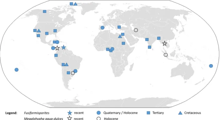
Freshwater ascomycetes have pan-tropical or pan-temperate distri-butions and may overlap in warm temperate or subtropical regions. The optimum temperature for tropical and temperate freshwater ascomy-cetes is 20–25 °C (Vijaykrishna et al., 2006). Recent documentation of Megalohypha aqua-dulces spores in Holocene peat cores from Indonesia (Fig. 2) confirms its pantropical distribution. Occurrence of the fungus in the peatland of Northern Iran and southern Brazil expands the known distribution of Megalohypha aqua-dulces to more arid subtropics, highlighting its ecological plasticity.
The modern distribution of Megalohypha aqua-dulces in the tropics, and its Holocene occurrence in the subtropics suggest that it is a good indicator of warm humid conditions in the Tertiary, as proposed from
geological records for Fusiformisporites (Elsik, 1968; Kumar, 1990; Oboh, 1992; Kalgutkar, 1997). Carrión and van Geel (1999) and
Carrión and Navarro (2002)use Fusiformisporites sp. as indicators of or-ganic matter decomposition and peaty layer formation in deposits of the Canal de Navarrés (Spain), whereas the presence of Megalohypha aqua-dulces provides evidence of decaying wood in the peat.
4.3. Geological evidence for the evolution of aquatic ascomycetes Freshwater fungi colonise streams around the world, and thus they are not restricted by geographical barriers (Wood-Eggenschwiler and Bärlocher, 1985; Vijaykrishna et al., 2006).Hyde and Goh (2003) sug-gest that fungi 1) might have evolved before the split of the continents, or 2) have been carried between continents on plant substrates, or 3) fungal spores may have dispersed by animals or wind. Ecological and molecular data suggest that freshwater ascomycetes should have evolved from their terrestrial ancestors (Vijaykrishna et al., 2006) and may have multiple origins (Belliveau and Bärlocher, 2005). Molecular studies propose that the earliest divergence of freshwater species in Jahnulales occurred at 380 ± 100 MYA (Paleozoic period), however most of the freshwater lineages appear to have diverged during the Me-sozoic period (66-245 MYA) (Vijaykrishna et al., 2006). In contrast, palaeontological studies reveal that fungal diversity increased after the Cretaceous–Tertiary boundary and that fungi underwent rapid speciali-sation during the Tertiary, connected with the evolution of angiosperms (Graham, 1962; Kalgutkar, 1993, 1997).
In the absence of the molecular data for the evolution of Megalohypha aqua-dulces, palaeontological data reveal the occurrence of Fusiformisporites since the Upper Cretaceous (Fig. 2), corresponding well with the diversification of flowering plants and pointing to a co-evolution of both groups. Thefirst description of Fusiformisporites, F. crabbii, was made from Upper Cretaceous to the Middle Eocene sedi-ments of the Burrard Formation of western British Columbia (Rouse, 1962). Further Upper Cretaceousfinds of Fusiformisporites are known from NW Bolivia (Vajda-Santivanez, 1999), NW Ellesmere Island in Canada (Falcon-Lang et al., 2004) and the north-western desert in Egypt (El Beialy et al., 2010).
During the Tertiary, Fusiformisporites evolves further and becomes more diverse and abundant (Fig. 2,Elsik, 1976). In the Paleogene, Fusiformisporites pseudocrabbii, Fusiformisporites crabbii, Fusiformisporites lineolatus, Fusiformisporites lineatus, Fusiformisporites annafrancescae, Fusiformisporites microstriatus, Fusiformisporites paucistriatus, Fusiformisporites rugosus and other Fusiformisporites spe-cies are documented in the USA (Elsik, 1968; Sheffy and Dilcher, 1971), Canada (Kalgutkar, 1997; Norris, 1997), coasts of North America (Mustard and Rouse, 1994; Jansonius and Kalgutkar, 2000), Jamaica (Germeraad, 1979), central Ecuador (Jaillard et al., 2004), India (Saxena, 2006; Singh et al., 2011), Northern Trace basin (Turkey) (Ediger and Alişan, 1989), Cameroon (Salard-Cheboldaeff, 1979) and the Ethiopian Plateau (Massini and Jacobs, 2011). Neogenefinds of Fusiformisporites are less diverse. Fusiformisporites pseudocrabbii was found in the Eastern Niger Delta in Nigeria (Ajaegwu et al., 2012) and in the Parana Formation of Argentina (Garralla, 1989). Fusiformisporites crabbii is known from the Niger Delta (Bankole et al., 2014) and in Mi-zoram of NE India (Nandi and Sinha, 2007; Kar et al., 2010). Fusiformisporites acutus is described from the Miocene Quilon Beds of Kerala State in India (Kumar, 1990). Other unidentified Fusiformisporites are documented in the Niger Delta (Oboh, 1992), Cameroon (Tchouatcha et al., 2010), the Gulf of Suez in Egypt (El Beialy et al., 2005), the Gulf of California (Helenes et al., 2009), and India (Singh and Chauhan, 2008).
During the Quaternary, Fusiformisporites was found in the late glacial sediments in southern Brazil (Medeanic and Silva, 2010) as well as in the Holocene sediments of the Changuinola peat deposit in Panama (Phillips, 1995), at Holland bay and Bowden in Jamaica (Germeraad, 1979), SW Pacific islands (Macphail and Stevenson, 2004) and in the late Quaternary– Holocene sediments of Spain (Carrión and van Geel, 1999; Carrión and Navarro, 2002).Rull and Vegas-Villarúbia (1999)
found Fusiformisporites in surface samples from a coastal basin in Vene-zuela. Al-Ameri and Jassim (2011) indicated the presence of Fusiformisporites in the late Quaternary sediments of southern Iraq, but a photo of the hyaline spore does not correspond to Fusiformisporites. In addition, Holocenefinds of Fusiformisporites, new records of Megalohypha aqua-dulces are documented for north-eastern Iran, southern Brazil and Indonesia (Fig. 2).
Based on Fusiformisporites, the evolution of Megalohypha aqua-dulces can be traced to the late Cretaceous, defining its divergence within Jahnulales (Vijaykrishna et al., 2006).
5. Conclusions
Several species of the form genus Fusiformisporites have been de-scribed from Cretaceous to Holocene times (Kalgutkar and Jansonius, 2000). Not all of them seem to fulfil the morphological criteria of the genus Fusiformisporites as erected byRouse (1962). The type species Fusiformisporites crabbii and the later erected Fusiformisporites annafrancescae, Fusiformisporites keralensis, Fusiformisporites paucistriatus, and Fusiformisporites pseudocrabbii are in close morpho-logical accordance to the extant fungi Megalohypha aqua-dulces (Ferrer et al., 2007). Megalohypha aqua-dulces is a lignicolous freshwater fungus from the tropics belonging to order Jahnulales. Its substrate and habitat preference is underlined by the co-occurrence of spores from additional lignicolous freshwater taxa as Sporoschisma saccardoi-type, Dictyosporium heptasporum, Dictyosporium digitatum, Zopfiella cf. submersa, Xylomyces chlamydosporus-type in the Holocene record from NE Iran (Shumilovskikh et al., 2016). Until now known recent and Holocenefinds of Megalohypha aqua-dulces indicate that it can be expected in appropriate habitats of the tropics and subtropics including semi-arid regions. Its evolutionary history reaches back into the late Cretaceous and is most probably associated with the diversification of angiosperms. In the fossil context, Megalohypha aqua-dulces is a good indicator for decaying wood, even if macroremains of trees and shrubs do not appear in the sediment.
Acknowledgements
We thank James Dalling and Bas van Geel for helpful comments on the manuscript. The study is conducted within the European Research Council project PERSIA (grant 295375) and partly supported by the Russian Foundation for Basic Research (16-35-60083) and the Tomsk State University competitiveness improvement programme (grant 8.1.19.2017).
References
Ajaegwu, N.E., Odoh, B.I., Akpunonu, E.O., Obiadi, I.I., Anakwuba, E.K., 2012.Late Miocene to early Pliocene palynostratigraphy and palaeoenvironments of ANE-1 Wall, Eastern Niger Delta, Nigeria. J. Min. Geol. 48, 31–43.
Al-Ameri, T.K., Jassim, S.Y., 2011.Environmental changes in the wetlands of Southern Iraq based on palynological studies. Arab. J. Geosci. 4, 443–461.
Bankole, S.I., Schrank, E., Osterloff, P.L., 2014.Palynostratigraphy, palaeoclimates and palaeodepositional environments of the Miocene aged Agbada Formation in the Niger Delta, Nigeria. J. Afr. Earth Sci. 95, 41–62.
Behling, H., Pillar, V., Bauermann, S.G., 2005.Late Quaternary grassland (Campos), gallery forest,fire and climate dynamics, studied by pollen, charcoal and multivariate analy-sis of the São Francisco de Asanaly-sis core in western Rio Grande do Sul (southern Brazil). Rev. Palaeobot. Palynol. 133, 235–248.
Belliveau, M.J.-R., Bärlocher, F., 2005.Molecular evidence confirms multiple origins of aquatic hyphomycetes. Mycol. Res. 109, 1407–1417.
Cai, L., Hyde, K.D., Tsui, C.K.M., 2006.Genera of freshwater fungi. Fungal Diversity Re-search Series 18. Fungal Diversity Press.
Campbell, J., Ferrer, A., Raja, H.A., Sivichai, S., Shearer, C.A., 2007.Phylogenetic relation-ships among taxa in the Jahnulales inferred from 18S and 28S nuclear ribosomal DNA sequences. Can. J. Bot. 85, 873–882.
Carrión, J.S., Navarro, C., 2002.Cryptogam spores and other non-pollen microfossils as sources of palaeoecological information: case-studies from Spain. Ann. Bot. Fenn. 39, 1–14.
Carrión, J.S., van Geel, B., 1999.Fine-resolution Upper Weichselian and Holocene palyno-logical record from Navarrés (Valencia, Spain) and a discussion about factors of Med-iterranean forest succession. Rev. Palaeobot. Palynol. 106, 209–236.
Clarke, R.T., 1965.Fungal spores from Vermejo Formation coal beds (Upper Cretaceous) of central Colorado. Mt. Geol. 2, 85–93.
Damon, S.C., 1952.Type studies in Dyctiosporium, Speira, and Cattanea. Lloydia 15, 110–124.
Ediger, V.Ş., Alişan, C., 1989.Tertiary fungal and algal palynomorph biostratigraphy on the Northern Thrace Basin, Turkey. Rev. Palaeobot. Palynol. 58, 139–161.
El Beialy, S.Y., Mahmoud, M.S., Ali, A.S., 2005.Insights on the age, climate and depositional environments of the Rudeis and Kareem formations, GS-78-1 well, Gulf of Suez, Egypt: a palynological approach. Rev. Esp. Micropaleontol. 37, 273–289.
El Beialy, S.Y., El Atfy, H.S., Zavada, M.S., El Khoriby, E.M., Abu-Zied, R.H., 2010. Palynolog-ical, palynofacies, paleoenvironmental and organic geochemical studies on the Upper Cretaceous succession of the GPTSW-7 well, North Western Desert, Egypt. Mar. Pet. Geol. 27, 370–385.
Elsik, W.C., 1968.Palynology of a Paleocene Rockdale lignite, Milam county, Texas. I. Mor-phology and taxonomy. Pollen Spores 10, 263–314.
Elsik, W.C., 1976.Microscopic fungal remains and cenozoic palynostratigraphy. Geosci. Man 15, 115–120.
Elsik, W.C., 1983.Annotated glossary of fungal palynomorphs. AASP Contributions Series 11, pp. 1–42.
Falcon-Lang, H.J., MacRae, R.A., Csank, A.Z., 2004.Palaeoecology of Late Cretaceous polar vegetation preserved in the Hansen Point Volcanics, NW Ellesmere Island, Canada. Palaeogeogr. Palaeoclimatol. Palaeoecol. 212, 45–64.
Ferrer, A., Sivichai, S., Shearer, C.A., 2007.Megalohypha, a new genus in the Jahnulales from aquatic habitats in the tropics. Mycologia 99, 456–460.
Garralla, S., 1989.Palinomorfos (fungi) de la formacion Parana (Mioceno superior) del Pozo Josefina, provincia de Santa Fe, Argentina. Rev. Asoc. Cienc. Nat. Litoral 20, 29–39.
Gelorini, V., Verbeken, A., van Geel, B., Cocquyt, C., Verschuren, D., 2011.Modern non-pol-len palynomorphs from East African lake sediments. Rev. Palaeobot. Palynol. 164, 143–173.
Germeraad, J.H., 1979.Fossil remains of fungi, algae and other organisms from Jamaica. Scr. Geol. 52, 1–41.
Goh, T.K., Ho, W.H., Hyde, K.D., Tsui, K.M., 1997a.Four new species of Xylomyces from sub-merged wood. Mycol. Res. 101, 1323–1328.
Goh, T.K., Ho, W.H., Hyde, K.D., Umali, T.E., 1997b.New records and species of Sporoschisma and Sporoschismopsis from submerged wood in the tropics. Mycol. Res. 101, 1295–1307.
Goh, T.K., Hyde, K.D., Ho, W.H., Yanna, 1999.A revision of the genus Dictyosporium, with descriptions of three new species. Fungal Divers. 2, 65–100.
Goos, R.D., Brooks, R.D., Lamore, B.J., 1977.An undiscribed hyphomycete from wood sub-merged in a Rhode Island stream. Mycologia 69, 280–286.
Graham, A., 1962.The role of fungal spores in palynology. J. Paleontol. 36, 60–68.
Guarro, J., Al-Saadoon, A.H., Gené, J., Abdullah, S.K., 1997.Two new cleistothecial ascomy-cetes from Iraq. Mycologia 89, 955–961.
Hawksworth, D.L., Webb, J.A., Wiltshire, P., 2016.Caryospora callicarpa: found in archaeo-logical and modern preparations– but not collected since 1865. Field Mycol. 11, 55–59.
Helenes, J., Carreño, A.L., Carrillo, R.M., 2009.Middle to late Miocene chronostratigraphy and development of the northern Gulf of California. Mar. Micropaleontol. 72, 10–25.
Hyde, K.D., Goh, T.K., 2003.Adaptation for dispersal infilamentous freshwater fungi. In: Tsui, C.K.M., Hyde, K.D. (Eds.), Freshwater Mycology. Fungal Diversity Press, Hong Kong, pp. 231–258.
Iturriaga, T., Pfister, D.H., 2006.A monograph of the genus Cookeina (Ascomata, Pezizales, Sarcoscyphaceae). Mycotaxon 95, 137–180.
Jaillard, E., Ordoñez, M., Suárez, J., Toro, J., Iza, D., Lugo, W., 2004.Stratigraphy of the late Cretaceous– paleogene deposits of the Cordillera Occidental of central Ecuador: geodynamic implications. J. S. Am. Earth Sci. 17, 49–58.
Jansonius, J., Kalgutkar, R.M., 2000.Redescription of some fossil fungal spores. Palynology 24, 37–47.
Kalgutkar, R.M., 1993.Paleogene fungal palynomorphs from Bonnet Plume Formation, Yukon Territory. Geol. Surv. Can. Bull. 444, 51–105.
Kalgutkar, R.M., 1997.Fossil fungi from the lower Tertiary Iceberg Bay Formation, Eukeka Sound Group, Axel Heiberg Island, Northwest Territories, Canada. Rev. Palaeobot. Palynol. 97, 197–226.
Kalgutkar, R.M., Jansonius, J., 2000.Synopsis of fossil fungal spores, mycelia and fructifica-tions. AASP Contributions Series 39.
Kar, R., Mandaokar, B.D., Kar, R.K., 2010.Fungal taxa from the Miocene sediments of Mi-zoram, northeast India. Rev. Palaeobot. Palynol. 158, 240–249.
Klymiuk, A.A., Taylor, T.N., Taylor, E.L., Krings, M., 2013.Paleomycology of the Princeton Chert I. Fossil hyphomycetes associated with the early Eocene aquatic angiosperm, Eorhiza arnoldii. Mycologia 105, 521–529.
Kohlmeyer, J., Volkmann-Kohlmeyer, B., 1998.A new marine Xylomyces on Rhizophora from the Caribbean and Hawaii. Fungal Divers. 1, 159–164.
Kumar, P., 1990.Fungal remains from the Miocene Quilon Beds of Kerala State, South India. Rev. Palaeobot. Palynol. 62, 13–28.
Macphail, M., Stevenson, J., 2004.Fungal Spores in Archaeological Contexts: Part 1: Back-ground Evidence. Centre for Archaeological Research, Canberra.
Malloch, D., Cain, R.F., 1971.New cleistothecial Sordariaceae and a new family, Coniochaetaceae. Can. J. Bot. 49, 869–880.
Marsh, P.E., Cohen, A.D., 2008.Identifying high-level salt marshes using a palynomorphic fingerprint with potential implications for tracking sea level change. Rev. Palaeobot. Palynol. 148, 60–69.
Massini, J.L.G., Jacobs, B.F., 2011.The effects of volcanism on Oligocene-age plant commu-nities from the Ethiopian Plateau, and implications for vegetational resilience in a heterogeneous landscape. Rev. Palaeobot. Palynol. 164, 211–222.
Medeanic, S., Silva, M.B., 2010.Indicative value of non-pollen palynomorphs (NPPs) and palynofacies for paleoreconstructions: Holocene peat, Brazil. Int. J. Coal Geol. 84, 248–257.
Mustard, P.S., Rouse, G.E., 1994.Stratigraphy and evolution of Tertiary Georgia Basin and subjacent Upper Cretaceous sedimentary rocks, southwestern British Columbia and northwestern Washington State. In: Monger, J.W.H. (Ed.), Geology and Geological Hazards of the Vancouver Region, Southwestern British Columbia. Geological Survey of Canada Bulletin 481, pp. 97–169.
Nandi, B., Sinha, A., 2007.Validation of the Miocene fungal spore Mediaverrunites from Mizoram, India. Palynology 31, 95–100.
Norris, G., 1997.Paleocene-Pliocene deltaic to inner shelf palynostratigraphic zonation, depositional environments and paleoclimates in the Imperial ADGO F-28 Well, Beau-fort-Mackenzie basin. Geol. Surv. Can. Bull. 523 (71 pp.).
Nuñez Otaño, N., Di Pasquo, M., Bianchinotti, M.V., 2016.The occurrence of Potamomyces palmarensis sp. nov. in the Late Holocene of El Palmar National Park (Colón, Entre Ríos, Argentina) and transfer of fossil species of Mediaverrunites to Potamomyces. Pal-ynology 41, 267–277.
Oboh, F.E., 1992.Multivariate statistical analyses of palynodebris from the Middle Mio-cene of the Niger Delta and their environmental significance. Palaios 7, 559–573.
Oliveira, M.S., Malosso, E., Barbosa, M.A., Araújo, M.A.G., Castañeda-Ruiz, R.F., 2015.
Xylomyces acerosisporus sp. nov. from submerged leaves from Brazil. Mycotaxon 130, 875–878.
Phillips, S., 1995.Holocene Evolution of the Changuinola Peat Deposit, Panama: Sedimen-tology of a Marine-influenced Tropical Peat Deposit on a Tectonically Active Coast. (PhD Thesis). The University of British Columbia.
Pirozynski, K.A., 1989.Methods in Quaternary ecology #9. Fungi. Geosci. Can. 16, 183–189.
Prager, A., Barthelmes, A., Theuerkauf, M., Joosten, H., 2006.Non-pollen palynomorphs from modern Alder carrs and their potential for interpreting microfossil data from peat. Rev. Palaeobot. Palynol. 141, 7–31.
Rouse, G.E., 1962.Plant microfossils from Burrard Formation of Western British Columbia. Micropaleontology 8, 187–218.
Rull, V., Vegas-Villarúbia, T., 1999.Surface palynology of a small coastal basin from Vene-zuela and potential paleoecological applications. Micropaleontology 45, 365–393.
Salard-Cheboldaeff, M., 1979.Palynologie maestrichtienne et tertiaire du Cameroun. Etudie qualitative et repartition verticale des principales especes. Rev. Palaeobot. Palynol. 28, 365–388.
Saxena, R.K., 2006.A Catalogue of Tertiary Fungi from India. Birbal Sahni Institute of Palaeobotany, Lucknow.
Schlütz, F., Shumilovskikh, L.S., 2013.On the relation of Potamomyces armatisporus to the fossil form-type Mediaverrunites and its taxonomical and ecological implications. Fungal Ecol. 6, 309–315.
Schlütz, F., Shumilovskikh, L.S., 2017.Non-pollen palynomorphs notes: 1. Type HdV-368 (Podospora-type), description of associated species, and thefirst key to related spore types. Rev. Palaeobot. Palynol. 239, 47–54.
Sheffy, M.V., Dilcher, D.L., 1971.Morphology and taxonomy of fungal spores. Palaeontogr. Abt. B 133, 34–51.
Sherwood-Pike, M.A., 1988.Freshwater fungi: fossil record and paleoecological potential. Palaeogeogr. Palaeoclimatol. Palaeoecol. 62, 271–285.
Shumilovskikh, L.S., Schlütz, F., Achterberg, I., Bauerochse, A., Leuschner, H.H., 2015.The development of the raised bog“Borsteler Moor” (Lower Saxony, Germany) based on non-pollen palynomorph data. Stud. Quat. 32, 5–18.
Shumilovskikh, L.S., Hopper, K., Djamali, M., Ponel, P., Demory, F., Rostek, F., Tachikawa, K., Bittmann, F., Golyeva, A., Guibal, F., Talon, B., Wang, L.-C., Nezamabadi, M., Bard, E., Lahijani, H., Nokandeh, J., Omrani Rekavandi, H., de Beaulieu, J.-L., Sauer, E., Andrieu-Ponel, V., 2016.Landscape evolution and agro-sylvo-pastoral activities on the Gorgan Plain (NE Iran) in the last 6000 years. The Holocene 26, 1676–1691.
Simonis, J.L., Raja, Huzefa A., Shearer, C.A., 2008.Extracellular enzymes and soft rot decay: are ascomycetes important degraders in fresh water? Fungal Divers. 31, 135–146.
Singh, S.K., Chauhan, M.S., 2008.Fungal remains from the Neogene sediments of Mahuadanr valley, Latehar district, Jharkhand, India and their palaeoclimatic signifi-cance. J. Palaeontol. Soc. India 53, 73–81.
Singh, S.D., Sinha, S., Shukla, S., Gupta, A., Shanmukhappa, M., 2011.Refinement of paleobathymentric curves of paleocene– early eocene sequences in selected wells of Cambay Basin. Abstract Proceeding. The 2nd South Asian Geoscience Conference and Exhibition, GeoIndia2011.
Sivichai, S., Sri-indrasudthi, V., Gareth Jones, E.B., 2011.Jahnula aquatica and its anamorph Xylomyces chlamidosporus on submerged wood in Thailand. Mycotaxon 116, 137–142.
Suetrong, S., Boonyuen, N., Pang, K.-L., Ueapattanakit, J., Klaysuban, A., Sri-indrasutdhi, V., Sivichai, S., Jones, E.B.G., 2011.A taxonomic revision and phylogenetic reconstruction of the Jahnulales (Dothideomycetes), and the new family Manglicolaceae. Fungal Di-vers. 51, 163–188.
Taylor, T.N., Krings, M., Taylor, E.L., 2015.Fossil fungi. Elsevier, Amsterdam.
Tchouatcha, S.M., Ricard, N.N.P., Salah, M.M., Said, D.A., Ekodeck, E.G., 2010.Existence of “late continental” deposits in the Mbere and Djerem sedimentary basins (North Cam-eroon): palynologic and stratigraphic evidence. J. Geol. Min. Res. 2, 159–169.
Vajda-Santivanez, V., 1999.Miospores from upper Cretaceous– paleocene strata in north-western Bolivia. Palynology 23, 181–196.
Van Geel, B., Aptroot, A., 2006.Fossil ascomycetes in Quaternary deposits. Nova Hedwigia 82, 313–329.
Van Geel, B., Gelorini, V., Lyaruu, A., Aptroot, A., Rucina, S., Marchant, R., Sinninghe Damsté, J.S., Verschuren, D., 2011.Diversity and ecology of tropical African fungal spores from a 25,000-year palaeoenvironmental record in southeastern Kenya. Rev. Palaeobot. Palynol. 164, 174–190.
Vijaykrishna, D., Jeewon, R., Hyde, K.D., 2006.Molecular taxonomy, origins and evolution of freshwater ascomycetes. Fungal Divers. 23, 351–390.
Weinstein, R.N., Pfister, D.H., Iturriaga, T., 2002.A phylogenetic study of the genus Cookeina. Mycologia 94, 673–682.
Wolf, F.A., 1967.Fungus spores in east African lake sediments. Mycologia 59, 397–404.
Wood-Eggenschwiler, S., Bärlocher, F., 1985.Geographical distribution of Ingoldian fungi. Verh. Int. Ver. Theor. Angew. Limnol. 22, 2780–2785.