HAL Id: hal-01529469
https://hal.archives-ouvertes.fr/hal-01529469
Submitted on 26 Sep 2017
HAL is a multi-disciplinary open access
archive for the deposit and dissemination of
sci-entific research documents, whether they are
pub-lished or not. The documents may come from
teaching and research institutions in France or
abroad, or from public or private research centers.
L’archive ouverte pluridisciplinaire HAL, est
destinée au dépôt et à la diffusion de documents
scientifiques de niveau recherche, publiés ou non,
émanant des établissements d’enseignement et de
recherche français ou étrangers, des laboratoires
publics ou privés.
melanoma in vivo
Cécile Campagne, Edouard Reyes Gomez, M.E. Picco, Sophia Loiodice, Paul
Salaun, Jacky Ezagal, Florence Bernex, P.H. Commere, Stéphanie Pons,
Diane Esquerre, et al.
To cite this version:
Cécile Campagne, Edouard Reyes Gomez, M.E. Picco, Sophia Loiodice, Paul Salaun, et al.. RACK1
cooperates with NRASQ61K to promote melanoma in vivo. Cellular Signalling, Elsevier, 2017, In
Press, �10.1016/j.cellsig.2017.03.015�. �hal-01529469�
UNCORRECTED
PROOF
Contents lists available at ScienceDirect
Cellular Signalling
journal homepage: www.elsevier.comRACK1 cooperates with NRAS
Q61K
to promote melanoma in vivo
C. Campagne
a, b, ⁎, E. Reyes-Gomez
a, b, c, M.E. Picco
d, S. Loiodice
a, b, 1, P. Salaun
a, b, J. Ezagal
a, b, F. Bernex
a, b, c, 2,
P.H. Commère
e, S. Pons
f, D. Esquerre
g, E. Bourneuf
h, i, J. Estellé
h, U. Maskos
f, P. Lopez-Bergami
d, j,
G. Aubin-Houzelstein
a, b, J.J. Panthier
a, b, k, l, &G. Egidy
a, b, h, ⁎⁎aINRA, UMR955 Génétique Fonctionnelle et Médicale, Ecole Nationale Vétérinaire d'Alfort, F-94704 Maisons-Alfort, France
bUniversité Paris-Est, Ecole Nationale Vétérinaire d'Alfort, UMR955 Génétique Fonctionnelle et Médicale, F-94704 Maisons-Alfort, France
cUniversité Paris-Est, Ecole Nationale Vétérinaire d'Alfort, Unité d'Embryologie, d'Histologie et d'Anatomie Pathologique, F-94704 Maisons-Alfort, France dInstituto de Medicina y Biologia Experimental, CONICET, Buenos Aires, Argentina
ePlateforme de Cytométrie, Département d'Immunologie, Institut Pasteur, F-75724 Paris, France
fUnité Neurobiologie Intégrative des Systèmes Cholinergiques, UMR 3571, CNRS, Institut Pasteur, F75724 Paris Cedex 15, France gGenPhySE, Université de Toulouse, INRA, INPT, ENVT, Castanet Tolosan, France
hGABI, INRA, AgroParisTech, Université Paris-Saclay, Jouy-en-Josas, France iLREG, CEA, Université Paris-Saclay, F-78352 Jouy-en-Josas, France
jCentro de Estudios Biomédicos, Biotecnologicos, Ambientales y Diagnostico, Universidad Malmonides, CONICET, Buenos Aires, Argentina kCNRS URM 3738, USC INRA 2026, F-75724, France
lInstitut Pasteur, Département de Biologie du Développement et Cellules Souches, Génétique fonctionnelle de la Souris, 25 rue du Docteur Roux, Paris F-75724, France
A R T I C L E I N F O Keywords: Scaffold MAPK pathways Melanocyte JNK STAT3 Angiogenesis A B S T R A C T
Melanoma is the deadliest skin cancer. RACK1 (Receptor for activated protein kinase C) protein was proposed as a biological marker of melanoma in human and domestic animal species harboring spontaneous melanomas. As a scaffold protein, RACK1 is able to coordinate the interaction of key signaling molecules implicated in both physiological cellular functions and tumorigenesis. A role for RACK1 in rewiring ERK and JNK signaling path-ways in melanoma cell lines had been proposed. Here, we used a genetic approach to test this hypothesis in
vivo in the mouse. We show that Rack1 knock-down in the mouse melanoma cell line B16 reduces
invasive-ness and induces cell differentiation. We have developed the first mouse model for RACK1 gain of function,
Tyr::Rack1-HA transgenic mice, targeting RACK1 to melanocytes in vivo. RACK1 overexpression was not
suffi-cient to initiate melanomas despite activated ERK and AKT. However, in a context of melanoma predisposition, RACK1 overexpression reduced latency and increased incidence and metastatic rate. In primary melanoma cells from Tyr::Rack1-HA, Tyr::NRasQ61Kmice, activated JNK (c-Jun N-terminal kinase) and activated STAT3 (signal
transducer and activator of transcription 3) acted as RACK1 oncogenic partners in tumoral progression. A sequen-tial and coordinated activation of ERK, JNK and STAT3 with RACK1 is shown to accelerate aggressive melanoma development in vivo.
1. Introduction
Cutaneous melanoma is the deadliest skin cancer. Melanoma has a high metastatic capacity. Despite recent clinical breakthroughs, the ma-jority of metastatic melanoma patients do not survive [1]. The study of a minipig melanoma model revealed an overexpression of RACK1 (Re-ceptor for activated protein kinase C) mRNA in melanoma cells [2].
RACK1 protein is strongly expressed in melanoma cells of primary tumors and metastases in different mammalian species: patients [2], horses [3] and dogs [4]. In sharp contrast, RACK1 is not detected in nor-mal skin melanocytes or in nævi by immunofluorescence [2–4]. Inter-estingly, RACK1 increased the survival of human melanoma MeWo cells following UV induced-apoptosis. Moreover, inhibition of RACK1 expres-sion using RNA interference was shown to reduce the tumorigenicity of MeWo cells in a xenograft model [5].
⁎ Correspondence to: C. Campagne, INRA, UMR955 Génétique Fonctionnelle et Médicale, Ecole Nationale Vétérinaire d'Alfort, F-94704 Maisons-Alfort, France. ⁎⁎ Correspondence to: G. Egidy, GABI, INRA, AgroParisTech, Université Paris-Saclay, 78350 Jouy-en-Josas, France.
Email addresses: cecile.campagne@curie.fr (C. Campagne); giorgia.egidy-maskos@inra.fr (G. Egidy)
1 Current address: Laboratoire de Physiopathologie Orale et Moléculaire, Centre de Recherche des Cordeliers, INSERM UMR1138, F-75006 Paris, France. 2 Current address: RHEM - Institut de Recherche en Cancérologie de Montpellier, INSERM U896, F-34298 Montpellier, France.
http://dx.doi.org/10.1016/j.cellsig.2017.03.015
UNCORRECTED
PROOF
RACK1 is a ubiquitous and abundant protein [6]. It is a scaffold con-taining seven WD40 repeats considered protein-protein interaction plat-forms. Through its ability to coordinate the interaction of key signaling molecules, RACK1 is thought to integrate various pathways involved in both physiological and tumorigenic cellular functions making it a sig-naling hub [7]. Yet, the extent to which the multiple binding partners of RACK1 are coordinated has not been much tested in vivo. In an attempt to alter RACK1 levels in mammals, the group of S. Biffo obtained one mouse line with a hypomorphic Rack1 allele. While homozygosity for that hypomorphic Rack1 allele resulted in a lethal phenotype, heterozy-gous adult mice showed no major phenotype except for a belly spot and hypopigmented tail and paws [8], typical features of a developmental defect in melanoblast migration.
A role for RACK1 in the crosstalk between ERK (Extracellular sig-nal-regulated kinase) and JNK (c-Jun N-terminal kinase) signaling in melanoma was proposed to set up a feed forward mechanism triggering tumoral progression [9]. In the light of these in vitro data, we hypothe-sized that gain of function of RACK1 targeted to melanocytes in the con-text of NRas constitutive activation would accelerate melanomagenesis by strengthening converging tumoral signaling.
As for other solid cancers, cutaneous melanoma development is con-sidered as a multistep process. Melanomagenesis requires a combination of gain of function mutations in oncogenes and loss of function muta-tions in tumor suppressor genes [10]. The first spontaneous
metastasiz-ing melanoma model harbored the NRasQ61Kmutation in a deleted
Cd-kn2a background [11]. To test whether an overexpression of RACK1 was sufficient to trigger melanoma, we created Tyr::Rack1-HA transgenic mice in which a hemaglutinin (HA) epitope-tagged-RACK1 is expressed off the Tyrosinase promoter. We show here that RACK1 overexpression is not sufficient to trigger nevi or melanomas despite ERK and AKT activa-tion. Yet, in a context of melanoma predisposition, RACK1 melanocytic overexpression reduced latency and increased incidence and metasta-tic rate. We found activated JNK and STAT3 as partners of RACK1 in melanomagenesis.
2. Materials and methods
2.1. Mice and genotyping
Mouse Rack1 cDNA was tagged with HA by PCR before insertion in a pBSK-UPT-Tyr-SV40 plasmid [12]. Micro-injection of the linearized vector was made in B6CBAF1/J fertilized oocytes. Tyr::Rack1-HA trans-genic founders were characterized by Southern blot analysis and PCR genotyping. Data come from the 7th backcross onwards on C57BL/6J
background. The Pax3GFPand -Cdkn2a alleles and Tyr::NRasQ61K
trans-gene have been backcrossed onto the C57BL/6J background for > 15 generations [13]. Animal care and use for this study were approved by the ethical board of Alfort Veterinary School in accordance with Euro-pean Union Standards (agreement number 16, notice 14/02/12-4). To identify the Tyr::Rack1-HA transgene, the following primers were used: forward: 5′-gtcgacatgaccgagcagatgacc-3′ and reverse 5′-tacatggttgcgc-catctgcgccgcgggtaccaatag-3′. PCR conditions were 30 s at 94 °C, 30 s at 65 °C, 30 s at 72 °C for 30 cycles and a final extension step at 72 °C for 10 min. The other genotyping conditions were as described [13]. 2.2. Histologic analysis and immunofluorescence in mouse samples
Complete necropsy and systematic pathological analysis were formed on all mice as described [14]. Immunofluorescence was per-formed with mouse monoclonal anti-RACK1 (Transduction Laborato-ries, dilution 1:150, BD Biosciences, Le Pont de Claix, France), chicken polyclonal anti-GFP (Abcam, 1:600, Paris, France), mouse monoclonal anti-HA (Covance, 1:600, Rueil-Malmaison, France), rabbit anti-cytok-eratin5 (Thermo Scientific, 1:100, Fisher Scientific, Illkirch, France)
and rabbit polyclonal anti-pERK (Thr202/Tyr204, 1:200) and anti-pAKT (Ser473, 1:50) (Cell Signaling, Ozyme, St Quentin, France) and rabbit anti-ERK 1:100, goat anti-Ki67 1:100, anti-STAT3, anti-JNK (D-2) (Santa Cruz, Heidelberg Germany) antibodies. Nuclear counter-staining was achieved with 4′,6′-diamidino-2-phenylindole (DAPI) (1:1000, Invitro-gen). Sections were examined with a Zeiss Axio Observer Z1M ApoTome microscope (Carl Zeiss S.A.S., Le Pecq, France). Controls without the first antibodies showed no unspecific labeling. Images were processed with the AxioVision computer program version 4.6 (Carl Zeiss). Figures are representative of the skin samples evaluated (n > 8 for each mouse line). All images shown are individual sections of z series stack. Final figures were assembled with Adobe Photoshop CS3 (Adobe Systems; USA). Quantification of Ki67/GFP positive nuclei was performed on im-ages obtained at 40 × in regions positive for Ki67, counting at least 30
GFP+cells per field, 2 fields per mouse, 6 mice per genotype.
2.3. Fluorescent activated cell sorting (FACS), cell culture, soft agar assays and immunofluorescence
Skin melanocytes and melanoma cells from primary tumors (n = 6) or metastases (lymph nodes n = 6; lung n = 4; liver n = 1; brain n = 1) were isolated, FACS-sorted and cultured as previously described [13]. B16 melanoma cell line was grown in DMEM medium with 10% fetal calf serum and penicillin/streptomycin. All cells were grown at
37 °C under 5% CO2 at pH 7.0–7.1. ERK inhibitor U0126, 5 μM and
10 μM, and JNK inhibitor SP600126 20 μM (Sigma-Aldrich, Saint-Quentin Fallavier, France) were incubated for 24 or 48 h. Human cells UACC903 were cultured as previously described [9]. Soft agar tests were made in 96-well plates as described [13].
For immunofluorescence on cells plated onto coverslips, fixation lasted 15 min in 2% PFA and permeabilization with ice-cold methanol, 10 min. Immunolabeling on cells or agar slices was performed like in tissue section with the omission of the antigen retrieval step. Antibodies used were mouse monoclonal anti-Ki67 (1:100, Novocastra, Newcastle
upon Tyne, UK), rabbit polyclonal anti-pPKCα/βII(Thr638/641, 1:100),
anti-pJNK (pSAPK Thr183/Tyr185, 1:25), anti-pSTAT3 (Tyr705, 1:100) (Cell Signaling) and as above.
2.4. RNA interference and transduction
Mouse Rack1 shRNA sequence (ID# 61854) corresponding to a se-quence inside exon 2, was obtained from Ambion (Invitrogen): GCACTCCCACTTCGTTATT and the scramble sequence used was GT-CACTCACCCTTCGGTTATT [15]. Lentiviral vectors with GFP reporter of infections were produced as previously described [16]. Three Stat3 shRNA (ID# 424803, 424802, 641819) were obtained from Open Biosystem (Thermo Fisher Scientific) as lentiviral vectors. Transduction was performed with at 0.45 ng/μl of lentiviral titer in presence of poly-brene. RNA was collected on the third day.
2.5. RNA extraction and quantitative RT-PCR
RNA extractions were performed on 20,000 FACS-sorted cells fol-lowing RNA XS kit manufacturer instructions (Macherey Nagel, Ger-many) as described [13]. RNA sequencing (RNA-seq) on shScramble
and shRack1-treated melanocytes, primary melanoma from Tyr::NRas⁎;
Pax3GFP/+cells with or without Tyr::Rack1-HA was performed on
tech-nical triplicates of viral infection. Libraries were prepared by select-ing polyadenylated mRNA usselect-ing the TruSeq RNA Sample Prep Kit (Il-lumina, San Diego, CA). When performed on plated infected cells, RNA
was prepared from 106cells in 6 well plates. qPCR assays on cDNA
from primary cells infected with shRack1 were performed using TATAA Granscript cDNA Supermix for reverse transcription and TATAA SYBR
UNCORRECTED
PROOF
GranMaster mix on a Light Cycler480 qPCR instrument (Roche) (TATAA Biocenter, Czech Republic). Actb, Gapdh, Tubb5 and Rnp2 were used as reference genes. Experiments were carried out at least twice in tripli-cates.
2.6. Protein extractions, immunoprecipitation, Western-blot analyses and JNK kinase assay
Experiments were performed as described [13], at least twice. An-tibodies used were anti-tubulin (Cell Signaling), anti-STAT3, anti-JNK (D-2) (Santa Cruz, Heidelberg Germany) and the same as above. JNK immunokinase assays were performed with endogenous JNK as previ-ously described [5].
2.7. Statistical methods
Error bars in the figures represent standard errors of the mean. The two-tailed Student's t-test or nonparametric Mann-Whitney U test were used to assess differences between groups. A P-value < 0.05 was con-sidered as statistically significant (***P < 0.001, **P < 0.01).
Other details regarding transgenesis construct, histological analysis, immunofluorescence in skin samples and cells, RNA interference and transduction, quantitative RT-PCR, RNA seq or Western blot are avail-able in the supplementary section.
3. Results
3.1. Effect of RACK1 knock-down on metastatic melanoma cell clonogenicity and differentiation
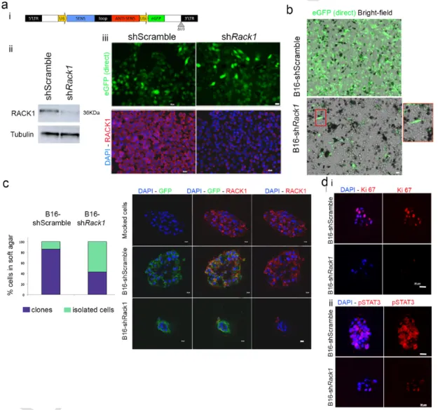
To test the importance of RACK1 in melanoma development we de-veloped a Rack1 shRNA lentivirus by inserting a previously validated sequence [15] into a backbone allowing visual control of infection [16] (Fig. 1a). The murine melan-a cell line, which is a nontransformed im-mortalized melanocytic line, and the highly metastatic B16 melanoma line were used to test RACK1 knock-down. With a transduction effi-cacy around 90%, RACK1 protein was efficiently reduced by shRack1 as evaluated by immunofluorescence and Western blot analyses without affecting unrelated proteins like Tubulin, three days post transduction of metastatic B16 cells (Fig. 1a). However, B16-shRack1 cells were pro
Fig. 1. RACK1 knock-down reduced B16 melanoma cells invasiveness and led to cell differentiation.a: i Lentiviral vector backbone for shRNA expression with GFP control of transduction;
ii Western-blot analysis for RACK1 and Tubulin in shRack1 and shScramble B16 cells 3 days post-transduction (3dpt); iii Fluorescent microscopy analysis of direct GFP signal (green) and immunolabeling for RACK1 (red) on 3 day-transducted cells. b: Aspect of shRack1 and shScramble-treated B16 cultures 14 days after transduction. Note the differentiation and loss of GFP+
cells in shRack1-treatd B16 cells. c: Soft agar assay of shRack1 and shScramble-treated B16 cells 3dpt, i Proportions of clones (purple) and isolated cells that did not proliferate (green); ii Immunolabeling for GFP (green) and RACK1 (red) in B16 clones mock-treated or treated with shRack1 or shScramble; d: Immunolabeling for i) the proliferation marker Ki67 (red) or ii) pSTAT3 (red) on soft agar clones. Nuclear counterstaining with DAPI (blue). Bars: 20 μm. (For interpretation of the references to colour in this figure legend, the reader is referred to the web version of this article.)
UNCORRECTED
PROOF
gressively lost from day 10 to day 20 post transduction, being re-placed by cells not transduced, hence not expressing the shRNA (Fig. 1b). Remaining fluorescent B16-shRack1 cells showed a differentiated phenotype. They switched from the typical rounded B16 shape to a melanocyte-like shape with dendritic extensions and higher melanin content (Fig. 1b), as did melan-a cells (Fig. S1).
We assessed the effect of RACK1 silencing on anchorage-indepen-dent cell growth of 3 day transduced cells. Only 40% of B16-shRack1 cells formed clones in soft agar compared to 86 and 88% in
B16-shScramble and B16-mock cells, respectively (χ2test; P < 0,0005)
(Fig. 1c). Furthermore, the B16-shRack1 clones were smaller than the controls (Fig. 1c). This reduced size of B16-shRack1 clones relates to a sharp reduction of Ki67 and pSTAT3 staining (Fig. 1d).
3.2. Overexpressed RACK1 in melanocytic cells from the Tyr::NRasQ61K
melanoma model
We next determined RACK1 expression in the Tyr::NRasQ61K;
Cd-kn2a−/−mouse melanoma model, referred to as Tyr::NRas⁎thereafter.
These mice carry a melanocyte-targeted NRasQ61Ktransgene which leads
to constitutive ERK activation [11]. Tyr::NRas⁎mice develop early
der-mal melanocytic proliferation responsible for skin hyperpigmentation, which can eventually progress to a malignant lesion [14]. We have
shown that introduction of the Pax3GFPallele allows the identification
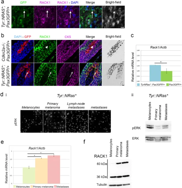
of fluorescent melanocytic cells in the skin without affecting melanoma development [13,17]. Immunofluorescence on cutaneous melanomas
showed a cytoplasmic RACK1 signal in GFP+ cells (Fig. 2a). RACK1
was immunodetected in all lesions from Tyr::NRas⁎; Pax3GFP/+ and
Tyr::NRas⁎mice.
We also tested melanoma-free skin. In control Pax3GFP/+; Cdkn2a−/−
mice, RACK1 protein was highly expressed in the cytoplasm of ker-atinocytes, here considered as positive controls. In contrast,
melanocytes identified as GFP+cells were negative for RACK1. We
ex-cluded the possibility that melanocytes displayed membrane RACK1 signal with triple immunostaining against RACK1, GFP and
cytoker-atin 5 (CK5), a marker of basal kercytoker-atinocytes. Instead, in Tyr::NRas⁎;
Pax3GFP/+mice, skin sections displayed a specific cytoplasmic RACK1
signal in GFP+, CK5−melanocytes (Fig. 2b). Follicular, interfollicular
and dermal melanocytes displayed this specific melanocytic RACK1 sig-nal (Fig. S2). To test whether RACK1 overexpression occurred at the
transcript level, GFP+melanocytes were sorted by FACS from
neona-tal skins. Higher Rack1 mRNA levels were observed in Tyr::NRas⁎;
Pax3GFP/+ melanocytes isolated from neonatal skin compared to
Cd-kn2a−/−; Pax3GFP/+control pup littermates (Fig. 2c). Thus, ERK
activa-tion together with RACK1 overexpression is associated with melanoma and melanoma predisposition in this model.
To study signaling associated with the progression stages, we estab-lished primary cultures from neonatal skin, primary tumor, locoregional
metastasis, and distant metastasis which, as expected, were pERK+
ac-cording to NRasQ61Kexpression when assayed by immunofluorescence
and Western blot (Fig. 2d) [13]. In these cells, overexpression of Rack1 mRNA (Fig. 2e) and high RACK1 protein level were identified (Fig. 2f). Interestingly, a 60 kDa band in addition to the predicted 36 kDa band was revealed in tumor cells compared to melanocytes (Fig. 2f). Denatur-ing conditions did not support dimerization of RACK1 (not shown). Inhi-bition of MEK1 with U0126 did not alter RACK1 reactivity in melanoma cells (Fig. S3).
3.3. Activated ERK and transiently increased skin melanocytes in pups with Rack1 gain of function in melanocytes
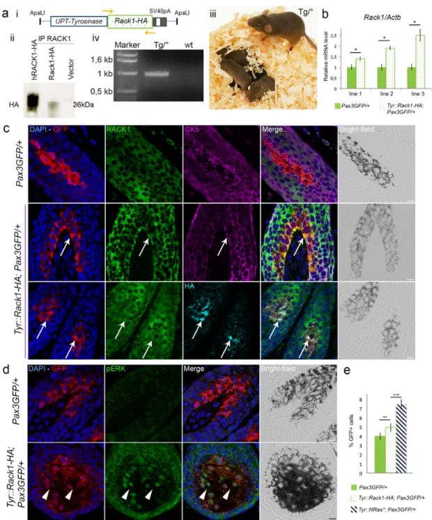
To address a causative role of RACK1 in melanoma development, we generated Tyr::Rack1-HA transgenic mice. We used the 6.1 kb pro-moter sequence of the mouse Tyrosinase gene in combination with the
3.6 kb distal control region [12] to target the expression of the mouse Rack1 gene (MGI:101849) that encodes RACK1, tagged by HA, to the melanocytic lineage (Fig. 3a). RACK1-HA expression from the Tyr::Rack1-HA transgene was detected when transfected into B16 cells in vitro (Fig. 3a).
Five Tyr::Rack1-HA transgenic founders were obtained using classi-cal transgenesis (Fig. 3aiii). All founders were viable, fertile and reached adulthood without displaying any overt phenotype. Three lines were es-tablished with offspring from three distinct founders and analysed in de-tail. In order to easily identify melanocytes, the three lines were crossed
with Pax3GFP/+mice. Melanocytes were FACS-sorted on GFP from back
skin of 3 day-old pups for each transgenic line and quantitative RT-PCR assays were performed. As expected, Rack1 mRNA levels in melanocytes
were higher in Tyr::Rack1-HA; Pax3GFP/+mice than in Pax3GFP/+
litter-mates in each transgenic line (Fig. 3b). Rack1/Actb ratios were
compa-rable to ratios measured in the Tyr::NRas⁎mice.
Protein expression of RACK1 in melanocytes in furry and glabrous skin was assessed by immunofluorescence (Fig. 3c, Fig. S4). In
Tyr::Rack1-HA; Pax3GFP/+ melanocytes, GFP and HA protein signals
were colocalized (Fig. 3c, third line). Moreover, a specific cytoplasmic RACK1 signal was detected in GFP and HA-positive melanocytes (Fig. 3c). Triple immunostaining against RACK1, GFP and CK5 confirmed the melanocytic origin of the RACK1 signal (Fig. 3c, fourth line). Thus, in our three Tyr::Rack1-HA transgenic mouse lines, Rack1 mRNA was over-expressed and RACK1 protein was detected in melanocytes. This ex-cluded a role of the integration site of the transgene.
RACK1 was reported to associate with the core kinases of the ERK pathway and RACK1 reduction resulted in lower ERK activity while RACK1 overexpression produced an increased ERK activation [18,19]. We tested ERK activation in melanocytes of transgenic Tyr::Rack1-HA; Pax3GFP/+and Pax3GFP/+control mice by immunostaining (Fig. 3d). In
control Pax3GFP/+ melanocytes, pERK signal was hardly detected.
In-stead, in Tyr::Rack1-HA; Pax3GFP/+melanocytes, nuclear GFP and pERK
signals were co-localized. Besides, pERK signal was also identified in keratinocytes. Noteworthy, total ERK expression was equivalent in both sample types (Fig. S5). We checked whether the PI3K/AKT pathway was also activated in the skin of Tyr::Rack1-HA transgenics. Nuclear pAKT signal was detected as well as in Tyr::NRas⁎, Pax3GFP/+skins, as opposed
to Pax3GFP/+skin (not shown). These data suggest that RACK1
overex-pression associated with ERK and AKT activation. We studied whether this pERK immunodetection corresponded to a proliferative signal
trans-lating efficient ERK activation. Tyr::Rack1-HA; Pax3GFP/+skin biopsies
presented 26% more GFP+cells than Pax3GFP/+skins (Fig. 3e). Yet, no
coat or skin hyperpigmentation was visible in any of the mouse lines. Over 17 months of follow-up, no melanocytic lesions were detected in any of the three transgenic lines (n > 15 mice/line). In addition, the morphology of the skin was normal indicating that RACK1 overexpres-sion alone was not sufficient to drive melanoma development (Fig. S6). 3.4. Accelerated melanoma appearance with RACK1-HA expression in a context of Tyr::NRas⁎melanoma predisposition
Melanoma penetrance is not complete in the Tyr::NRas⁎model [11].
In our colony, 33% of mice develop melanoma [14]. To assess the effects of RACK1 expression in a genetic background predisposing to
melanoma, we produced Tyr::Rack1-HA; Tyr::NRas⁎; Pax3GFP/+mice for
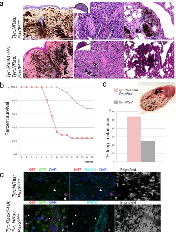
the three Tyr::Rack1-HA independent transgenic lines. These mice devel-oped melanocytic lesions ranging from benign to malignant tumors [14] (Fig. 4a). No differences in latency and incidence were seen between the three transgenic lines. Nevertheless, 78% of the Tyr::Rack1-HA; Tyr::NRas⁎;Pax3GFP/+mice in each line showed reduced latency within
about 10 months, mean 7.5 months (Mann-Whitney test; P < 0.002)
UNCORRECTED
PROOF
Fig. 2. The Tyr::NRas⁎, Pax3GFP/+mouse melanoma model displays increased RACK1 in hyperplastic melanocytes.ApoTome microscopy analysis of triple labeling for GFP, RACK1 and
CK5 in melanoma (a) and dorsal skin from Tyr::NRas⁎Pax3GFP/+and control Cdkn2a−/−; Pax3GFP/+mice (b). GFP (green), RACK1 (magenta) in a; GFP (red), RACK1 (green) and CK5
(magenta) in b. Arrows point to melanoma cells or melanocytes with RACK1 signal. Arrowheads point to melanocytes without RACK1 signal. DAPI nuclear counterstaining in blue. Bars: 10 μm. c: Reverse transcription-qPCR for Rack1 mRNA in melanocytes isolated from Tyr::NRas⁎, Pax3GFP/+(n = 5) mice compared to control Pax3GFP/+(n = 5). Results shown are Rack1
expression normalized to Actb. d: i: pERK immunolabeling in GFP-FACS sorted cells from neonatal skin and cutaneous melanomas at different progression stages in Tyr::NRas⁎, Pax3GFP/+
mice. ii: Western blot of pERK and ERK on the same cells as in i. e: Quantitative RT-PCR for Rack1 mRNA in GFP+sorted melanocytic cells from skin, primary melanoma and metastases
in Tyr::NRas⁎; Pax3GFP/+normalized to Actb from triplicates. f: Western-blot analysis for RACK1 in GFP + sorted cells as in e. Note a 60 kDa band specific to tumoral cells on top of the
36 kDa expected band. * P < 0.05. (For interpretation of the references to colour in this figure legend, the reader is referred to the web version of this article.)
melanomas, indicating a clear increase in incidence of primary
cu-taneous malignant lesions (n = 22/31, χ2 test; P < 0.05) (Fig. 4a).
Moreover, distant metastases were more frequently identified in
Tyr::Rack1-HA; Tyr::NRas⁎; Pax3GFP/+ mice (55%, n = 12/22)
com-pared to Tyr::NRas⁎; Pax3GFP/+ mice (36%, n = 9/25) (Fig. 4c).
His-tological analysis of the lesions detected no differences between Tyr::Rack1-HA lesions and those of control Tyr::NRas⁎; Pax3GFP/+
litter-mates (Fig. 4a). However, when the proliferation status of melanocytic cells was analysed using Ki67 labeling, Tyr::Rack1-HA bearing melanomas presented a higher index than controls (Fig. 4d) (32.9 ± 17.8 versus 22.3 ± 8.1 respectively, Student's t-test; P < 0.05).
The higher incidence, lower latency and higher frequency of mi-tosis provide the first in vivo evidence of a contribution of RACK1 to melanoma development. Histological data point to an acceleration of the proliferative status of the lesions.
3.5. JNK and STAT3 as oncogenic partners of RACK1 in melanoma development
To investigate the clinical advantage conferred by the
overexpres-sion of RACK1 in the Tyr::NRas⁎model, we explored candidate
teins related to metastasis. STAT3 activation has been shown to pro-mote metastasis in melanoma [20]. To test whether PKC and JNK acti-vation were involved in melanomagenesis induced by activated NRAS as modeled in vitro [9], we isolated primary melanocytic cells from
neona-tal skin and at different stages of tumoral progression from Tyr::NRas⁎;
Pax3GFP/+mice. pPKCα/βII,pJNK and pSTAT3 were analysed by
im-munofluorescence. Phosphorylation of PKCα/βII was detected both in
melanocytes and in tumoral cells, independently of their tumoral pro-gression stage. pJNK, instead, started to be observed in
UNCORRECTED
PROOF
Fig. 3. Targeted RACK1 overexpression in melanocytes is not sufficient to initiate melanoma.a, i Scheme of the Tyr::Rack1-HA construct and genotyping strategy (orange arrows), ii
Western-blot analysis for HA after RACK1 immunoprecipitation in B16 cells transfected with the above construct and the human RACK1-HA. iii Phenotype of the transgenic mice. iv Detection of the transgene by PCR. b, Reverse transcription-qPCR for Rack1 RNA in melanocytes isolated by FACS on GFP from Tyr::Rack1-HA Pax3GFP/+mice from each of the three
transgenic lines (dotted bar) compared to littermates (green bar). Results shown are Rack1 expression normalized to Actb from triplicates. c, ApoTome microscopy analysis of triple labeling for GFP (red), RACK1 (green) and CK5 (magenta) or GFP (red), HA (cyan) and RACK1 (green) in control Pax3GFP/+mice and in Tyr::Rack1-HA; Pax3GFP/+mice of transgenic line 1.
Arrows point to melanocytes overexpressing RACK1. Bar: 10 μm.d, ApoTome microscopy analysis of double labeling for GFP (red) and phospho-ERK (green) in control Pax3GFP/+mice
and in Tyr::Rack1-HA; Pax3GFP/+mice of transgenic line 1. Arrowheads point to melanocytes overexpressing pERK. Nuclear counterstaining in blue. Bars: 10 μm. e, Percentages of GFP
cells detected in the dorsal skin of 6 day-old pups from Pax3GFP/+(green), Tyr::Rack1-HA; Pax3GFP/+(dotted) and Tyr::NRas*; Pax3GFP/+(hatched) lines. ** P < 0.01, *** P < 0.001. (For
interpretation of the references to colour in this figure legend, the reader is referred to the web version of this article.)
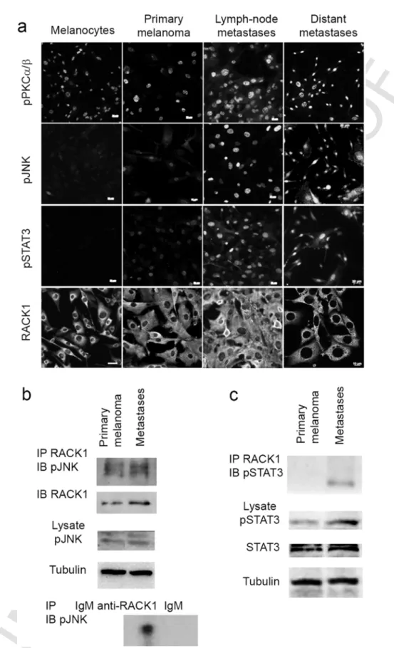
melanoma cells from primary lesions and was stronger in cells iso-lated from metastases (Fig. 5a). While no signal was visible on neonatal melanocytes and only a small number of primary cutaneous melanoma cells showed a signal, a high proportion of metastatic cells were found positive for pSTAT3 (Fig. 5a). RACK1 cytoplasmic immunoreactivity in-stead is detected in all cells (Fig. 5a).
Next, we analysed whether JNK and RACK1 interacted in Tyr::NRas⁎
melanoma cells. Positive interaction was shown between
pJNK and RACK1 by Western blot analysis after RACK1 immunoprecip-itation in primary melanoma and metastases. RACK1-pJNK interaction occurred at basal levels, without specific induction suggesting consti-tutive activation of JNK in these cells (Fig. 5b). We also validated the activation of STAT3 in melanoma cells by Western blot as well as its binding to RACK1, after RACK1 immunoprecipitation. Despite the ac-tivation of STAT3 in primary melanoma cells RACK1–pSTAT3 interac
UNCORRECTED
PROOF
Fig. 4. Targeted RACK1 overexpression reduces latency and increases incidence of melanoma development in Tyr::NRas⁎mice.a, Histological features (haematoxylin-eosin-saffron staining)
of melanocytic nevi, primary cutaneous melanoma and lung metastases in Tyr::NRas⁎; Cdkn2a+/−; Pax3GFP/+mice and Tyr::Rack1-HA; Tyr::NRas⁎; Cdkn2a+/−; Pax3GFP/+mice. Note that
lesions are histologically indistinguishable among groups.b, Survival (Kaplan-Meier plot) for Tyr::Rack1-HA; Tyr::NRas⁎; Pax3GFP/+mice (n = 22, pink curve) compared to Tyr::NRas⁎;
Pax3GFP/+control mice (n = 25, grey curve). Mice from the three transgenic lines were pooled in the Kaplan-Meier graph.c, Percentages of lung metastases in Tyr::Rack1-HA; Tyr::NRas⁎;
Pax3GFP/+mice (pink bar) compared to control Tyr::NRas⁎; Pax3GFP/+(grey bar). Gross feature of a Tyr::Rack1-HA; Tyr::NRas⁎; Pax3GFP/+lung with multiple melanoma metastases.d,
ApoTome microscopy analysis of double labeling for Ki67 (red), GFP (green) and RACK1 (cyan) in control Tyr::NRas⁎; Pax3GFP/+mice and in Tyr::Rack1-HA; Tyr::NRas⁎; Pax3GFP/+mice.
Nuclear counterstaining in blue. Bars: 10 μm. Arrowheads point to Ki67+, GFP+, RACK1+melanocytic cells. Arrow points to a mitosis, RACK1−. In the lower row, intense RACK1+cells
UNCORRECTED
PROOF
Fig. 5. JNK and STAT3 are oncogenic partners of RACK1 in ERK melanoma development.a, Fluorescent microscopy analysis of immunolabeling for pPKCα/βII, pJNK, pSTAT3 and RACK1
in Tyr::NRas⁎; Pax3GFP/+cells from each stage of melanoma progression. Bars: 10 μm. pPKCα/β
IIwas detected at all stages, pJNK was negative in nontransformed melanocytes and pSTAT3
appeared in metastases.b, Western-blot analysis for pJNK after RACK1 immunoprecipitation in melanoma and lymph node metastases cells. Western blot for pJNK on the 10% inputs used for immunoprecipitation. Below: control immunoprecipitation without RACK1 antibody. Both cell types showed constitutive pJNK-RACK1 interaction.c, Western blot analysis for pSTAT3 after RACK1 immunoprecipitation in lung metastasis cells. Protein interaction between RACK1 and pSTAT3 appears only in metastatic melanoma cells. Western-blot analysis for pSTAT3 and STAT3 in primary melanoma and metastasis cells. Tubulin was used as loading control.
UNCORRECTED
PROOF
tion was only detected in metastases (Fig. 5c). These data suggest that JNK and STAT3 are activated prior or during the metastatic process. 3.6. Coordinated expression of RACK1, JNK and STAT3 regulates invasive potential
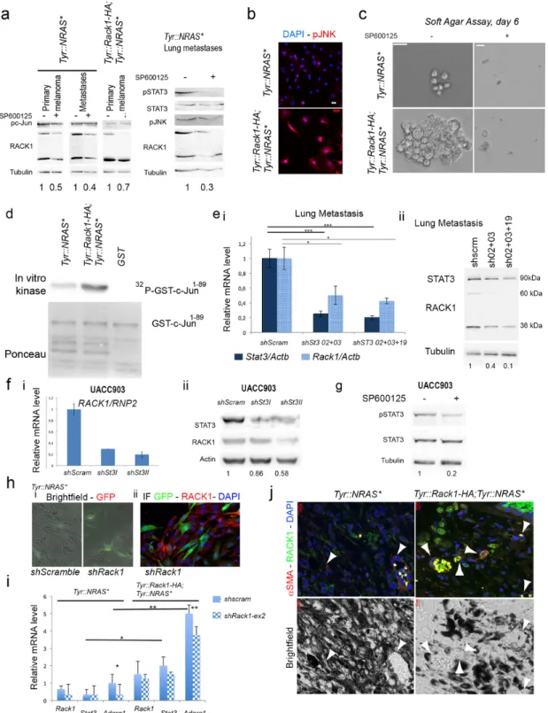
To investigate RACK1, JNK and STAT3 coordination, we verified whether RACK1 levels were under c-Jun transcriptional control treat-ing cells with a JNK inhibitor, SP600125. SP600125 reduced the lev-els of both 36 kDa and 60 kDa forms of RACK1 in all melanoma cells types (Fig. 6a). A reduction of pJNK was also found (Fig. 6a). When we compared FACS-sorted cells derived from primary melanomas of
Tyr::Rack1-HA; Tyr::NRas⁎; Pax3GFP/+ mice with the ones from
Tyr::NRas⁎; Pax3GFP/+ mice, we observed pJNK immunoreactivity in
Tyr::Rack1-HA melanoma cells not only in the nucleus but also in the cytosol (Fig. 6b). We reasoned that if RACK1 overexpression accelerated the appearance of tumorigenic properties, a correlation with JNK activ-ity would be found in Tyr::Rack1-HA melanoma cells. To test this hy-pothesis we carried out clonogenic and JNK activity assays. In soft agar assay, cells overexpressing Rack1 started forming clones earlier than control cells (Fig. 6c). SP600125 treatment of Tyr::Rack1-HA melanoma cells prevented colony formation in agar over a 6 days period (Fig.
6c). When immunokinase assays using GST-c-Jun1–89as substrate were
performed on primary cells of melanomas, an increased JNK activity was revealed in Tyr::Rack1-HA melanoma cells compared to controls Tyr::NRas⁎ (Fig. 6d). These data point to a stronger JNK activity in
RACK1 overexpressing melanomas.
Out of 5 enhancer regions described by ENCODE and identified on human RACK1 genecard (www.genecards.org), three (GHO5E181172, GH05E181215, GH05E181240) present binding sites for STAT3. To test
whether STAT3 transcriptionally regulates RACK1 in Tyr::NRas⁎cells,
we transduced interfering lentiviral shStat3 vectors. STAT3 knock-down in lung metastasis cells led to Rack1 mRNA (Fig. 6ei) and RACK1 pro-tein reduction on both the 36 kDa and the 60 kDa bands (Fig. 6eii). STAT3 knock-down effect on RACK1 mRNA and protein levels was also effective in human UACC903 melanoma cell lines as illustrated in Fig. 6f. As the JNK pathway was shown to activate STAT3 in epithelial cells [21], we tested whether JNK was regulating STAT3 in melanoma cells. In agreement with above findings, pSTAT3 signal was reduced in SP600125-treated mouse cells (Fig. 6a). Inhibition of JNK activation did also reduce STAT3 activation in human melanoma cell lines (Fig. 6g).
In order to further dissect the mechanism engaged by RACK1 over-expression to allow melanoma cells to metastasize, we examined the
transcriptome of melanoma cells from primary lesions in Tyr::NRas⁎and
Tyr::Rack1-HA, Tyr::NRas⁎by RNA-seq. Ingenuity Pathway Analysis on
numerically differentially expressed genes resulted in the enrichment of tissue morphology, cellular movement, growth and proliferation
path-ways. Stat3 appeared increased in Tyr::Rack1-HA, Tyr::NRas⁎with a fold
change of 1.3. We intended to analyse the effect of shRack1. In these heterogeneous primary cultures, estimation of the efficacy of lentivi-ral vector transduction was around 50% for both shRack1 and shScram-ble (Fig. 6h). By immunohistochemistry RACK1 protein downregula-tion was detected in the transduced cells (Fig. 6h) but no reducdownregula-tion of Rack1 mRNA was noticeable for any of the 6 transcripts of Rack1, from which only 2 (ENSMUST00000020640 and ENSMUST00000125166) can be translated. Among the genes that exhibited increased
expres-sion in Tyr::Rack1-HA, Tyr::NRas⁎cells and decreased expression
follow-ing shRack1 treatment, Adgrg1 encodfollow-ing the adhesion G protein-cou-pled receptor G1, has been shown to dually regulate angiogenesis dur-ing melanoma progression [22]. Quantitative RT-PCR on Rack1 exon 2, targeted by the interfering sequence, showed a slight reduction upon shRack1 treatment (Fig. 6i). Reduction of RACK1 was related to a clear decrease in Adgrg1 expression in melanoma cells from
Tyr::Rack1-HA, Tyr::NRas⁎and Tyr::NRas⁎mice. The Adgrg1 reduction
was stronger on Tyr::Rack1-HA, Tyr::NRas⁎cells, in which Stat3
reduc-tion was observed, in contrast to Tyr::NRas⁎ cells (Fig. 6i). In
agree-ment with these results, RACK1 knock-down in B16 cell agar colonies led to pSTAT3 reduction (Fig. 1dii). To test whether angiogenesis could
be responsible for the clinical outcome of Tyr::Rack1-HA, Tyr::NRas⁎
melanomas, we performed α-smooth muscle actin (αSMA) immunoflu-orescence on the mouse primary melanoma lesions of Tyr::Rack1-HA, Tyr::NRas⁎and Tyr::NRas⁎models. αSMA is a marker of myofibroblasts
and pericytes used to identify capillaries and vascular structures. Fig. 6j
shows the higher density of αSMA+and vascularisation in heavily
pig-mented lesions in the Tyr::Rack1-HA genotype.
4. Discussion
RACK1 overexpression has been described in different solid cancers. Its detection in situ was proposed as a diagnostic marker in melanoma [2] and several carcinomas [23–25]. Many studies have shown that RACK1 is involved in multiple aspects of cancer progression in vitro (re-viewed by [7]). As cell monolayers are far from in vivo complexity, we
wanted to test whether the model, in which NRASQ61Ktriggering ERK/
c-Jun accumulation with subsequent RACK1 increase and JNK activa-tion, was valid in the mouse. In this study, we generated Tyr::Rack1-HA transgenic mice to test a RACK1 role in melanoma initiation and pro-gression in vivo. Our results support the model of RACK1 as a major player in melanomagenesis.
We showed that transgenic Tyr::Rack1-HA mice overexpressed Rack1 in melanocytes with concomitant detection of pERK and pAKT. Never-theless, Tyr::Rack1-HA mice did not develop either hyperpigmentation nor melanoma suggesting that additional alterations are required for
both events. In Tyr::NRAS⁎ transgenic mice, NRASQ61Ktriggers MAPK
and PI3K/AKT pathways activation [10]. This mutation leads to hy-perproliferation of skin melanocytes and spontaneous melanoma de-velopment [11]. We showed here that these mice also overexpress RACK1 in melanocytes. Thus, the difference in phenotypes could be as-cribed to a differential compartmentalization of the proteins in the two melanocyte-types engaging then distinct genetic programs [26]. Alter-natively, it could hint at the existence of additional pathways triggered
by the NRasQ61Kmutation, independent of ERK, JNK, AKT, STAT3
path-ways and scaffold RACK1.
Breeding Tyr::Rack1-HA mice with Tyr::NRAS⁎ mice enhanced
melanoma initiation and progression, with reduced latency and in-creased incidence as well as distant metastases in three independent mouse lines. We provided a first evidence of a role for RACK1 in melanomagenesis in vivo.
RACK1 serves as a signaling hub that has been implicated in the regulation of several pathways. To identify proteins mediating a role for RACK1 in melanomagenesis, we determined the activation status of
PKCα/βII, STAT3 and JNK in primary cultures. pPKCα/βIIwas detected
since the early stages as described [2], whereas STAT3 and JNK pro-teins were found activated in all lymph node metastatic cells which are representative of the first metastatic stage. Clinically, improved therapy for melanoma will benefit from understanding the metastatic process at a molecular level. In our primary culture cells, JNK activa-tion marked progression to lymph-node metastases. Fittingly, a pathol-ogy study showed JNK activation associated with poor prognosis [27]. However, an oncogenic role of the JNK/c-Jun pathway in melanoma development is still under debate [28]. The tumor suppressor p16/Cd-kn2a was reported to exert its inhibitory role on tumor cells by sup-pressing JNK activity [29]. In our system, Cdkn2a deletion was not suf-ficient alone to trigger JNK activation. Lopez-Bergami et al. described that RACK1 mediates JNK activation by PKC in vitro [5]. Moreover, in human melanoma cell lines, constitutively active ERK provides signals to increase the activity of JNK via a rewired signaling [9]. In the pri
UNCORRECTED
PROOF
Fig. 6. RACK1 regulation and angiogenesis in melanoma.a, Western blot analysis of pc-Jun and RACK1 after SP600125 inhibition of JNK on protein lysates from primary melanoma and
metastases from Tyr::NRas⁎; Pax3GFP/+mice and primary melanoma cells in Tyr::Rack1-HA; Tyr::NRas⁎; Pax3GFP/+. SP600125 20 μM reduced RACK1 levels after 48 h treatment. pc-Jun
reduction is shown. Western blot analysis of RACK1, pSTAT3, STAT3 and pJNK, in Tyr::NRas⁎; Pax3GFP/+melanoma lung metastatic cells after treatment with 20 μM SP600125 for 48 hs
and quantification. b, pJNK immunoreactivity on cells from primary melanomas of Tyr::Rack1-HA; Tyr::NRas⁎; Pax3GFP/+mice compared to the ones from Tyr::NRas⁎; Pax3GFP/+mice.
Bars: 10 μm. Note a higher pJNK signal on Rack1 overexpressing cells.c, Soft agar assay over 6 days with melanoma cells overexpressing Rack1-HA and controls without or with SP600125 (20 μM). Rack1-HA cells started forming clones earlier, and SP600125 prevented it. Bars: 10 μm.d, In vitro JNK kinase assay. Cell lysates from Tyr::NRas⁎; Pax3GFP/+and
Tyr::Rack1-HA; Tyr::NRas⁎; Pax3GFP/+melanomas and Tyr::NRas⁎; Pax3GFP/+melanocytes were subjected to immunoprecipitation using JNK antibody followed by kinase assay using
GST-c-Jun1–89as substrate.e, i Quantitative RT-PCR for Stat3 (dark blue bars) and Rack1 (hatched light bars) mRNAs on lung melanoma metastasis cells after treatment with lentiviral
vectors coding for shStat3 (02, 03, 19) or control. Results shown are Stat3 or Rack1 expression normalized to Actb from triplicates. * P < 0.05, *** P < 0.001. ii Western-blot for RACK1, STAT3 and Tubulin after Stat3 knock-down on mouse melanoma cells.f, i Quantitative RT-PCR for RACK1 mRNA on human UACC903 melanoma cells after treatment with lentiviral vectors expressing shSTAT3 or shScramble. RACK1 levels are normalized to RNP2. e; ii Western-blot for RACK1, STAT3, and Actin after STAT3 knock-down. g, Western blot analysis for
UNCORRECTED
PROOF
pSTAT3, STAT3 and Tubulin in human UACC903 cell line after SP600125 treatment. h, i Fluorescent microscopy analysis of GFP signal (red) for infection visualization; ii immunolabeling for RACK1 (green) on Tyr::NRas⁎; Pax3GFP/+transducted cells with the shRack1 lentiviral vector as in Fig. 1.i, Quantitative RT-PCR for Rack1, Stat3 and Adgrg1 mRNAs on primary
melanoma metastasis from Tyr::Rack1-HA; Tyr::NRas⁎; Pax3GFP/+or Tyr::NRas⁎; Pax3GFP/+mice, after treatment with the lentiviral vector expressing shRack1. * P < 0.05, ** P < 0.01.j,
Immunolabeling of αSMA (red) and RACK1 (green) in primary melanoma from Tyr::NRas⁎; Pax3GFP/+and Tyr::Rack1-HA; Tyr::NRas⁎; Pax3GFP/+mice. Arrowheads point to αSMA+vascular
structures. Bars: 10 μm. Brightfields show higher vascularisation in Tyr::Rack1-HA; Tyr::NRas⁎; Pax3GFP/+tissue. (For interpretation of the references to colour in this figure legend, the
reader is referred to the web version of this article.)
mary melanoma cells from Tyr::Rack1-HA; Tyr::NRas⁎; Pax3GFP/+mice,
pJNK was detected both in the nucleus and cytoplasm. JNK activity was increased as determined in the kinase assay. Although we have not ex-plored the underlying mechanism of JNK activation, our data support the previous in vitro model where JNK activation is being driven by PKC, ERK and RACK1. Data are consistent with the positive correlation observed between cytoplasmic pJNK and pERK in human melanomas [27], where RACK1 is overexpressed [2]. Phospho-STAT3 on Ser705 was mainly found in lymph-node metastatic cells, which is consistent with progressive activation found in human melanoma cell lines [20] [30] and in tumor samples. In melanoma cells, activated STAT3 was re-ported to sustain expression of the anti-apoptotic genes, Bcl-xL (Bcl2-like 1) and Mcl-1 (Myeloid cell leukemia sequence 1) [31]. Constitutive STAT3 activation might be a crucial event in metastasis development. We iden-tified STAT3 as a direct RACK1 partner in melanoma development. Con-firming the functional interaction between STAT3 and RACK1 we found that RACK1 was regulated at the mRNA level by STAT3. Noteworthy, STAT3-mediated maintenance of NF-κB activity occurs in human A2058 melanoma cell line [32]. In turn, NF-κB is known to upregulate the tran-scription of RACK1 through direct interaction with its promoter thus contributing to cell survival in PC12 cells [33]. Besides, inhibition of JNK activity led to decreased activation of STAT3. This was reported in colon cancer cells after treatment with AS601245, another JNK inhibitor [34].
B16 mouse melanoma cells harbor both STAT3 activation and JNK activation (data not shown). RACK1 silencing in B16 cell line led to re-duced invasive capacities and cell differentiation and loss. The differen-tiated shape with dendritic extensions closely resembles the in situ sta-tus of melanocytes in normal skin where no RACK1 is detected [2,3]. Yet, our results seem to be in apparent contradiction with the recent publication of Marubashi et al. which showed that Rack1 knock-down in melan-a cells decreased their dendricity [35]. In fact, this specific phenotype corresponded to forskolin treated cells, and it might depend upon the extent of RNA silencing and the time before observation. Pre-vious observations showed that mouse fibroblast cells with silenced RACK1 contained more and longer focal adhesions [18,19].
We observed an additional 60 kDa band revealed by RACK1 anti-body only in transformed cells, which does not appear in the litera-ture. This band decreased in the presence of the JNK inhibitor or STAT3 knock down suggesting it could reflect a form of RACK1 related to ma-lignancy. Further investigation must be carried out to understand the significance of this isoform.
RNA-seq data were produced in order to determine the targets of sig-naling pathways induced by RACK1 overexpression that cause the phe-notype changes between normal and tumoral melanocytes with different
potential. Tyr::Rack1-HA; Tyr::NRas⁎primary melanoma cells expressed
a higher basal level of Adgrg1 mRNA. Interestingly, Adgrg1 has been related to angiogenesis [22] and RACK1 overexpressing melanomas
did appear to contain more αSMA+ cells, a marker of angiogenesis.
Adgrg1 activation was recently shown to promote melanoma migra-tion [37]. Melanomas have a high angiogenic potential. These observa-tions are in agreement with early findings on RACK1 expression dur-ing angiogenesis [38]. Biological replicates of tumors should be used to formulate new hypotheses on transcriptional targets of RACK1-in-volving pathways. The wide implication of RACK1 in physiological processes precludes the possibility to directly act on this protein [7]. Identification of RACK1-protein interactions holds the oppor
tunity to develop new therapeutics by the design of peptides interfering with specific binding partners of RACK1 [39,40].
In summary, we demonstrated that RACK1 is implicated in melanomagenesis. RACK1 enhanced melanoma initiation and progres-sion in vivo. We propose that activated STAT3 adds to JNK as RACK1 partners in the metastatic process. Specific interference between RACK1 and its partners must be further analysed as a potential therapeutic im-provement in melanoma treatment.
Supplementary data to this article can be found online at http://dx. doi.org/10.1016/j.cellsig.2017.03.015.
Authors contributions
JJP, GEM designed the study, CC performed all studies on the mouse and mouse cell assays, MEP and PLB on human cell lines and the JNK assay, SL, PS performed mouse transgenesis, FB and ERG scored histo-logical samples, JEz performed immunofluorescence, PHC carried out FACS, SP, UM provided lentiviral vectors expressing shRack1, GAH ad-vised with constructs, DE, performed RNA-seq, JE bioinformatic analy-sis, EB converted gene names. CC and GEM wrote the manuscript.
Uncited reference
[36]
Acknowledgements
We wish to thank A. Casanova and C. Koenen for dedicated animal husbandry. We are grateful to A. Champeix and P. Wattier for sample inclusions in paraffin. We thank S. Bibi for technical help with qPCR. We thank Pierre Adenot (Microscopie et Imagerie des Microorganismes, Animaux et Aliments, MIMA2, INRA) for access to confocal microscopy facilities. We are thankful to Z. Ronaï for control plasmid used in pre-liminary experiments. We specially thank A. Eychene and N. Mach for helpful discussions and D. Milan for support. This work was funded by grants from Institut National de la Recherche Agronomique, INRA Crédits incitatifs du département de génétique animale (GEM), l'Agence Nationale de la Recherche Emergence Bio ANR 09-EBIO-006-01 to GEM, Ecos-Sud to JJP, GEM and PLB and Association pour la Recherche contre le Cancer (grants 99/7468 and 1002) to GAH and GEM. CC received a grant (Allocation de Recherche MENRT) from the Ministère de la Recherche et de l'Enseignement Supérieur. MEP received a grant from the CONICET.
References
[1] P.A. Ascierto, S. Agarwala, G. Botti, A. Cesano, G. Ciliberto, M.A. Davies, S. De-maria, R. Dummer, A.M. Eggermont, S. Ferrone, et al., Future perspectives in melanoma research: meeting report from the "Melanoma Bridge". Napoli, Decem-ber 1st–4th 2015, J. Transl. Med. 14 (2016) 313.
[2] G. Egidy, S. Julé, P. Bossé, F. Bernex, C. Geffrotin, S. Vincent-Naulleau, V. Horak, X. Sastre-Garau, J.J. Panthier, Transcription analysis in the MeLiM swine model identifies RACK1 as a potential marker of malignancy for human melanocytic pro-liferation, Mol. Cancer 7 (2008) 34.
[3] C. Campagne, S. Julé, F. Bernex, M. Estrada, G. Aubin-Houzelstein, J.J. Panthier, G. Egidy, RACK1, a clue to the diagnosis of cutaneous melanomas in horses, BMC Vet. Res. 8 (2012) 95.
[4] C. Campagne, S. Julé, C. Alleaume, F. Bernex, J. Ezagal, S. Château-Joubert, M. Estrada, G. Aubin-Houzelstein, J.J. Panthier, G. Egidy, Canine melanoma diagno-sis: RACK1 as a potential biological marker, Vet. Pathol. 50 (2013) 1083–1090.
UNCORRECTED
PROOF
[5] P. López-Bergami, H. Habelhah, A. Bhoumik, W. Zhang, L.H. Wang, Z. Ronai, RACK1 mediates activation of JNK by protein kinase C [corrected], Mol. Cell 19 (2005) 309–320.
[6] D. Ron, C.H. Chen, J. Caldwell, L. Jamieson, E. Orr, D. Mochly-Rosen, Cloning of an intracellular receptor for protein kinase C: a homolog of the beta subunit of G proteins, Proc. Natl. Acad. Sci. U. S. A. 91 (1994) 839–843.
[7] D.R. Adams, D. Ron, P.A. Kiely, RACK1, a multifaceted scaffolding protein: struc-ture and function, Cell Commun. Signal 9 (2011) 22.
[8] V. Volta, A. Beugnet, S. Gallo, L. Magri, D. Brina, E. Pesce, P. Calamita, F. Sanvito, S. Biffo, RACK1 depletion in a mouse model causes lethality, pigmentation deficits and reduction in protein synthesis efficiency, Cell. Mol. Life Sci. 70 (2013) 1439–1450.
[9] P. Lopez-Bergami, C. Huang, J.S. Goydos, D. Yip, M. Bar-Eli, M. Herlyn, K.S. Smal-ley, A. Mahale, A. Eroshkin, S. Aaronson, Z. Ronai, Rewired ERK-JNK signaling pathways in melanoma, Cancer Cell 11 (2007) 447–460.
[10] D.C. Bennett, How to make a melanoma: what do we know of the primary clonal events?, Pigment Cell Melanoma Res. 21 (2008) 27–38.
[11] J. Ackermann, M. Frutschi, K. Kaloulis, T. McKee, A. Trumpp, F. Beermann, Metastasizing melanoma formation caused by expression of activated N-RasQ61K on an INK4a-deficient background, Cancer Res. 65 (2005) 4005–4011. [12] A. Camacho-Hübner, F. Beermann, Increased transgene expression by the mouse
tyrosinase enhancer is restricted to neural crest-derived pigment cells, Gene-sis 29 (2001) 180–187.
[13] C. Campagne, E. Reyes-Gomez, S. Loiodice, S. Gadin, J. Ezagal, F. Bernex, M. Abit-bol, A. Louise, F. Beermann, J.J. Panthier, et al., Haplosufficiency of PAX3 for melanoma development in Tyr: NRASQ61K; Cdkn2a −/− mice allows identifica-tion and sorting of melanoma cells using a Pax3GFP reporter allele, Melanoma Res. 26 (2016) 12–20.
[14] C. Campagne, E. Reyes-Gomez, M. Battistella, F. Bernex, S. Château-Joubert, H. Huet, F. Beermann, G. Aubin-Houzelstein, G. Egidy, Histopathological atlas and proposed classification for melanocytic lesions in Tyr:: NRas(Q61K); Cd-kn2a(−/−) transgenic mice, Pigment Cell Melanoma Res. 26 (2013) 735–742. [15] M. Mourtada-Maarabouni, L. Kirkham, F. Farzaneh, G.T. Williams, Functional
ex-pression cloning reveals a central role for the receptor for activated protein kinase C 1 (RACK1) in T cell apoptosis, J. Leukoc. Biol. 78 (2005) 503–514.
[16] M.E. Avale, P. Faure, S. Pons, P. Robledo, T. Deltheil, D.J. David, A.M. Gardier, R. Maldonado, S. Granon, J.P. Changeux, U. Maskos, Interplay of beta2* nicotinic re-ceptors and dopamine pathways in the control of spontaneous locomotion, Proc. Natl. Acad. Sci. U. S. A. 105 (2008) 15991–15996.
[17] J. Djian-Zaouche, C. Campagne, E. Reyes-Gomez, S. Gadin-Czerw, F. Bernex, A. Louise, F. Relaix, M. Buckingham, J.J. Panthier, G. Aubin-Houzelstein, Pax3(GFP), a new reporter for the melanocyte lineage, highlights novel aspects of PAX3 ex-pression in the skin, Pigment Cell Melanoma Res. 25 (2012) 545–554. [18] T. Vomastek, M.P. Iwanicki, H.J. Schaeffer, A. Tarcsafalvi, J.T. Parsons, M.J.
We-ber, RACK1 targets the extracellular signal-regulated kinase/mitogen-activated protein kinase pathway to link integrin engagement with focal adhesion disassem-bly and cell motility, Mol. Cell. Biol. 27 (2007) 8296–8305.
[19] H.C. O'Donovan, P.A. Kiely, R. O'Connor, Effects of RACK1 on cell migration and IGF-I signalling in cardiomyoctes are not dependent on an association with the IGF-IR, Cell. Signal. 19 (2007) 2588–2595.
[20] T.X. Xie, F.J. Huang, K.D. Aldape, S.H. Kang, M. Liu, J.E. Gershenwald, K. Xie, R. Sawaya, S. Huang, Activation of stat3 in human melanoma promotes brain metas-tasis, Cancer Res. 66 (2006) 3188–3196.
[21] B. Chen, J. Liu, Q. Chang, K. Beezhold, Y. Lu, F. Chen, JNK and STAT3 signaling pathways converge on Akt-mediated phosphorylation of EZH2 in bronchial epithe-lial cells induced by arsenic, Cell Cycle 12 (2013) 112–121.
[22] L. Yang, G. Chen, S. Mohanty, G. Scott, F. Fazal, A. Rahman, S. Begum, R.O. Hynes, L. Xu, GPR56 regulates VEGF production and angiogenesis during melanoma progression, Cancer Res. 71 (2011) 5558–5568.
[23] Z. Wang, B. Zhang, L. Jiang, X. Zeng, Y. Chen, X. Feng, Y. Guo, Q. Chen, RACK1, an excellent predictor for poor clinical outcome in oral squamous carcinoma, simi-lar to Ki67, Eur. J. Cancer 45 (2009) 490–496.
[24] X.X. Cao, J.D. Xu, J.W. Xu, X.L. Liu, Y.Y. Cheng, W.J. Wang, Q.Q. Li, Q. Chen, Z.D. Xu, X.P. Liu, RACK1 promotes breast carcinoma proliferation and invasion/ metastasis in vitro and in vivo, Breast Cancer Res. Treat. 123 (2010) 375–386. [25] S. Shi, Y.Z. Deng, J.S. Zhao, X.D. Ji, J. Shi, Y.X. Feng, G. Li, J.J. Li, D. Zhu, H.P.
Koeffler, et al., RACK1 promotes non-small-cell lung cancer tumorigenicity through activating sonic hedgehog signaling pathway, J. Biol. Chem. 287 (2012) 7845–7858.
[26] A. Herrero, B. Casar, P. Colón-Bolea, L. Agudo-Ibáñez, P. Crespo, Defined spa-tiotemporal features of RAS-ERK signals dictate cell fate in MCF-7 mammary ep-ithelial cells, Mol. Biol. Cell 27 (2016) 1958–1968.
[27] K. Jørgensen, B. Davidson, V.A. Flørenes, Activation of c-jun N-terminal kinase is associated with cell proliferation and shorter relapse-free period in superficial spreading malignant melanoma, Mod. Pathol. 19 (2006) 1446–1455. [28] P. Lopez-Bergami, The role of mitogen- and stress-activated protein kinase
path-ways in melanoma, Pigment Cell Melanoma Res. 24 (2011) 902–921. [29] B.Y. Choi, H.S. Choi, K. Ko, Y.Y. Cho, F. Zhu, B.S. Kang, S.P. Ermakova, W.Y. Ma,
A.M. Bode, Z. Dong, The tumor suppressor p16(INK4a) prevents cell transforma-tion through inhibitransforma-tion of c-Jun phosphorylatransforma-tion and AP-1 activity, Nat. Struct. Mol. Biol. 12 (2005) 699–707.
[30] M. Sakaguchi, M. Oka, T. Iwasaki, Y. Fukami, C. Nishigori, Role and regulation of STAT3 phosphorylation at Ser727 in melanocytes and melanoma cells, J. Invest. Dermatol. 132 (2012) 1877–1885.
[31] G. Niu, T. Bowman, M. Huang, S. Shivers, D. Reintgen, A. Daud, A. Chang, A. Kraker, R. Jove, H. Yu, Roles of activated Src and Stat3 signaling in melanoma tu-mor cell growth, Oncogene 21 (2002) 7001–7010.
[32] H. Lee, A. Herrmann, J.H. Deng, M. Kujawski, G. Niu, Z. Li, S. Forman, R. Jove, D.M. Pardoll, H. Yu, Persistently activated Stat3 maintains constitutive NF-kappaB activity in tumors, Cancer Cell 15 (2009) 283–293.
[33] D.S. Choi, H. Young, T. McMahon, D. Wang, R.O. Messing, The mouse RACK1 gene is regulated by nuclear factor-kappa B and contributes to cell survival, Mol. Pharmacol. 64 (2003) 1541–1548.
[34] A. Cerbone, C. Toaldo, S. Pizzimenti, P. Pettazzoni, C. Dianzani, R. Minelli, E. Ciamporcero, G. Roma, M.U. Dianzani, R. Canaparo, et al., AS601245, an anti-in-flammatory JNK inhibitor, and clofibrate have a synergistic effect in inducing cell responses and in affecting the gene expression profile in CaCo-2 colon cancer cells, PPAR Res. 2012 (2012) 269751.
[35] S. Marubashi, N. Ohbayashi, M. Fukuda, A Varp-binding protein, RACK1, regu-lates dendrite outgrowth through stabilization of Varp protein in mouse melanocytes, J. Invest. Dermatol. 136 (2016) 1672–1680.
[36] X.J. Yang, S. Grégoire, A recurrent phospho-sumoyl switch in transcriptional re-pression and beyond, Mol. Cell 23 (2006) 779–786.
[37] N.Y. Chiang, Y.M. Peng, H.H. Juang, T.C. Chen, H.L. Pan, G.W. Chang, H.H. Lin, GPR56/ADGRG1 activation promotes melanoma cell migration via NTF dissocia-tion and CTF-mediated Gα12/13/RhoA signaling, J. Invest. Dermatol. (2016). [38] H. Berns, R. Humar, B. Hengerer, F.N. Kiefer, E.J. Battegay, RACK1 is
up-regu-lated in angiogenesis and human carcinomas, FASEB J. 14 (2000) 2549–2558. [39] K.J. Smith, G.S. Baillie, E.I. Hyde, X. Li, T.M. Houslay, A. McCahill, A.J. Dunlop,
G.B. Bolger, E. Klussmann, D.R. Adams, M.D. Houslay, 1H NMR structural and functional characterisation of a cAMP-specific phosphodiesterase-4D5 (PDE4D5) N-terminal region peptide that disrupts PDE4D5 interaction with the signalling scaffold proteins, beta-arrestin and RACK1, Cell. Signal. 19 (2007) 2612–2624. [40] M. Kiely, D.R. Adams, S.L. Hayes, R. O'Connor, G.S. Baillie, P.A. Kiely, RACK1
sta-bilises the activity of PP2A to regulate the transformed phenotype in mammary epithelial cells, Cell. Signal. (2016).