Antidepressant Actions of Histone Deacetylase Inhibitors
The MIT Faculty has made this article openly available.
Please share
how this access benefits you. Your story matters.
Citation
Covington, Herbert E. et al. “Antidepressant Actions of Histone
Deacetylase Inhibitors.” J. Neurosci. 29.37 (2009): 11451-11460. ©
2009 Society of Neuroscience.
As Published
http://dx.doi.org/10.1523/jneurosci.1758-09.2009
Publisher
Society for Neuroscience
Version
Final published version
Citable link
http://hdl.handle.net/1721.1/56634
Terms of Use
Article is made available in accordance with the publisher's
policy and may be subject to US copyright law. Please refer to the
publisher's site for terms of use.
Behavioral/Systems/Cognitive
Antidepressant Actions of Histone Deacetylase Inhibitors
Herbert E. Covington III,
1Ian Maze,
1Quincey C. LaPlant,
1Vincent F. Vialou,
1Yoshinori N. Ohnishi,
1Olivier Berton,
2Dan M. Fass,
3William Renthal,
4Augustus J. Rush III,
4Emma Y. Wu,
4Subroto Ghose,
4Vaishnav Krishnan,
4Scott J. Russo,
1Carol Tamminga,
4Stephen J. Haggarty,
3,5and Eric J. Nestler
1,41Fishberg Department of Neuroscience, Mount Sinai School of Medicine, New York, New York 10029,2Department of Psychiatry, University of Pennsylvania Medical School, Philadelphia, Pennsylvania 19104,3Stanley Center for Psychiatric Research, Broad Institute of Harvard and Massachusetts Institute of Technology, Boston, Massachusetts 02142,4Department of Psychiatry, University of Texas Southwestern Medical Center, Dallas, Texas 75390, and5Center for Human Genetic Research, Massachusetts General Hospital, Boston, Massachusetts 02114
Persistent symptoms of depression suggest the involvement of stable molecular adaptations in brain, which may be reflected at the level
of chromatin remodeling. We find that chronic social defeat stress in mice causes a transient decrease, followed by a persistent increase,
in levels of acetylated histone H3 in the nucleus accumbens, an important limbic brain region. This persistent increase in H3 acetylation
is associated with decreased levels of histone deacetylase 2 (HDAC2) in the nucleus accumbens. Similar effects were observed in the
nucleus accumbens of depressed humans studied postmortem. These changes in H3 acetylation and HDAC2 expression mediate
long-lasting positive neuronal adaptations, since infusion of HDAC inhibitors into the nucleus accumbens, which increases histone acetylation,
exerts robust antidepressant-like effects in the social defeat paradigm and other behavioral assays. HDAC inhibitor [
N-(2-aminophenyl)-4-[N-(pyridine-3-ylmethoxy-carbonyl)aminomethyl]benzamide (MS-275)] infusion also reverses the effects of chronic defeat stress on global
pat-terns of gene expression in the nucleus accumbens, as determined by microarray analysis, with striking similarities to the effects of the standard
antidepressant fluoxetine. Stress-regulated genes whose expression is normalized selectively by MS-275 may provide promising targets for the
futuredevelopmentofnovelantidepressanttreatments.Together,thesefindingsprovidenewinsightintotheunderlyingmolecularmechanisms
of depression and antidepressant action, and support the antidepressant potential of HDAC inhibitors and perhaps other agents that act at the
level of chromatin structure.
Introduction
Currently available antidepressants exert initial and rapid effects
on monoaminergic systems in brain, whereas the mood-elevating
effects of these agents require several weeks of administration.
These observations have suggested the involvement of altered
gene expression in antidepressant action. In recent years, a role
for chromatin remodeling in antidepressant action has also been
proposed: chronic exposure to antidepressant drugs alters
his-tone acetylation and methylation in specific brain regions (Lee et
al., 2006; Tsankova et al., 2006). Likewise, given the broad range
of symptoms and multiple brain regions involved in depression,
it has been postulated that transcriptional regulation of large gene
sets may be altered by the disorder (Drevets, 2001; Charney and
Manji, 2004; Krishnan and Nestler, 2008).
Histone acetylation at lysine residues [e.g., acetylated histone
H3 at lysine 14 (acH3K14)] is controlled by the actions of histone
acetyltransferases (HATs) and histone deacetylases (HDACs)
(Shahbazian and Grunstein, 2007; Tsankova et al., 2007).
acH3K14 deacetylation serves to condense areas of chromatin,
limiting access to transcriptional machinery at gene promoters
(Kurdistani et al., 2004). In neuronal tissue, HDAC inhibitors
prevent histone deactylation through the selective inactivation of
class I or II HDACs, resulting in increased levels of histone
acet-ylation. Two previous studies (Tsankova et al., 2006; Schroeder et
al., 2007) reported modest antidepressant-like effects of sodium
butyrate, a weak and highly nonspecific inhibitor of class I and
class II HDACs, when given systemically. Whether the HDAC
inhibitory effect of sodium butyrate is responsible for these
antidepressant-like effects remains unknown. Investigations
into the brain regions and target genes that mediate the
antidepressant-like actions of HDAC inhibition could provide
novel insight into the molecular mechanisms controlling
depressive-like symptoms.
The nucleus accumbens (NAc) has been implicated in the
development of depressive-like behaviors and in antidepressant
action (Willner, 1983; Zacharko and Anisman, 1991; Willner et
al., 1992; Drevets, 2001; Charney and Manji, 2004; Nestler and
Carlezon, 2006). The current experiments used the chronic social
defeat model (Berton et al., 2006; Krishnan et al., 2007) to explore
the impact of chronic stress on histone acetylation in this brain
region. We also examined the antidepressant potential, across
behavioral and genomic assays, of two HDAC inhibitors
admin-istered directly into the NAc: suberoylanilide hydroxamic acid
(SAHA) (i.e., Vorinostat), a selective inhibitor of class I and II
HDACs, and
N-(2-aminophenyl)-4-[N-(pyridine-3-ylmethoxy-carbonyl)aminomethyl]benzamide (MS-275), a selective
in-Received April 12, 2009; revised July 23, 2009; accepted July 27, 2009.
This work was supported by grants from the National Institute of Mental Health and a research alliance with AstraZeneca.
E.J.N. reports consulting income from Merck Research Laboratories and PsychoGenics, Inc.
Correspondence should be addressed to Eric J. Nestler, Fishberg Department of Neuroscience, Mount Sinai School of Medicine, One Gustave L. Levy Place, Box 1065, New York, NY 10029-6574. E-mail: eric.nestler@mssm.edu.
DOI:10.1523/JNEUROSCI.1758-09.2009
Copyright © 2009 Society for Neuroscience 0270-6474/09/2911451-10$15.00/0
hibitor of class I enzymes. Our results
demonstrate that global levels of histone
acetylation and of one particular HDAC
enzyme, HDAC2, a class I HDAC, are
regulated in the NAc by chronic social
defeat stress in mice as well as in
de-pressed humans examined postmortem.
Moreover, we show that infusion of
HDAC inhibitors into the NAc exerts
potent antidepressant-like effects in
several behavioral assays and at the level
of gene expression. These studies
pro-vide direct support for the hypothesis
that regulation of histone acetylation,
specifically within the NAc, is part of an
individual’s adaptation to chronic stress
and that targeting HDACs with selective
inhibitors may provide a novel
ap-proach for treating depression.
Materials and Methods
Animals
Nine- to 11-week-old C57BL/6J male mice (The Jackson Laboratory) were used in all ex-periments. Four days before the beginning of experiments, all mice were singly housed and maintained on a 12 h light/dark cycle with ad
libitum access to food and water. Behavioral
assessments and tissue collection were per-formed 1 h into the animals’ dark phase. Mouse procedures were performed in accor-dance with the Institutional Animal Care and Use guidelines of University of Texas South-western and Mount Sinai School of Medicine.
Chronic social defeat stress
Social defeat stress was performed as described previously (Berton et al., 2006; Tsankova et al., 2006; Krishnan et al., 2007). Briefly,
experi-mental C57BL/6J mice were exposed to a novel CD1 aggressor for 5 min daily over 10 consecutive days. Each CD1 mouse was a retired breeder (Charles River Laboratories) and⬍5 months of age (⬃11–20 weeks of age). During bouts of physical contact, experimental C57BL/6J mice dis-played visible signs of subordination including hallmark stress responses such as social escape, submissive posturing, and freezing. After 5 min of physical contact, experimental mice were removed from contact with the aggressor and placed on the opposite side of the aggressor’s home cage behind a “protective” partition that was perforated with holes to allow for sensory contact during the following 24 h. Nondefeated control mice were housed as two animals per cage under the same conditions as their experimental counterparts but without the presence of an aggressive CD1 mouse. Experimental mice were relocated to a new cage each day immediately before the commencement of social defeat. Twenty-four hours after the final social defeat, all mice were housed individually.
Immunohistochemical quantification of acetylated H3 after
chronic social stress
One hour, 24 h, or 10 d after chronic social defeat stress, mice (n⫽ 6/group) were anesthetized and perfused intracardially with 4% parafor-maldehyde in 1⫻ PBS. Brains were removed and postfixed by immersion overnight in 4% paraformaldehyde and cryoprotected in 30% sucrose in 1⫻ PBS. Coronal sections (35m) were cut on a freezing microtome and processed for infrared immunohistochemistry using a LI-COR system as described previously (Hawes and Picciotto, 2004). Briefly, brain sections including the NAc were preincubated in a blocking buffer containing 0.1% Triton and 3% normal donkey serum. A mixture of antibodies raised against acetyl-histone H3 Lys14 (Millipore) and histone H1 (Millipore) at respective dilutions of 1:30,000 and 1:20,000 were used
overnight at 4°C in blocking buffer. After washing, sections were incu-bated with IRDye 680 donkey anti-mouse antibody and IRDye 800 don-key anti-rabbit (both diluted to 1:5000; LI-COR Biosciences) in PBS with 0.05% Triton for 2 h at room temperature. Fluorescent immunocom-plexes were then detected using a LI-COR Odyssey Infrared Imager (21 m resolution, 1 mm offset at the highest quality). Integrated intensities of acetylated H3 and total H1 were determined using the Odyssey soft-ware. Results are given as integrated intensity values per square millime-ter. Values for H1 were used as a normalization control. To validate the quantification of acH3K14 using this method, a dot blot assay was per-formed using an acH3K14 peptide (Millipore). We confirmed a linear increase in the relative intensity of the LI-COR signal with increasing concentrations of peptide. In addition, the H1 protein was verified as a useful loading control as no differences in this protein were detectable when compared between defeated and control mice. The dot blot analysis and H1 protein data are shown in supplemental material S4, B and C (available at www.jneurosci.org as supplemental material).
RNA isolation, reverse transcription, and quantitative PCR
Bilateral NAc punches (15 gauge) were dissected from C57BL/6J mice as previously described (Renthal et al., 2007), 24 h after either 10 d of chronic social defeat stress or 10 d of control handling. Frozen brain tissue was later thawed in Trizol (Invitrogen) and processed according to the manufacturer’s protocol. RNA was further purified using RNAeasy Micro columns (QIAGEN) and processed as indicated via the manufac-turer’s instructions. Spectroscopy confirmed that the RNA had 260/280 and 260/230 ratios⬎1.8. Total RNA was reverse transcribed using Su-perscript III (Invitrogen) and random hexamers following the instruc-tions provided by the manufacturer. Quantitative PCR was performed
Figure 1. Chronic social defeat stress in mice and clinical depression are associated with increased levels of acetylated H3 in the NAc. A, Brain sections were collected from C57BL/6J mice at 1 h, 24 h, or 10 d after chronic (10 d) social defeat or control conditions. acH3K14, and total levels of histone H1 as a control, were then quantified using immunohistochemical analysis in the NAc (indicated by the square). Shown here is a representative section collected 10 d after the last social defeat stress or control condition. B, Acetylated H3 is dynamically regulated after chronic defeat stress: acH3K14 (normalized to total H1) is decreased 1 h after the final stress episode but significantly increased 24 h and 10 d later compared with nondefeated controls. Differences between nonstressed controls and chronically defeated mice at each time point are denoted by triple asterisks (***) to indicate significance at p⬍ 0.001. C, Increased levels of acH3K14 in the NAc 15 d after chronic social defeat stress are also evident by Western blot (left) Similarly, clinical depression is accompanied by an increase in levels of acH3K14 in the NAc compared with matched controls (right) (*p⬍ 0.05, **p ⬍ 0.01). Error bars indicate SEM.
using⬃25 ng of cDNA for each reaction plus primers and SYBR Green (ABI). Reactions were performed in triplicate, and quantified using the ⌬⌬Ct method described previously (Tsankova et al., 2006). A list of primers used is included in supplemental material S1 (available at www. jneurosci.org as supplemental material).
Western blot quantification of acetylated H3 and HDAC2
Frozen tissue punches from mouse (n⫽ 6–8/group) or pulverized sam-ples dissected from human (n⫽ 8/group) NAc were assessed for protein analyses. For Western blotting, protein samples were subjected to boiling SDS extraction (1%) for 5 min, and protein quantification was subse-quently performed using a DC protein assay (Bio-Rad). Thirty micro-grams of total cell lysates were used to assess acH3K14 (Millipore; 1:1000) and HDAC2 (Santa Cruz Biotechnology; 1:500) levels via a standard Western blotting protocol (4 –20% SDS-PAGE).-Actin (MP Biomedi-cals; 1:40,000) was used to normalize acH3K14 bands to ensure for proper gel loading. Western blots were further quantified using standard densitometric analysis (NIH ImageJ software).
Human brain tissue analyses
Human specimens were obtained from the Dallas Brain Collection (Stan et al., 2006). With next of kin permission, tissue samples were collected from cases examined by the Dallas County Medical Examiner’s Office and the Transplant Service Center at University of Texas Southwestern. Blood toxicology screens were conducted in each case, and subjects with a recent or past history of drug abuse, neurological disorders, or head injury were excluded. None of the cases used sustained agonal factors at the time of death (for details, see supplemental material S2, available at www.jneurosci.org as supplemental material). Clinical records and col-lateral information from telephone interviews with a primary caregiver were obtained for each case. Two psychiatrists performed extensive reviews of the clinical records and made independent diagnoses fol-lowed by a consensus diagnosis using Diagnostic and Statistical
Manual of Mental Disorders, Fourth Edition, criteria. Specimens of
hu-man NAc were obtained, and RNA extracted, exactly as described previ-ously (Krishnan et al., 2007). The RNA integrity of each sample was determined by isolating total RNA using Trizol (Invitrogen) followed by analysis with an Agilent 2100 Bioanalyzer.
Osmotic delivery of HDAC inhibitors into limbic brain structures
While under a combination of ketamine (100 mg/kg) and xylazine (10 mg/kg) anesthesia, mice were surgically implanted with two subcutane-ous Alzet minipumps (model 1002; Durect) and bilateral guide cannulae (Plastics One) targeting the NAc. One day before surgery, two cannulae (28 gauge stainless steel) were filled with MS-275 (100 or 10M; provided
by the Broad Institute), SAHA (100M; provided by the Broad Institute),
or 5% hydroxypropyl-cyclodextrin vehicle (Trappsol; CTD), and each pedestal within the assembly was separately affixed via vinyl tubing to a minipump, each loaded with drug or vehicle. The minipumps were acti-vated on the evening before surgery (by incubating them at 40°C) to initiate a continuous delivery at 0.25l/h over 14 d. Briefly, the surgical procedure began with an incision over the skull, and the skin was spread apart under the scapulae to create an area for positioning the minipumps on the back. Bilateral cannulae were delivered into the NAc according to bregma: anteroposterior,⫹1.5; mediolateral, ⫹1.0; dorsoventral, ⫺4.5. Cannulae were permanently fixed to the skull with Loctite skull adhesive (Henkel). Cannulae, tubing, and minipumps were all secured under the skin using Vetbond tissue adhesive (3M) and two staples. Mice were allowed to recover from surgery for at least 5 d before beginning behav-ioral tests, or 9 d before molecular assessments.
Behavioral assessments
Five days after surgically implanting minipumps and cannula in defeated and control mice (n⫽ 10–12/condition), behavioral assessments were examined in the following order under red lighting conditions.
Social interaction. Social interaction was performed as previously
de-scribed (Berton et al., 2006). Briefly, mice were placed into a novel arena with a small animal cage at one end. Their movement was monitored for 2.5 min in the absence of an aggressive CD1 mouse (used to determine open field behavior), followed by 2.5 min in the presence of the caged
aggressor. We obtained information pertaining to the distance traveled (in centimeters), duration spent in the interaction zone (in seconds), as well as other measures using Ethovision 3.0 software.
Sucrose preference. For sucrose preference testing, 50 ml tubes
contain-ing stoppers fitted with ballpoint sipper tubes (Ancare) were filled with solutions containing either 1% sucrose diluted in drinking water, or drinking water alone. All animals were acclimatized for 2 consecutive days to two-bottle choice conditions before 2 additional days of choice testing. On test days, fluid levels were noted, and the position of the tubes were interchanged. Sucrose preference was calculated as a percentage of sucrose/water consumed and was averaged over the 2 d of testing.
Forced-swim task. As described previously (Krishnan et al., 2007), the
forced-swim test was performed in a 4 L beaker containing⬃3 L of tap water at a temperature of 25⫾ 1°C. Videotracking-based methods were used to record the duration of time spent “immobile” in the arena over a 6 min trial.
Light/dark box. The light/dark transition test was conducted in a two
sided chamber divided by a small door in the center (20⫻ 40 ⫻ 20 cm). One side of the chamber was brightly lighted (⬃300 lux) and the other chamber was protected from light (⬍5 lux). At the start of each test session, mice were gently placed into the dark compartment, and the door to the lighted compartment was opened 2 min later. Movement of the mouse into the lighted side is detected by photocells. Mice can move freely between the lighted and dark sides of the chamber for an additional 10 min. The total time spent in the lighted side of the chamber was calculated as the dependent measure.
Subcutaneous fluoxetine pellets
Custom-made pellets (Innovative Research) were used to deliver fluox-etine (20 mg䡠 kg⫺1䡠 d⫺1; provided by Eli Lilly) continuously, for up to 20 d. The day after the last social defeat stress episode, mice were
im-Figure 2. Chronic social defeat stress in mice and clinical depression are associated with decreased levels of HDAC2 in the NAc. A, Chronic (10 d) social defeat stress decreases mRNA levels of HDAC2, but not of HDAC1 or HDAC3, in the NAc relative to nondefeated controls exam-ined 24 h after the last defeat episode (**p⬍ 0.01). B, Likewise, HDAC2 protein levels are significantly decreased 15 d after the last social defeat episode (left) and also downregulated in human postmortem NAc collected from a clinically depressed population compared with matched controls (right) (*p⬍ 0.05). Error bars indicate SEM.
planted subcutaneously with one fluoxetine (or placebo) pellet while under brief isoflurane anesthesia. Twenty days after implantation of the fluoxetine pellet, this procedure was found to result in blood levels of fluoxetine plus nor-fluoxetine (mean, 180 ng/ml) that are in the clinical range (150 –250 ng/ml) (Brunswick et al., 2002).
RNA isolation and microarrays
Eight groups of mice with four biological rep-licates per group were used for the microarray study (totaling 32 microarrays; results from two arrays were discarded because they did not meet quality control standards). Four separate groups of mice received direct intra-NAc deliv-ery of MS-275 or vehicle, whereas an additional four groups received placebo or fluoxetine pellets. One-half of each cohort for the two drug treatments included nondefeated con-trol mice, and one-half had previously under-gone chronic (10 d) social defeat. Ten days after the start of intra-NAc infusion of HDAC inhibitor or fluoxetine pellets implantation, animals were rapidly decapitated and brains were removed and placed on ice. Dissections of NAc were taken using a 15 gauge needle punch and quickly frozen on dry ice until RNA was extracted. Bilateral punches were pooled from four animals per replicate, totaling 16 mice per group. RNA isolation, microarray processing, and data analysis were performed as previously described (Krishnan et al., 2007). Briefly, RNA was isolated using Trizol reagent (Invitrogen), was further purified with the RNAeasy micro kit from QIAGEN, and was checked for quality using Agilent’s Bioanalyzer. Reverse transcrip-tion, amplificatranscrip-tion, labeling, and hybridization to Illumina MouseWG-8 V2.0 arrays were per-formed using standard procedures by Univer-sity of Texas Southwestern’s microarray core. Raw data were background subtracted and quantile normalized using Beadstudio soft-ware (Illumina). Normalized data were then analyzed using GeneSpring software (Agi-lent), and gene lists were generated using sig-nificance criteria of 1.3-fold change cutoff coupled with a nonstringent p value cutoff of
p⬍ 0.05. We have a high degree of
confi-dence in these data for several reasons. First, all animals were handled, treated, and killed at the same time, under the same conditions. As well, all RNA and array processing was performed at the same time. Second, we per-formed quadruplicate arrays, with tissue pooled from multiple animals analyzed for each array sample, thereby minimizing dif-ferences attributable to individual variability and increasing statistical power (Peng et al., 2003). Third, the data analysis criteria used for our study are recommended by the Mi-croArray Quality Control (MAQC) project because these criteria have been validated to
have the highest intersite reproducibility and intraplatform repro-ducibility (Guo et al., 2006; Shi et al., 2006).
Statistics
For assessing acetylated H3 in tissue slices after social defeat, ratios of acetylated H3 over total H1 were calculated, and a 2⫻ 3 ANOVA (stress by time) was used. mRNA levels of class I HDACS after social defeat were
analyzed using an ANOVA. The relative density of acetylated H3 in the NAc after 10 d of in vivo infusion of vehicle, or the HDAC inhibitors MS-275 or SAHA were analyzed using an ANOVA. Western blot analyses of HDAC2 and acetylated H3 in the NAc of postmortem human tissue (depressed vs control) and in mice (defeated vs control) were compared using two-tailed t tests. For analyzing each depressive-like behavior, groups of nondefeated controls and socially defeated mice were
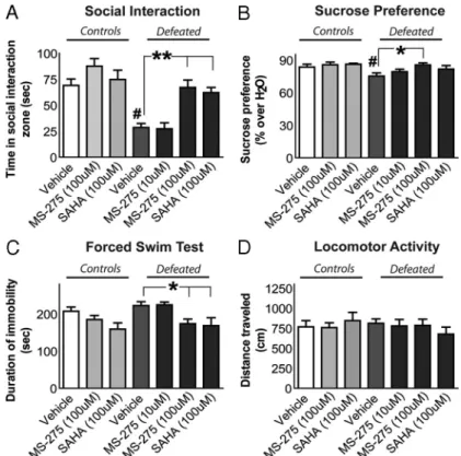
com-Figure 3. HDAC inhibitors have robust antidepressant-like effects when infused into the NAc. After chronic (10 d) social defeat stress or control conditions, separate groups of mice were fitted with subcutaneous minipumps and bilateral guide cannulae to receive a continuous infusion of the HDAC inhibitor MS-275 or SAHA, or vehicle, into the NAc. A, On experimental day 15 (5 d after surgery), each mouse was examined for total time spent within the “interaction zone” when a social target was present. When receiving vehicle infusion, stressed mice spent significantly less time engaged in social interaction when compared with nonde-feated controls. In contrast, the infusion of either MS-275 (100M) or SAHA (100M) restored the amount of time spent interacting socially. B, Over the next 4 d, preference for a sweetened 1% sucrose solution (over water alone) was examined across these same groups. Defeated mice infused with vehicle had a reduced preference for sucrose compared with their controls, and this effect of stress was reversed by the MS-275 (100M) infusion. C, On the next day, a forced-swim test was conducted. Chronic stress increased the total duration of immobility recorded within vehicle-treated mice. MS-275 (100M) and SAHA (100M) both decreased the duration of stress-induced immobility during this test. D, During 2.5 min of exposure to an open field, no differences in distance traveled were observed across the various treatment groups. Differences between vehicle and drug infusions in socially defeated mice are denoted by an asterisk (*) and double asterisks (**) to indicate significance at p⬍ 0.05 or 0.01, respectively. Differences between vehicle-treated nondefeated controls and socially defeated groups are denoted by a pound sign (#) to indicate significance at p⬍ 0.05. Error bars indicate SEM.
Figure 4. Continuous infusion of MS-275 (100M) or SAHA (100M) into the NAc significantly increases acetylated H3 in vivo. acH3K14 was quantified directly where the HDAC inhibitor was delivered after 10 continuous days of treatment via immunohis-tochemistry (A) and Western blotting (B) (*p⬍ 0.01, SAHA; **p ⬍ 0.001, MS-275). Error bars indicate SEM.
pared using a 2⫻ 3 ANOVA (stress by drug). An additional group of defeated mice was included for examining a lower molar concentration of MS-275 infusion on depressive-like behaviors, but this group of mice was not included in the statistical analysis. For comparing MS-275 to vehicle treatments in light/dark box behavior, two-tailed t tests were used. For all one-way ANOVAs, post hoc tests were performed using Dunnett’s multiple-comparisons test to selectively assess significant changes from the control condition. For all two-way ANOVAs, Bonferroni’s
post hoc tests were used to assess isolated comparisons. All differences were
considered significant when p values were⬍0.05. Statistical analyses were performed with GraphPad Prism software.
Results
Regulation of histone acetylation in the NAc in mouse social
defeat and in human depression
The NAc, which plays an important role in the expression of
reward- and stress-related behavioral responses (Anisman and
Zacharko, 1986; Wise, 1987; Koob, 1996), has been implicated
more recently in depression (Nestler and Carlezon, 2006). Changes
in histone acetylation within the NAc after chronic social stress
may contribute to significant changes in gene expression that
either sustain or oppose depressive-like behaviors (Berton and
Nestler, 2006; Krishnan and Nestler, 2008). To directly assess this
possibility, brains were collected from stressed and control mice
at either 1 h, 24 h, or 10 d after chronic (10
d) social defeat stress (Berton et al., 2006).
Using immunohistochemical analysis,
lev-els of acH3K14 were quantified in the NAc.
Shown in Figure 1A are representative
sec-tions at the level of the NAc obtained in
chronically defeated or nondefeated
con-trol mice stained for acH3K14 (top) or
to-tal histone H1 (bottom). We selected the
Lys14 residue for analysis since it is tightly
associated with increases in gene
tran-scription (Shahbazian and Grunstein,
2007). We found that levels of acH3K14
are dynamically regulated after chronic
social defeat stress: acH3K14 levels are
de-creased
⬃50% 1 h after the final stress
event but are significantly increased at
both 24 h and 10 d after social defeat stress
(Fig. 1 B), as indicated by a main effect of
stress (F
(1,30)⫽ 51.3; p ⬍ 0.0001), a main
effect of time (F
(2,30)⫽ 946.4; p ⬍ 0.0001),
and a significant interaction (F
(2,30)⫽
946.4; p
⬍ 0.0001). A significant increase
in acH3K14 levels in the NAc was also
verified by Western blotting, using
tis-sue collected 15 d after the last social
defeat episode (t
(7)⫽ 2.4; p ⫽ 0.047)
(Fig. 1C, left).
To study the relevance of these altered
levels of acH3K14 induced in the NAc of
mice subjected to chronic social defeat
stress to human depression, we examined
acH3K14 levels in human postmortem
NAc tissue obtained from depressed and
extensively matched control individuals.
Equivalent to the persistent increase in
acH3K14 found in mouse NAc after
chronic defeat stress, we found that
hu-man depression is associated with a
signif-icant increase (t
(14)⫽ 3.7; p ⫽ 0.001) in
levels of acH3K14 in the NAc (Fig. 1C,
right). This analysis included subjects who were and were not on
antidepressants at their time of death. However, chronic
admin-istration of fluoxetine had no impact on levels of acH3K14 in the
mouse NAc (t
(10)⫽ 1.3; p ⫽ 0.2) (data not shown), arguing
against a drug effect in our depressed human sample.
Regulation of HDAC2 in the NAc in mouse social defeat and
in human depression
acH3K14 has been shown to be regulated by class I HDACs
(Rundlett et al., 1996). Accordingly, we next examined whether
levels of expression of class I HDACs, namely, HDAC1, HDAC2,
or HDAC3, are altered in the NAc after chronic social defeat
stress in mice. HDAC2, but not HDAC1 or HDAC3, mRNA levels
were significantly decreased (F
(2,21)⫽ 27.8; p ⬍ 0.0001) 24 h after
chronic defeat stress (Fig. 2 A), an adaptation that could mediate
the observed increase in acH3K14 levels at this time point. This
effect of chronic social defeat on HDAC2 expression is a
long-lasting phenomenon, as protein levels of the enzyme are
signifi-cantly reduced (t
(8)⫽ 2.5; p ⫽ 0.04) for up to 15 d after the last
defeat episode (Fig. 2 B, left).
Since human depression, like mouse social defeat, is
associ-ated with increased acH3K14 levels in the NAc, we hypothesized
Figure 5. Gene expression arrays provide novel insight into the molecular mechanisms of antidepressant action in the NAc. Extending previous reports, chronic (10 d) social defeat stress induces a unique pattern of gene expression in the NAc that is mostly reversed by fluoxetine or MS-275 administration. Vehicle or MS-275 (100Minfusion into the NAc), or placebo or fluoxetine (20 mg䡠 kg⫺1䡠 d⫺1, subcutaneous pellet) were administered for 10 continuous days after chronic (10 d) social defeat stress or control conditions, and representative patterns of gene expression in the NAc were analyzed using Illumina microarrays. A, Chronic stress-induced genomic regulation is mostly reversed by MS-275 or fluoxetine, with differences between the two treatments indicated by the red bars and similarities indicated by blue bars. B, Within MS-275-treated mice, gene expression is robustly regulated in both nonstressed controls (⬃435genes)anddefeatedmice(⬃413).Incontrast,fluoxetineregulatesalargenumber of genes in previously stressed mice only (⬃206), with many fewer genes regulated in nonstressed controls (⬃84). C, When analyzing only those genes that are upregulated by fluoxetine after chronic stress, MS-275 regulates many of them in a similar way (above the top blue bar), although some are differently regulated. In much the same way, fluoxetine similarly regulates a sizable percentage of the MS-275-regulated genes (above the bottom blue bar).
that HDAC2 could be similarly reduced in this region of clinically
depressed individuals. Using the same postmortem tissue
exam-ined in Figure 1 B, HDAC2 protein expression was found to be
downregulated (t
(14)⫽ 2.2; p ⫽ 0.02) in the NAc of depressed
humans (Fig. 2 B, right), further corroborating that the effects
observed after social defeat stress in mice are clinically relevant. In
mouse, chronic treatment with fluoxetine did not significantly
alter levels of HDAC2 protein in the NAc (t
(10)⫽ 1.9; p ⫽ 0.1)
(data not shown), further suggesting that antidepressant
treat-ment in our human sample did not affect HDAC2 expression.
HDAC inhibitors have antidepressant-like effects when
infused into the NAc
To test the functional role played by altered levels of acH3K14 in
the NAc, groups of mice subjected to chronic (10 d) social defeat
stress and nonstressed control mice were, on day 11, surgically
prepared with Alzet subcutaneous minipumps connected to
bi-lateral guide cannulae allowing for continuous infusion of HDAC
inhibitors or vehicle into the NAc over extended periods of time.
After 5 d of infusion, each mouse was tested for its time spent
interacting with a novel mouse under controlled conditions. A
reduction in social interaction is one of the most robust and
lasting sequelae of chronic social defeat stress and can be reversed
by chronic (not acute) treatment with antidepressants (Berton et
al., 2006; Tsankova et al., 2006). We observed a significant effect
of social defeat (F
(1,54)⫽ 20.5; p ⬍ 0.0001) and a significant effect
of HDAC inhibitor (F
(2,54)⫽ 9.7; p ⫽ 0.0002) on this measure of
social interaction when infused into the NAc. As reported
previ-ously for unoperated animals, we found that chronically stressed
mice, which received intra-NAc vehicle infusions over 5 d, spent
significantly less time engaged in social interaction compared
with that of controls (Fig. 3A). In contrast, continuous infusion
of either MS-275 (100
M) or SAHA (100
M) into the NAc
reversed the stress-induced social avoidance in defeated mice and
restored the amount of time the animals spent interacting socially
(Fig. 3A).
Over the next 4 d, mice were examined for their preference for
a sucrose solution over water alone. We observed a significant
effect of social defeat (F
(1,54)⫽ 4.6; p ⬍ 0.04) and a significant
effect of HDAC inhibitor (F
(2,54)⫽ 3.4; p ⫽ 0.04) on sucrose
preference when infused into the NAc. Defeated mice chronically
infused with vehicle displayed a significantly reduced preference
for sucrose solution compared with nondefeated vehicle
con-trols, as reported previously for unoperated animals (Krishnan et
al., 2007), and this effect of chronic stress was also reversed by
infusion of MS-275 (100
M) (Fig. 3B). Subsequent to the sucrose
preference test, the forced-swim task was conducted. A
signifi-cant effect of HDAC inhibitor infused into the NAc was found on
the duration of immobility (F
(2,54)⫽ 5.8; p ⫽ 0.005). Specifically,
time spent swimming was increased by infusion of either MS-275
(100
M) or SAHA (100
M) into the NAc in previously defeated
and control mice (Fig. 3C). Such an increase in swimming
repre-sents an antidepressant-like effect in the forced-swim test. We
observed a slight (and insignificant) reduction in the duration of
time spent swimming after chronic stress. During a 3 min
expo-sure to an open field, no differences were found between any
groups of mice with regard to the total amount of locomotor
activity recorded (Fig. 3D), an important control for the
forced-swim and other behavioral measures. To complement these
be-havioral studies on depressive-like behavior, we also compared
the effects of continuously infusing MS-275 (100
M) into the
NAc on the expression of anxiety-like behavior as measured
dur-ing a light/dark test (Bourin and Hascoe¨t, 2003). After 7 d of
infusion with either MS-275 or vehicle, mice were placed into the
dark compartment of the light/dark box, and the amount of time
spent in the light compartment was assessed. MS-275 had no
effect on anxiety-like behavior when infused into the NAc (data
not shown).
MS-275 and SAHA increase levels of acH3K14 in the NAc
To confirm that HDAC inhibitors are reliably delivered into the
NAc via osmotic minipump infusion, and to validate that these
drugs are biochemically active at the site of infusion, levels of
acH3K14 were quantified under the infusion site after 10
contin-uous days of treatment. Immunohistochemistry and Western
blotting (F
(2,10)⫽ 9.9; p ⫽ 0.004) confirmed that acH3K14 levels
are robustly elevated in the NAc of mice that received continuous
infusion of MS-275 (100
M) or SAHA (100
M) compared with
vehicle-treated controls (Fig. 4 A, B).
Intra-NAc infusion of MS-275 reverses chronic stress-induced
patterns of gene expression similar to fluoxetine
One consequence of chronic social defeat stress is the
promulga-tion of a distinctive pattern of gene expression in the NAc, which
can be mostly normalized to that of nonstressed control mice by
chronic fluoxetine treatment (Berton et al., 2006). Given that
MS-275, when infused directly into the NAc, promotes an
antidepressant-like behavioral profile similar to that of chronic,
systemic fluoxetine administration, we tested whether the two
treatments also exert a similar regulatory impact on gene
expres-sion in the NAc. To directly compare the effect of MS-275
treat-ment to that of the conventional antidepressant fluoxetine on
patterns of gene expression in the NAc, both drugs were
admin-istered continuously for 10 d (MS-275 via intra-NAc infusion and
fluoxetine via systemic pellet implantations) after chronic (10 d)
social defeat stress, and gene expression patterns in the NAc were
studied using microarray technology. We confirmed that this
10 d course of treatment with MS-275 or with fluoxetine induced
equivalent antidepressant-like effects in the social interaction test
(data not shown). We found that social defeat stress, as observed
previously, induces a unique pattern of gene expression in the
NAc and that this global pattern of gene transcription is mostly
reversed by treatment with either MS-275 or fluoxetine (Fig. 5A).
In addition to genes affected similarly by MS-275 and fluoxetine,
each treatment also uniquely reversed smaller, distinct subsets of
defeat-regulated genes in the NAc. Overall, MS-275 altered
ex-pression of a larger number of genes than fluoxetine, and
regu-lated approximately the same number of genes in chronically
stressed and control animals (Fig. 5B). Close to one-third of all
regulated genes showed similar regulation by MS-275 in stressed
versus control mice. In contrast, fluoxetine altered expression of
more than twofold more genes in chronically stressed mice than
in control mice, with
⬍10% overlap between the two conditions.
These data are consistent with reports that fluoxetine causes little
to no mood-elevating effect in humans in the absence of
depres-sion or a related syndrome (Tsankova et al., 2006).
We next compared in greater detail the effects of MS-275 and
fluoxetine on gene expression in the NAc after chronic social
defeat stress. Focusing on genes that are upregulated by
fluox-etine in chronically stress mice, we identified a set of genes
(
⬃37%) that are similarly upregulated by MS-275 (Fig. 5C, above
the top blue bar). Other fluoxetine-regulated genes are either not
affected by MS-275 (
⬃15%) or are regulated in the opposite
direction by this HDAC inhibitor (48%). Conversely, a larger set
of genes are upregulated by MS-275 after chronic social defeat, as
mentioned previously, with fluoxetine causing similar regulation
for 48% of the genes, no effect for 9%, and opposite effects to
MS-275 for 43% (Fig. 5C, above the bottom blue bar).
Examples of genes that exhibit these varying patterns of
regu-lation are listed in Table 1 (for full gene lists, see supplemental
material S3, available at www.jneurosci.org as supplemental
ma-terial). Fifteen genes regulated on the microarrays by MS-275
after chronic social stress were randomly selected for validation
by quantitative PCR (qPCR) analysis. Of these, regulation of 12
genes (80%) was confirmed on independent tissue samples,
in-dicating a relatively low false-positive rate consistent with
previ-ous microarray studies of microdissected brain tissue. Among the
MS-275- and fluoxetine-regulated genes are several that would be
expected to exert antidepressant-like effects based on
informa-tion in the literature, as well as many novel genes that provide
new insight into the molecular basis of defeat stress and its
rever-sal by established antidepressants or HDAC inhibition (see
Dis-cussion). Portrayed in Figure 6 are examples of genes, and their
molecular pathways, that were significantly regulated in the NAc
by either social stress alone or the poststress treatment with
MS-275. Many of the genes regulated by MS-275 after social defeat
stress provide insight into potentially novel targets for reversing
stress-induced depressive-like behavior. The identification of
genes whose products regulate cellular morphology via effects
on actin reorganization [e.g., SLIT, TGF␣, p38, JNK (c-Jun
N-terminal kinase), and Rho] is very interesting because of the
emerging significance of dendritic restructuring in the
persis-tence of depression (Krishnan and Nestler, 2008). The complex
impact of chronic stress and MS-275 treatment on intracellular
signaling cascades is further evidenced by the regulation of
nu-merous molecular pathways that control gene transcription. For
instance, regulation of the transcription factors CREB (cAMP
response element-binding protein), ICER (inducible cAMP early
repressor), REST (RE1 silencing transcription factor), co-REST,
STAT (signal transducer and activator of transcription), and
NFAT (nuclear factor of activated T-cells) presumably contribute
to stress- and MS-275-induced changes in histone acetylation at
regulated genes.
Discussion
Histone acetylation is a promising target for novel treatments of
psychiatric illness because of its control over patterns of gene
transcription that ultimately establish and stabilize cognitive
and behavioral processes. The current study reveals a robust
antidepressant-like effect of HDAC inhibitors delivered directly
into the NAc after chronic social defeat stress. Our results also
demonstrate that chronic social stress induces a complex pattern
of changes in H3 acetylation in the NAc, with a dramatic decrease
observed 1 h after the last stress, followed by a smaller, but
sus-tained increase in acetylated H3 that persists for at least 2 weeks. The
observation that inhibition of HDACs and the consequent increase
in H3 acetylation in the NAc induces robust antidepressant-like
ef-fects suggests that the transient reduction in H3 acetylation after
social defeat stress may contribute to depressive symptoms,
whereas the sustained increase in H3 acetylation represents a
positive adaptation that serves to restore normal functioning.
The mechanism underlying the transient decrease in H3
acetyla-tion remains unknown, but the lasting increase in levels of
acety-lated H3 in the NAc could be mediated via a persistent and
selective decrease in expression of the class I HDAC, HDAC2, in
the NAc. The notion that downregulation of HDAC2 in the NAc
may be an adaptive mechanism is interesting in light of recent
findings that, in the hippocampus, HDAC2 is an important
me-diator of synaptic plasticity and morphological changes necessary
for associative learning (Guan et al., 2009). This underscores the
very different effects that HDAC2, like many other proteins
(Nes-tler and Carlezon, 2006), play in distinct brain regions.
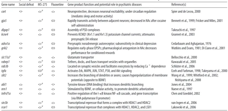
Table 1. Examples of genes regulated in the NAc by chronic social defeat stress and the effect of intra-NAc MS-275 and systemic fluoxetine
Gene name Social defeat MS-275 Fluoxetine Gene product function and potential role in psychiatric diseases Reference(s)
cort 佧 佦a 佦 Neuroprotective, decreases neuronal excitability; under circadian regulation
(mediates sleep and motor activity)
Spier and de Lecea, 2000
gja5 佧 佦a
N Rapidly transmits activity between adjacent neurons; decreased in NAc after cocaine self-administration
Bennett et al., 1999; Fricker and Miles, 2001
dlgap1 佧 佦a
N Assembly of PSD complexes Takeuchi et al., 1997
kcne4 佧 佦 N Prevents KCNQ1 (Kv1.1 and Kv1.3) potassium channel currents; attenuates presynaptic DA release
Grunnet et al., 2003
adra1a 佧 佦a N Presynaptic noradrenergic autoreceptor; subsensitivity in clinical depression Cedarbaum and Aghajanian, 1976
grik2 佧 佦a N Regulates early-phase EPSPs; pharmacological antagonism in NAc decreases performance for conditioned rewards
Watkins and Evans, 1981; Di Ciano et al., 2001
slc17a7 佧 佦 佦 Glutamate transporter Bellocchio et al., 2000
rabep1 佧 佦a
N Tethers, docks, and fuses transport vesicles with organelles Kawasaki et al., 2005
rab3b 佧 佦a 佦 Located on synaptic vesicles and facilitates exocytosis by reducing Ca⫹dependence Schlüter et al., 2006
tgfa 佧 Na 佦 Activates Erk, MAPK, JUN, STAT, P38, and Akt signaling Ezeh and Farbman, 1998; Takeyama et al., 2000 slit2 佧 佦a 佧 Increases the branching of dendrites or axons; causes hyperpolarization of membrane
potentials (opposite to BDNF)
Wang et al., 1999; Whitford et al., 2002; Nishiyama et al., 2008
abl1 佦 佧a 佧 Tyrosine kinase (DNA binding) that increases dendritic branching Jones et al., 2004 nrn1 佧 佦 佦 Stimulated by BDNF, or cellular activity, to promote dendritic arborization Naeve et al., 1997
tnfrsf1a 佧 佦 佦 Positive regulation of the I-B kinase/NF-B cascade, and gene transcription (via RNA polymerase II promoter)
Chen and Goeddel, 2002
sin3b 佧 佦a 佦 Transcriptional repressor that forms a complex with HDAC1 and HDAC2 van Ingen et al., 2006
rcor1 佧 佦a
N Transcriptional repressor that complexes with HDAC1, HDAC2, and LSD1 Lakowski et al., 2006
The table lists some of the genes that were regulated in the NAc by chronic (10 d) social defeat stress and the influence of chronic intra-NAc infusion of MS-275 or of chronic systemic fluoxetine administration. Genes regulated in the same direction by MS-275 and fluoxetine could represent part of a common, shared mechanism of antidepressant action. Genes uniquely affected by MS-275 may be useful for identifying novel biochemical pathways that promote antidepressant effects. Each gene listed contributes to forms of cellular plasticity, as indicated in the table. Some genes have previously been observed to contribute to stress-related illnesses. Gene abbreviations: cort, Cortistatin; gja5, gap junction protein ␣ 5; dlgap1, discs (large homolog-associated protein 1); kcne4, potassium voltage-gated channel; adra1a, adrenergic ␣ 1A receptor; grik2, ionotropic glutamate receptor (kainate 2); slc17a7, solute carrier family 17 (sodium-dependent inorganic phosphate cotransporter member 7); rabep1, rabaptin (RAB GTPase binding effector protein 1); rab3b, member RAS oncogene family; tgfa, transforming growth factor␣; slit 2, slit homolog 2; abl1, c-abl oncogene 1 (receptor tyrosine kinase); nrn1, neuritin 1; tnfrsf1a, tumor necrosis factor receptor superfamily member 1A; sin3b, SIN3 homolog B (transcription regulator); rcor1, REST corepressor 1.
aDenotes randomly selected genes whose regulation by MS-275 after social stress was validated by qPCR on independent tissue samples.
Importantly, we show here that human depression, like
chronic social defeat in mice, is also associated with increased
levels of H3 acetylation and reduced levels of HDAC2 expression
in the NAc. These findings further establish the validity of
chronic social defeat as a bona fide model of depression
(Kudryavtseva et al., 1991; Rygula et al., 2005; Berton et al., 2006;
Krishnan et al., 2007; Miczek et al., 2008). Based on the
antidepressant-like effects of HDAC inhibition in the mouse
NAc, we hypothesize that reduced HDAC2 expression in the NAc
is an adaptive neuronal response that emerges with clinical
de-pression. We speculate that this adaptive response may, in some
individuals, promote neuronal plasticity and contribute to
recov-ery. The fact that chronic fluoxetine treatment does not increase
H3 acetylation, or decrease HDAC2 levels, in the mouse NAc
indicates that HDAC inhibition represents a distinct and
funda-mentally novel mechanism of antidepressant action that now
warrants direct study in clinical populations.
The NAc is important for processing reward-related stimuli
(Wise, 1987; Koob, 1996; Carelli, 2002). Clinical depression often
includes anhedonic, motivational, and arousal deficits,
suggest-ing a role for the NAc in mediatsuggest-ing these symptoms (Nestler and
Carlezon, 2006). As such, we find that infusing HDAC inhibitors
into the NAc reverses the effect of chronic defeat stress on social
avoidance, decreased sucrose preference, and decreased mobility
during the forced-swim task. The persistent increase in H3
acet-ylation in this brain area combined with the antidepressant-like
effect of HDAC inhibitor infusion verifies the importance of the
NAc in the emergence of stress-induced depressive symptoms
and their reversal during effective treatment. To complement the
behavioral studies, we compared global patterns of gene
expres-sion by microarray analysis in the NAc after local MS-275
infu-sion with that of systemic fluoxetine treatment in stressed and
control mice. These gene arrays provide novel insight into
poten-tial molecular targets for the development of new treatments for
depression, particularly symptoms that pertain to NAc function.
Social defeat stress produces a distinct pattern of gene expression,
in the NAc, which is generally reversed by treatment with either
MS-275 or fluoxetine. For example, the neuropeptide cortistatin
is downregulated by stress in the NAc, an effect reversed by both
treatments. Cortistatin is normally regulated by circadian
vari-ables, and disruptions in this gene may be related to the severe
disruptions in sleep and arousal among depressed individuals
(Spier and de Lecea, 2000). In addition, stress-induced
disrup-tion of glutamatergic signaling, synaptic plasticity, inflammatory
responses, and chromatin remodeling may serve to promote and
sustain depressive-like behaviors: genes such as slc17a (glutamate
transporter protein), abl1 (tyrosine kinase), nrn1 (neuritin),
rab3b (Ras associated protein 3b), tnfrsf1a (tumor necrosis factor
Figure 6. Molecular pathway analysis of genes regulated in the NAc by the HDAC inhibitor MS-275 after chronic social defeat stress. Examples of highly regulated molecular pathways include genes that encode presynaptic vesicular proteins, plasma membrane receptors, intracellular signaling molecules, proteins that regulate the actin cytoskeleton, and the transcriptional regulatory machinery.
receptor superfamily, member 1A), and sin3b (transcriptional
regulator), involved in these various molecular pathways, are
each regulated by chronic stress and reversed by both drug
treat-ments. Importantly, these genes may be part of a shared
mecha-nism of antidepressant action for MS-275 and fluoxetine.
In an attempt to reveal novel targets for antidepressant action,
we examined genes whose regulation by chronic stress was
re-versed by MS-275, but not by fluoxetine. Examples of such genes
include gja5 (involved in gap junction formation) and dlgap1
(assembles postsynaptic density complexes). A decrease in gap
junctions after cocaine administration (Bennett et al., 1999) may
indicate a possible mechanism for cross-sensitization between
stress and stimulants. Insensitivity to the
␣-adrenergic receptor
(encoded by the gene adra1a) has been implicated in depression
(Cedarbaum and Aghajanian, 1976), and this gene was also
downregulated by stress and restored by MS-275. Interestingly,
the observation that many genes induced by chronic defeat stress
were also restored by MS-275 indicates the complexity of
tran-scriptional regulation in the brain (Sapolsky, 2003). Overall,
these results define the complex patterns of gene regulation in the
NAc during the emergence of depressive-like symptoms and their
reversal by antidepressant treatments.
Depression involves the persistent expression of diverse
symptoms, suggesting the involvement of stable molecular
adap-tations in the NAc, a brain region important for processing
emo-tional stimuli. Here, we report that chronic social stress leads to
prolonged increases in levels of acetylated histone H3 in the NAc
with a corresponding decrease in levels of HDAC2. The effects of
social stress on acetylated H3 and HDAC2 in the current mouse
model of depression were also observed in the NAc of
postmor-tem depressed humans, which further validates the social defeat
model and demonstrates the relevance of chromatin remodeling
in human depression. Furthermore, HDAC inhibitor infusion
into the NAc after chronic stress produces robust
antidepressant-like effects across several behavioral assays. Presumably, the
antidepressant-like effects of HDAC inhibition occur by
increas-ing histone acetylation at certain gene promoters and thereby
exerting complex effects on gene expression, as revealed here by
gene arrays for MS-275. These effects overlap significantly with
those of standard antidepressants but also reveal additional
po-tential targets of HDAC inhibition. Together, these findings
sup-port the utility of HDAC inhibitors as antidepressants and
provide novel insight regarding the molecular mechanisms
un-derlying antidepressant responses.
References
Anisman H, Zacharko RM (1986) Behavioral and neurochemical conse-quences associated with stressors. Ann N Y Acad Sci 467:205–225. Bellocchio EE, Reimer RJ, Fremeau RT Jr, Edwards RH (2000) Uptake of
glutamate into synaptic vesicles by an inorganic phosphate transporter. Science 289:957–960.
Bennett SA, Arnold JM, Chen J, Stenger J, Paul DL, Roberts DC (1999) Long-term changes in connexin32 gap junction protein and mRNA ex-pression following cocaine self-administration in rats. Eur J Neurosci 11:3329 –3338.
Berton O, Nestler EJ (2006) New approaches to antidepressant drug discov-ery: beyond monoamines. Nat Rev Neurosci 7:137–151.
Berton O, McClung CA, Dileone RJ, Krishnan V, Renthal W, Russo SJ, Graham D, Tsankova NM, Bolanos CA, Rios M, Monteggia LM, Self DW, Nestler EJ (2006) Essential role of BDNF in the mesolimbic dopamine pathway in so-cial defeat stress. Science 311:864 – 868.
Bourin M, Hascoe¨t M (2003) The mouse light/dark box test. Eur J Pharma-col 463:55– 65.
Brunswick DJ, Amsterdam JD, Fawcett J, Quitkin FM, Reimherr FW, Rosenbaum FJ, Beasley CM Jr (2002) Fluoxetine and norfluoxetine
plasma concentrations during relapse-prevention treatment. J Affect Disord 68:243–249.
Carelli RM (2002) The nucleus accumbens and reward: neurophysiological investigations in behaving animals. Behav Cogn Neurosci Rev 1:281–296. Cedarbaum JM, Aghajanian GK (1976) Noradrenergic neurons of the locus coeruleus: inhibition by epinephrine and activation by the alpha-antagonist piperoxane. Brain Res 112:413– 419.
Charney DS, Manji HK (2004) Life stress, genes, and depression: multiple pathways lead to increased risk and new opportunities for intervention. Sci STKE 2004:re5.
Chen G, Goeddel DV (2002) TNF-R1 signaling: a beautiful pathway. Sci-ence 296:1634 –1635.
Di Ciano P, Cardinal RN, Cowell RA, Little SJ, Everitt BJ (2001) Differential involvement of NMDA, AMPA/kainate, and dopamine receptors in the nucleus accumbens core in the acquisition and performance of pavlovian approach behavior. J Neurosci 21:9471–9477.
Drevets WC (2001) Neuroimaging and neuropathological studies of de-pression: implications for the cognitive-emotional features of mood dis-orders. Curr Opin Neurobiol 11:240 –249.
Ezeh PI, Farbman AI (1998) Differential activation of ErbB receptors in the rat olfactory mucosa by transforming growth factor-alpha and epidermal growth factor in vivo. J Neurobiol 37:199 –210.
Fricker D, Miles R (2001) Interneurons, spike timing, and perception. Neuron 32:771–774.
Grunnet M, Jespersen T, MacAulay N, Jørgensen NK, Schmitt N, Pongs O, Olesen SP, Klaerke DA (2003) KCNQ1 channels sense small changes in cell volume. J Physiol 549:419 – 427.
Guan JS, Haggarty SJ, Giacometti E, Dannenberg JH, Joseph N, Gao J, Wang X, Mazitschek R, Bradner JE, DePhino RA, Jaenisch R, Tsai LH (2009) HDAC2 negatively regulates memory formation and synaptic plasticity. Nature 459:55– 60.
Guo L, Lobenhofer EK, Wang C, Shippy R, Harris SC, Zhang L, Mei N, Chen T, Herman D, Goodsaid FM, Hurban P, Phillips KL, Xu J, Deng X, Sun YA, Tong W, Dragan YP, Shi L (2006) Rat toxicogenomic study reveals analytical consistency across microarray platforms. Nat Biotechnol 24:1162–1169.
Hawes JJ, Picciotto MR (2004) Characterization of GalR1, GalR2, and GalR3 immunoreactivity in catecholaminergic nuclei of the mouse brain. J Comp Neurol 479:410 – 423.
Jones SB, Lu HY, Lu Q (2004) Abl tyrosine kinase promotes dendrogenesis by inducing actin cytoskeletal rearrangements in cooperation with Rho family small GTPases in hippocampal neurons. J Neurosci 24:8510 – 8521. Kawasaki M, Nakayama K, Wakatsuki S (2005) Membrane recruitment of effector proteins by Arf and Rab GTPases. Curr Opin Struct Biol 15:681– 689.
Koob GF (1996) Hedonic valence, dopamine and motivation. Mol Psychi-atry 1:186 –189.
Krishnan V, Nestler EJ (2008) The molecular neurobiology of depression. Nature 455:894 –902.
Krishnan V, Han MH, Graham DL, Berton O, Renthal W, Russo SJ, Laplant Q, Graham A, Lutter M, Lagace DC, Ghose S, Reister R, Tannous P, Green TA, Neve RL, Chakravarty S, Kumar A, Eisch AJ, Self DW, Lee FS, et al. (2007) Molecular adaptations underlying susceptibility and resistance to social defeat in brain reward regions. Cell 131:391– 404.
Kudryavtseva NN, Bakshtanovskaya IV, Koryakina LA (1991) Social model of depression in mice of C57BL/6J strain. Pharmacol Biochem Behav 38:315–320.
Kurdistani SK, Tavazoie S, Grunstein M (2004) Mapping global histone acetylation patterns to gene expression. Cell 117:721–733.
Lakowski B, Roelens I, Jacob S (2006) CoRest-like complexes regulate chro-matin modification and neuronal gene expression. J Mol Neurosci 29: 227–239.
Lee MG, Wynder C, Schmidt DM, McCafferty DG, Shiekhattar R (2006) Histone H3 lysine 4 demethylation is a target of nonselective antidepres-sive medications. Chem Biol 13:563–567.
Miczek KA, Yap JJ, Covington HE 3rd (2008) Social stress, therapeutics and drug abuse: preclinical models of escalated and depressed intake. Phar-macol Ther 120:102–128.
Naeve GS, Ramakrishnan M, Kramer R, Hevroni D, Citri Y, Theill LE (1997) Neuritin: a gene induced by neural activity and neurotrophins that pro-motes neuritogenesis. Proc Natl Acad Sci U S A 94:2648 –2653. Covington et al.• Antidepressant Actions of HDAC Inhibitors J. Neurosci., September 16, 2009•29(37):11451–11460 • 11459
Nestler EJ, Carlezon WA Jr (2006) The mesolimbic dopamine reward cir-cuit in depression. Biol Psychiatry 59:1151–1159.
Nishiyama M, von Schimmelmann MJ, Togashi K, Findley WM, Hong K (2008) Membrane potential shifts caused by diffusible guidance signals direct growth-cone turning. Nat Neurosci 11:762–771.
Peng X, Wood CL, Blalock EM, Chen KC, Landfield PW, Stromberg AJ (2003) Statistical implications of pooling RNA samples for microarray experiments. BMC Bioinformatics 4:26.
Renthal W, Maze I, Krishnan V, Covington HE 3rd, Xiao G, Kumar A, Russo SJ, Graham A, Tsankova N, Kippin TE, Kerstetter KA, Neve RL, Haggarty SJ, McKinsey TA, Bassel-Duby R, Olson EN, Nestler EJ (2007) Histone deacetylase 5 epigenetically controls behavioral adaptations to chronic emotional stimuli. Neuron 56:517–529.
Rundlett SE, Carmen AA, Kobayashi R, Bavykin S, Turner BM, Grunstein M (1996) HDA1 and RPD3 are members of distinct yeast histone deacety-lase complexes that regulate silencing and transcription. Proc Natl Acad Sci U S A 93:14503–14508.
Rygula R, Abumaria N, Flu¨gge G, Fuchs E, Ru¨ther E, Havemann-Reinecke U (2005) Anhedonia and motivational deficits in rats: impact of chronic social stress. Behav Brain Res 162:127–134.
Sapolsky RM (2003) Stress and plasticity in the limbic system. Neurochem Res 28:1735–1742.
Schlu¨ter OM, Basu J, Su¨dhof TC, Rosenmund C (2006) Rab3 superprimes synaptic vesicles for release: implications for short-term synaptic plastic-ity. J Neurosci 26:1239 –1246.
Schroeder FA, Lin CL, Crusio WE, Akbarian S (2007) Antidepressant-like effects of the histone deacetylase inhibitor, sodium butyrate, in the mouse. Biol Psychiatry 62:55– 64.
Shahbazian MD, Grunstein M (2007) Functions of site-specific histone acetylation and deacetylation. Annu Rev Biochem 76:75–100.
Shi L, Reid LH, Jones WD, Shippy R, Warrington JA, Baker SC, Collins PJ, de Longueville F, Kawasaki ES, Lee KY, Luo Y, Sun YA, Willey JC, Setterquist RA, Fischer GM, Tong W, Dragan YP, Dix DJ, Frueh FW, Goodsaid FM, et al. (2006) The MicroArray Quality Control (MAQC) project shows inter- and intraplatform reproducibility of gene expression measure-ments. Nat Biotechnol 24:1151–1161.
Spier AD, de Lecea L (2000) Cortistatin: a member of the somatostatin
neu-ropeptide family with distinct physiological functions. Brain Res Brain Res Rev 33:228 –241.
Stan AD, Ghose S, Gao XM, Roberts RC, Lewis-Amezcua K, Hatanpaa KJ, Tamminga CA (2006) Human postmortem tissue: what quality markers matter? Brain Res 1123:1–11.
Takeuchi M, Hata Y, Hirao K, Toyoda A, Irie M, Takai Y (1997) SAPAPs. A family of PSD-95/SAP90-associated proteins localized at postsynaptic density. J Biol Chem 272:11943–11951.
Takeyama K, Dabbagh K, Jeong Shim J, Dao-Pick T, Ueki IF, Nadel JA (2000) Oxidative stress causes mucin synthesis via transactivation of epidermal growth factor receptor: role of neutrophils. J Immunol 164:1546 –1552. Tsankova N, Renthal W, Kumar A, Nestler EJ (2007) Epigenetic regulation
in psychiatric disorders. Nat Rev Neurosci 8:355–367.
Tsankova NM, Berton O, Renthal W, Kumar A, Neve RL, Nestler EJ (2006) Sustained hippocampal chromatin regulation in a mouse model of de-pression and antidepressant action. Nat Neurosci 9:519 –525.
van Ingen H, Baltussen MA, Aelen J, Vuister GW (2006) Role of structural and dynamical plasticity in Sin3: the free PAH2 domain is a folded mod-ule in mSin3B. J Mol Biol 358:485– 497.
Wang KH, Brose K, Arnott D, Kidd T, Goodman CS, Henzel W, Tessier-Lavigne M (1999) Biochemical purification of a mammalian slit protein as a positive regulator of sensory axon elongation and branching. Cell 96:771–784.
Watkins JC, Evans RH (1981) Excitatory amino acid transmitters. Annu Rev Pharmacol Toxicol 21:165–204.
Whitford KL, Marillat V, Stein E, Goodman CS, Tessier-Lavigne M, Che´dotal A, Ghosh A (2002) Regulation of cortical dendrite development by Slit-Robo interactions. Neuron 33:47– 61.
Willner P (1983) Dopamine and depression: a review of recent evidence. II. Theoretical approaches. Brain Res 287:225–236.
Willner P, Muscat R, Papp M (1992) Chronic mild stress-induced anhedo-nia: a realistic animal model of depression. Neurosci Biobehav Rev 16:525–534.
Wise RA (1987) The role of reward pathways in the development of drug dependence. Pharmacol Ther 35:227–263.
Zacharko RM, Anisman H (1991) Stressor-induced anhedonia in the meso-corticolimbic system. Neurosci Biobehav Rev 15:391– 405.