Volume 74 (2018)
Supporting information for article:
Solid-state structure and antimicrobial and cytotoxicity studies of a
cucurbit[6]uril-like Cu6L4 constructed from
3,5-bis[(1H-tetrazol-5-yl)methyl]-4H-1,2,4-triazol-4-amine
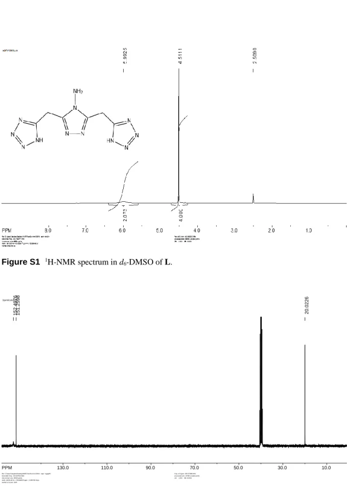
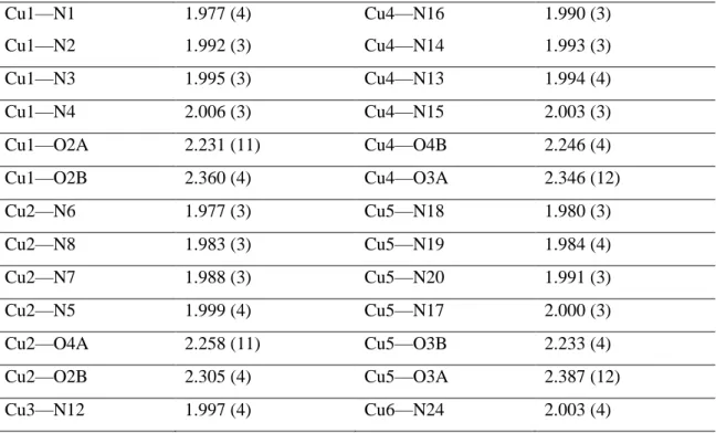
Acta Cryst. (2018). C74, doi:10.1107/S2053229618013670 Supporting information, sup-1 Figure S1 1H-NMR spectrum in d 6-DMSO of L. Figure S2 13C-NMR spectrum in d 6-DMSO of L. SpinW orks 2.5: PPM 130.0 110.0 90.0 70.0 50.0 30.0 10.0 152. 4976 151. 2598 20.0 226
file: C:\Users\Vasylevs\Desktop\NMR Pure\Sv-trtr-tr2\4\fid expt: <zgpg30> transmitter freq.: 100.637918 MHz
time domain size: 65536 points width: 24038.46 Hz = 238.860879 ppm = 0.366798 Hz/pt number of scans: 1024
freq. of 0 ppm: 100.627856 MHz processed size: 32768 complex points LB: 1.000 GB: 0.0000
Figure S3 XPRD of Cu6-complex (1): magenta-simulated; blue-obtained.
Table S1 Selected geometric parameters (Å)
Cu1—N1 1.977 (4) Cu4—N16 1.990 (3) Cu1—N2 1.992 (3) Cu4—N14 1.993 (3) Cu1—N3 1.995 (3) Cu4—N13 1.994 (4) Cu1—N4 2.006 (3) Cu4—N15 2.003 (3) Cu1—O2A 2.231 (11) Cu4—O4B 2.246 (4) Cu1—O2B 2.360 (4) Cu4—O3A 2.346 (12) Cu2—N6 1.977 (3) Cu5—N18 1.980 (3) Cu2—N8 1.983 (3) Cu5—N19 1.984 (4) Cu2—N7 1.988 (3) Cu5—N20 1.991 (3) Cu2—N5 1.999 (4) Cu5—N17 2.000 (3) Cu2—O4A 2.258 (11) Cu5—O3B 2.233 (4) Cu2—O2B 2.305 (4) Cu5—O3A 2.387 (12) Cu3—N12 1.997 (4) Cu6—N24 2.003 (4)
Acta Cryst. (2018). C74, doi:10.1107/S2053229618013670 Supporting information, sup-3
Cu3—N10 2.012 (3) Cu6—N21 2.017 (3)
Cu3—N9 2.025 (3) Cu6—N23 2.018 (4)
Cu3—N11 2.040 (4) Cu6—N22 2.022 (4)
Cu3—O3 2.165 (3) Cu6—O6 2.182 (3)
This is a table footnote (style: IUCr table footnote)
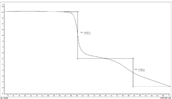
Figure S5 TGA of the complex, recorded in range 298 to 873 K.
Acta Cryst. (2018). C74, doi:10.1107/S2053229618013670 Supporting information, sup-5
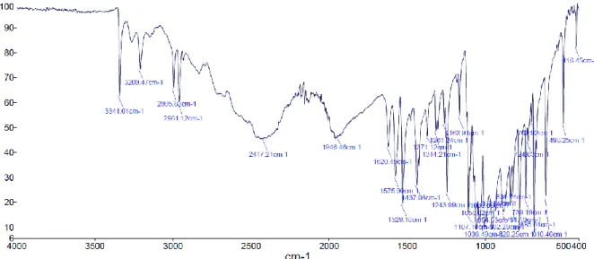
Figure S7 FT-IR of the ligand recorded in range 4000 – 400 cm-1.
Figure S9 Comparison of complex in a non-dried and dried forms. Blue – measured in glass
capillary with mother liquor, and red – dried crystals measured in range 2θ = 2 to 86.9o.
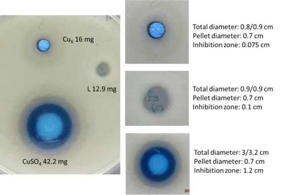
Figure S10 . Kirby Bauer test of the Cu6-complex; L –ligand; and blank CuSO4 against bacteria E.
Acta Cryst. (2018). C74, doi:10.1107/S2053229618013670 Supporting information, sup-7
Table S2 Overview of the amount of substance used and the ZOI`s for the triplicates
CuSO4·5H2O L Cu6
Amount of substance [mmol]
0.17 0.29 0.16 0.05 0.04 0.04 0.05 0.03 0.02
ZOI [mm] 12 13 13 1 1.3 1. 0.8 0.5 0.5
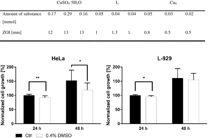
Figure S11 . The effect of 0.4% DMSO on the HeLa and L-929 cell growth; cell growth was
normalized to the 24 h Ctrl (in %); the bar chart represents the mean ± SD cell growth of 3
experiments (in triplicates) after 24 h and 48 h of 0.4% DMSO incubation time, statistical significance was determined via the multiple t-test (*p < 0.05, **p < 0.01, ***p < 0.001)
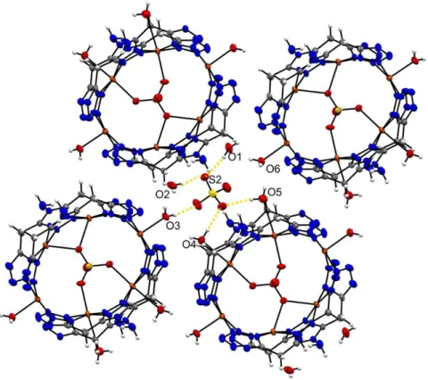
Figure S12 . Formation of intermolecular hydrogen bonds between coordinated water molecules, O1