Publisher’s version / Version de l'éditeur:
Current Opinion in Colloid & Interface Science, 15, 6, pp. 489-498, 2010-12-01
READ THESE TERMS AND CONDITIONS CAREFULLY BEFORE USING THIS WEBSITE.
https://nrc-publications.canada.ca/eng/copyright
Vous avez des questions? Nous pouvons vous aider. Pour communiquer directement avec un auteur, consultez la première page de la revue dans laquelle son article a été publié afin de trouver ses coordonnées. Si vous n’arrivez pas à les repérer, communiquez avec nous à [email protected].
Questions? Contact the NRC Publications Archive team at
[email protected]. If you wish to email the authors directly, please see the first page of the publication for their contact information.
NRC Publications Archive
Archives des publications du CNRC
This publication could be one of several versions: author’s original, accepted manuscript or the publisher’s version. / La version de cette publication peut être l’une des suivantes : la version prépublication de l’auteur, la version acceptée du manuscrit ou la version de l’éditeur.
For the publisher’s version, please access the DOI link below./ Pour consulter la version de l’éditeur, utilisez le lien DOI ci-dessous.
https://doi.org/10.1016/j.cocis.2010.06.003
Access and use of this website and the material on it are subject to the Terms and Conditions set forth at
Ceramide-enriched microdomains in planar membranes
Zou, Shan; Johnston, Linda J.
https://publications-cnrc.canada.ca/fra/droits
L’accès à ce site Web et l’utilisation de son contenu sont assujettis aux conditions présentées dans le site LISEZ CES CONDITIONS ATTENTIVEMENT AVANT D’UTILISER CE SITE WEB.
NRC Publications Record / Notice d'Archives des publications de CNRC:
https://nrc-publications.canada.ca/eng/view/object/?id=8bd8728b-b3cd-469e-b163-741084bb979b
https://publications-cnrc.canada.ca/fra/voir/objet/?id=8bd8728b-b3cd-469e-b163-741084bb979b
Ceramide-enriched microdomains in planar membranes
Shan Zou, Linda J. Johnston
⁎
Steacie Institute for Molecular Sciences, National Research Council of Canada, Ottawa, ON, Canada, K1A 0R6
a b s t r a c t
a r t i c l e
i n f o
Article history:
Received 8 April 2010
Received in revised form 8 June 2010 Accepted 9 June 2010
Available online 16 June 2010
Keywords: Ceramide Monolayers Supported bilayers Microdomains Sphingomyelinase
Ceramide has a large effect on the properties of biological membranes, increasing lipid order and promoting lateral phase separation, and plays an important role in cell signaling. This review provides an overview of recent studies of the effects of direct ceramide incorporation and enzymatic ceramide generation on planar supported membranes, including lipid monolayers and supported lipid bilayers. Recent studies have focused on understanding the nucleation, growth and morphology of ceramide gel domains, characterizing the properties of ceramide-rich membrane phases and investigating the effects of ceramide on phase-separated membranes with co-existing liquid-ordered and fluid phases, as models for cellular membranes.
Crown Copyright © 2010 Published by Elsevier Ltd. All rights reserved.
1. Introduction
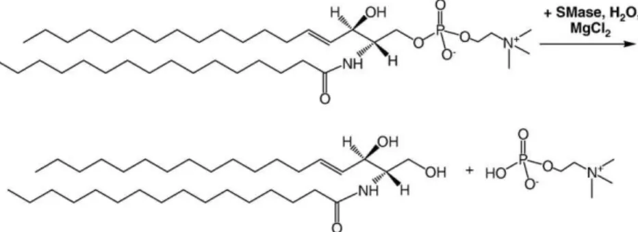
Ceramide is one of the simplest sphingolipids found in cell membranes. Although ceramide accounts for a relatively small fraction of the total lipid content in resting cells, its concentration increases rapidly in response to various stresses and can reach levels as high as 10% of the total lipid of apoptotic cells[1]. Ceramide is also a major component of the stratum corneum which serves as a hydrophobic barrier to prevent water evaporation through the skin. The production of ceramide in cells occurs by two main routes, de novo synthesis and the action of sphingomyelinase enzymes that hydrolyse the phosphor-ylcholine head group of cellular sphingomyelins (Fig. 1). Additional pathways include enzymatic hydrolysis of complex glycosphingolipids or ceramide-1-phosphate and via action of ceramide synthase[2]. A significant fraction of cellular ceramide is present as intermediates in the metabolism of more complex sphingolipids. The last decade has seen a major surge in interest in ceramide, largely due to its important role in cell signaling [2–5]. Ceramides are believed to function as second messengers, thus influencing a diverse range of biological processes, including apoptosis, immune response, bacterial and viral pathogenesis and cell cycle arrest. Although several ceramide binding proteins have been identified, ceramide can also regulate cell signaling through its ability to modulate the physical properties of membranes, leading to membrane reorganization in response to stress signals[3]. For example,
the generation of ceramide within sphingomyelin and cholesterol-rich plasma membrane rafts has been shown to induce coalescence of small rafts to give larger signaling platforms[6,7]. These ceramide-enriched membrane platforms provide a mechanism for organizing signaling molecules to facilitate and amplify signaling via specific membrane receptors. It has been hypothesized that the transformation of small inactive rafts to larger signaling platforms explains the function of ceramide in cellular activation by many receptors, in stress stimulation and in developmental processes[2,6]. As noted in a recent review[4], “the precise role of ceramide in biological processes remains to be well defined, but likely involves the intersection of classical signaling paradigms with the unique membrane-modifying properties of ceramide”.
The importance of ceramide in modulating membrane physical properties and biological function has provided the motivation for numerous investigations of ceramide incorporation in model mem-branes. In this review, we focus on studies of ceramide in planar lipid membranes, including monolayers formed at the air–water interface and supported lipid bilayers. We first outline background information on the ability of ceramide to modulate membrane properties and the use of phase-separated supported membranes to model lipid organization in cellular membranes. We then provide an overview of results obtained by both direct ceramide incorporation and enzymatic ceramide generation in planar membranes from 2007 to early 2010. Coverage is restricted to simple ceramides (not sphingomyelins or glycosphingoli-pids), primarily the physiologically relevant derivatives with C16or C18 chains. Although the focus is on monolayers and supported lipid bilayers, comparisons to data obtained in other model systems (e.g., vesicles) and in cellular membranes are also provided. Note that much useful information on the characteristics of ceramide-enriched domains ⁎ Corresponding author. Tel.: +1 613 990 0973.
E-mail address:[email protected](L.J. Johnston).
1359-0294/$ – see front matter. Crown Copyright © 2010 Published by Elsevier Ltd. All rights reserved. doi:10.1016/j.cocis.2010.06.003
Contents lists available atScienceDirect
Current Opinion in Colloid & Interface Science
and phase diagrams for ceramide-containing mixtures has been obtained in vesicles.
2. Ceramide properties
A comprehensive review by Goni and Alonso in 2006 summarizes the effects of ceramide (Cer) and several of its derivatives on both model and cellular membranes[8]. Several more focused updates on Cer-induced lateral phase separation of membranes have been published during the last two years, clearly illustrating the high level of interest and activity in Cer biophysics[9–11]. Many of the consequences of Cer incorporation on membranes are directly related to its structure. Ceramides are formed by acylation of the amino group of sphingosine with fatty acids. Naturally occurring Cers have fatty acid chains that vary from 2 to 28 carbons in length, with C16to C24being the most common, and are frequently saturated, although mono-unsaturated derivatives are also found. With their small polar head group, Cers are among the most hydrophobic lipids and can induce negative membrane curvature. They have high melting temperatures (e.g., ∼90 °C for C16Cer[12]) and a strong propensity for hydrogen bonding, functioning as both hydrogen bond donors and acceptors. These properties contribute to the strong impact of Cer on membrane properties, even at relatively low concentrations[8]. One of the main effects of Cer is an increase in molecular order of phospholipid mixtures and the promotion of lateral phase separation and domain formation. Ceramides also enhance transmembrane lipid motion (flip-flop), lead to membrane permeabi-lization via membrane defects and promote membrane fusion and budding processes, possibly by formation of non-lamellar phases. Lastly, Cer has been reported to induce order in fluid membrane phases and to displace cholesterol (Chol) from liquid-ordered domains [13,14], although recent results (described below [15]) indicate that Cer solubility in the fluid phase is enhanced in the presence of Chol. 3. Models for understanding membrane organization
The lateral heterogeneity of cell membranes plays an important role in regulating biological processes such as signal transduction, cell adhesion, lipid trafficking and viral and bacterial entry. Much of the current interest in membrane organization has been prompted by the lipid raft hypothesis [16,17]. Although the raft hypothesis remains controversial, a recent consensus definition states that “Lipid rafts are small (10–200 nm), heterogeneous, dynamic, sterol and sphingolipid-enriched domains that compartmentalize cellular processes. Small rafts can sometimes be stabilized to form larger platforms through protein– protein and protein–lipid interactions.”[18,19]. Rafts are generally believed to exist in a liquid-ordered phase with properties distinct from the surrounding fluid disordered membrane. Because of the complexity of a natural membrane and the difficulty of visualizing small, dynamic lipid domains in cells, most of our knowledge on the manner in which lipid–lipid interactions modulate membrane organization comes from simple model systems such as phospholipid monolayers, vesicles and
supported bilayers. Although an artificial membrane does not reproduce the complexity of a natural membrane, it does provide a tractable system for understanding how molecular interactions control lipid domain formation and the association of specific peptides or proteins with lipid domains.
Many recent studies have utilized phase-separated binary and ternary lipid mixtures (vesicles and planar membranes) to model the domains that are believed to exist in cellular membranes [20–22]. Binary lipid mixtures typically have co-existing gel and liquid phases. The gel phase consists of a lipid with a high melting temperature and is characterized by well-packed acyl chains in an extended conformation and slow lateral lipid diffusion. The liquid or fluid phase consists of a lipid with a low melting temperature, usually with either short or unsaturated acyl chains, with disordered packing and a higher lateral diffusion coefficient. Ternary lipid mixtures comprised of a saturated phosphatidylcholine (PC) or sphingomyelin (SM), an unsaturated PC and Chol have been shown to exhibit two co-existing fluid phases: a liquid-ordered (Lo) phase that is rich in Chol and saturated lipid and a liquid-disordered (Ld, fluid) phase that is predominantly unsaturated PC (Fig. 2A). The Lophase is believed to be analogous to membrane rafts and is characterized by highly ordered lipid acyl chains, but a degree of lateral lipid mobility that is similar to a fluid membrane.
There are several challenges associated with extrapolating from relatively simple model membranes to the more complex behavior of natural membranes[23]. First, a natural membrane has a complex mixture of lipids with a wide range of head groups and chain lengths/ unsaturation and with an asymmetric distribution of lipids between the two bilayer leaflets. By contrast, most model membrane studies focus on relatively simple lipid mixtures and have domains coupled between the two bilayer leaflets. Second, it is evident that domains in cells are typically small, well below the diffraction-limited resolution of optical microscopy [17]. However, Lo domains in most model membranes (for example, giant unilamellar vesicles, GUVs, which have been widely studied) are large since line tension generated by hydrophobic mismatch is minimized by merger of small domains. Third, experiments in model membranes are typically carried out under equilibrium conditions, which is rarely the case in cells where lipids are constantly trafficked to and from the membrane and the presence of proteins can modulate the membrane's phase separation behavior. Notwithstanding these limitations, much useful and complementary information has been derived from studies in vesicles and planar membranes. One advantage of planar supported mem-branes is the applicability of interface sensitive characterization tools that are compatible with detecting and characterizing the small nanoscale domains that are relevant to heterogeneity of natural membranes. Furthermore, domains in supported membranes are often smaller and rearrange or equilibrate more slowly than those in vesicles. This review will highlight the utility of these factors for investigating the nanoscale membrane morphology induced by Cer incorporation (Section 4) and the dynamic effects induced by enzymatic generation of Cer (Section 5).
4. Direct ceramide incorporation
The consequences of direct Cer incorporation in membranes have been studied for many binary, ternary and quaternary lipid mixtures, providing convincing evidence for formation of Cer-rich gel phase domains for a range of lipid compositions and temperatures, as reviewed in [8,10]. Planar supported membranes are particularly useful for studies of lateral phase separation, since they can be imaged over a wide range of length scales (e.g., from tens of nanometers using atomic force microscopy (AFM) to several hundred micrometers using fluorescence microscopy). During the last 3 years, studies in mono-layers and supported bimono-layers have probed the effects of Cer on lateral phase separation in membranes with an emphasis on the following three areas, each of which is described below: (1) understanding the factors that control nucleation, growth and morphology of Cer-rich domains in binary lipid mixtures, (2) investigating the effects of Cer addition to ternary lipid mixtures with co-existing phases and (3) probing the mechanical properties of Cer-enriched membranes. Although model membranes are usually assumed to be in thermody-namic equilibrium, this is not always the case for planar membranes, as noted in some examples below. This provides an opportunity to probe the non-equilibrium effects that occur during enzymatic Cer generation (Section 5).
4.1. Nucleation, growth and morphology of ceramide domains The formation of Cer gel phase domains in binary lipid mixtures with PC and SM is well-documented and recent studies have focused on detailed investigations of domain nucleation, growth and morphology. In one example, the effect of changing the length of the N-acyl chain of Cer on domain morphology was systematically studied in monolayers by Kartunnen et al.[24]using fluorescence microscopy. Shorter chain Cers (Cer2, Cer6and Cer8) were miscible in DMPC at a 1:2 molar ratio, whereas Cer10–Cer14/DMPC mixtures formed flower-like domains. A further increase in the N-acyl chain length changed the domain morphology from flower-like to round and bean-like shapes for Cer16 and then to round domains for Cer18, Cer20, and Cer24/DMPC mixtures. The size of the domains decreased with increasing N-acyl chain length at all surface pressures and slightly larger domains formed at higher surface pressures. The authors concluded that the domains are not at equilibrium and that their different morphologies arise from a subtle interplay between nucleation and growth [24]. Surface pressure provides the driving force for the formation of Cer-rich gel phase
domains that grow by excluding DMPC molecules. This type of growth is known to lead to formation of morphological instabilities and flower-like patterns when the nucleation rates are slow[25]. At fixed surface pressure an increase in the Cer chain length increases the driving force for the phase transformation due to attractive van der Waals interactions. Increasing Cer chain length leads to changes in its conformational and hydrogen bonding properties, which favor cluster-ing at low surface pressures. Thus, nucleation is enhanced, and the domains saturate in size before diffusional instabilities take over.
Gel phase domains were also obtained for mixtures of a natural brain Cer with POPC. Fidorra et al.[26]observed either striped or flower-shaped domains in AFM images of supported bilayers for POPC/Cer mixtures with up to ∼25 mol% Cer. Interestingly, solid Cer domains were also formed for ternary mixtures composed of POPC/Chol/Cer with low Chol content (b20%). A detailed investigation of the same ternary lipid mixture in GUVs by the Prieto group showed that the Cer domains disappeared at higher Chol concentrations and that Cer solubility in the fluid phase was increased by the presence of Chol[15]. Thus, the strong propensity for Cer-rich domain formation in binary mixtures cannot be extrapolated to ternary mixtures with Chol, an important point in the context of the raft model systems discussed below.
The Longo group has employed high resolution AFM imaging to examine domain nucleation and growth when Cer-containing mixtures are cooled below their miscibility temperature[27–29]. In one study, the steady-state domain nucleation rates were measured as a function of temperature for supported bilayers of two phase-separated binary lipid systems, DOPC/galactosylceramide (GalCer) and DOPC/DSPC[28]. The DSPC mixture produced symmetric domains superimposed in the two bilayer leaflets, whereas GalCer gave asymmetric domains that formed only in the leaflet distal to the substrate. Faster domain nucleation rates were measured for GalCer than for DSPC and the data were used to calculate line tensions for symmetric DSPC and asymmetric GalCer domains using classical nucleation theory. The larger line tension for the symmetric DSPC domains is consistent with the significantly larger hydrophobic mismatch between symmetric domains and the surrounding fluid phase. A second study focused on the kinetics of lipid domain growth for the same binary lipid mixtures; depending on the lipid composition and temperature, either diffusion limited growth, giving fractal domain shapes, or reaction limited growth of round domains was observed[30]. These studies, combined with data on the effects of Chol on domain growth in GalCer/PC mixtures[29], demonstrated that lipid domain formation and growth are regulated by lipid Fig. 2. Schematic illustration (A), AFM height (B) and fluorescence (C) images of phase-separated supported lipid bilayers prepared from the following lipid mixtures: DOPC/eggSM/ C16Cer/Chol at a 4:3:1:2 ratio in (B) and DOPC/eggSM/Chol/C16Cer (7:7:4:2 ratio) with 0.5 mol% NBD-Chol dye in (C). Images were reproduced from references[37]and[46], with
composition, domain symmetry and temperature. Extension of this method for studying domain formation to Cer without the sugar head group would be of significant interest.
4.2. Ceramide-enriched domains in model raft mixtures
The incorporation of Cer in ternary lipid mixtures with co-existing ordered and fluid phases has been examined by several groups as a model for Cer effects on membrane rafts. The first direct visualization of the effects of Cer in supported bilayers prepared from the canonical lipid raft mixture (1:1:1 DOPC/C18SM/Chol) was reported by the Schwille group[31]. AFM images showed that the bilayers had three distinct phases at 8% and 12% C18Cer, whereas only Lo domains surrounded by Ldphase were observed at 4% Cer. The tallest regions were assigned to a Cer-enriched phase that formed small subdomains within the large SM-Chol-rich Lodomains, which were of intermedi-ate height (Fig. 2A). The DOPC-rich Ldphase was the thinnest region of the membrane. Decreases in lipid diffusion coefficient for the fluid phase with increasing Cer content were attributed to Chol enrichment in this phase, by analogy to earlier reports on the displacement of Chol from SM-rich domains by Cer[13,14]. Comparison of several different acyl chain lengths demonstrated that C16 and C18 Cer form Cer-enriched domains that segregate from the Lodomains in raft mixtures, whereas shorter chain Cer are solubilized in the existing Lodomains
[32]. Fluorescence correlation spectroscopy (FCS) indicated that the relative lipid diffusion coefficients varied only slightly (∼20%) as a function of Cer chain length for the Ldphase in 1:0.7:0.3:0.67 DOPC/ SM/Cer/Chol mixtures. By contrast, diffusion coefficients for Lo domains increased by more than a factor of two in the presence of C6and C2Cer, demonstrating the ability of short chain Cer to disrupt lipid packing and decrease domain stability and viscosity.
Similarly to Schwille's work, our group used AFM to show that a mixture of 3 co-existing phases was formed when Cer replaced a fraction of the SM in bilayers of a different ternary lipid mixture (2:2:1 DOPC/eggSM/Chol)[33,34]. A typical image of the three co-existing phases in the presence of 10% Cer is shown inFig. 2B. At a lower Chol concentration (5:5:1 DOPC/eggSM/Chol) bilayers had small ordered domains in the absence of Cer (b200 nm in diameter; assigned to a SM-rich gel phase)[35]. The domains increased in size with increasing Cer concentration, with some domains as large as 1 μm in diameter at 9.1% Cer, but there was no evidence for a third Cer-enriched phase[35]. Although the Cer content is similar in the two egg SM mixtures, the SM/ Cer ratio is considerably higher in the 5:5:1 bilayer, accounting for the lack of Cer-enriched domains.
AFM is ideal for probing the nanoscale morphology of bilayers in the presence of Cer but provides limited information on the lipid composition of the various phases, especially for mixtures with 4 (or more) components. This has been addressed by employing multi-modal approaches that combine AFM with FCS (as noted above), force mapping (seeSection 4.3) or fluorescence microscopy. The utility of two novel dye labeled Chol probes for labeling ordered domains for fluorescence microscopy studies for bilayers containing Cer has been examined
[36,37]. In one case, a probe with BODIPY attached to the alkyl side chain of Chol was used to visualize the loss of Chol from Lodomains after Cer incorporation[36]. In a second example, a probe with NBD attached to the Chol hydroxyl group via a carbamate linker was capable of distinguishing between the three co-existing phases produced by adding Cer to ternary lipid mixtures, albeit with small differences in contrast between Loand Ld phases[37].Fig. 2C shows a fluorescence image for a DOPC/eggSM/Chol/ Cer bilayer with Cer-rich domains (dark), Lodomains (grey) and Ldphase (bright). As an alternative approach for obtaining information on domain composition, AFM has been combined with chemically-selective imaging using time of flight secondary ion mass spectrometry (Tof-SIMS) to probe the lipid distribution in Langmuir–Blodgett monolayers[38]. AFM images of DOPC/eggSM/Chol/Cer monolayers showed heterogeneous liquid condensed (LC) domains that were similar to the Lodomains with
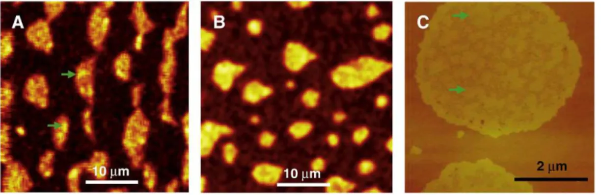
Cer-enriched subdomains obtained for bilayers of the same mixtures. Tof-SIMS demonstrated that SM was uniformly distributed throughout the domains whereas Cer localized predominantly in small clusters within the domains (Fig. 3).
Although the formation of three co-existing phases after Cer addition to ternary lipid mixtures was first visualized in supported bilayers[31], similar observations were subsequently reported for GUVs[39,40]and LUVs[41]. The formation of tightly-packed Cer-enriched gel phase has been attributed to the strong intermolecular interactions between Cer molecules [42]. The stability of Cer/SM interactions is evident from the persistence of highly ordered structures probed by electron spin resonance (ESR) spectroscopy and the appearance of a sharp wide-angle X-ray reflection at temperatures higher than the gel–fluid transition for Cer in egg PC bilayers. The Slotte group has examined the effect of Cer chain length (from 4 to 24 carbons) on the sterol distribution within LUVs prepared from quaternary mixtures (PC/SM/Cer/Chol)[43]. The overall mem-brane distribution reflects the differential partitioning of sterol into ordered domains where Cer and sterol compete for association with SM.
Finally, the effect of Cer on protein distribution in raft lipid bilayers has been investigated by AFM imaging combined with FCS and confocal microscopy[36]. Chiantia et al. showed that two typical raft-associated proteins (glycosylphosphatidylinositol placental alkaline phosphatase and the cholera toxin–GM1 complex) had a preference for localization within Cer-enriched domains in DOPC/SM/Chol/Cer bilayers. Both proteins showed a dramatic reduction in lateral diffusion within the Cer domains. By contrast, a non-raft protein (synaptobrevin 2) was almost completely excluded from the Cer-enriched domains. The results for the two lipid-anchored proteins are consistent with the hypothesis that Cer domains in cells can recruit specific proteins and reduce their lateral diffusion, providing a mechanism for amplified signaling. It remains to be tested whether the same trend applies to raft-associated integral membrane proteins.
4.3. Mechanical stability of ceramide-enriched supported lipid bilayers Tightly-packed Cer domains are expected to have a significant effect on the mechanical properties of membranes, as reviewed recently by Lopez-Montero et al.[11]. For example, the mechanical stabilization of Cer-enriched membranes has been hypothesized to be an important factor[11] in the clustering of membrane signaling receptors and in the strong enhancement of detergent resistance upon addition of low concentrations of Cer to SM or DPPC vesicles[44]. The high Cer content of the stratum corneum also plays a central role in determining its mechanical properties and low permeability[45]. By contrast, enzymatic generation of Cer decreases the mechanical stability of membranes, leading to membrane defects and permeabil-ity and to vesicle budding.
AFM-based force mapping coupled with AFM imaging provides direct correlation of the organization of multicomponent lipid membranes with their nanomechanical properties, although until very recently it had not been applied to complex phase-separated membranes. Our group has developed a novel approach where 2D visual maps of the intrinsic breakthrough forces, elastic moduli, and adhesion can be used to correlate the nanoscale structure of multicomponent lipid mixtures with their mechanical stability[34]. We showed that the direct incorporation of 10% Cer into DOPC/ eggSM/Chol bilayers increases the breakthrough force and the Young's modulus in both Ldand Lodomains (Fig. 4), an effect that was attributed to the influence of Cer on the lipid organization, as well as the displacement of Chol in response to the generation of Cer-enriched domains[34,46]. Furthermore, Cer-enriched domains ex-hibit a different packing behavior from the well-known gel phases, presenting a high breakthrough force (N12 nN). The mechanical stability and compactness of the Cer-enriched bilayers is relevant to
the Cer-induced formation of signaling platforms in cell membranes and, hence, AFM force mapping is a valuable complement to other biophysical techniques currently used to study multicomponent lipid bilayers.
It is worth noting that the presence of 10% Cer in DOPC/eggSM/ Chol bilayers increased the breakthrough forces from 1.4 to 4.1 nN in the Ldphase and from 3.2 to 5.0 nN in the Lodomains (Fig. 4E,[34,46]). Our force mapping results also demonstrated that the Cer-enriched domains require both methyl β-cyclodextrin (MβCD) and chloroform treatments to weaken their highly ordered organization, suggesting a lipid packing that is different from that in typical gel states[46]. A
lower limit of 8–12 nN for the breakthrough force for the ceramide-rich phase was estimated[34,46]. These results are consistent with earlier measurements of the resistance to compression for mono-layers[11]. For example, the compressibility modulus is higher for Cer (300 mN/m for bovine brain Cer and 600 mN/m for C16Cer)[47]than for fluid lipids such as POPC (80 mN/m) or SM (200 mN/m for C16SM)
[48].
The Maggio group has recently measured diffusion coefficients for Cer LC domains in C16SM:C16Cer monolayers and used the data to calculate apparent viscosities of 10−9–10−7N s/m for lateral surface pressures between 15–30 mN/m[49]. This viscosity change is caused Fig. 3. Tof-SIMS (A, Cer signal; B, SM signal) and AFM (C) images for DOPC/eggSM/Chol/Cer monolayers transferred at 30 mN/m; each image is for an independently prepared monolayer. Cer is heterogeneously distributed in clusters (green arrows) within the domains (A, C), while SM is uniformly distributed throughout the domains (B). Reproduced from reference[38]with permission.
Fig. 4. AFM height image (A), corresponding maps of breakthrough force (B), Young's modulus (C), and force curves (D) measured on supported lipid bilayers prepared from DOPC/ eggSM/C16Cer/Chol (4:3:1:2 ratio) after treatment with 1 mM methyl-β-cyclodextrin and chloroform vapor. Histograms of breakthrough forces for co-existing Loand Ldphases in
by an increase in both the intrinsic viscosity and the domain crowding. This work provides a convenient approach for analyzing the influence of domains on the mechanical properties of lipid monolayers and is relevant to Cer domain growth and equilibration for enzyme treated monolayers (seeSection 5.1).
5. Enzymatic ceramide generation in supported membranes
Sphingomyelinases (SMase) are a group of phospholipases that are responsible for the hydrolysis of SM to give Cer and the water-soluble phosphocholine[4,5]. In common with other phospholipases, they are active at the membrane–water interface and their activity is regulated by membrane properties, including lipid composition, phase separa-tion, membrane curvature and defects, and dipole potential. SMase activation provides a mechanism for the rapid generation of Cer in specific membrane locations. This spatial and temporal control is a key aspect of Cer modulation of cell signaling, ultimately allowing it to trigger a variety of cellular responses, including cell proliferation, differentiation and death. Supported membranes are ideal models for probing the consequences of Cer generation on membrane organiza-tion since optical microscopy and interface-selective characterizaorganiza-tion methods can be used to visualize enzyme-mediated reorganization.
Studies of SMase activity in model membranes typically use the readily available, water-soluble, Mg2+-dependent bacterial SMases from Bacillus cereus or S. aureus; both bacterial enzymes are part of a large family that includes the mammalian neutral SMases and has a conserved catalytic domain. Enzyme activity generally shows an initial lag period, during which the enzyme partitions to the interface, binds to the SM substrate and undergoes a slow (precatalytic) activation step. The lag time depends on the enzyme concentration and can vary from tens of seconds to tens of minutes. This is followed by a period of steady-state catalysis during which interfacially active enzyme is associated irreversibly with the interface and the rate is independent of SM concentration[50,51]. Enzyme activity eventually halts due to either substrate depletion or product accumulation. Catalytic activity is affected by the phase of the lipid, with gel phase SM vesicles showing relatively low activity. By contrast, mixed vesicles (SM plus PCs, phosphatidylethanolamines or Chol) or SM vesicles above the lipid melting temperature exhibit significantly higher activity[52,53].
5.1. Complex morphologies for sphingomyelin-ceramide monolayers Maggio, Fanani and coworkers have reported detailed fluo-rescence microscopy studies of the enzymatic generation of Cer in SM monolayers in the liquid-expanded (LE) phase at the air–water interface [9,51,54–56]. Their work represents one of the most comprehensive examinations of the factors that influence membrane reorganization during and after SMase activity. The kinetics for enzyme activity are monitored by the reduction in surface area that occurs when the larger SM is converted to Cer at constant surface pressure (10 mN/m). Fluorescence microscopy using DiIC12to label the LE phase of the monolayer provides simultaneous information on membrane morphology, one of the key advantages of this approach. Early studies examined the changes in membrane topography at high rates of Cer production and provided the first direct visualization of SMase-induced formation of Cer-enriched domains in monolayers
[50,51]. Detailed morphology changes of the monolayer at each stage of SMase activity led to the following conclusions. The nucleation and growth of small circular Cer-enriched domains occurs at the end of a short lag time after injection of enzyme to the aqueous phase below the monolayer. During the steady-state catalysis regime, the initial domains grow in size and undergo two distinct changes in morphology, first to give irregular star-shaped domains and second to produce increasingly branched fractal structures. Further evolution of the monolayer gives hexagonal domain lattices and finally the
condensed domains reach the percolation threshold, resulting in a discontinuous SM-rich LE phase. The variations in domain shape and organization were rationalized on the basis of competing line tension and dipolar electrostatic repulsion and illustrated the ability of local enzyme activity to influence organization on a range of length scales. This work also demonstrated that premixed SM/Cer monolayers did not reproduce the complex morphology of enzyme treated monolayers.
The monolayer studies described above illustrate the complex interplay between monolayer morphology and enzyme activity and raise several mechanistic questions that have been addressed during the last two years[54–56]. The correlation between the formation of Cer-enriched domains and the development of full catalytic capacity at the end of the lag period was examined[56]. The transition from a precatalytic state to full catalytic capacity depended strongly on the extent of the interface between LE and LC phases, leading to the conclusion that the amount of lateral interface provided by the Cer-rich domains is one of the main parameters that allows the enzyme to attain full activity.
The observation that the Cer domain interface favors the activa-tion steps required for full activity is consistent with the border-(or perimeter-) activated mechanism which postulates that an enzyme becomes fully active only when bound to a domain boundary or defect. Although a border-activated mechanism has been hypoth-esized for a number of phospholipases, other evidence suggests that some phospholipases are active over the entire fluid surface of a membrane, rather than at domain boundaries (an excellent literature summary is provided in a recent review [9]). To probe which mechanism applies to SMase hydrolysis of SM monolayers, the distribution of Alexa-488-labeled SMase was examined on SM and 9:1 SM/Cer monolayers at various stages of enzyme activity[55]. SMase was found to localize preferentially in the LE phase of both SM and SM/Cer monolayers, with no evidence for higher enzyme concentration at domain boundaries. The possibility of higher enzyme activity at the interface was excluded based on the lack of dependence of the catalytic rate on the amount of interface between LE and LC phases. The authors concluded that an area-activated mechanism with enzyme distributed uniformly throughout the LE phase best accounts for SMase action. Homogeneous generation of Cer was postulated to yield a supersaturated mixed monolayer, resulting in product-induced inhibition of SMase during the lag period. Nucleation of Cer-enriched domains then lowers the Cer concentration in the LE phase, allowing enzyme activity to proceed at a steady-state rate. Thus, the kinetic barrier for the nucleation of Cer-enriched domains contributes to the lag time for achieving full catalytic activity.
Several aspects of the SMase hydrolysis of SM monolayers suggest that enzymatic reaction leads to monolayer morphologies that are far from equilibrium[54–56]. These include the observation of different shapes and sizes for Cer-enriched domains prepared by direct mixing and enzyme action and the formation of a supersaturated Cer-enriched phase during the initial lag period. Furthermore, other studies (seeSection 5.2) indicate that bilayers subjected to enzymatic generation of Cer continue to reorganize after enzyme activity has been halted. The consequences of the “out-of-equilibrium” conditions produced by enzymatic generation of Cer have been investigated by monitoring the evolution of domain morphology for SM monolayers in which enzyme activity was stopped by adding ETDA during the steady-state catalysis regime [54]. The initial irregularly-shaped domains annealed to give rounded shapes, reaching near-equilibrium values after 80–100 min. Round domains were also formed at very low rates of Cer production. Correlation of domain shapes and perimeter lengths with growth rates led to the conclusion that the domains contain varying concentrations of Cer with a competition between line tension and dipole moment determining their shape. At high rates of Cer production domains contain predominantly Cer which has a higher dipole moment than SM; the increased
intermolecular repulsion leads to domain branching and irregular perimeters. However, these domains gradually equilibrate to give rounded domains by incorporation of additional SM. By contrast, at slow rates of Cer production the domain composition is close to the equilibrium composition of χCer∼ 0.53 observed for mixed SM/Cer monolayers; the dipole moment is lower and the effects of line tension dominate, leading to rounded domains. Overall, the experi-ments on SM monolayers provide a detailed model for the relationship between membrane organization and enzyme activity, with domain formation and morphology regulated by enzyme activity, but activity also modulated by domain formation.
5.2. Restructuring of bilayers with liquid-ordered domains
Investigations of enzymatic generation of Cer in supported bilayers have focused on phase-separated lipid membranes with co-existing ordered (Loor gel) and fluid phases as a model to understand the effects of Cer on membrane rafts. Initial AFM studies of SMase treatment of phase-separated bilayers were published within months of each other by three groups, each reporting the formation of new Cer-enriched ordered domains. In work from the Schwille group, SMase treatment of DOPC/C18SM/Chol (1:1:1) bilayers resulted in reduction in the size of some Lodomains and formation of higher regions that were assigned to Cer-enriched domains, by analogy with results for bilayers containing premixed Cer[31]. The formation of raised edges on many domains was interpreted as evidence for initial enzyme activity at the Lo/Ldinterface. Related work from our group similarly showed the formation of Cer-enriched subdomains located mostly at the edges of the original Lodomains after SMase treatment of DOPC/eggSM/Chol (2:2:1) bilayers[33]. However, the enzyme also led to a pronounced bilayer restructuring that has some analogies to the proposed coalescence of rafts in cell membranes. This included (1) the formation of clusters of domains within a new region that had an intermediate height between those of the original Lodomains and fluid phase and (2) the generation of large regions of a lower (fluid) phase with occasional defects. Similar results were obtained for several enzyme concentrations (varying over two orders of magni-tude) and for enzymes from two bacterial sources. Although Cer-rich subdomains and domain clusters appeared within the first several minutes after enzyme addition, the membrane morphology continued to evolve over a period of more than 1 h, even when the enzyme was deactivated by extensive rinsing with water. In a third example, the Devaux group examined the effects of SMase in a more complex phase-separated mixture (PE/PC/SM/Chol, 1:1:1:1 molar ratio)[57]. Disappearance of the initial ordered domains, formation of higher Cer-enriched domains and formation of small membrane defects were observed. Parallel studies in phase-separated GUVs with Lo/Ldphases also showed Cer domains after enzyme exposure; however, SMase treatment of GUVs without Lophase led to vesicle collapse and was attributed to membrane tension and defects caused by asymmetric generation of Cer in a single bilayer leaflet.
The observations of new Cer-enriched domains and a large scale membrane reorganization are common to each of the above studies, independent of incubation conditions (time, enzyme source, concen-tration) and membrane composition. Although AFM provides unpar-alleled capability to monitor dynamic changes in membrane morphology at high spatial resolution, its lack of molecular specificity and the heterogeneous membranes produced by SMase make it crucial to employ other complementary methods. Thus, the effect of the initial domain size, the kinetics for enzyme activity and the nature of the domain clusters have been investigated using a combination of AFM and fluorescence with multiple labeling strategies. For example, we showed that DOPC/eggSM/Chol (5:5:1) bilayers with SM-rich nanodomains behaved similarly to bilayers with micrometer-sized Lo domains [35]. The initial SM domains enlarged and became heterogeneous in height due to formation of Cer-enriched areas
after SMase treatment. They also clustered together, leaving uniform areas of a lower phase that was considerably less viscous than the original DOPC-rich phase, based on its ability to maintain bilayer defects. Both the large domain clusters and surrounding lower phase could be visualized by fluorescence allowing measurement of the kinetics for membrane restructuring. The bilayer restructuring varied with different lipid compositions and domain sizes and it was postulated that Cer-induced Chol expulsion from the domains accounted for some of the observations.
A multi-modal imaging approach was employed to examine the SMase-induced clustering of domains for DOPC/eggSM/Chol bilayers (2:2:1)[58]. Fluorescence imaging using Texas Red-DHPE to label the fluid phase showed that membrane restructuring involves the formation of large dye-excluded regions that initially form around the pre-existing Lo domains. The kinetics for formation of these regions were consistent with activity of different numbers of enzyme molecules in various bilayer regions. Correlated AFM and fluorescence images of the same bilayer region before and after enzyme addition demonstrated that the large dye-excluded patches correspond to new membrane regions that have strong contrast in lateral deflection AFM images but are not always detectable in topographic images (Fig. 5). These areas either form around existing domains or appear in regions where the original domains have partly or completely disappeared. Although it was initially postulated that these regions were generated by loss of Chol from domains in response to Cer formation, the correlated imaging approach indicated that the situation was more complex and that these areas may also contain Cer and residual SM.
Attempts to label Loand/or Cer domains with GM1-Cholera toxin (as used successfully for bilayers with premixed Cer[36]) during in situ SMase treatment were not successful, apparently because the cholera toxin interfered with either enzyme activity or with subsequent membrane restructuring[58]. More definitive conclusions were obtained by using a NBD-Chol probe that was designed to model the Lo/Ldpartitioning behavior of natural Chol[37]. Correlated AFM and fluorescence imaging provided evidence for the dynamic displacement of Chol from the Lo domains and demonstrated that Chol was also excluded from the large dye-excluded regions, leading to the conclusion that these were Cer-enriched regions. Note that, as pointed out for SM monolayers [54], these new regions must represent “out-of-equilibrium” membrane compositions.
Jensen and coworkers have used fluorescence microscopy with labeling of multiple membrane components to address the complex-ities of enzyme-promoted reorganization of bilayers containing brain SM (2:2:1 DOPC/bSM/Chol)[59]. Bilayers were prepared in micro-fluidic channels, an approach adopted to facilitate reproducible bilayer fabrication and a more uniform distribution of enzyme than can be achieved by manual addition. A combination of fluorescence labeling strategies was used to identify SM, Cer, fluid phase domains and SMase during the multistage evolution of the bilayer. The initial stage (termed a reaction-induced phase transformation) occurred during the first 5 min and generated large SM-enriched domains and smaller Cer-enriched domains. This morphology was stable for approximately 20 min, possibly due to limited accessibility of the enzyme to the SM domains. The second stage of bilayer reorganization was initiated by nucleation of large 3-D features (several micrometers in diameter and height) that were enriched in enzyme, Texas Red-DHPE, SM and Cer, followed by rapid dissolution of SM domains and the appearance of more Cer-enriched domains. This “solvent-mediated phase transformation” continued until most of the SM had been consumed. The large 3-D features were hypothesized to represent a new enzyme-enriched phase with increased SMase activity that acts as a sink to absorb SM from the surrounding membrane. The authors compared their results to a previous monolayer study in which electrostatic absorption of phospholipase A to phase-separated regions of charged reaction products produced an enzyme-enriched phase[60]. Alternately, it has been suggested
that the 3-D structures may represent nucleation of a Cer-induced inverted phase that has increased enzyme activity[9]. Although the formation of large 3-D structures must create membrane defects, the effects of defects and Chol on the various bilayer transformations were not explicitly considered in this study.
Bilayers of the same composition have been examined by Schwille and coworkers[61]. They observed that a phase-separated (Lo/Ld) bilayer was transformed initially to a homogeneous bilayer (as viewed by fluorescence using BODIPY-Chol which localizes preferentially in the Ldphase) and then to a heterogeneous bilayer with dark domains, some of which were surrounded by brighter rings. Some areas of bilayer detached from the surface at long times, but no evidence for formation of the bright features observed by Jensen was reported. The Jensen and Schwille studies on identical starting bilayers highlight the importance of being able to visualize the fate of the initial domains and to label multiple membrane components in order to understand the complex changes induced by the enzyme. Interestingly, the Schwille paper is the only example to examine the effects of enzymatic generation of Cer on protein localization in supported bilayers. The GPI-anchored protein placental alkaline phosphatase initially associates with Lo(raft) domains and after enzyme treatment partitions preferentially into the Cer-enriched regions.
6. Comparison of ceramide effects in various model membranes
Studies of membranes containing premixed Cer indicate that Cer forms both Cer and Cer/SM gel phases and has significant solubility in Chol-enriched fluid membrane phases. The direct incorporation of Cer leads to membrane morphologies that are much closer to equilibrium than are typically observed when Cer is generated enzymatically, thus providing important information on lipid partitioning and domain composition. Recently published phase diagrams[15,41]for ternary lipid mixtures containing Cer (with SM, Chol, or unsaturated PC) have illustrated the complex regulation of Cer behavior by the Chol content of the membrane. It is noteworthy that Chol increases the solubility of Cer in the fluid phase, independent of the presence of SM. These results contrast with earlier observations that Cer displaces Chol from SM-rich liquid-ordered domains [13,14], indicating the complex partitioning behavior of both Cer and Chol. Extrapolation of the data for membranes with premixed Cer to the non-equilibrium behavior in enzyme treated bilayers will require information on the lipid composition of various regions of enzyme-restructured bilayers. Interestingly, an antibody that specifically recognizes ordered Chol/ Cer domains (but not the individual lipids) has provided evidence for the formation of crystalline Cer/Chol domains in cell membranes[62]. Such an approach could be used to help resolve the complexity of Cer-enriched model membranes.
Enzyme treatment of GUVs generally shows less complex changes in domain morphology than do monolayers and supported bilayers. For example, an initial study of GUVs prepared from ternary lipid mixtures provided evidence for a SMase-induced transformation from a mixture of Lo/Ldphases to gel and liquid phases, followed by vesicle collapse[63]. In two other cases, budding of small vesicles from the interior bilayer leaflet was observed after SMase application to the aqueous solution surrounding the GUVs [39,64], a result that is analogous to earlier observations by Holopainen in SM/PC GUVs[65]. Transformation of Loto gel phase was also observed at early times after enzyme addition[39,57]; co-existing gel, Loand Ldphases were not observed, although they have been reported for GUVs with premixed Cer[39]. Thus, it appears that the use of planar membranes allows for observation of more of the intermediate steps in SMase-induced membrane reorganization.
For comparison, a recent study in LUVs demonstrated that SMase activity depends strongly on the membrane composition and properties [66]. Raft mixtures exhibited the highest activity, but activity did not correlate directly with either substrate concentration or the presence of an interface between two phases. One of the most interesting conclusions is that the impact of Cer on the membrane depends strongly on the Chol content: at high Chol, Cer does not form a gel phase but is solubilized in the fluid phase, whereas at low Chol, Cer/SM gel phase domains form. This is consistent with studies of POPC/Chol/Cer mixtures where Cer is shown to be more soluble in Chol-rich membranes than in Chol-poor membranes[15].
There are several common trends in the various studies of enzymatic generation of Cer. First, it is clear that enzyme treatment leads to membranes that are far from equilibrium and thus very different from membranes prepared by direct Cer incorporation. Recent comparisons of direct and enzyme incorporated Cer provide a clear picture of the consequences of these out-of-equilibrium conditions for relatively simple SM/Cer monolayers. However, the situation is considerably more complex for supported bilayers with co-existing Lo/Ldphases, where the enzyme-induced reorganization gives several new bilayer regions and where exchange of lipids between bilayer leaflets may also occur. Changes in cell membranes may be even more complicated, although non-equilibrium membrane compositions are likely to be equally important.
Second, regulation of enzyme activity by membrane phase and morphology is tightly coupled to the modification of membrane organization by enzyme activity. A convincing case has been made for an area-activated mechanism for SM monolayers[9]. Although the formation of Cer-enriched subdomains at the edges of Lodomains in bilayers was interpreted as evidence for initial enzyme activity at domain interfaces[31,33], it is likely that this reflects lipid mixing and domain nucleation, since later studies indicate no preference for Fig. 5. Correlated fluorescence image (A), AFM height images (B, C), and height profile (D) for a DOPC/eggSM/Chol (2:2:1) bilayer with 0.5% Texas Red-DHPE after treatment with 0.3 U/mL SMase for 37 min, followed by washing to remove the enzyme. Reproduced from reference[58]with permission.
SMase binding to domain interfaces[59]. This is also consistent with studies in LUVs.
Third, changes in membrane morphology occur over a wide range of length scales. The use of planar membranes is particularly advantageous for following these processes, since it is possible to study formation of nanodomains as well as larger scale reorganizations that involve clustering of domains. The much faster reorganization in response to enzymatic generation of Cer in GUVs frequently permits visualization of only large scale changes, such as vesicle budding and collapse.
7. Future perspectives
The results summarized above indicate the considerable progress that has been made using planar membranes to explore the con-sequences of Cer incorporation. We have focused mainly on Cer-promoted formation or reorganization of membrane domains, an area thought to be relevant to Cer-induced coalescence of rafts to form large platforms that facilitate signaling via membrane receptors. In closing, it is interesting to speculate on which types of membrane rearrangement are likely to be most relevant to enzyme-induced changes in cell membranes and to the postulated Cer-induced platforms. A number of reports have suggested that Cer-enriched gel domains, for which there is now abundant evidence in model membranes, are similar to Cer platforms in cells. Although it has been shown that these domains incorporate lipid-anchored proteins, the Cer gel domains formed in quaternary lipid mixtures are generally smaller than the platforms observed so far in cells. Other studies have shown clustering of domains in a new membrane phase that incorporates Cer, suggesting that platforms in cells could be regions where small rafts or Cer-enriched domains cluster in areas that are formed by Cer-enrichment of an originally fluid membrane. It may be possible to distinguish between these possibilities using the emerging family of super-resolution methods for nanoscale imaging of live cells[67], coupled with some of the labeling strategies employed to probe model membranes. Extension of model studies to more complex membranes incorporating proteins or prepared from plasma membrane vesicles may help to bridge the gap between model and natural membranes. Finally, it would also be useful to develop methods to generate Cer with better spatial and temporal control than can be achieved enzymatically.
Acknowledgements
We thank the coworkers who have contributed to the NRC studies on ceramide-enriched domains in planar membranes. Activities in our group were supported in part by student stipends from the Natural Sciences Engineering Research Council.
References
[1] Hannun YA. Functions of ceramide in coordinating cellular responses to stress. Science 1996;274:1855–9.
[2] Zhang Y, Li X, Becker KA, Gulbins E. Ceramide-enriched membrane domains— structure and function. Biochim Biophys Acta-Biomembr 2009;1788:178–83.• An overview of the biological consequences of receptor clustering in ceramide-enriched platforms.
[3] Grassme H, Riethmuller J, Gulbins E. Biological aspects of ceramide-enriched membrane domains. Prog Lipid Res 2007;46:161–70.
[4] Jenkins RW, Canals D, Hannun YA. Roles and regulation of secretory and lysosomal acid sphingomyelinase. Cell Signal 2009;21:836–46.
[5] Clarke CJ, Snook CF, Tani M, Matmati N, Marchesini N, Hannun YA. The extended family of neutral sphingomyelinases. Biochemistry 2006;45:11247–56. [6] Bollinger CR, Teichgraber V, Gulbins E. Ceramide-enriched membrane domains.
Biochim Biophys Acta-Biomembr 2005;1746:284–94.
[7] Cremesti AE, Goni FM, Kolesnick R. Role of sphingomyelinase and ceramide in modulating rafts: do biophysical properties determine biologic outcome? FEBS Lett 2002;531:47–53.
[8] Goni FM, Alonso A. Biophysics of sphingolipids I. Membrane properties of sphingosine, ceramides and other simple sphingolipids. Biochim Biophys Acta-Biomembr 2006;1758:1902–21.•• A thorough review of the effects of sphingoli-pids on membrane properties.
[9] Fanani ML, Hartel S, Maggio B, De Tullio L, Jara J, Olmos F, et al. The aciton of sphingomyelinase in lipid monolayers as revealed by microscopic image analysis. Biochim Biophys Acta-Biomembr 2010;1798:1309–23.• A comprehensive review of sphingomyelinase activity in monolayers.
[10] Goni FM, Alonso A. Effects of ceramide and other simple sphingolipids on membrane lateral structure. Biochim Biophys Acta-Biomembr 2009;1788:169–77. [11] Lopez-Montero I, Monroy F, Velez M, Devaux PF. Ceramide: from lateral segregation to mechanical stress. Biochim Biophys Acta-Biomembr 2010;1798: 1348–56.• A review that rationalizes ceramide effects on the basis of changes in the mechanical properties of membranes.
[12] Shah J, Atienza JM, Duclos RI, Rawlings AV, Dong Z, Shipley GG. Structural and thermotropic properties of synthetic C16 (palmitoyl) ceramide: effect of hydration. J Lipid Res 1995;36:1936–44.
[13] Ali MR, Cheng KH, Huang J. Ceramide drives cholesterol out of the ordered lipid bilayer phase into the crystal phase in 1-palmitoyl-2-oleoyl-sn-glycero-3-phosphocholine/cholesterol/ceramide ternary mixtures. Biochemistry 2006;45: 12629–38.
[14] Megha, London E. Ceramide selectively displaces cholesterol from ordered lipid domains (rafts): implications for lipid raft structure and function. J Biol Chem 2004;279:9997–10004.
[15] Castro BM, Silva LC, Fedorov A, de Almeida RFM, Prieto M. Cholesterol-rich fluid membranes solubilize ceramide domains. Implications for the structure and dynamics of mammalian intracellular and plasma membranes. J Biol Chem 2009;284:22978–87.•• A phase diagram for POPC/ceramide/cholesterol mixtures shows that cholesterol regulates ceramide solubility.
[16] Simons K, Ikonen E. Functional rafts in cell membranes. Nature 1997;387:569–72. •• A seminal paper on the lipid raft hypothesis.
[17] Lingwood D, Simons K. Lipid rafts as a membrane-organizing principle. Science 2010;327:46–50.• An update focusing on technology advances that provide support for raft-based heterogeneity in cells.
[18] Pike LJ. The challenge of lipid rafts. J Lipid Res 2009;50:S323–8.• A review that summarizes progress towards questions raised by early reports on the organization of lipid domains.
[19] Pike LJ. Rafts defined: a report on the Keystone symposium on lipid rafts and cell function. J Lipid Res 2006;47:1597–8.
[20] Johnston LJ. Nanoscale imaging of domains in supported lipid membranes. Langmuir 2007;23:5886–95.
[21] London E. How principles of domain formation in model membranes may explain ambiguities concerning lipid raft formation in cells. Biochim Biophys Acta 2005;1746:203–20.
[22] Veatch SL, Keller SL. Seeing spots: complex phase behavior in simple membranes. Biochim Biophys Acta 2005;1746:172–85.
[23] Garcia-Saez AJ, Schwille P. Stability of lipid domains. FEBS Lett 2010;584:1653–8.• A recent review that summarizes current knowledge on the stability of lipid domains and their relevance to biological membranes.
[24] Kartunnen M, Haataja M, Saily M, Vattulainen I, Holopainen J. Lipid domain morphologies in phosphatidylcholine-ceramide monolayers. Langmuir 2009;25: 4595–600.• A detailed study of the effects of ceramide chain length on domain nucleation and morphology.
[25] Mullins WW, Sekerka RF. Morphological stability of a particle growing by diffusion or hear flow. J Appl Phys 1963;34:323.
[26] Fidorra M, Duelund L, Leidy C, Simonsen AC, Bagatolli LA. Absence of fluid-ordered/fluid-disordered phase coexistence in ceramide/POPC mixtures contain-ing cholesterol. Biophys J 2006;90:4437–51.
[27] Goksu EI, Vanegas JM, Blanchette CD, Lin W-C, Longo ML. AFM for structure and dynamics of biomembranes. Biochim Biophys Acta-Biomembr 2009;1788: 254–66.
[28] Blanchette CD, Lin W-C, Orme CA, Ratto TV, Longo MJ. Using nucleation rates to determine the interfacial line tension of symmetric and asymmetric lipid bilayer domains. Langmuir 2007;23:5875–7.• An interesting approach for determining line tensions.
[29] Blanchette CD, Lin W-C, Orme CA, Ratto TV, Longo ML. Domain nucleation rates and interfacial line tensions in supported bilayers of ternary mixtures containing galactosylceramide. Biophys J 2008;94:2691–7.
[30] Blanchette C, Orme C, Ratto T, Longo M. Quantifying growth of symmetric and asymmetric lipid bilayer domains. Langmuir 2008;24:1219–24.
[31] Chiantia S, Kahya N, Ries J, Schwille P. Effects of ceramide on liquid-ordered domains investigated by simultaneous AFM and FCS. Biophys J 2006;90:4500–8. • The first visualization of co-existing gel, liquid-ordered and liquid-disordered phases in ceramide-enriched membranes.
[32] Chiantia S, Kahya N, Schwille P. Raft domain reorganization driven by short- and long-chain ceramide: a combined AFM and FCS study. Langmuir 2007;23:7659–65. [33] Ira, Johnston LJ. Ceramide promotes restructuring of model raft membranes.
Langmuir 2006;22:11284–9.
[34] Sullan RMA, Li JK, Zou S. Direct correlation of structures and nanomechanical properties of multicomponent lipid bilayers. Langmuir 2009;25:7471–7.• Force mapping is used to correlate nanomechanical properties with structure for bilayers with co-existing Loand Ldphases.
[35] Ira, Johnston LJ. Sphingomyelinase generation of ceramide promotes clustering of nanoscale domains in supported bilayer membranes. Biochim Biophys Acta-Biomembr 2008;1778:185–97.• AFM and fluorescence are used to observe domain clustering after enzymatic ceramide generation.
[36] Chiantia S, Ries J, Chwastek G, Carrer D, Li Z, Bittman R, et al. Role of ceramide in membrane protein organization investigated by combined AFM and FCS. Biochim Biophys Acta-Biomembr 2008;1778:1356–64.•• The first report of protein localization in ceramide-enriched domains in a model membrane.
[37] Carter Ramirez DM, Ogilvie WW, Johnston LJ. NBD cholesterol probes to track cholesterol distribution in model membranes. Biochim Biophys Acta-Biomembr 2010;1798:558–68.
[38] Popov J, Vobornik D, Coban O, Keating E, Miller D, Francis J, et al. Chemical mapping of ceramide distribution in sphingomyelin-rich domains in monolayers. Langmuir 2008;24:13502–8.
[39] Staneva G, Momchilova A, Wolf C, Quinn PJ, Kouman K. Membrane microdomains: role of ceramides in the maintenance of their structure and functions. Biochim Biophys Acta-Biomembr 2009;1788:666–75.
[40] Sot J, Ibarguren M, Busto JV, Montes LR, Goni FM, Alonso A. Cholesterol displacement by ceramide in sphingomyelin-containing liquid-ordered domains, and generation of gel regions in giant lipidic vesicles. FEBS Lett 2008;582:3230–6. [41] Silva LC, de Almeida RFM, Castro BM, Fedorov A, Prieto M. Ceramide-domain formation and collapse in lipid rafts: membrane reorganization by an apoptotic lipid. Biophys J 2007;92:502–16.
[42] Staneva G, Chachaty C, Wolf C, Koumanov K, Quinn PJ. The role of sphingomyelin in regulating phase coexistence in complex lipid model membranes: competition between ceramide and cholesterol. Biochim Biophys Acta-Biomembr 2008;1778: 2727–39.
[43] Nyholm TKM, Grandell P-M, Westerlund B, Slotte JP. Sterol affinity for bilayer membranes is affected by their ceramide content and the ceramide chain length. Biochim Biophys Acta-Biomembr 2010;1798:1008–13.
[44] Sot J, Bagatolli LA, Goni FM, Alonso A. Detergent-resistant, ceramide-enriched domains in sphingomyelin/ceramide bilayers. Biophys J 2006;90:903–14. [45] Han C, Sanftleben R, Wiedmann T. Phase properties of mixtures of ceramides.
Lipids 1995;30:121–8.
[46] Sullan RMA, Li JK, Zou S. Quantification of the nanomechanical stability of ceramide-enriched domains. Langmuir 2009;25:12874–7.
[47] Holopainen JM, Brockman HL, Brown RE, Kinnunen PKJ. Interfacial interactions of ceramide with dimyristoylphosphatidylcholine: impact of the N-acyl chain. Biophys J 2001;80:765–75.
[48] Lopez-Montero I, Arriaga L, Monroy F, Rivas G, Tarazona P, Velez M. High fluidity and soft elasticity of the inner membrane of Escheria coli revealed by the surface rheology of model Langmuir monolayers. Langmuir 2008;24:4065–76. [49] Wilke N, Maggio B. The influence of domain crowding on the lateral diffusion of
ceramide-enriched domains in a sphingomyelin monolayer. J Phys Chem B 2009;113:12844–51.
[50] Hartel S, Fanani ML, Maggio B. Shape transitions and lattice structuring of ceramide-enriched domains generated by sphingomyelinase in lipid monolayers. Biophys J 2005;88:287–304.
[51] Fanani ML, Hartel S, Oliveira RG, Maggio B. Bidirectional control of sphingomye-linase activity and surface topography in lipid monolayers. Biophys J 2002;83: 3416–24.
[52] Ruiz-Arguello MB, Veiga MP, Arrondo JLR, Goni FM, Alonso A. Sphingomyelinase cleavage of sphingomyelin in pure and mixed lipid membranes. Influence of the physical state of the sphingolipid. Chem Phys Lipids 2002;114:11–20.
[53] Contreras FX, Sot J, Ruiz-Arguello MB, Alonso A, Goni FM. Cholesterol modulation of sphingomyelinase activity at physiological temperatures. Chem Phys Lipids 2004;130:127–34.
[54] Fanani ML, De Tullio L, Hartel S, Jara J, Maggio B. Sphingomyelinase-induced domain shape relaxation driven by out-of-equilibrium changes of composition. Biophys J 2009;96:67–76.•• Rapid ceramide production is shown to give non-equilibrium domain compositions.
[55] De Tullio L, Maggio B, Fanani ML. Sphingomyelinase acts by an area-activated mechanism on the liquid-expanded phase of sphingomyelin monolayers. J Lipid Res 2008;49:2347–55.
[56] De Tullio L, Maggio B, Hartel S, Jara J, Fanani ML. The initial surface composition and topography modulate sphingomyelinase-driven sphingomyelin to ceramide conversion in lipid monolayers. Cell Biochem Biophys 2007;47:169–77. [57] Lopez-Montero I, Velez M, Devaux PF. Surface tension induced by sphingomyelin
to ceramide conversion in lipid membranes. Biochim Biophys Acta-Biomembr 2007;1768:553–61.
[58] Ira, Zou S, Carter Ramirez D, Vanderlip S, Ogilvie W, Jakubek Z, et al. Enzymatic generation of ceramide induces membrane restructuring: correlated AFM and fluorescence imaging of supported bilayers. J Struct Biol 2009;168:78–89.• A multi-modal approach for investigating complex bilayer morphologies.
[59] Chao L, Gast AP, Hatton TA, Jensen KF. Sphingomyelinase-induced phase transformations: causing morphology switches and multiple time-domain ceramide generation in model raft membranes. Langmuir 2010;26:344–56.• Multiple labeling of 3 membrane lipids and sphingomyelinase provides new insight into bilayer reorganization.
[60] Maloney KM, Grandbois M, Grainger DW, Salesse C, Lewis KA, Roberts MF. Phospholipase A2 domain formation in hydrolyzed asymmetric phospholipid mono-layers at the air–water interface. Biochim Biophys Acta-Biomembr 1995;1235:395–405. [61] Carrer DC, Kummer E, Chwastek G, Chiantia S, Schwille P. Asymmetry determines the effects of natural ceramides on model membranes. Soft Matter 2009;5:3279–86. [62] Scheffer L, Futerman AH, Addadi L. Antibody labeling of cholesterol/ceramide
ordered domains in cell membranes. Chembiochem 2007;8:2286–94.
[63] Taniguchi Y, Ohba T, Miyata H, Ohki K. Rapid phase change of lipid microdomains in giant vesicles induced by conversion of sphingomyelin to ceramide. Biochim Biophys Acta-Biomembr 2006;1758:145–53.
[64] Trajkovic K, Hsu C, Chiantia S, Rajendran L, Wenzel D, Wieland F, et al. Ceramide triggers budding of exosome vesicles into multivesicular endosomes. Science 2008;319:1244–7.
[65] Holopainen JM, Angelova MI, Kinnunen PKJ. Vectorial budding of vesicles by asymmetrical enzymatic formation of ceramide in giant liposomes. Biophys J 2000;78:830–8.
[66] Silva LC, Futerman AH, Prieto M. Lipid raft composition modulates sphingomye-linase activity and ceramide-induced membrane physical alterations. Biophys J 2009;96:3210–22.• A combination of approaches provides information on cholesterol regulation of ceramide-induced membrane restructuring.