HAL Id: tel-01945575
https://tel.archives-ouvertes.fr/tel-01945575
Submitted on 5 Dec 2018
HAL is a multi-disciplinary open access
archive for the deposit and dissemination of
sci-entific research documents, whether they are
pub-lished or not. The documents may come from
teaching and research institutions in France or
L’archive ouverte pluridisciplinaire HAL, est
destinée au dépôt et à la diffusion de documents
scientifiques de niveau recherche, publiés ou non,
émanant des établissements d’enseignement et de
recherche français ou étrangers, des laboratoires
Mathematical models and numerical methods for the
analysis of medical images and complex data.
Laurent Risser
To cite this version:
Laurent Risser. Mathematical models and numerical methods for the analysis of medical images and
complex data.. Machine Learning [stat.ML]. Université Toulouse 3 Paul Sabatier, 2018. �tel-01945575�
Universit´e Paul Sabatier - Toulouse III
Manuscrit
pr´esent´e pour l’obtention de
L’Habilitation `
a Diriger des Recherches
par
Laurent Risser
de l’Institut de Math´ematiques de Toulouse
Mathematical models and numerical
methods for the analysis of medical
images and complex data.
Soutenance le 30 novembre 2018 devant le jury compos´e de :
Rapporteurs : Jean-Fran¸cois Aujol Professeur, Universit´e de Bordeaux Gabriel Peyr´e Directeur de Recherche, ENS Paris
Laurent Younes Professeur, Johns Hopkins University, Etats-Unis Examinateurs : Agn`es Desolneux Directrice de Recherche, ENS Paris-Saclay
Jean-Michel Loubes Professeur, Universit´e Toulouse 3
Xavier Pennec Directeur de Recherche, INRIA Sophia-Antipolis
Abstract
This manuscript synthesizes my scientific activity between September 2007 (end of my PhD thesis) and July 2018. This scientific activity was first carried out in the context of three postdoctoral positions at Neurospin - CEA Saclay, the departments of Applied Mathematics and of Biomedical Image Analysis at Imperial College London, and the department of Biomedical Image Analysis at University of Oxford. It was then pursued at the Mathematics Institute of Toulouse, where I obtained a CNRS Research Engineer position in 2012. This has lead me to work on different projects in collaboration with various researchers and PhD students. A selection of relevant scientific contributions related to Mathematical models and numerical methods in medical image and complex data analysis are synthesized in this manuscript and other contributions are only mentioned.
A general synthesis of my scientific activity is first developed Chapter 1. It first gives a global overview of my carrier and my PhD work. It then describes the different scientific projects in which I have been involved and who were my main collaborators. As almost all my contributions were carried out in the context of collaborative projects, so it also develops what was my personal con-tribution to selected communications. It finally gives my bibliographic record. A more detailed presentation of the selected research projects is given in the following chapters. I distinguish my contributions in Mathematical models in medical image analysis, Numerical methods for stochastic modeling and Statis-tical learning on complex data. Chapters 2, 3 and 4 then deal with each these fields.
Acknowledgements
Je voudrais d’abord remercier Fabrice Gamboa et Jean Michel Loubes de m’avoir particuli`erement aid´e dans ce joli projet qu’est la pr´eparation d’une HDR. Je souhaite de mˆeme remercier chaleureusement mes rapporteurs Jean-Fran¸cois Aujol, Gabriel Peyr´e et Laurent Younes pour leurs rapports ainsi qu’Agn`es Desolneux et Xavier Pennec qui ont accept´e de participer `a mon jury de soute-nance. Un gros merci aussi `a Laure Coutin pour son support dans ce projet ainsi que S´ebastien Gadat et Gersende Fort pour m’avoir impliqu´e dans leurs projets et ainsi relancer mon activit´e scientifique sur des th´ematiques `a la crois´ee entre programmation num´erique et al´eatoire. Un merci aussi `a J´erˆome Fehrenbach et Fran¸cois Bachoc pour leurs conseils dans la r´edaction du manuscrit et `a Mich`ele Antonin pour son support administratif.
La liste des personnes qui ont compt´e dans ma carri`ere ces 11 derni`eres ann´ees est bien longue et je leur suis infiniment reconnaissant. Mes encadrants de postdoc ont notamment eu un impact significatif sur ma carri`ere : Philippe Ciuciu m’a fait d´ecouvrir le monde de l’imagerie m´edicale, m’a beaucoup pouss´e et ainsi largement fait progresser scientifiquement parlant. Darryl D. Holm m’a permis de d´ecouvrir une vision plus Anglo-Saxonne de la recherche que celle que j’avais connu jusqu’alors. Travailler avec Daniel Rueckert et Julia A. Schnabel a ´et´e un r´eel plaisir de par leur vision pragmatique et efficace de la maturation d’une intuition vers une communication scientifique. Merci beaucoup ! Thank you very much! Danke sehr!
Une rencontre d´eterminante pour moi a aussi ´et´e celle de Fran¸cois-Xavier Vialard avec qui j’ai ´et´e particuli`erement compl´ementaire sur plusieurs projets li´es au recalage d’images. Ceci nous a permis de d´evelopper des id´ees que je consid`ere parmis les plus belles de ma carri`ere. Un gros merci !
Je n’oublie aussi pas tous ceux qui m’ont permis de me lancer dans la recherche, avant et pendant ma th`ese. Je pense en particulier `a C´eline Badufle, Benjamin Vidal et Renaud Marty sans qui je ne me serais sans doute jamais ori-ent´e vers une th`ese `a la fin de mon Master. Patrice Dalle et Jean-Denis Duroux ont ensuite ´et´e les premiers `a me guider dans le monde de la recherche. Ensuite, Franck Plourabou´e, Caroline Fonta et Xavier Descombes ont ´et´e les piliers pour me former au m´etier de chercheur. Merci `a tous !
Enfin, je voulais avoir une pens´ee pour tout l’amour et le support que m’apporte ma famille. Merci !
Contents
Manuscript organization 1 1 General synthesis 3 1.1 Preamble . . . 3 1.1.1 Career overview . . . 3 1.1.2 PhD thesis work . . . 41.2 Projects and collaborations . . . 6
1.2.1 Projects in medical image analysis . . . 6
1.2.2 Projects in numerical methods for stochastic modeling . . 9
1.2.3 Projects in statistical learning on complex data . . . 10
1.3 Personal contributions . . . 10
1.4 Teaching activity and students supervision . . . 19
1.5 Bibliographic record . . . 21
2 Mathematical models in medical image analysis 29 2.1 Introduction . . . 29
2.1.1 Medical image analysis . . . 29
2.1.2 Medical image registration . . . 29
2.1.3 LDDMM image registration . . . 32
2.1.4 LogDemons image registration . . . 36
2.2 Medical image registration models . . . 37
2.2.1 Summary of contributions . . . 37
2.2.2 Diffeomorphic image matching using geodesic shooting . . 38
2.2.3 Karcher mean estimations for 3D images . . . 39
2.2.4 Left-invariant metrics for diffeomorphic image matching . 42 2.2.5 Image matching based on a reaction-diffusion model. . . . 44
2.3 Regularization metrics in medical image registration . . . 46
2.3.1 Summary of contributions . . . 46
2.3.2 Multi-scale metrics . . . 48
2.3.3 Diffeomorphic image registration with sliding conditions . 52 2.3.4 Learning optimal regularization metrics . . . 54
2.4 Similarity metrics in medical image registration . . . 55
2.4.1 Summary of contributions . . . 55
2.4.2 Local estimation of mutual information gradients . . . 56
2.5 Image segmentation models . . . 59
2.5.1 Summary of contributions . . . 59
2.5.2 Regularization model for the Fast Marching segmentation 60 2.6 Outlook . . . 62
3 Numerical methods for stochastic modeling 63
3.1 Summary of contributions . . . 63
3.2 Numerical methods for the analysis of the brain activity . . . 64
3.2.1 A general model for the analysis of fMRI time series . . . 64
3.2.2 Estimation of 3D Ising and Potts field partition functions 67 3.2.3 Results and discussion . . . 69
3.3 A stochastic framework for the online graph barycenter estimation 71 3.3.1 Motivation . . . 71
3.3.2 Methodology . . . 72
3.3.3 Results and discussion . . . 75
3.4 Outlook . . . 77
4 Statistical learning for complex data 79 4.1 Summary of contributions . . . 79
4.2 Regularization models on 3D image domains . . . 80
4.2.1 Motivation . . . 80
4.2.2 Methodology . . . 80
4.2.3 Results . . . 82
4.3 Distribution regression with a RKHS approach . . . 82
4.3.1 Motivation . . . 82
4.3.2 Methodology . . . 82
4.3.3 Results and discussion . . . 83
4.4 Representative variable detection for complex data . . . 84
4.4.1 Motivation . . . 84
4.4.2 Methodology . . . 85
4.4.3 Results and discussion . . . 86
4.5 Outlook . . . 87
Bibliography
88
Main publications
98
A Mathematical models in medical image analysis 99 A.1 Simultaneous Multiscale Registration using Large Deformation Diffeomorphic Metric Mapping [IJ-6] . . . 99A.2 Diffeomorphic 3D Image Registration via Geodesic Shooting us-ing an Efficient Adjoint Calculation [IJ-7] . . . 114
A.3 Diffeomorphic Atlas Estimation using Geodesic Shooting on Vol-umetric Images [IJ-8] . . . 128
A.4 Mixture of Kernels and Iterated semidirect Product of Diffeomor-phisms Groups [IJ-10] . . . 141
A.5 Piecewise-Diffeomorphic Image Registration: Application to the Motion Estimation between 3D CT Lung Images with Sliding Conditions [IJ-11] . . . 167
A.6 Construction of Diffeomorphic Spatio-temporal Atlases using K¨archer means and LDDMM [IC-22] . . . 180
A.7 Piecewise-diffeomorphic registration of 3D CT/MR pulmonary images with sliding conditions [IC-27] . . . 189
A.8 Hybrid Feature-based Diffeomorphic Registration for Tumour Track-ing in 2-D Liver Ultrasound Images [IJ-13] . . . 194 A.9 Diffeomorphic image matching with left-invariant metrics [B-2] . 205 A.10 Spatially-varying metric learning for diffeomorphic image
regis-tration. A variational framework [IC-32] . . . 226 A.11 Diffeomorphic registration with self-adaptive spatial
regulariza-tion for the segmentaregulariza-tion of non-human primate brains [IC-33] . 235 A.12 Filling Large Discontinuities in 3D Vascular Networks using
Skeleton-and Intensity-based Information [IC-34] . . . 240 A.13 A DCE-MRI Driven 3-D Reaction-Diffusion Model of Solid
Tu-mour Growth [IJ-20] . . . 249 A.14 Regularized Multi-Label Fast Marching and Application to
Whole-Body Image Segmentation [IC-37] . . . 261
B Numerical methods for stochastic modeling 266
B.1 Unsupervised spatial mixture modelling for within-subject anal-ysis of fMRI data [IJ-4] . . . 267 B.2 Min-max extrapolation scheme for fast estimation of 3D Potts
field partition functions [IJ-5] . . . 284 B.3 How to calculate the barycenter of a weighted graph [IJ-19] . . . 299 B.4 Online Barycenter Estimation of Large Weighted Graphs [SJ-6] . 333
C Statistical learning on complex data 356
C.1 Longitudinal deformation models, spatial regularizations and learn-ing strategies to quantify Alzheimer’s disease progression [IJ-15] . 356 C.2 A representative variable detection framework for complex data
based on CORE-clustering [SC-1] . . . 369 C.3 Distribution regression model with a Reproducing Kernel Hilbert
Manuscript organization
This manuscript contains a synthesis of my scientific activity between September 2007 (end of my PhD thesis work) and July 2018. A selection of representative communications is also given in appendix.
A general synthesis of my scientific activity is first developed Chapter 1. Its first section is a preamble giving a global overview of my carrier and my PhD work. Section 1.2 then describes the different scientific projects in which I have been involved after my PhD work and who were my main collaborators. Note that almost all my research projects were carried out in collaboration with other researchers or PhD students. My personal contribution to selected journal papers and conference proceedings is then developed in Section 1.3. Then, a pre-sentation of the students I have formally supervised and of the different courses and practicals I have taught is given in Section 1.4. My bibliographic record is finally given in Section 1.5, where I distinguish different kinds of contribu-tions (Refereed international journal papers, Refereed international conference proceedings, . . .). In this manuscript, all these communications are cited with a different style as the other citations. Table 1 represents their style.
A more detailed presentation of selected research projects is then given in Chapters 2 to 4. For clarity purposes, I only present the communications in which I consider that my scientific contribution was significant. Among them, I distinguished scientific contributions related to three themes: Mathematical models in medical image analysis, Numerical methods for stochastic modeling and Statistical learning on complex data. Explanations related to these themes will be developed in Chapters 2, 3 and 4, respectively. Each of these chapters starts with an introductory section and its following sections develop the con-tributions of specific projects.
Refereed international journal papers [IJ-.] Refereed national journal papers [NJ-.]
Papers submitted to journals [SJ-.]
Books and book chapters [B-.]
Submitted book chapters [SB-.]
Refereed international conference proceedings [IC-.] Refereed national conference proceedings [NC-.] Submitted international conference proceedings [SC-.]
Table 1: Citation types of the references in the bibliographic record of Sec-tion 1.5. The dots (.) correspond to the communicaSec-tion numbers in chronolog-ical order.
Chapter 1
General synthesis
1.1
Preamble
1.1.1
Career overview
After I graduated with my bachelor degree, I have for a long time hesitated be-tween studying applied mathematics or computer science. I finally found myself between the two with a Master 1 degree in applied mathematics and a Master 2 degree in computer science. I discovered the fields of Scientific Computing (PDEs, Optimization) and Statistics during my Master 1 degree and realized this year that I wanted to make my career in numerical mathematics. Then, I decided to be even more specialized in programming for data analysis. This has lead me to go to a Master 2 degree in computer science applied to signal and image analysis.
This profile in-between applied mathematics and computer science gave me the opportunity to get a funding for a PhD thesis in engineering science with Pr F. Plourabou´e at the Fluid Mechanics Institute of Toulouse. My PhD work consisted in analyzing large 3D images of the cerebral micro-vasculature, in order to quantify the blood flow properties at the brain scale. In terms of pro-gramming, I coded image analysis algorithms where the memory management and the algorithmic complexity were critical constraints. Different statistical issues related to small datasets in high dimension also became concrete for me. This finally gave me the taste for applications in life sciences. In parallel to my PhD thesis work, I was also junior lecturer (moniteur ) in fluid mechanics at the Paul Sabatier University. I gave a Master 1 level course in computa-tional models for fluid dynamics (non-linear 2D/3D PDEs) during three years, which strengthened my knowledge in numerical simulation. I also gave different courses in mechanics and realized how important are pertinent approximations when one mathematically solves a real-life problem. Selecting the most influ-ential properties of a modeled phenomenon is indeed what often makes such problems solvable with a negligible approximation error.
My goal after my PhD thesis work was to focus on numerical mathematics applied to medical image analysis, in order to find either an academic or an industrial position in this field. I first obtained a postdoctoral position at
Neu-rospin/CEA Saclay with Dr P. Ciuciu where I worked for almost two years on Bayesian optimization models to estimate the brain activity in functional Mag-netic Resonance Imaging. Then obtained two postdoctoral positions at Imperial College London (with Pr D. Rueckert and Pr D.D. Holm) and at University of Oxford (with Dr J.A. Schnabel), where I worked for three years on the develop-ment of image registration strategies in medical imaging. In addition to work experiences in data analysis, these almost five years of postdoctoral positions were probably those where I learned my most important lessons about how to communicate in science and how to build a research project. I then wished to continue my career at the interaction between numerical mathematics and real-life applications.
In 2012, I obtained a CNRS Research Engineer position at the Toulouse In-stitute of Mathematics (IMT). My two main missions there were (and are still) first to give a high level technical support to the scientific activity at IMT, and to additionally have my own scientific activity. I have then continued different collaborations in medical imaging with former colleagues. Medical imaging was however a very minor research theme at IMT at the time, so I involved myself in other research projects of the Probability and Statistics team (ESP) of IMT. In particular, I have initiated collaborations related to statistical learning and the analysis of complex data. These themes are indeed close to medical imaging from a methodological point of view and were also interesting to me. After working on several projects with technical contributions in statistical learning (C++, Python, OpenCL and R programming; results interpretation; trainees co-supervision; . . .), I was gradually more and more involved in their scientific aspects, so I also developed a research activity in this field.
This path has lead me to have scientific contributions in relatively varied fields, although being all related to Mathematical models and numerical methods in medical image and complex data analysis. In this manuscript, I will synthesize the scientific contributions I have made after my PhD work (September 2007) and before July 2018. I will focus on my most significant scientific contributions, in my opinion, and mention the other ones.
1.1.2
PhD thesis work
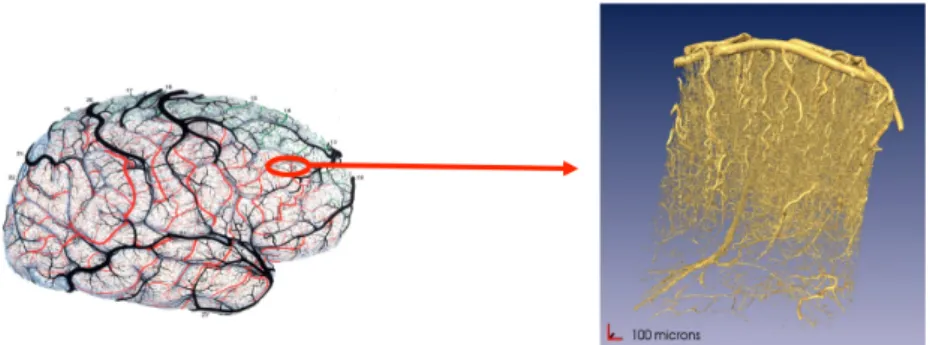
My PhD thesis work [B-1] was carried out at the Fluid Institute of Toulouse (IMFT, UMR 5502) between 2003 and 2007, under the supervision of Franck Plourabou´e. My goal was to quantify the anatomy of intracortical vascular net-works in order to evaluate their blood flow properties using statistical mechanics techniques. Image resolution was 1.43microns per voxel for acquired volumes of
about 3 cubic millimeters, as shown Fig. 1.1. The whole vasculature was then captured in the acquired images which made it possible to estimate the blood flow properties at the millimeter scale. This scale is interesting as it is similar to the cortical areas size. The results of this work were then of importance to compare the blood flow properties of different brain regions, or of a single brain region in different groups of subjects. This also made it possible to quantify the local impact of a brain stroke in terms of nutriment supply for the brain tissues, and to quantify how a tumor strongly increases its nutriment supply by transforming the vascular network in its neighborhood. Remark that these
Figure 1.1: Data studied during my PhD work: Samples of the intracortical vasculature of about 3 mm3 were acquired using synchrotron tomography at a
resolution of 1.4µm per voxel. All vessels are then distinguished in the volumes, which makes it possible to evaluate blood flow properties at the cortical region scale using statistical mechanics techniques. Illustration out of [B-1].
results were also of interest to understand the observed signal in BOLD fMRI as developed in Section 3.2. This work was rewarded in 2008 by the French national prize La recherche with the human health mention. Although I had a minor research activity in vascular image analysis after my PhD thesis [IJ-9, IC-34, SJ-3], this experience had an important impact on the research themes I developed later:
1. I used various tools of statistical mechanics and image analysis in [IJ-3,IJ-1, IC-9, IC-7, IC-6, IC-5, IC-3, IC-2, IC-[IJ-3,IJ-1, NC-1]. This made me familiar with the statistical analysis of 3D medical images.
2. The project which had the highest impact on my future career was the one published in [IJ-2,IC-4]. I developed a novel image processing strategy to fill the discontinuities that can be obtained when segmenting vascular networks in 3D images. These developments were made in a scientific community different to the one of F. Plourabou´e. He therefore put me in contact with X. Descombes (INRIA Sophia-Antipolis) who is specialist of bio-medical imaging and who advised me in this context. This allowed me to move from the fluid mechanics community to the medical imaging community after my PhD.
3. In the very end of my PhD thesis, I also worked on graph representations of the vascular networks in order to detect clusters of influent vessels using graph clustering techniques (see Fig. 1.2). This work is the last method-ological part of my PhD manuscript [B-1] and was only published four years later in a journal [IJ-9], after further bio-mechanical developments. Although it had little impact in my PhD work, it was the basis for the development of a new research activity in graphs analysis eight years later, at the Mathematics Institute of Toulouse [IJ-19,SJ-6,SC-1].
4. I finally worked on large data volumes: each 3D image was about 8 times larger than what I could allocate on my workstation. This developed my taste for low-level algorithms suitable for real-data analysis, which I kept later in all my methodological developments.
Figure 1.2: Automatic detection of the most influent structures in intracortical vascular networks, based on graph clustering. (Left) A segmented and skele-tonized sample of vascular network. The vascular network is represented by a graph augmented with spatial coordinates and a local diameter at each node. The colors represent here local vessels diameters. (Right) Subgraphs extracted out of the graph on the left. Each color represent an influent cluster of vessels with respect to the blood flow properties in the sample. Illustration out of [B-1].
The rest of this manuscript focuses on the scientific contributions I developed during my postdoctoral works and at the Mathematics Institute of Toulouse between 2007 and 2018. I refer to [B-1] as well as the citations of this section for further details about my PhD work.
1.2
Projects and collaborations
This section presents the main research projects in which I have been involved after my PhD work. In particular, I explain their working environment and who were my main collaborators. The scientific contributions and overviews of the main publications out of these projects is developed in Chapters 2 to 4.
1.2.1
Projects in medical image analysis
My projects related to medical image analysis are developed in Chapter 2. Here is below a synthesis of their motivations and contributions.
Medical image registration in the LDDMM framework An important
project for me has been the development of methodologies in the Large Deforma-tion Diffeomorphic Metric Mapping (LDDMM) framework (see SecDeforma-tion 2.1.2). I started working on this framework during my postdoctoral work at Imperial College London where my goal was to develop medical image registration strate-gies in interaction with D.D. Holm at the Department of Applied Mathematics and D. Rueckert in the Biomedical Image Analysis department. F.X. Vialard (former PhD student of A. Trouv´e at ENS Cachan) was hired as a postdoctoral researcher in the same project as me and we started a long term collaboration.
Our starting point was to deeply understand [BMTY05a] and how to make it work on practical cases given by D. Rueckert.
The first work in this collaboration with F.X. Vialard, was [IJ-6,IJ-10,IC-20,IC-18,IC-24] (Subsection 2.3.2). We defined multi-scale metrics in LDDMM, motivated by real applications in 3D medical imaging. From the application side, I collaborated with M. Murgasova (former PhD student with D. Rueck-ert) who motivated the problems related to the registration of pre-term babies MR images. I also collaborated with M. Bruveris (former PhD student with D.D. Holm) who extended [IJ-6] in [IJ-10] with a more mathematically rigor-ous approach. We then worked on an extension of [BMTY05a] where sliding constraints could be modeled in the context of my postdoctoral work at Univer-sity of Oxford with J.A. Schnabel (former researcher in the Biomedical image analysis department of Univ. Oxford) and published this work in [IJ-11,IC-24] (Subsection 2.3.3). This work motivated the development of alternative for-mulation to LDDMM, where spatially-varying metrics would make sense. This was done in collaboration with T. Schmah (Univ. Toronto) and published in [B-2,IC-31], where left-invariant metrics mathematically justified the use of LD-DMM with spatially-varying metrics (Subsection 2.2.4). After having justified the use of spatially-varying registration in LDDMM, we finally built a strategy that learns optimal spatially-varying regularization metrics with respect to a learning set of reference images [IC-32] (Subsection 2.3.4). Remark that these methods were recently summarized in [SB-1].
An alternative project on which I worked with F.X. Vialard, based on the same starting point, was the development of a formulation of LDDMM where geodesic shooting is used to register the images [IJ-7] (Subsection 2.2.2). This strategy was then used to define the Karcher means of shapes (average shapes) in 3D images [IJ-8,IC-22,IC-21] (Subsection 2.2.3). An original statistical learning pipeline based on the initial momenta computed using [IJ-7] and the averaged shapes of [IJ-8] was also presented in [IC-28,IJ-15] in collaboration with J.B. Fiot (former PhD student with L.D. Cohen at Univ. Paris Dauphine).
Medical image registration in other frameworks Personal contributions in medical image registration, outside of the LDDMM framework, were made mostly by collaborating with different PhD students. Tight collaborations with PhD students are much more frequent for postdoctoral researchers in the UK than in France. One of their role is indeed to give a scientific support to the PhD students of their advisor. I worked for three years as a postdoctoral researcher in the UK, which explains these collaborations. Note that I also continued having such collaborations after having been hired at IMT, in particular with J.A. Schnabel’s team. In [IJ-17,IC-30], I worked with B. Papiez (former PhD student of J.A. Schnabel) to extend the sliding motion strategy of [IJ-11]. A simpler image registration formulation was used but the location of the sliding constraints was automatically detected. I also worked with J. Ferhenbach in this project to justify the developments. In parallel, I also had also a strong scientific implication in the PhD work of A. Cifor (former PhD student of J.A. Schnabel) were we worked on the robust tracking of liver tumors in 2D Ultra-sound image series [IJ-13,IC-29,IC-26]. In the same vein, I also had a strong implication in the PhD thesis work of T. Roque (former PhD student of J.A. Schnabel), in particular in [IJ-20] where image deformations are driven by a
physiologically motivated reaction-diffusion model (Subsection 2.2.5). By regu-larly talking with M.P. Heinrich (former PhD student of J.A. Schnabel) about the definition of new realistic frameworks for multi-modal image registration, I also defined the mutual information gradient estimation technique of [IC-27,IC-23]. More secondary collaborations with J.A. Schnabel’s students were first in [IJ-12] with H. Baluwala (former PhD student of J.A. Schnabel) were we mostly shared image pre-processing tasks with [IJ-11]. I also worked with M. Bhushan [IC-25] in order to make diffeomorphic his motion correction strategy.
In addition to these collaborations with J.A. Schnabel’s students, I also collaborated with the team of C. Fonta (DR CNRS) and M. Mescam (lecturer, Univ. Toulouse) at the Brain and Cognition (CerCo) laboratory of Toulouse. In this context, I developed the image registration with automatic selection of the deformations scale in [IC-33] for marmoset monkeys brains.
Image segmentation Several image segmentation projects on which I have contributed are related to image registration: A reference segmentation can be throughly performed once for all on a template (average) image containing the shape of interest. The segmented image is then registered to the template and the reference segmentation is transported from the template domain to the segmented image using the mapping. Using such techniques I had a strong implication in the PhD thesis work of D.P. Zhang (former PhD student of D. Rueckert) where we developed different strategies for the segmentation of the coronary artery in 3D+time cardiac CT sequences [IC-19,IC-17,IC-16]. More recently, I also developed a plugin for the 3Dslicer software in order to perform the template-based segmentation of marmoset brain images [SJ-2] with C. Fonta and M. Mescam from CerCo.
As an extension of my PhD thesis gap filling strategy of [IJ-2], I also worked with R. Bates (former PhD student of J.A. Schnabel) on a post-treatment strat-egy for tubular structures segmentation. We indeed extended [IJ-2] to micro-CT images of tumorous vascular networks [IC-34]. By collaborating with J.M. Mire-beau (CR CNRS, Univ. Paris Dauphine) and J. Fehrenbach in the context of an ANR project, we also presented in [IJ-16] the ITK implementation of an efficient anisotropic non-linear diffusion technique for 2D or 3D images.
I also work with F. Gamboa (Pr Univ. Toulouse, IMT), A. Goss´e (CR CEA Saclay) and A. Quaini (CR CEA Saclay) since 2015 on the segmentation and the feature extraction of 2D image sequences representing rotating and levitat-ing balls which are extremely warmed-up. The goal here is to understand the mechanical properties of the balls under extreme conditions. The main techni-cal issues deal with artifacts, occlusions and low boundary contrasts observed during the experimental protocol. A communication explaining the results of this work should be submitted over the following months.
Finally, I currently collaborate with F. Malgouyres (Pr Univ. Toulouse, IMT) and colleagues from the Toulouse Cancer University Institute, in particu-lar S. Ken (Research Engineer INSERM, IUCT), on a 4 years INSERM project where we work on segmentation strategies for nodules and other structures in multi-modal whole body images. For now, we have published a regularization strategy for the Fast Marching algorithm [IC-37] and I co-supervise with F. Mal-gouyres V.K. Ghorpade (postdoctorate IMT) who works on these developments since January 2018.
1.2.2
Projects in numerical methods for stochastic
mod-eling
I mainly developed original numerical methods for stochastic modeling in the context of two projects and had besides minor contributions. Here is below a synthesis of their motivations and contributions. Further explanations are developed Chapter 3.
My first work experience with numerical methods for stochastic modeling was during my postdoctoral work at CEA Saclay with P. Ciuciu (former CR CEA Saclay). This project was related to the analysis of the brain activity in functional Magnetic Resonance Imaging (fMRI) time series, (Section 3.2). An original Bayesian model was first developed in collaboration with T. Vincent (former PhD student of P. Ciuciu) to analyze fMRI time series [IJ-4,IJ-14,NJ-1,IC-11,IC-15,IC-8]. I have been particularly involved in the development of a strategy to efficiently compute the partition function of Potts field with respect to their inverse temperature β [IJ-5,IC-13,IC-14,IC-12,NC-3], in order to make unsupervised the spatial regularization of this model. Insights about the pro-posed methodology were also developed with F. Forbes (DR INRIA Grenobles) and J. Idier (Pr Central Nantes), who temporarily hired me for two months in the end of my postdoctoral work at CEA Saclay and before my postdoctoral work at Imperial College London. Note that I also supervised the Master 2 project of A.L. Fouque (ENS Cachan) during this postdoctoral work and we developed a statistical clustering strategy for fMRI time series [IC-10,NC-2].
More recently, I collaborated with S. Gadat (Pr Toulouse School of Eco-nomics, IMT) and I. Gavra (former PhD student of S. Gadat) on the definition of a barycenter estimation strategy for graphs in which a probability measure reflects observation occurrences on the graph nodes (Section 3.3). We first pub-lished [IJ-19] and extended this work to the online and high dimensional context in [IJ-6]. I. Gavra has recently obtained a lecturer position at University of Rennes and we plan continuing our collaboration.
I also worked with S. Ribes who was PhD student under the supervision of O. Caselles at a laboratory of Univ. Toulouse 3 (SIMAD). She had the potential and the material to write a paper in a medical image analysis journal but her supervisors had little experience in this field. After talking together about her project, we agreed that I would informally advise her in this part of her PhD thesis work. I mainly helped her to develop the image segmentation pipeline based on Bayesian model as in [IJ-4], and led the paper redaction at the IEEE Trans. Medical Imaging format [IJ-18].
I have finally started a collaboration last year with G. Fort (DR CNRS, IMT) about Maximum Likelihood inference algorithms in statistical models. My goal in this collaboration is to develop original numerical methodologies to make scalable Gibbs sampling strategies. For now, my contributions were mostly technical and I worked on an efficient implementation of the algorithms in [IC-36,NC-4]. Note that, I also had a similar technical contribution with S. Gadat and M. Costa (Lecturer at Univ. Toulouse, IMT) [SJ-1] which deals with atomic deconvolution i.e. with deconvolution in density estimation.
1.2.3
Projects in statistical learning on complex data
The third field in which I had a scientific activity is statistical learning applied to complex data. My research activity in this field is developed Chapter 4 and is synthesized hereafter. Note that I give different links between complex data analysis and medical image registration in Section 4.1. In this manuscript, I distinguish my research activity in these fields by considering medical image registration as a specific subfield of complex data analysis.
As mentioned Subsections 1.2.1 and 1.2.2, I first had a minor contribution in this field while supervising the M2 project of A.L. Fouque with P. Ciuciu during my postdoctoral work at CEA Saclay [IC-10,NC-2]. This contribution was about statistical learning techniques to clusterize hemodynamic parameters out of fMRI time series. Several years later, I worked with J.B. Fiot and F.X. Vialard on the exploration of different spatial regularization models for logistic regression on 3D image domains [IC-28,IJ-15] (Section 4.2). I also developed an image registration strategy with automatic selection of the deformations scale in [IC-33] based on LASSO regularization. In these contributions, statistical learning techniques were mostly applied to specific applicative cases.
I started developing new models in statistical learning by co-supervising two PhD theses with J.M. Loubes (Pr Univ. Toulouse, IMT). I first work with T. Bui since September 2016 on the development of statistical models for the classification of 3D coiled shapes out of the inner ear as well as distributions of the response of the ear to otoacoustic emission (OAE) [SJ-5] (Section 4.3). I also work with C. Champion since September 2017 on the extraction of representative variables in complex systems [SC-1,NC-5] (Section 4.4).
Other collaborations in this theme have also started with F. Gamboa (Pr Univ Toulouse, IMT), F. Bachoc (Lecturer Univ Toulouse, IMT), and S. D´ejean (Research Engineer, Univ. Toulouse, IMT) and should lead to new developments in the future.
1.3
Personal contributions
Almost all my scientific contributions were developed in the context of collab-orative works. In this section, I therefore make clear what was my personal contribution to selected journal papers and conference proceedings. These se-lected communications are presented in chronological order and correspond to the communications given in appendix.
Unsupervised spatial mixture modelling for within-subject analysis of fMRI data [IJ-4]
(Motivation) Within-subject analysis of the brain activity in BOLD fMRI con-sists in detecting patterns of energy consumption in 3D+time image series. Each of these patterns is located at an image point and smoothly evolves in time dur-ing several seconds after an onset. It represent a brain activation and is related to local variations of oxygen consumption in the brain due to a cognitive task. In 2010, all existing approaches either detected the activations using a predefined energy consumption pattern, or estimated this pattern at specific locations and times. There is however a physiological evidence that these two tasks should be performed simultaneously as the energy consumption pattern strongly depends
of the local vasculature which varies across brain regions [IJ-1,IJ-3]. (Main pa-per contributions) This papa-per develops an original Bayesian model in which the activations detection and the energy consumption patterns are simultane-ously estimated. The model makes physiologically realistic hypotheses to con-strain the minimized energy, making it possible to detect local activations that are lost using more generic regularization models. (Personal contributions) T. Vincent (who was a PhD student of P. Ciuciu at CEA Saclay) was the main contributor to this work. I developed the unsupervised spatial regularization model and strategy to estimate the 3D Potts field partition functions. The par-tition function strategy was generalized later in [IJ-5]. This paper is shown in Appendix B.1.
Min-max extrapolation scheme for fast estimation of 3D Potts field partition functions [IJ-5]
(Motivation) Potts models are typically used as Hidden Markov Fields with K labels/colors when segmenting an image using a Bayesian formalism. They indeed allow to spatially regularize the optimal segmentation with a strength which is controlled by an inverse temperature β. When the regularization level is unsupervised, it is however mandatory to compute the partition function of the Potts field w.r.t. β, which can be extremely demanding in terms of compu-tations. (Main paper contributions) In [IJ-5], we proposed a fast partition function estimation strategy for 2D and 3D Potts fields with irregular shapes. This technique was applied to the estimation of the brain activity in func-tional MRI time-series. In this application, about 100 Potts fields were used to spatially regularize the detection of brain activation/deactivation/inactivation, where the regularization level was automatically-tuned and region-wise. (Per-sonal contributions) I was the main contributor to this work. I collaborated with T. Vincent (who was a PhD student of P. Ciuciu at CEA Saclay) to in-tegrate the partition function estimation strategy to the brain activity analysis pipeline. I also worked with F. Forbes, J. Idier and P. Ciuciu to develop my insights about the proposed method. This paper is shown in Appendix B.2. Simultaneous Multiscale Registration using Large Deformation Dif-feomorphic Metric Mapping [IJ-6]
(Motivation) This paper was motivated by the lack of literature in 2011 on the choice of physiologically realistic regularizing metrics to register medical im-ages with LDDMM [BMTY05b]. (Main paper contributions) The impact of the regularizing metric in medical image registration was first discussed. In particular, we have made clear that using unsuitable regularizing metrics with respect to the registered structures leads to physiologically implausible defor-mations, even if the shape boundaries are accurately matched. Motivated by real-life medical image registration cases, a strategy to define multi-scale metrics in LDDMM was presented, assessed and discussed. (Personal contributions) I was the main contributor to this work. The key ideas came by discussing with F.X. Vialard when developing our implementation of [BMTY05b] on 3D medi-cal images. I realized that very little literature was dealing with the choice of the metric in LDDMM although this choice is fundamental in practice. This paper is shown in Appendix A.1.
Diffeomorphic 3D Image Registration via Geodesic Shooting using an Efficient Adjoint Calculation [IJ-7]
(Motivation) This work was motivated by the need for an accurate tool to com-pute the initial momenta that compare 3D images in the LDDMM framework. Initial momenta are indeed important in LDDMM, as they compactly encode local differences between the registered images and can therefore be used for further statistics on shape spaces or for the estimation of average shapes. Note that they are specific to LDDMM in the image registration community and are one of the main reasons that make this formalism appealing. (Main paper contributions) A new variational strategy for the diffeomorphic registration of 3D images is defined. It performs the optimization on the set of geodesic paths instead of on all the possible curves, and therefore directly estimates the initial momenta comparing two images. (Personal contributions) I tightly collaborated with F.X. Vialard on this paper. My main contributions have dealt with the resolution of implementation and numerical issues related to the use of the geodesic shooting strategy on 3D medical images. This paper is shown in Appendix A.2.
Diffeomorphic Atlas Estimation using Geodesic Shooting on Volumet-ric Images [IJ-8]
(Motivation) Computing the average shape of a given organ is fundamental for many medical image applications, in particular in brain imaging. It indeed makes it natural to propagate local information measured on a reference set of imaged organs into this average shape, denoted template. This information (typically a probabilistic segmentation) can then be propagated to other images. Another important application is to quantify the local variability of the reference images. The motivation of this paper is to define a computationally tractable strategy to compute average shapes out of 3D medical images. (Main paper contributions) A new algorithm to compute intrinsic means of organ shapes from 3D medical images was defined. This algorithm is based on the geodesic shooting algorithm of [IJ-7] and is fully diffeomorphic. Contrary to other tem-plate definition strategies, the intensities of the average shapes are then not the average intensities of several images registered to each other, leading to sharper region boundaries. This strategy also offers interesting properties for further statistical studies by using the information contained in initial momenta. (Per-sonal contributions) F.X. Vialard and me contributed equally to this work. F.X. Vialard computed the gradients of the optimized energy and I developed the gradient descent based strategy. This paper is shown in Appendix A.3. Mixture of Kernels and Iterated semidirect Product of Diffeomor-phisms Groups [IJ-10]
(Motivation) This work directly follows [IJ-6], where we defined and discussed a practical method to use multi-scale metrics in LDDMM. A first attempt to dis-tinguish scale-dependent deformations out of an optimal deformation between two shapes was given in [IJ-6], but this contribution was secondary compared with other ones. As it may be useful for further statistical studies, the work of [IJ-10] strongly develops this discussion. (Main paper contributions) The influence of different scales when comparing two shapes using LDDMM with
multi-scale kernels is studied with a more rigorous model than in [IJ-6]. A variational approach is developed for the multiscale analysis of diffeomorphisms and the semidirect product representation is generalized to several scales. (Per-sonal contributions) F.X. Vialard and M. Bruveris (who was a PhD student of D.D. Holm at Imperial College London) were the main contributors to the developed model. My first contribution was to give the research directions of this work and to specifically make sure that the mathematical developments would have a practical impact from a image analysis point of view and would be algorithmically realistic on 3D images. I also implemented and assessed the strategy on 3D medical images. This paper is shown in Appendix A.4.
Piecewise-Diffeomorphic Image Registration: Application to the Mo-tion EstimaMo-tion between 3D CT Lung Images with Sliding CondiMo-tions [IJ-11]
(Motivation) Standard medical image registration models make the hypoth-esis that the deformations between registered images are smooth (and then continuous) everywhere. However, sliding conditions can be observed in medi-cal images, for instance at the lung boundaries. This paper was then motivated by the need for diffeomorphic image registration models with sliding conditions. (Main paper contributions) We first defined a general strategy for model-ing slidmodel-ing conditions when registermodel-ing 3D images in a piecewise-diffeomorphic framework. Compared with existing literature in 2012, this strategy ensured that the estimated deformations were invertible everywhere although they could be locally discontinuous. We also integrated the proposed strategy to the LD-DMM [BMTY05b] and the LogDemons [VPPA08] diffeomorphic registration frameworks. (Personal contributions) I was the main contributor to this work. I worked with F.X. Vialard to define the admissible Reproducing Kernel Hilbert Space in the LDDMM context. We also shared image pre-processing tasks with H.A. Baluwala (who was a PhD student of J.A. Schnabel at Univ. Oxford) that were also used in [IJ-12]. This paper is shown in Appendix A.5. Construction of Diffeomorphic Spatio-temporal Atlases using K¨archer means and LDDMM [IC-22]
(Motivation) This work directly extends [IJ-8] where a fully diffeomorphic strategy was proposed to compute average shapes (atlases) out of 3D images. It was motivated by the need for the definition of fully diffeomorphic spatio-temporal atlases. In this context, each reference image is associated to an acquisition time and the average template evolves in time. The spatio-temporal atlas also spatially moves smoothly in time with no intensity change and any intensity blurring, which allows to preserve the sharpness of region boundaries, or to make move an atlas associated to a single segmentation. (Main paper contributions) Compared with [IJ-8], a straightforward contribution was to use a time-dependent kernel to weight the influence of each reference image at a given time. In order to make the temporal evolution of the template fully diffeomorphic and dense in time, our key contribution was to perform the spatio-temporal shape averaging on the tangent space of the evolution rather than on the space of images. (Personal contributions) I was the main contributor to this work. F.X. Vialard formalized the intuition I had about the spatio-temporal
averaging strategy on tangent spaces. This paper is shown in Appendix A.6. Piecewise-diffeomorphic registration of 3D CT/MR pulmonary im-ages with sliding conditions [IC-27]
(Motivation) The driving motivation of this paper was to make it possible to register multimodal 3D images with sliding conditions. The two technical issues addressed in this paper were (1) to make the estimation of local similar-ity gradients computationally tractable on large multimodal images, and (2) to strongly regularize the deformations, as the registered structures have a strongly different representations in the CT and MR images, while modeling local slid-ing conditions. (Main paper contributions) This paper directly applies the regularization strategy of [IJ-11] to locally constrain sliding deformations. Its main contribution is the use of approximated local gradient of mutual informa-tion to match the images which was first presented in [IC-23] and is developed here. (Personal contributions) I was the main contributor to this work. M.P. Heinrich (who was a PhD student of J.A. Schnabel at Univ. Oxford) helped me to pre-process the images and to assess the registration quality. This paper is shown in Appendix A.7.
Hybrid Feature-based Diffeomorphic Registration for Tumour Track-ing in 2-D Liver Ultrasound Images [IJ-13]
(Motivation) Ultrasound (US) imaging is a widely accessible and low-cost im-age acquisition modality but also opens various questions in imim-age analysis. This is due to the fact that it generates different artifacts and that it acquires 2D image sequences in a 3D domain. The specific driving motivation of [IJ-13] is to define a robust and accurate method to compensate for the breathing mo-tion when tracking liver tumors in US imaging. (Main paper contribumo-tions) A whole diffeomorphic image registration pipeline was defined to follow the tu-mors. The PDE-based deformation model was inspired from the LogDemons framework of [VPPA08]. The main contribution of [IJ-13] was the definition of new matching forces that allow to robustly follow the tumor in 2D US im-age sequences. (Personal contributions) My main contribution to this paper was to scientifically lead the work of A. Cifor (who was a PhD student of J.A. Schnabel at Univ. Oxford) to integrate the image features she defined in an image registration framework and to assess the results. I also found a mathe-matical justification to the algorithm and its parameters through a PDE-based formulation of the registration algorithm. This paper is shown in Appendix A.8. Longitudinal deformation models, spatial regularizations and learning strategies to quantify Alzheimer’s disease progression [IJ-15]
(Motivation) The early detection of Alzheimer’s disease (AD) is an important challenge for its efficient treatment through adapted drug delivery. In this pa-per, we worked on its detection based on local hippocampal shape changes in time. The hippocampus is indeed a subcortical structure which is known by the clinicians to be anatomically impacted by AD. (Main paper contributions) In this paper, we explored the use of different spatial regularization models in logistic regression to learn which local shape deformations optimally discrim-inate AD subjects from subject with Mild Cognitive Impairment. (Personal
contributions) J.B. Fiot (who was a PhD student of L.D. Cohen at Univ. Paris Dauphine) and F.X. Vialard were the main contributors to this work. I helped J.B. Fiot defining an average shape and aligning the images. We have also made the link between the regularization strategies and their physiological interpretation. This project is the one in which I started developing a scientific activity in machine learning. This paper is shown in Appendix C.1.
Diffeomorphic image matching with left-invariant metrics [B-2] (Motivation) The Large Deformation by Diffeomorphic Metric Mapping (LD-DMM) framework of [BMTY05b] was designed to regularize the deformations with the same smoothing properties in the whole image domain. This contribu-tion presented an alternative problem formulacontribu-tion, denoted Left-LDM or LIDM, in which spatially-varying metrics make sense. (Main paper contributions) We first explored the use of left-invariant metrics on diffeomorphism groups based on reproducing kernels defined in the body coordinates of the source im-age. This approach differs from LDDMM, where right-invariant metric on a diffeomorphism group are used. A link with LDDMM was also established and a practical algorithm to register 3D images with LIDM was given. (Personal contributions) My main contribution was to guide the mathematical devel-opments in strong collaboration with F.X. Vialard and T. Schmah so that the registration strategy would be realistically applied on 3D medical images. This has lead to a computationally tractable 3D image registration algorithm (very close to [BMTY05b]) where the final deformation is analytically the same as us-ing LIDM although the path is different. This paper is shown in Appendix A.9. Spatially-varying metric learning for diffeomorphic image registra-tion. A variational framework [IC-32]
(Motivation) In medical image registration, it makes obvious sense that the deformations of different organs should be ideally regularized with different smoothing properties. Standard medical image registration algorithms how-ever use spatially homogeneous smoothing properties for two main reasons: (1) This is mathematically and algorithmically much simpler, and (2) tuning spa-tially varying smoothing properties requires prior information on the registered structures that is generally not available. (Main paper contributions) In this paper, we build on the diffeomorphic registration model of [B-2] to define a strategy that learns optimal spatially-varying regularization metrics with re-spect to a learning set of reference images. The learning strategy is defined in a variational framework. (Personal contributions) I tightly collaborated with F.X. Vialard in this project. I gave research directions, so that the strategy would be computationally tractable on 3D medical images and would lead to meaningful results. I also defined a numerical solution to keep the learning di-mension reasonable, implemented the strategy and tested it on real 3D medical images. This paper is shown in Appendix A.10.
Diffeomorphic registration with self-adaptive spatial regularization for the segmentation of non-human primate brains [IC-33]
(Motivation) The motivation of this paper is close to the one of [IC-32] and deals with the semi-automatic tuning of the smoothing properties in the
reg-istration of template images. Contrary to [IC-32], the goal of this paper is to alleviate the need for scale definition in the regularizing metric of a med-ical image registration algorithm. (Main paper contributions) The main methodological contribution of this paper is to explore a new strategy to auto-matically tune the spatial regularization of the deformations in medical image registration. To do so, the image registration model is an optimization strategy in which the deformations of the template are the weighted sum of reference deformations at different scales, and the weights are penalized with a L1 norm
(LASSO). Sparse non-null weights are then computed, leading to optimal scale selection. (Personal contributions) I was the main contributor to this work. L. Dolius, C. Fonta and M. Mescam acquired the images and helped me to interpret the results. This paper is shown in Appendix A.11.
Filling Large Discontinuities in 3D Vascular Networks using Skeleton-and Intensity-based Information [IC-34]
(Motivation) The segmentation of vascular networks often leads to discon-tinuities in the segmented vessels. For tumorous networks, no hypotheses can additionally be made on the network structures, due to the chaotic arrangement of their vessels. This makes it impossible to use standard gap filling algorithms for such vascular networks. (Main paper contributions) This paper extended [IJ-2], that I wrote during my PhD thesis work, with a gap filling strategy that combines both skeleton- and intensity-based information to fill large disconti-nuities. (Personal contributions) My main contribution to this paper was to scientifically lead the work of R. Bates (who was a PhD student of J.A. Schnabel at Univ. Oxford) in order to extend [IJ-2] and make it efficient with the data he had. This paper is shown in Appendix A.12.
How to calculate the barycenter of a weighted graph [IJ-19]
(Motivation) Undirected graphs with weighted edges and probability measures on their nodes are of particular interest to model complex phenomena. For in-stance, they may represent a social network with individuals talking about a given topic. In this case, the individuals are the nodes, the strength of the relation between two individuals is an edge weight, and the probability mea-sures reflect the occurrences of a tag (e.g. an hashtag in twitter). There was no algorithm to compute the barycenter such structures in 2017 although this may be statistically informative. (Main paper contributions) In this pa-per, we introduced an original stochastic algorithm to find the Fr´echet mean of such graphs. It relies on a noisy simulated annealing algorithm. (Personal contributions) I. Gavra (who was a PhD student of S. Gadat at Univ. Paul Sabatier/IMT) was the main contributor to this work. S. Gadat worked with here on the algorithm definition and its convergence. My main contribution was to lead I. Gavra’s work to make her strategy usable on real data. This allowed us to make it algorithmically efficient on reasonably large graphs, to de-velops insights about its parametrization, and to identify practical issues which make this algorithm not scalable to large graphs. These issues were treated in the follow-up paper [SJ-6] where my scientific involvement was stronger. This paper is shown in Appendix B.3.
A DCE-MRI Driven 3-D Reaction-Diffusion Model of Solid Tumour Growth [IJ-20]
(Motivation) This work was motivated by the need for tumor growth predic-tion models to estimate the response to therapies. (Main paper contribu-tions) This paper introduced an image-driven 3D reaction-diffusion model of avascular tumor growth in order to predict spatio-temporal tumor evolution. The model is calibrated using information derived from follow-up DCE-MRI images. It indeed consists in registering follow-up multi-layer images with con-straints encoded in a non-linear reaction-diffusion model. The registration then consists in estimating the model parameters. Note that it can also be seen as a PDE-constrained optimization problem. (Personal contributions) I had two major contributions in this paper. The first one was to scientifically lead the work of T. Roque (who was a PhD student of J.A. Schnabel at Univ. Oxford) to transform the tumor growth equations she collected in the literature into a reaction-diffusion model which can be used based on DCE-MRI image infor-mation. I also discretized the equations to make the resolution scheme stable and sufficiently fast on 3D image domains. In addition, I advised T. Roque on a simple and pragmatic optimization strategy to automatically tune tumor specific model parameters. This paper is shown in Appendix A.13.
Regularized Multi-Label Fast Marching and Application to Whole-Body Image Segmentation [IC-37]
(Motivation) The segmentation of multiple structures such as lymph nodes in whole-body MR images of patients with tumors is a task which can be hardly automatized for two main reasons: (1) Structures boundaries are not visible ev-erywhere, and (2) the patients and the structures to segment may have a large anatomical variability. User interventions are then necessary but should be as limited as possible, and related to particularly responsive algorithms. (Main paper contributions) We proposed a computationally efficient regularization strategy for the Fast Marching (FM) segmentation of multiple organs. The regularization stabilizes the segmentation of complex structures and has a low computational impact. We also integrated this regularized segmentation strat-egy to the 3Dslicer software so that clinicians could validate the methodology on real cases. (Personal contributions) I supervised this project in collabora-tion with F. Malgouyres with whom we deepened the first intuicollabora-tions about the regularization strategy, S. Ken who gave us its driving motivation and partici-pated to the results assessment, and S. Lebreton who integrated the algorithms to 3DSlicer. This paper is shown in Appendix A.14.
A representative variable detection framework for complex data based on CORE-clustering [SC-1]
(Motivation) Discovering representative information in high dimensional spaces with a limited number of observations is a recurrent problem in data analy-sis. Heterogeneity between the variables behavior and multiple similarities be-tween variable subsets make the analysis of complex systems an ambiguous task. (Main paper contributions) This paper presents a formalism to robustly es-timate the representative variables in such complex systems. The formalism is based on a novel graph clustering strategy, denoted CORE-clustering, adapted
to the addressed problem. The graphs encode the relations between different observed variables and the clusters are selected based on the number of variables they contain. The representative variables are finally the cluster centers, so the number of variables in each cluster can be seen as a regularization parameter. The method is additionally designed to be scalable to large datasets. (Personal contributions) The original idea of the CORE-clustering algorithm came from the PhD thesis work of A.C. Brunet with J.M. Loubes as a supervisor, but was only published in Arxiv [BAL+16]. I supervised C. Champion (PhD student
IMT, co-supervised by J.M. Loubes and me) to totally re-design the methodol-ogy. We developed mathematical and algorithmic insights to make it efficient in the general complex data case. I also advised her in the experimental validation. We finally wrote together [SC-1], with advice from J.M Loubes. This paper is shown in Appendix C.2.
Distribution regression model with a Reproducing Kernel Hilbert Space approach [SJ-5]
(Motivation) Regression analysis is a predictive modeling technique that has been widely studied over the last decades with the goal to investigate relation-ships between predictors and responses. Extensions of the Reproducing Kernel Hilbert Space (RKHS) framework became popular to extend the results of the statistical learning theory in the context of regression of functional data as well as to develop estimation procedures of functional valued functions f . As far as the authors know, It has however not been extended so far to probability distribution spaces. (Main paper contributions) This paper introduces a strategy to solve the regression problem where the inputs belong to probability distribution spaces and the output predictors are real values. The regression function is composed of an unknown function f and an element ofH(K), where H(K) is the RKHS induced by the kernel K defined on the set of mean em-beddings of distributions to RKHSH(k). (Personal contributions) My main contribution in this paper was to guide the work of T. Bui (PhD student IMT, co-supervized by J.M. Loubes, P. Balaresque and me) in order to establish the link between the mathematical formalism she developed and the auto-acoustic response curves she studied. This was critical to understand the model and to obtain pertinent results. This paper is shown in Appendix C.3.
Online Barycenter Estimation of Large Weighted Graphs [SJ-6] (Motivation) This paper follows [IJ-19], where an original strategy was pro-posed to compute the barycenter of undirected weighted graphs. The method of [IJ-19] has strong mathematical foundations but is not scalable to large graphs. In addition, although the formalism is general enough to address the online estimation of online graphs, this application is not clearly discussed in [IJ-19]. (Main paper contributions) In this paper, we extend [IJ-19] to efficiently estimate the barycenter of very large graphs. The online case, where empirical observations of the graph node probability measures are made in parallel to the barycenter estimation, is also discussed. Algorithmic aspects of the strategy are highlighted as they are directly related to the scalability of the method. (Per-sonal contributions) I have tightly collaborated with I. Gavra to develop the methodology and we have similar contributions in [SJ-6]. This paper is shown
in Appendix B.4.
1.4
Teaching activity and students supervision
This section briefly develops the teaching activity I had in during career and gives an overview of the students I have supervised. Note that I have mixed in this section the activity I had during and after my PhD thesis work (defended in September 2007).
Teaching activity
• 2017-2018: Lectures and practical courses in Image Analysis (32 hours). Master 2 MAPI3 (Applied Mathematics). Paul Sabatier University, Toulouse. • 2017-2018: Lectures and practical courses in Machine Learning (18 hours).
Master students to University lecturers (context of a two weeks spring school). VNUHCM - University of Science, Ho-Chi-Minh city, Vietnam. • 2017-2018: Practical courses in Statistics (16 hours). Master 2 of ISAE /
Supaero, Toulouse.
• 2017-2018: Lecture and practical courses in GPU computing (4 hours). Master 2 of ISAE / Supaero, Toulouse.
• 2016-2017: Lectures and practical courses in Image Analysis (8 hours). Master 2 MAPI3 (Applied Mathematics). Paul Sabatier University, Toulouse. • 2016-2017: Practical courses in Statistics (16 hours). Master 2 of ISAE /
Supaero, Toulouse.
• 2006: Lectures and practical courses of Numerical Simulation in Fluid Me-chanics (32 hours). Master 1 in MeMe-chanics and Energetics, Paul Sabatier University, Toulouse.
• 2006: Practical courses of Stochastic Process applied to heterogeneous media (14 hours). Master 1 in Mechanics and Energetics, Paul Sabatier University, Toulouse.
• 2005: Lectures and practical courses of Numerical Simulation in Fluid Me-chanics (44 hours). Master 1 in MeMe-chanics and Energetics, Paul Sabatier University, Toulouse.
• 2005: Practical courses of Point Mechanics (20 hours). Licence 1 in Math-ematics and Computer Science applied to Science (DEUG MIAS), Paul Sabatier University, Toulouse.
• 2004: Lectures and practical courses of Numerical Simulation in Fluid Me-chanics (44 hours). Master 1 in MeMe-chanics and Energetics, Paul Sabatier University, Toulouse.
• 2004: Practical courses of Point Mechanics (20 hours). Licence 1 in Math-ematics and Computer Science applied to Science (DEUG MIAS), Paul Sabatier University, Toulouse.
Students supervision
Postdoc supervision
• 02/2018-.: V. K. Ghorpade. Postdoctoral researcher in Applied Mathe-matics for medical image analysis: Mutli-modal image registration of 3D whole-body medical images. Co-supervision with F. Malgouyres (Pr IMT). PhD student supervision
• 09/2017-.: C. Champion (Applied Mathematics at l’INSERM/IMT): De-velopment of new strategies for the analysis of complex data. Co-supervision with J.-M. Loubes (Pr IMT) and R. Burcelin (DR INSERM).
• 09/2016-.: T. Trang Bui (Applied Mathematics at INSA Toulouse/IMT): Regularization models for the analysis of auditory data. Co-supervision with J.-M. Loubes (Pr IMT) and P. Balaresque (CR1 CNRS, UMR5288). Master students supervision
• 2018: R. Vaysse (M1 Applied Mathematics/Data Analysis, Paul Sabatier University - 5 months): Statistical analysis of data out of speech samples for Parkinson’s disease detection. Co-supervision with S. D´ejean (IR UPS) and J. Farinas (Mcf UPS - UMR5505)
• 2017: V. Br`es (M2 Applied Mathematics/Computer Science, ENSEEIHT - 6 months): GPU computing with OpenCL to speed-up large graph clus-tering algorithms.
• 2017: S. Lebreton (M2 Applied Mathematics/Computer Science, EN-SEEIHT - 6 months): Development of a C++ plugin in 3DSlicer for the semi-interactive segmentation of 3D medical images. Co-supervision with F. Malgouyres (Pr IMT).
• 2017: N. Artigouha (M1 Computer Science, INSA Toulouse - 2 months): Using the C++ Boost Graph Library to for the analysis of large graphs. • 2016: D. Grasselly (M1 Applied Mathematics, INSA Toulouse - 3 months):
Development of a Matlab code for the registration of lung images with sliding conditions. Co-supervision with J. Fehrenbach (Mcf UPS/IMT). • 2016: M. Verdier (M1 Applied Mathematics, INSA Toulouse - 3 months):
Induction of Bayesian networks from medical data.
• 2016: M. Ralle (M2 Math´ematiques, Paris Orsay University - 4 months): Statistical analysis of the cochlear coil. Co-supervision with J.M. Loubes (PR UPS/IMT).
• 2015: T. Berriat (M1 Applied Mathematics, INSA Toulouse - 3 months): Statistical analysis of the cochlear coil. Co-supervision with J.M. Loubes (PR UPS/IMT).
• 2014: A. Choury (M2 Applied Mathematics, INSA Toulouse - 6 months): Statistical analysis of seismic wave propagation measures. Co-supervision with J.M. Loubes (PR UPS/IMT) and P. Besse (PR INSA/IMT).
• 2013: L. Dolius (M2 Medical Imaging and Radiophysics, Paul Sabatier University - 6 months): Quantitative Analysis of 3D brain images. Co-supervision with C. Fonta (DR CerCo/CNRS) and M. Mescam (McF UPS/CerCo).
• 2010: A. Camphuis (M1 Supelec - 2 months): Validation of a medical image registration algorithm. Co-supervision with F.X. Vialard (Postdoc-toral researcher at Imperial College London).
• 2008: A.L. Fouque (M2 ENS Cachan - 5 months): Analysis of the BOLD signal in functional MRI. Co-supervision with P. Ciuciu (CR CEA Saclay). • 2005: V. Gratsac (M2 Computer Science, Nantes University - 6 months):
Segmentation of large vascular network images using Tensor Voting. I have finally supervised 8 trainees with License 2 and 3 levels from pr´epa INPT, ENS Lyon and IUT Toulouse on the implementation of different algo-rithms.
1.5
Bibliographic record
Refereed international journal papers
[IJ-20] T. Roque, L. Risser, V. Kersemans, S. Smart, D. Allen, P. Kinchesh, S. Gilchrist, A. Gomes, J. A. Schnabel, and M. Chappell. A DCE-MRI driven 3-d reaction-diffusion model of solid tumour growth. IEEE Trans-actions on Medical Imaging, 37(3):712–23, 2018.
[IJ-19] S. Gadat, I. Gavra, and L. Risser. How to calculate the barycenter of a weighted graph. Informs: Mathematics of Operations Research, 2018. [IJ-18] S. Ribes, D. Didierlaurent, N. Decoster, E. Gonneau, L. Risser, V. Feillel,
and O. Caselles. Automatic segmentation of breast MR images through a markov random field statistical model. IEEE Transactions on Medical Imaging, 2014.
[IJ-17] B. W. Papiez, M. P. Heinrich, J. Fehrenbach, L. Risser, and J. A. Schnabel. An implicit sliding-motion preserving regularisation via bilateral filtering for deformable image registration. Medical Image Analysis, 2014.
[IJ-16] J. Mirebeau, J. Fehrenbach, L. Risser, and S. Tobji. Anisotropic Diffusion in ITK. The Insight Journal, 2014.
[IJ-15] J. B. Fiot, H. Raguet, L. Risser, L. D. Cohen, J. Fripp, F. X. Vialard, and ADNI. Longitudinal deformation models, spatial regularizations and learning strategies to quantify alzheimer’s disease progression. NeuroIm-age: Clinical, 2014.
[IJ-14] T. Vincent, S. Badillo, L. Risser, L. Chaari, C. Bakhous, F. Forbes, and P. Ciuciu. Flexible multivariate hemodynamics fMRI data analyses and simulations with pyhrf. Frontiers in Neuroscience, 2014.
[IJ-13] A. Cifor, L. Risser, D. Chung, E. M. Anderson, and J. A. Schnabel. Hybrid feature-based diffeomorphic registration for tumour tracking in 2-d liver ultrasound images. IEEE Transactions on Medical Imaging, 2013. [IJ-12] H. Y. Baluwala, L. Risser, J. A. Schnabel, and K. A. Saddi. Towards a
physiologically motivated registration of diagnostic CT and PET/CT of lung volumes. Medical Physics, 40(2), 2013.
[IJ-11] L. Risser, F. X. Vialard, H. Y. Baluwala, and J. A. Schnabel. Piecewise-diffeomorphic image registration: Application to the motion estimation between 3d CT lung images with sliding conditions. Medical Image Anal-ysis, 2012.
[IJ-10] M. Bruveris, L. Risser, and F. X. Vialard. Mixture of kernels and iterated semidirect product of diffeomorphisms groups. SIAM Multiscale Modeling and Simulation, 10(4):1344–68, 2012.
[IJ-9] R. Guibert, C. Fonta, L. Risser, and Plourabou´e F. Coupling and robust-ness of intra-cortical vascular territories. NeuroImage, 2012.
[IJ-8] F. X. Vialard, L. Risser, D. Rueckert, and D. Holm. Diffeomorphic atlas estimation using geodesic shooting on volumetric images. Annals of the British Machine Vision Association, 2012.
[IJ-7] F. X. Vialard, L. Risser, D. Rueckert, and C. J. Cotter. Diffeomorphic 3d image registration via geodesic shooting using an efficient adjoint cal-culation. International Journal of Computer Vision, 2011.
[IJ-6] L. Risser, F. X. Vialard, R. Wolz, M. Murgasova, D. Holm, D. Rueckert, and ADNI. Simultaneous multiscale registration using large deformation diffeomorphic metric mapping. IEEE Transactions on Medical Imaging, 2011.
[IJ-5] L. Risser, T. Vincent, F. Forbes, J. Idier, and P. Ciuciu. Min-max extrap-olation scheme for fast estimation of 3d potts field partition functions. application to the joint detection-estimation of brain activity in fMRI. Journal of Signal Processing Systems, 60(1), 2010.
[IJ-4] T. Vincent, L. Risser, P. Ciuciu, and J. Idier. Unsupervised spatial mix-ture modelling for within-subject analysis of fMRI data. IEEE Transac-tions on Medical Imaging, 29(4):1059–75, 2010.
[IJ-3] L. Risser, F. Plourabou´e, P. Cloetens, and C. Fonta. A 3d-investigation shows that angiogenesis in primate cerebral cortex mainly occurs at capil-lary level. International Journal of Developmental Neuroscience, 27(2):185– 96, 2008.
[IJ-2] L. Risser, F. Plourabou´e, and X. Descombes. Gap filling in vessel networks by skeletonization and tensor voting. IEEE Transactions on Medical Imaging, 27(5):674–87, 2008.
[IJ-1] L. Risser, F. Plourabou´e, A. Steyer, P. Cloetens, G. Le Duc, and C. Fonta. From homogeneous to fractal normal and tumorous micro-vascular net-works in the brain. Journal of Cerebral Blood Flow and Metabolism, 27:293–303, 2006.


![Figure 2.2: Registration of synthetic images using the methods of [BMTY05b]](https://thumb-eu.123doks.com/thumbv2/123doknet/14705790.747909/50.892.218.669.186.493/figure-registration-synthetic-images-using-methods-bmty-b.webp)
![Figure 2.3: Estimation of the average spatio-temporal development of the cortex A τ , τ ∈ [τ init , τ end ] from segmented 3D images I s , s ∈ [1,](https://thumb-eu.123doks.com/thumbv2/123doknet/14705790.747909/52.892.277.608.352.599/figure-estimation-average-spatio-temporal-development-cortex-segmented.webp)
![Fig. 2.5 illustrates the main result of [IC-31] on a synthetic example. In this example, LIDM registered the images using a kernel which is defined accord-ingly to a partition of unity which smoothly splits the spatial domain into two sub-regions](https://thumb-eu.123doks.com/thumbv2/123doknet/14705790.747909/55.892.213.681.413.653/illustrates-synthetic-example-example-registered-defined-partition-smoothly.webp)
![Figure 2.8: Representation of scale-dependent deformations ϕ i out of a defor- defor-mation ϕ obtained between two brain images using the method of [IJ-10]](https://thumb-eu.123doks.com/thumbv2/123doknet/14705790.747909/62.892.241.650.186.414/figure-representation-dependent-deformations-mation-obtained-images-method.webp)
