Machine
ARCHIVES
MAS'SACHUSETS INSTIby OF TECHNOLOGY
Jonathan Dean Grabenstatter
SEP 2
8
2015
B.A., Miami University (2004)
LIBRARIES
Submitted to the Department of Earth, Atmospheric and Planetary Sciences in Partial Fulfillment of the Requirements for the Degree of
Doctor of Philosophy at the
MASSACHUSETTS INSTITUTE OF TECHNOLOGY
September 2015
Massachusetts Institute of Technology. All rights reserved
Signature of Author...Signature
redacted
/epartment of Earth, Atmospheric and Planetary Sciences August 27, 2015
Certified by...Signature
redacted
Robert T. Sauer Salvador E. Luria Professor of Biology Thesis Supervisor
Signature redacted
A ccepted by ... ...
Robert D. van der Hilst Schlumberger Professor of Earth Sciences Head, Department of Earth, Atmospheric and Planetary Sciences
TUTE
Cooperativity and Communication in Archaeal Cdc48-20S, An Ancient Proteolytic Machine
by
Jonathan Dean Grabenstatter
Submitted to the Department of Earth, Atmospheric and Planetary Sciences on June 18,
2015, in partial fulfillment of the requirements for the degree of
Doctor of Philosophy
Abstract
ATP dependent proteolysis is a process essential for life and is carried out by AAA+ proteases. AAA+ unfoldases use the energy of ATP hydrolysis to power the unfolding and translocation of protein substrates into compartmentalized peptidases for regulated proteolysis. Cdc48 is a highly conserved AAA+ homohexameric unfoldase which is made up of two AAA+ rings. Each ring can, in principle, bind and hydrolyzing ATP, but it is unclear what roles are played by each ring and how they coordinate their activities. A regulatory N domain functions to control the activity of the enzyme and binding to its partner peptidase, the 20S proteasome. In this thesis I present experiments which investigate the role of inter-ring communication in ATP hydrolysis, protein unfolding, and allosteric interactions with the 20S and show how these features affect enzyme function. Experiments also show how the N domain controls D1-D2 interactions that govern ATP hydrolysis and substrate unfolding. Finally, I present experiments that take steps toward developing a system for screening protein substrates of Cdc48-20S and identify several substrates from E. coli lysates.
Thesis Supervisor: Robert T. Sauer
Acknowledgements
The last thing anyone needs is another overwrought acknowledgements section. So I will limit this to a few sentences thanking those most important and influential in my graduate experience.
The bulk of the thanks must go to my wife, Christine, for her loving support during graduate school. Without her all of this would be meaningless. Thank you to my parents for making it possible for me to follow my interests. Thank you to my advisor Bob Sauer for helping at many stages along the way and for providing a great environment in which to do science. Thanks to Roger Summons for support during my initial years at MIT and for allowing me to explore crazy ideas. Thanks to my friends in EAPS and Biology departments. I will not list you all by name, but you know who you are. It was great going through these important years with you.
Table of Contents
Abstract 3
Acknowledgements 4
Table of Contents 5
Chapter 1: Cdc48: A Functionally Diverse Protein Unfoldase 6
Protein Degradation 7
Ubiquitin proteasome system 10
Architecture of the 20S proteasome 12
AAA+ partners of the 20S 13
Cdc48/p97 function 15
Cdc48/p97, autophagy, membrane sorting, and chromatin 18
AAA+ family ATPases: A functionally diverse group of 21
molecular motors
Single and double ring AAA+ enzymes: 22
similarities and differences
Archaeal Cdc48-20S forms and alternative 26
proteolytic complex
References 29
Chapter 2: Domain interactions govern function of Cdc48-20S 35
Introduction 36
Results 37
Discussion 50
Materials and Methods 54
References 58
Chapter 3: Screening Methods for Identifying Substrates 60
of Cdc48* 20S
Introduction 61
Results 62
Discussion 68
Materials and Methods 70
References 73
Chapter 1:
Protein degradation
Protein synthesis involves several anabolic steps which must occur in order to yield a properly folded and functional protein. First an mRNA is translated by the ribosome which begins the formation of a nascent polypeptide. As the nascent chain grows, specific chaperones assist in holding the incomplete protein in an unfolded state until translation of a domain or the entire protein the transcript is complete, at which point additional chaperones often help the domain or full-length protein to fold into is final and active structure (Preissler and Deuerling, 2012).
Protein degradation, the regulated process by which proteins are removed from the cell, is essentially the opposite process of protein synthesis. A family of broadly conserved proteases, with at least one component belonging to the class of AAA+ enzymes (ATPases Associated with Diverse Cellular Activities), recognizes specific proteins as substrates for degradation (Sauer and Baker, 2011). In eubacteria, recognition usually involves binding of at least one amino-acid sequence, known as a degron or degradation tag, on the target protein to the AAA+ enzyme. Next, the AAA+ enzyme unfolds the tagged protein, one domain at a time, and then translocates the unfolded peptide chain into a partner peptidase. Upon entry into the peptidase, the protein is degraded into small peptide fragments, which are further hydrolyzed to single amino acids by other cellular enzymes allowing recycling.
This process of protein degradation can consume from 30 to more than 1000 molecules of ATP per 100 amino acids (Kenniston et al., 2003; Gur and Sauer, 2009; losefson et
al., 2015). Thus, in some cases, more ATP is used to degrade a protein than was required for its synthesis (four ATP/GTP per amino acid). As a consequence, it is important for intracellular degradation to be accurate, degrading only the correct protein targets; efficient, consuming only as much energy as necessary; and relatively fast on cellular-time scales. Archaea Eubacteria Eukarya Nucleus Cytoplasm -- Mitochondiron
Figure 1. AAA+ proteases across the three domains of life.
AAA+ proteases are found in all three domains of life (Figure 1), with the eubacterial
enzymes being the best studied (Striebel et al., 2009; Sauer and Baker, 2011; Matyskiela and Martin, 2013). The Escherichia coli CIpXP system has been used as a model for understanding the principles of AAA+ protease structure and machine function (Baker and Sauer, 2012). The AAA+ CIpX enzyme assembles as a hexamer of
six identical subunits, each with the ability to bind and hydrolyze ATP. CIpP is its proteolytic partner. An axial pore in the ClpX hexamer binds the degradation tags of protein substrates (Figure 2). Repeated cycles of ATP binding and hydrolysis then pull the attached native domain against the CIpX ring, creating an unfolding force. Once the substrate unfolds, which can require 100's of pulling events, the unfolded polypeptide is
actively translocated through the axial pore and into CIpP for degradation (Fig. 2).
A
SubstrateB
C
protein Degradation tag AAA+ unfoldase ATP ATPPartner protease ADP P
Figure 2. Basic mechanism of a AAA+ protease. (A) A protein substrate possess a short degradation tag that targets it to the AAA+ unfoldase. (B) The degradation tag binds in the axial channel of the ATP-bound AAA+ unfoldase and is engaged by flexible pore loops. (C) ATP hydrolysis powers unfolding and translocation of the substrate into the peptidase chamber (After Sauer and Baker, 2011).
CIpP is a serine protease. The active enzyme consists of two homo-heptameric rings,
sites (Wang et al., 1997). A gated axial portal in each CIpP7 ring prevents folded
proteins from entering the degradation chamber in the absence of CIpX (Lee et al., 2010). One ClpX hexamer can bind to each CIpP7 ring in an ATP-dependent reaction
that involves docking of conserved loops on CIpX into clefts of the surface of the CIpP ring (Grimaud et al., 1998; Kim et al., 2001; Martin et al., 2007). Some of the CIpX-CIpP binding energy drives a conformational change of the gating residues of CIpP, opening the axial portal and allowing the unfoldase to thread substrates into the proteolytic chamber for peptide-bond hydrolysis.
The mode of action exemplified by CIpXP is broadly conserved among AAA+ proteases.
CIpXP, CIpAP, HsIUV, Mpa-20S, Lon, and FtsH are the major AAA+ proteases in
eubacteria, and also function in some eukaryotic organelles (Striebel et al., 2009; Sauer and Baker, 2011). The first four enzymes consist of separate self-compartmentalized peptidases and AAA+ partner enzymes, whereas the AAA+ and protease domains are encoded on single polypeptides for Lon and FtsH. Archaea contain the AAA+ Lon,
PAN-20S, and Cdc48-20S AAA+ proteases, whereas the 26S proteasome is the major
ATP-dependent protease in eukaryotes (Barthelme and Sauer, 2012; Gur, 2013; Matyskiela and Martin, 2013).
Ubiquitin proteasome system
In the eukaryotic cytosol and nucleus, the ubiquitin-proteasome system (UPS) serves as a major means of protein degradation and plays important roles in cell-cycle control, regulation of transcription, signal transduction, inflammation, apoptosis, and general
protein-quality control (Finley, 2009; Matyskiela and Martin, 2013). The 26S proteasome recognizes and degrades protein substrates that have been targeted for degradation by addition of polyubiquitin chains. A set of enzymes known as El, E2 and E3 ligases -work in series to attach multiple copies of ubiquitin, a small protein, to a target protein destined for degradation (Figure 3). The initial linkage of one ubiquitin to the substrate is mediated by an isopeptide bond between a C-terminal diglycine of ubiquitin and one or more lysine side chains on the substrate. Subsequent ubiquitins are attached through isopeptide bonds to ubiquitins already attached to the substrate. A minimum of four ubiquitin chains is thought to be required a substrate to be recognizes and degraded by the 26S proteasome. Proteasomal degradation Signaling Structural remodeling* ATP
Figure 3. Summary of the ubiquitin-proteasome system. A three-enzyme cascade catalyzes the covalent addition of the small protein ubiquitin to substrate proteins. El ubiquitin activating enzymes form a high-energy thiol-ester E1-Ub linkage that is transferred to the E2-ubiquitin conjugating enzyme. Finally, E3 ubiquitin protein ligases transfer the ubiquitin moiety from E2 to a protein substrate, numerous times if
necessary. Depending on the length and location of the ubiquitin linkage, substrates are targeted for proteasomal degradation, cellular signaling pathways or for structural remodeling. Deubiquitinating (DUB) enzymes can shorten and edit the Ub chains to alter targeting (After Matyskiela and Martin, 2013).
Before conjugation to targets, the C-terminus of ubiquitin is activated by an El enzyme, first using ATP to create an adenylated form of ubiquitin, which is then attached to El via a thioester linkage. This activated form of ubiquitin is handed off to an E2 enzyme in
a multidomain complex with an E3 enzyme. These E2-E3 complexes are responsible for the specificity of ubiquitin addition to protein substrates. Humans contain -35 E2 enzymes and more than 600 E3 enzymes. The end result of their activity is addition of ubiquitin chains to target protein, which can then be degraded by the 26S proteasome.
Architecture of the 20S proteasome
The 20S proteasome consists of four stacked heptameric rings (a7I7P7a7), which
enclose a proteolytic chamber (see Fig. 4A). In bacteria and archaea, the U7 and P7
rings consist of single types of a and
P
subunits, respectively, and theP
subunits contain the threonine catalytic sites for peptide-bond cleavage. The active-site threonine is at the N-terminus of thep
subunit and is normally revealed by autocleavage of a propeptide segment. In eukaryotes, by contrast, seven distinct a and seven distinctP
subunits assemble to form the a7 and P7 rings, and only a subset ofP
subunits are catalytically active (Borissenko and Groll, 2007). N-terminal residues in the a subunits form a gate that restricts access to the proteolytic chamber (Groll et al., 2000). By itself,the 20S peptidase can cleave small peptides but larger peptides or unfolded peptides are cleaved very slowly. However, deletion of residues 1-11 of the cc subunits removes the gate and allows faster cleavage of larger peptides and polypeptides (Smith, et al.,
2007).
AAA+ partners of 20S
The archaeal PAN enzyme was initially identified as a proteolytic partner of archaeal
20S (Benaroudj and Goldberg, 2000). PAN is a type-2 AAA+ enzyme, defined as
containing one AAA+ module or nucleotide-binding domain (NBD) per subunit. The active oligomeric form of PAN is a hexamer, which appears to be stabilized by an N-terminal domain (Figure 4). PAN associates with the 20S peptidase in a reaction requiring ATP or ATPyS, with the latter analog being hydrolyzed extremely slowly or not at all by PAN (Zwickle et al. 1999). ADP does not support complex formation. Tripeptides at the C-terminus of PAN have a hydrophobic (Hb), tyrosine (Y), any-residue (X) sequence and dock into hydrophobic pockets on the cC7 ring of the 20S peptidase. Addition of synthetic HbYX peptides to 20S stimulates degradation of peptides, a reaction called "gate" opening, and deletion of the PAN HbYX residues is sufficient to abolish gate-opening activity (Smith et al. 2005). The bacterial Mpa enzyme, a PAN homolog, docks with bacterial 20S using related HbYX motifs (Darwin et al., 2003).
The eukaryotic 26S proteasome consists of the 20S peptidase and the 19S regulatory particle (RP) (Matyskiela and Martin, 2013). The 19S complex contains a AAA+ Rpti.6
ring, in which six distinct PAN-like proteins assemble to form a hexamer that mediates interactions with 20S (again through C-terminal tails), as well as substrate unfolding and translocation. In addition, the RP contains several scaffold proteins, ubiquitin receptors, and enzymes that can cleave ubiquitin from the substrate polypeptide chain prior to degradation. The ability of 19S to bind and stimulate peptide cleavage by the 20S enzyme is also ATP dependent (Lander et al., 2012). The complexity of the 19S RP compared to PAN/Mpa seems to be an evolutionary adaptation that allows protein degradation to depend upon marking substrates for degradation by poly-ubiquitination. In fact, ubiquitin and the E1-E3 ligase system is absent in bacteria and most archaea, but some ubiquitin ligases were found in the genome of a complex archeon bearing several other cellular features thought to be hallmarks of eukaryotes (Spang et al.,
2015). Although, recognition of poly-ubiquitin chains is a major determinant of
degradation by the 26S proteasome, protein substrates must also contain an unstructured polypeptide that can bind in and be engaged by the axial pore of the Rpt1.6
ring to allow unfolding (Prakash et al., 2004).
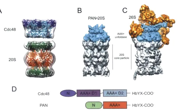
Recently, archaeal Cdc48 (known as p97 in mammals) was also found to be capable of binding the 20S peptidase, stimulating gate opening, and catalyzing protein degradation (Barthelme and Sauer, 2012). Cdc48 is a type-1 or double-ring AAA+ enzyme, defined as containing two AAA+ modules that form distinct D1 and D2 rings in the active hexamer as observed in crystal structures and cryo-EM structures. Both archaeal and eukaryotic Cdc48/p97 enzymes contain C-terminal HbYX motifs which are necessary for robust 20S-gate-opening activity. Cdc48/p97 also contains an N-terminal domain
that contacts the D1 ring in crystal and EM structures. Deletion of this N-terminal domain increases basal ATP-hydrolysis activity markedly (Gerega et al., 2005).
Cdc48/p97 function
Cdc48/p97 is found in all eukaryotes and archaea. It was initially purified from Xenopus
extracts by size-exclusion chromatography and was shown to form a 97-kDa protein complex with ATP-hydrolysis activity (Peters et al., 1990). Negative-stain electron microscopy revealed ring-shaped oligomers. Cdc48/p97 is highly abundant, accounting for -1 % of total cellular protein. The cdc48 gene is essential in yeast and mice (Moir et al., 1982; Muller et al., 2007). From archaea to humans, Cdc48/p97 orthologs display
-50% amino-acid sequence identity.
The broad conservation and distribution of Cdc48/p97 enzymes suggest a fundamentally important role in cell biology, and experiments link Cdc48/p97 to functions as diverse as membrane dynamics, ubiquitin-mediated degradation, the ER-stress response, cell-cycle regulation, and chromatin remodeling (Baek et al., 2013). These activities underlie a multitude of cellular pathways which control cellular
physiology and are factors in human health and disease.
Currently, Cdc48/p97 is viewed as a molecular motor whose function is directed and modulated by association with a large set of adaptor proteins. Some of these adaptor proteins interact with the N-terminal domain of Cdc48/p97, whereas others interact with the C-terminal HbYX tails (Meyer et al., 2012). A major group of adaptors contain a UBX
domain, which adopts a ubiquitin-like fold and interacts with the Cdc48/p97 N-domain. Although the ubiquitin-like fold suggests a link to the UPS, the lack of C-terminal diglycine motif makes it unlikely that UBX domains are covalently attached to substrates.
The p47 protein was the first member of the UBX family found to interact with Cdc48/p97. The UBX fold defines a modular domain that can interact with the N-terminal domain of Cdc48/p97. Through structure-based alignments, proteins containing
UBX domains (including p47, FAF1, SAKS1, and UBXD1) were found in all eukaryotic
species. Mutational studies identified a conserved binding motif consisting of an
R... FPR surface patch that binds to a hydrophobic surface at the junction of two
sub-domains of the Cdc48/p97 N-terminal domain. Several other families of adaptors have been found to function in similar fashion, including the UBX-like (UBX-L) containing
Ufd1-Np14 complex, which directs Cdc48/p97 to the cytoplasmic side of the ER, where it
extracts poly-ubiquitinated substrates from the membrane for eventual delivery to the proteasome. Most studies have linked Cdc48/p97 to binding ubiquitinated clients which are then remodeled or unfolded, thus facilitating subsequent downstream steps. Cdc48/p97 can also organize the ubiquitination and ubiquitin-editing steps by interacting with a series of E3 ubiquitin ligases and cullin RING scaffolds. For example, the N-terminal domain of Cdc48/p97 N-domain recruits the E4B/Ufd2 complex, which functions to lengthen short ubiquitin chains (Jentsch and Rumpf, 2007). Alternatively, several deubiquitinating enzymes can function with Cdc48/p97 to edit the length of
ubiquitin chains, possibly to tailor different substrates for recognition by specific proteasome adaptors (Wang et al., 2004; Rumpf and Jentsch, 2006).
A network of adaptors functionalize Cdc48/p97 to control important cellular processes
(Schuberth and Buchberger, 2006; Yeung et al., 2008). One of the best-studied pathways is endoplasmic-reticulum-associated-degradation (ERAD), in which a poly-ubiquitin tag mediates Cdc48/p97 binding of misfolded or aberrant proteins in the ER membrane which then catalyzes retrotranslocation into the cytosol, where the proteins undergo degradation by the 26S proteasome. In eukaryotic cells, the ER is the organelle where proteins fold and assemble as part of the secretory pathway or for membrane insertion, handling roughly one third of all cellular proteins. Thus, a robust quality-control system has evolved to maintain protein homeostasis within this organelle. The final and crucial step of this quality-control pathway requires the extraction of a misfolded polypeptide from the ER membrane. This step relies on the
Ufdl-Npl4 cofactor which binds polyubiquitin chains and mediates Cdc48/p97 substrate
binding in a nucleotide-independent fashion (Yi et al., 2012). Interestingly, Cdc48/p97 can interact with non-ubiquitinated ER substrates, but in this case ATP binding is required, suggesting that adaptors recruit substrates via binding motifs, but engagement
by Cdc48/p97 requires nucleotide binding. In a fashion similar to ERAD, Cdc48/p97
participates in the mitochondrial protein-quality control pathway. Here, the Vms1 protein forms a heterodimer with Npl4 to direct Cdc48/p97 to ubiquitinated substrates on the cytoplasmic side of the mitochondrial outer membrane (Xu et al., 2011).
Cdc48/p97, autophagy, membrane sorting, and chromatin
After discussing the role of Cdc48/p97 in 26S-mediated degradation of ERAD substrates it is surprising to then learn of its requirement for lysosomal degradation. The lysosomal pathway is the alternate means of protein degradation in the eukaryotic cell, but shares little in common with the UPS system except for the use of ubiquitin as a signal for recognition by Cdc48/p97. Both the endocytic pathway and autophagy also make use of ubiquitin as a cargo tag and as a sorting signal. In the case of endocytosis, the protein caveolin-1 (CAV1) becomes mono-ubiquitinated which mediates recognition
by the Cdc48/p97-UBXD1 complex, a requirement for late endosome formation and
fusion with the lysosome. For autophagy, a double membrane first forms and binds a lipidated form of the ubiquitin-like protein LC3 (Bug and Meyer, 2012). This complex forms an autophagosome upon engulfment of a cellular target, be it mitochondria, protein aggregates, ribosomes or any number of other cellular structures. Cdc48/p97 is required for proper maturation of autophagosomes and their eventual fusion with the lysosome. The exact mechanism of Cdc48/p97 in these two cellular pathways has yet to be determined, but the evidence suggests it acts to dislocate target proteins associated with membranes.
Lysosomes are organelles which contain hydrolytic enzymes capable of breaking down many kinds of biomolecules and are the ATP-independent alternative to 26S proteasomal degradation for recycling of proteins. Genetic studies of human disorders and yeast show that functional Cdc48/p97 is required for the proper targeting of autophagosomes to the lysosome (Tresse et al., 2010). In patients with Paget's
disease, a disorder caused by missense mutations in Cdc48/p97, an accumulation of incomplete autophagy intermediates results in inclusion body myopathy and early onset dementia associated with neurodegeneration of the frontal lobe (Guinto et al., 2007; Watts et al., 2004; Kimonis et al., 2008). In yeast it has been shown that the adaptors
Ufd3 and Ubp3 are required for autophagy of ribosomes. Together, these phenotypes
imply that Cdc48/p97 is required for autophagosome formation and their fusion with lysosomes. In autophagy, a multi-step pathway directs cytoplasmic contents targeted for turnover to be engulfed by autophagosomes whose contents are then shuttled to the lysosome where degradation takes place. Cells lacking functional Cdc48/p97 accumulate autophagy intermediates that fail to fuse with the lysosome. Similarly, Cdc48/p97 functions in targeting substrates to the lysosome through endosomal trafficking.
In addition to its function in the cytosol, Cdc48/p97 has also has been shown to act on chromatin and nucleoprotein complexes. In response to DNA damage, Cdc48/p97 was shown to control DNA replication by coordinating the degradation of replication initiation factor Cdtl (Raman et al., 2011). Cdc48/p97 also has been shown to interact with several other DNA repair proteins. The yeast mating-type switching, a regulated DNA repair process, has been linked to Cdc48/p97-Ufdl-Npl4 activity. The Mat-a2 transcriptional repressor undergoes degradation by the 26S proteasome during switching to the a-mating type, but Mat-a2 must be first removed from promoters in a Cdc48/p97-dependent process that requires ubiquitination of Mat-a2 (Dantuma and Hoppe, 2012).
Several aspects of the eukaryotic cell cycle depend upon functional Cdc48/p97. Most significantly, during mitosis Cdc48/p97 is needed for accurate and faithful segregation of chromosomes. Depletion of Cdc48/p97-Ufdl-Np4 results in failure of mitotic-spindle disassembly during the mitotic-exit pathway (Cao, 2003). At this point in the cell cycle, removal of Aurora B from the chromatin reduces local kinase activity, which then allows decondensation of chromosomes and reformation of the nuclear envelope. Aurora B is modified with ubiquitin chains and then becomes a substrate for the
Cdc48/p97-Ufd-Npl4 complex. In the absence of Cdc48/p97, Aurora B persists on the
chromatin and proper chromosome segregation cannot occur.
It is clear that Cdc48/p97 serves many biological roles in eukaryotes, and more pathways may be found to rely on Cdc48/p97. However, patterns have begun to emerge that suggest a unified picture of Cdc48/p97 biology. Whether functioning within the scope of the UPS or in proteasome-independent pathways, Cdc48/p97 functions by recognizing specific tags on target complexes, which it then remodels to facilitate downstream events. Depending on the cellular pathway, Cdc48/p97 will cooperate with a specific adaptor or groups of adaptors to recognize ubiquitin or ubiquitin-like moieties. Upon recognition and engagement, ATP-dependent movements catalyze the extraction of targets which can either be recycled, degraded, or further modified. Sometimes the target protein is the functionally important part of the pathway, as in ERAD when misfolded proteins must be removed from the ER membrane to restore protein homeostasis. Other times, the Cdc48/p97 substrates are part of large complexes that must be remodeled or dislocated from their parent complex so that organelle maturation
pathways can proceed, as in autophagy and endocytosis. At the heart of these biological functions is Cdc48/p97, which functions with a diverse repertoire of adaptors as the upstream arbiter of pathways that require the power of a molecular motor to extract or remodel client proteins.
AAA+ family ATPases: A functionally diverse group of molecular motors
Most AAA+ ATPases function as oligomers, with hexamers being most common, and contain at least one core ATP-binding module of roughly 200-250 amino acids (Neuwald et al., 1999). Within the AAA+ module, conserved Walker-A and Walker-B sequence motifs play essential roles in enzyme function. For example, a lysine in the Walker-A sequence contacts one or more phosphates of an ATP molecule and is required for strong nucleotide binding. The Walker-B motif consists of two conserved acidic residues following four hydrophobic residues (hhhhDE). The glutamic acid (E) is thought to activate a water molecule that is critical for ATP hydrolysis (Hanson and Whitehart,
2005). A third important sequence feature of the AAA+ module is an arginine finger,
which resides in a region of homology on the C-terminal side of the Walker-B motif. The Arg finger from one subunit can make contacts with an ATP bound to a neighboring subunit, which is believed to communicate nucleotide occupancy and to coordinate of ATP hydrolysis within the AAA+ ring.
At the axis of Cdc48/p97 and other AAA+ rings that function in protein unfolding, translocation, or remodeling is a channel or pore, which is lined with conserved loops that help to engage and apply force to a polypeptide substrate (Sauer and Baker, 2011).
For example, the pore-1 loops typically contain a conserved aromatic-hydrophobic dipeptide, and mutations of these residues results in defects in substrate unfolding and translocation (Martin et al., 2008; losefson et al., 2015). All AAA+ enzymes couple the chemical energy of ATP binding, hydrolysis, and product release to conformational changes that can perform mechanical work. The molecular details of this chemo-mechanical coupling are being actively investigated but are not yet clear for any AAA+ enzyme.
Single and double-ring AAA+ enzymes: similarities and differences
The operating principles of AAA+ enzymes have been most intensively characterized using bacterial ClpXP as a model (Baker and Sauer, 2012). CIpX contains a single
AAA+ module and assembles into a homohexamer. The axial pore of the AAA+ ring can
recognize a peptide sequence in the ssrA tag, which is added as a degradation signal to proteins whose synthesis on ribosomes cannot be completed normally (Keiler et al.,
1996). ClpX alone is capable of recognizing, unfolding, and translocating ssrA-tagged
substrates, and ClpXP can degrade these proteins (Gottesman et al., 1998; Kim et al, 2000; Baytshtok et al., 2015).
Studies of several AAA+ enzymes show that mutation of either the A or Walker-B motifs abolishes ATPase activity, rendering the enzyme inactive (Hanson and Whiteheart, 2005). Mutations in the Walker-B motif of ClpX permit ATP binding, binding to ssrA-tagged substrates, and binding to CIpP (Hersch et al., 2005). By contrast Walker-A mutations in ClpA unfoldase and the ClpB chaperone prevent ATP binding
and activities that require nucleotide binding (Kim et al., 1998; Singh and Maurizi, 1994)
By engineering, six CIpX subunits can be genetically linked to produce a covalent
hexamer, which allows testing the effects of different numbers and combinations of ATPase-dead subunits within the hexameric ring (Martin et al., 2005). Interestingly,
CIpX hexamers with only one or two ATPase-active subunits retain some unfolding,
translocation, and degradation activity. This result suggests that CIpX can hydrolyze ATP and unfold substrates in a probabilistic manner in which individual subunits can function relatively independently of other subunits in the ring. Although all CIpX subunits normally have the same sequence, structural studies show that only a subset adopt conformations that allow ATP binding (Glynn et al., 2009). Moreover, CIpX subunits must be able to switch between ATP-binding and non-binding conformations to allow mechanical functions like unfolding and translocation (Stinson et al., 2013; 2015). During the unfolding of very stable proteins, the vast majority of power strokes fail. For example, CIpX hydrolyzes an average of -600 ATPs during unfolding of an ssrA-tagged titin127 domain (Kenniston et al., 2003). Thus, ATP hydrolysis, conformational movements, and unfolding cannot be tightly coupled. The switching of CIpX subunits between ATP-binding and non-binding conformations may help to create an enzyme that is robust to failure and thus capable of unfolding/translocating substrates with very different amino-acid sequences and a wide range of native protein stabilities (Stinson et al., 2013).
In the CIpAP protease, CIpA, a different AAA+ partner, functions with CIpP (Grimaud et al., 1998; Striebel et al., 2009). Like Cdc48/p97, CIpA is a double-ring or type-1 AAA+
enzyme, consisting of discrete D1 and D2 rings, in addition to a family specific N-terminal domain, which helps modulate substrate specificity and adaptor binding. An outstanding question in the field is what is the functional difference between single and double-ring AAA+ ATPases. Do both AAA+ modules function in ATP hydrolysis? If so, are the functions independent or linked cooperatively? In CIpA, ATP binding to the D1 ring appears to be required to form a functional hexamer, whereas ATP hydrolysis is carried out primarily by the D2 ring. For example, Weber-Ban and colleagues found that Walker-B mutations in the D1 ring had little effect on ATPase rates, whereas the same mutations in the D2 ring resulted in a 10-fold reduction of the ATP-turnover rate (Kress et al., 2009). Not only was the D2 ring of CIpA responsible for most ATP hydrolysis, its activity was also required to unfold and degrade very stable proteins, such as GFP-ssrA.
Bacterial CIpB and its yeast homolog, Hspl04, are double-ring AAA+ enzymes that function to solubilize and refold aggregated proteins following heat shock and other cellular stresses (Mogk et al., 2015). Unlike CIpA, CIpB/Hsp1O4 show allosteric communication between the D1 and D2 rings. For example, mutation in the D1 and D2
rings of Hsp104 reduced Vmax for ATP hydrolysis to -10% and -25% of the wild-type value, respectively (Hattendorf and Lindquist, 2002) Interestingly, these mutations had only small effects on cooperativity as measured by Hill constant, suggesting that cooperativity arises from interactions within a given AAA+ ring. In subunit-mixing experiments where wild-type monomers were mixed with ATPase mutants, it was found that ATPase activity decreased non-linearly whether single or double mutants were
titrated, revealing strong coupling between rings (Werbeck et al., 2008). Interestingly, different results were found when mixed complexes were assayed for chaperone activity.
N-ethylamine sensitive factor (NSF) is a double-ring AAA+ enzyme found in eukaryotic organisms (Nagiec et al., 1995). It functions in heterotypic membrane-fusion pathways
by binding and disassembling SNARE complexes to resolve membrane fusion events. NSF is a homo-hexameric ring with an N-terminal domain responsible for SNAP-SNARE binding. The basal ATPase of this enzyme is very low but is stimulated upon
addition of its substrate. Mutational studies have shown that the D1 ring of NSF is dominant in ATPase activity, resulting in approximately 80% loss of activity, whereas the same mutations in the D2 domain have minimal effects on ATP hydrolysis (Zhao et al., 2012). Additionally, nucleotide occupancy in the D2 ring helps promote hexamer formation. Although NSF and Cdc48/p97 share significant sequence and structural homology in their N-domains and D1 and D2 AAA+ modules, the D2 ring of eukaryotic Cdc48/p97 appears to be responsible for most ATP hydrolysis, whereas the D1 ring appears to be responsible for hexamerization (Wang et al., 2003). In fact the D1 ring and the linker region spanning D1-D2 is sufficient for hexamer formation, even in the absence of nucleotide. However, it was recently shown that nucleotide binding to the D2
domain increases the catalytic efficiency of D1 ATP hydrolysis markedly, both by increasing Vmax and decreasing KM (Chou et al., 2014).
A
B
C
PAN-20S 26S Cdc48 AAA+ unfoldase core particleD
Cdc48 HbYX-COO PANHbYX-COO-Figure 4. Structural representations of archaeal and eukaryotic proteasomes. (A) Model of archaeal Cdc48-20S based on a low-resolution EM structure (Barthelme et al., 2014) and crystal structures of mammalian Cdc48/p97 (N domain colored in blue and D1-D2
in cyan) and archaeal 20S (a subunits in green and
P subunits in orange). (B) Structural
model of a docked complex based on crystal structures of archaeal PAN (cyan) and the20S core particle (gray). (C) Cryo-EM reconstruction of the yeast 26S proteasome. The
unfoldase subunits (cyan) are PAN homologs, but additional 19S regulatory subunits (orange) are also present. (D) Domain structure of the archaeal proteasomal ATPases, Cdc48 and PAN. (panels B and C adapted from Matyskiela and Martin, 2013)
Archaeal Cdc48-20S forms an alternative proteolytic complex
Many conflicting reports exist in the literature on the exact mechanism of Cdc48/p97 function, the role of structural rearrangements in the ATPase cycle, and its putative
function in concert with the 20S peptidase. Recent work from our lab has shown that archaeal Cdc48 forms a proteolytic complex with the 20S peptidase (Barthelme and Sauer, 2012; Figure 4). It uses ATP hydrolysis to unfold and translocate model substrates and has a conserved mechanism of 20S association. In Chapter 2 of this thesis, I explore the enzymatic relationship between the D1 and D2 rings of archaeal Cdc48 and how their activities and interactions govern functions such as ATP hydrolysis, protein unfolding, and 20S binding. Specifically, I use A and Walker-B mutations to selectively inhibit nucleotide binding or hydrolysis in each ring and then investigate the functional consequences. I also address the mechanism of D1-D2
communication by mutating the linker connecting these domains. My results show that ATP binding and hydrolysis in the D1 ring play an important allosteric role in Cdc48-20S
complex formation. Deleting the N-domain of Cdc48 activates ATP hydrolysis and strengthens 20S binding. I explore the linkage between the N-domain, D1-D2
communication, and ATP hydrolysis, and propose a mechanism for control of 20S binding. Another question is how archaeal Cdc48 targets specific proteins for unfolding and degradation. In chapter 3, I make use of a heterologous system of substrate identification to begin to explore substrate recognition by Cdc48-20S. Together these experiments help extend and deepen our knowledge of Cdc48, a biologically essential and medically important protein. Although Cdc48 is a complicated enzyme with many layers of regulation, the experiments presented in this thesis provide a biochemical foundation for understanding the archaeal Cdc48-20S system.
References
Baek, G.H., Cheng, H., Choe, V., Bao, X., Shao, J., Luo, S., Rao, H. (2013). Cdc48: a swiss army knife of cell biology. J. Amino. Acids. 183421.
Baker, T.A., Sauer, R.T. (2012). CIpXP, an ATP-powered unfolding and protein-degradation machine. Biochim. Biophys. Acta. 1823, 15-28.
Barthelme D., Sauer, R.T. (2012). Identification of the Cdc48-20S proteasome as an ancient AAA+ proteolytic machine. Science 337, 843-846.
Baytshtok, V., Baker, T.A., Sauer, R.T. (2015). Assaying the kinetics of protein denaturation catalyzed by AAA+ unfolding machines and proteases. Proc. NatI. Acad. Sci. U.S.A. 112, 5377-5382.
Benaroudj, N., Goldberg, A.L. (2000). PAN, the proteasome-activating nucleotidase from archaebacteria, is a protein-unfolding molecular chaperone. Nat. Cell. Biol. 2,
833-839.
Borissenko, L., Groll, M. (2007). 20S proteasome and its inhibitors: crystallographic knowledge for drug development. Chem. Rev. 107, 687-717.
Bug, M., Meyer, H. (2012). Expanding into new markets--VCP/p97 in endocytosis and autophagy. J. Struct. Biol. 179, 78-82.
Chou, T.F., Bulfer, S.L., Weihl, C.C., Li, K., Lis, G., Walters, M.A., Schoenen, F.J., Lin,
H.J., Deshaies, R.J., Arkin, M.R. (2014). Specific inhibition of p97/VCP ATPase and
kinetic analysis demonstrate interaction between D1 and D2 ATPase domains. J. Mol. Biol. 426, 2886-2899.
Cao, K., Nakakima, R., Meyer, H.H., Zheng, Y. (2003). The AAA-ATPase Cdc48/p97 regulates spindle disassembly at the end of mitosis. Cell 155,
355-367.
Dantuma, N.P., Hoppe, T. (2012). Growing sphere of influence: Cdc48/p97 orchestrates ubiquitin-dependent extraction from chromatin. Trends Cell Biol. 22, 483-491.
Darwin, K.H., Ehrt, S., Gutierrez-Ramos, J.C., Weich, N., Nathan, C.F. (2003). The proteasome of Mycobacterium tuberculosis is required for resistance to nitric oxide. Science 302, 1963-1966.
Finley, D. (2000). Nat. Struct. Biol. 7, 1062-1067.
Finley, D. (2009). Recognition and processing of ubiquitin-protein conjugates by
Gerega, A., Rockel, B., Peters, J., Tamura, T., Baumeister, W., Zwickl, P. (2005) VAT, the thermoplasma homolog of mammalian p97/VCP, is an N domain-regulated protein
unfoldase. J. Biol. Chem. 280, 42856-42862.
Glynn, S.E., Martin, A., Nager, A.R., Baker, T.A., Sauer, R.T. (2009). Structures of asymmetric ClpX hexamers reveal nucleotide-dependent motions in a AAA+ protein-unfolding machine. Cell 139, 744-756.
Grimaud, R., Kessel, M., Beuron, F., Steven, A.C., Maurizi, M.R. (1998) Enzymatic and structural similarities between the Escherichia coli ATP-dependent proteases, ClpXP and ClpAP. J. Biol. Chem. 273, 12476-12481.
Gottesman, S., Roche, E., Zhou, Y., Sauer, R.T. (1998). The ClpXP and CIpAP proteases degrade proteins with carboxy-terminal peptide tails added by the SsrA-tagging system. Genes Dev. 12, 1338-1347.
Groll, M., Bajorek, M., Kohler, A., Moroder, L., Rubin, D.M., Huber, R., Glickman, M.H., Guinto, J.B., Ritson, G.B., Taylor, J.P., Forman, M.S. (2007). Valosin-containing protein and the pathogenesis of frontotemporal dementia associated with inclusion body myopathy. Acta Neuropathol. 114, 55-61.
Gur, E., Sauer, R.T. (2009). Degrons in protein substrates program the speed and operating efficiency of the AAA+ Lon proteolytic machine. Proc. Natl. Acad. Sci. U.S.A.
106, 18503-12508.
Gur, E. (2013). The Lon AAA+ protease. Subcell. Biochem. 66, 35-51.
Hanson, P.I., Whiteheart, S.W. (2005). AAA+ proteins: have engine, will work. Nat. Rev. Mol. Cell Biol. 6, 519-529.
Hattendorf, D.A., Lindquist, S.L., (2002). Cooperative kinetics of both Hsp104 ATPase domains and interdomain communication revealed by AAA sensor-1 mutants. EMBO J. 21,12-21.
Hersch, G.L., Burton, R.E., Bolon, D.N., Baker, T.A., and Sauer, R.T. (2005). Asymmetric interactions of ATP with the AAA+ ClpX6 unfoldase: Allosteric control of a protein machine. Cell 121, 1017-1027.
losefson, 0., Nager, A.R., Baker, T.A., Sauer, R.T. (2015). Coordinated gripping of substrate by subunits of a AAA+ proteolytic machine. Nat. Chem. Biol. 11, 201-206. Jentsch, S., Rumpf, S. (2007). Cdc48 (p97): a "molecular gearbox" in the ubiquitin
Keiler, K.C., Waller, P.R., Sauer, R.T. (1996). Role of a peptide tagging system in degradation of proteins synthesized from damaged messenger RNA. Science 271,
990-993.
Kenniston, J.A., Baker, T.A., Sauer, R.T. (2003). Linkage between ATP consumption and mechanical unfolding during the protein processing reactions of an AAA+ degradation machine. Cell 114, 511-520.
Kim, K.I., Woo, K.M., Seong, I.S., Lee, Z.W., Baek, S.H., Chung, C.H. (1998). Mutational analysis of the two ATP-binding sites in ClpB, a heat shock protein with a protein-activated ATPase activity in Escherichia coli. Biochem. J. 333, 671-673.
Kim, Y.I., Burton, R.E., Burton, B.M., Sauer, R.T., Baker, T.A. (2000). Dynamics of substrate denaturation and translocation by the CIpXP degradation machine. Mol. Cell
5, 639-648.
Kim, T.I., Levchenko, I., Fraczkowska, K., Woodruff, R.V., Sauer, R.T., Baker, T.A. (2001). Molecular determinants of complex formation between Clp/Hsp100 ATPases and the CIpPpeptidase. Nat. Struct. Biol. 8, 230-233
Kimonis, V.E., Fulchiero, E., Vesa, J., Watts, G. (2008). VCP disease associated with myopathy, Paget disease of bone and frontotemporal dementia: review of a unique disorder. Biochim. Biophys. Acta. 1782, 744-748.
Kress, W., Mutschler, H., Weber-Ban, E. (2009). Both ATPase domains of ClpA are critical for processing of stable protein structures. J. Biol. Chem. 284, 31441-31452. Lander, G.C., Estrin, E., Matyskiela, M.E., Bashore, C., Nogales, E., Martin, A. (2012). Nature 482, 186-191.
Matyskiela, M.E., Martin, A. (2013). Design principles of a universal protein degradation machine. J. Mol. Biol. 425, 199-213.
Martin, A., Baker, T.A., Sauer, R.T. (2005). Rebuilt AAA + motors reveal operating principles for ATP-fuelled machines. Nature 437, 1115-1120.
Martin, A., Baker, T.A., Sauer, R.T. (2007). Distinct static and dynamic interactions control ATPase-peptidase communication in a AAA+ protease. Mol. Cell 27, 41-52. Martin, A., Baker, T.A., Sauer, R.T. (2008). Pore loops of the AAA+ ClpX machine grip substrates to drive translocation and unfolding. Nat. Struct. Mol. Biol. 15, 1147-1151. Meyer, H., Bug, M., Bremer, S. (2012). Emerging functions of the VCP/p97 AAA-ATPase in the ubiquitin system. Nat. Cell. Biol. 14, 117-123.
Moir, D., Stewart, S.E., Osmond, B.C., Botstein, D. (1982). Cold-sensitive
cell-division-cycle mutants of yeast: isolation, properties, and pseudoreversion studies. Genetics
100, 547-563.
Mogk, A., Kummler, E., Bukau, B. (2015). Cooperation of Hsp70 and
Hsp1OO chaperone machines in protein disaggregation. Front. Mol Biosci. 2, 22.
Muller, J.M., Deinhardt, K., Rosewell, I., Warren, G., Shima, D.T. (2007) Targeted deletion of p97 (VCP/CDC48) in mouse results in early embryonic lethality. Biochem.
Biophys. Res. Common. 354, 459-465.
Nagiec, E.E., Bernstein, A., Whiteheart, S.W. (1995). Each domain of the N-ethylmaleimide-sensitive fusion protein contributes to its transport activity. J. Biol. Chem. 270, 29182-29188.
Neuwald, A.F., Aravind, L., Spouge, J.L., Koonin, E.V. (1999). AAA+: A class of chaperone-like ATPases associated with the assembly, operation, and disassembly of
protein complexes. Genome Res. 9, 27-43.
Peters, J.M., Walsh, M.J., Franke, W.W. (1990). An abundant and ubiquitous homo-oligomeric ring-shaped ATPase particle related to the putative vesicle fusion proteins
Secl8p and NSF. EMBO J. 9, 1757-1767.
Prakash, S., Tian, L., Ratliff, K.S., Lehotzky, R.E., Matouschek, A. (2004). An unstructured initiation site is required for efficient proteasome-mediated degradation. Nat. Struct. Mol. Biol. 11, 830-837.
Preissler, S., Deuerling, E. (2012). Ribosome-associated chaperones as key players in proteostasis. Trends. Biochem. Sci. 37, 274-283.
Raman, M., Havens, C.G., Walter, J.C., Harper, J.W. (2011) A genome-wide screen identifies p97 as an essential regulator of DNA damage-dependent CDT1 destruction. Mol. Cell 44, 72-84.
Rumpf, S., Jentsch, S. (2006). Functional division of substrate processing cofactors of
the ubiquitin-selective Cdc48 chaperone. Mol. Cell 21, 261-269.
Sauer, R.T., Baker, T.A. (2011). AAA+ proteases: ATP-fueled machines of protein destruction. Annu. Rev. Biochem. 8, 587-612.
Schuberth. C., Buchberger, A. (2008). UBX domain proteins: major regulators of the
AAA ATPase Cdc48/p97. Cell. Mol. Life Sci. 65, 2360-2371.
Singh, S.K., Maurizi, M.R. (1994). Mutational analysis demonstrates different functional roles for the two ATP-binding sites in CIpAP protease form Escherichia coli. J. Biol. Chem. 269, 29537-29545
Smith, D.M., Kafri, G., Cheng, Y., Ng, D., Walz, T., Goldberg, A.L. (2005). ATP binding to PAN or the 26S ATPases causes association with the 20S proteasome, gate opening, and translocation of unfolded proteins. Mol. Cell 20, 687-698.
Smith, D.M., Chang, S.C., Park, S., Finley, D., Cheng, Y., Goldberg, A.L. (2007). Docking of the proteasomal ATPases' carboxyl termini in the 20S proteasome's alpha ring opens the gate for substrate entry. Mol. Cell 27, 731-44.
Stinson, B.M., Nager, A.R., Glynn, S.E., Schmitz, K.R., Baker, T.A., Sauer, R.T. (2013). Nucleotide binding and conformational switching in the hexameric ring of a AAA+ machine. Cell 153, 628-639.
Stinson, B.M., Baytshtok, V., Schmitz, K.R., Baker, T.A., Sauer, R.T. (2015). Subunit asymmetry and roles of conformational switching in the hexameric AAA+ ring of CIpX.
Nat. Struct. Mol. Biol. 22, 411-416
Werbeck, N.D., Schlee, S., Reinstein, J. (2008). Coupling and dynamics of subunits in the hexameric AAA+ chaperone CIpB. J. Mol. Biol. 378, 178-190.
Striebel, F., Kress, W., Weber-Ban, E. (2009). Controlled destruction: AAA+ ATPases in protein degradation from bacteria to eukaryotes. Curr. Opin. Struct. Biol. 19, 209-217. Tresse, E., Salomons, F.A., Vesa, J., Bott, L.C., Kimonis, V., Yao, T.P., Dantuma, N.P., Taylor, J.P. (2010). VCP/p97 is essential for maturation of
ubiquitin-containing autophagosomes andthis functionis impaired by mutations that cause IBMPF
D. Autophagy 6, 217-227.
Wang, J., Hartling, J.A., Flanagan, J.M. (1997). The structure of CIpP at 2.3 A resolution suggests a model for ATP-dependent proteolysis. Cell 91, 447-456.
Wang, Q., Song, C., Yang, X., Li, C.C. (2003). D1 ring is stable and nucleotide-independent, whereas D2 ring undergoes major conformational changes during the ATPase cycle of p97-VCP. J. Biol. Chem. 278, 32784-32793.
Watts, G.D., Wymer, J., Kovach, M.J., Mehta, S.G., Mumm, S., Darvish, D., Pestronk,
A., Whyte, M.P., Kimonis, V.E. (2004). Inclusion body myopathy associated with Paget
disease of bone and frontotemporal dementia is caused by mutant valosin-containing protein. Nat. Genet. 36, 377-381.
Xu, S., Peng, G., Wang, Y., Fang, S., Karbowski, M. (2011). The AAA-ATPase p97 is essential for outer mitochondrial membrane protein turnover. Mol. Biol.
Yeung, H.O., Kloppsteck, P., Niwa, H., Isaacson, R.L., Matthews, S., Zhang, X., Freemont, P.S. (2008). Insights into adaptor binding to the AAA protein p97. Biochem. Soc. Trans. 36, 62-67.
Yi, L., Donstante, A., Kennerson, M.L., Mercer, J.F., Garbern, J.Y., Kaler, S.G. (2012). Altered intracellular localization and valosin-containing protein (p97 VCP) interaction underlie ATP7A-related distal motor neuropathy. Hum. Mol. Genet. 21, 1794-1807. Zhao, C., Smith, E.C., Whiteheart, S.W. (2012). Requirements for the catalytic cycle of the N-etheylmaleimide-sensitive factor (NSF). Biochim. Biophys. Acta. 1823, 159-171. Zwickl P., Ng, D., Woo, K.M., Klenk, H.P., Goldberg, A.L. (1999). An archaebacterial ATPase, homologous to ATPases in the eukaryotic 26 S proteasome, activates protein breakdown by 20 S proteasomes. J. Biol. Chem. 274, 26008-26014.
Chapter 2:
Introduction
The first archaeal proteasomal ATPase discovered was PAN, which functions as single-ring AAA+ hexamer to catalyze protein unfolding and translocation into the 20S peptidase for degradation (Zwickl et al., 1999). Recently, the double-ring AAA+ Cdc48 ATPase was found to be an alternative partner for the archaeal 20S proteasome
(Barthelme and Sauer, 2012; Matouschek and Finley, 2012). The a7P7P7aC architecture
of the 20S peptidase is similar in archaea and eukaryotes. However, archaeal 20S is simpler, as each ring is composed of a single type of a or
p
subunit. In eukaryotic 20S,by contrast, seven distinct types of a or
P
subunits comprise each ring. Archaeal andeukaryotic Cdc48 share substantial sequence and structural homology (Barthelme et al., 2014), raising the possibility that a Cdc48-20S proteasome might also function in eukaryotic organisms. However, although mammalian Cdc48 can bind and open the pore of mammalian 20S (Barthelme and Sauer, 2013), there is no compelling evidence that this complex is active in protein degradation.
All Cdc48 homologs consist of an N-terminal domain, a AAA+ module that forms the D1
ring in the hexamer, another AAA+ module that forms the D2 ring, and an unstructured C-terminal tail. In this chapter, I study the enzymology of archaeal Cdc48 to better understand this highly conserved and essential ATPase. Biochemical studies of eukaryotic Cdc48 have largely been limited to assaying changes in ATP hydrolysis, as direct assays for other activities are lacking. In contrast, assays have been developed for protein unfolding by archaeal Cdc48, binding of archaeal Cdc48 to 20S, and protein degradation by archaeal Cdc48-20S (Gerega et al., 2005; Barthelme and Sauer, 2012;
Barthelme et al., 2014). Using these assays and wild-type and mutant enzymes, I probe how the major domains of Cdc48 work together. I show that the D1 and D2 rings of archaeal Cdc48 collaborate in a cooperative ATPase cycle that is critical for robust mechanical activity and that the N domain regulates machine function, at least in part,
by controlling nucleotide binding.
Results
Mutations affecting ATP hydrolysis
Archaeal Cdc48 must hydrolyze ATP to power mechanical unfolding and translocation
of proteins. To test the degree to which the D1 and D2 AAA+ rings cooperate in these
enzymatic activities, I introduced Glu+Gln mutations' into the Walker-B motifs of the ATP-binding pockets in either the D1 ring (E291Q), or the D2 ring (E568Q). The Walker-B glutamic acid is a conserved feature of all P-loop ATPases, with the side-chain carboxylic acid thought to activate a water molecule for nucleophilic attack on the gamma phosphate of ATP, leading to hydrolysis. In other AAA+ enzymes, Glu+Gln Walker-B mutations allow ATP binding but substantially reduce the rate of hydrolysis (Hersch et al., 2005; Schaupp et al., 2007).
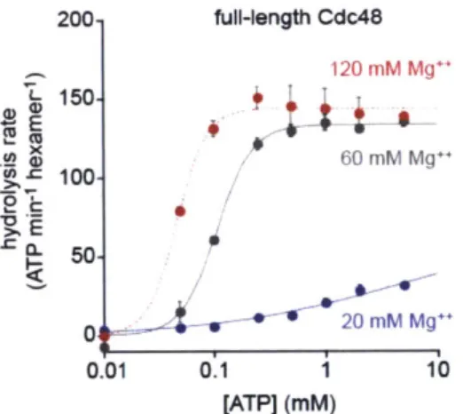
I first measured basal ATP hydrolysis rates in a standard buffer containing 20 mM
MgC 2 (Fig. 1A). The rate for wild-type Cdc48 was only modestly higher than for the
E291Q or E568Q variants. Thus, preventing robust ATP hydrolysis in either the D1 ring
or the D2 ring causes a relatively small reduction in the overall hydrolysis rate under
1 Replacing the Glu carboxylate with the isosteric Gin amide is the most conservative mutation possible at these positions.
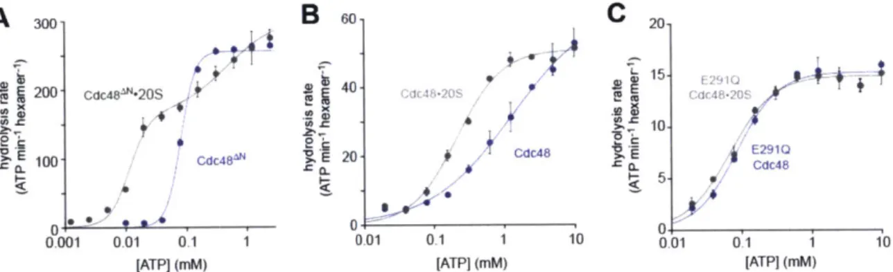
these conditions. The ATPase activity of wild-type Cdc48 is stimulated by high concentrations of Mg"* (Gerega et al., 2005). In buffer containing 120 mM MgC 2, I found that the hydrolysis rate for wild-type Cdc48 was -6-fold higher than for either
E291Q Cdc48 or E568Q Cdc48 (Fig. 1B). Thus, collaboration between the D1 and D2
rings is required to reach maximum ATPase rates in response to an activating stimulus. Next, I tested the effects of the Walker-B mutations in Cdc48AN, a variant lacking the wild-type N-terminal domain, using buffer with 20 mM MgC 2 (Fig. 1C). Under these conditions, the hydrolysis rate for the parental enzyme was -9-fold higher than the
E291Q or E568Q variants. Again, both the D1 ring and the D2 ring are required for rapid
rates of ATP hydrolysis. It is also notable that deletion of the N-terminal domain affects the pattern of ATP hydrolysis by the parent enzymes and Walker-B mutants in a fashion
similar to high Mg"* concentration (Fig. 1B and 1C).
C 300- Cdc48AN 20 mM Mg++ 250
A
200 full-length Cdc48 20 mM Mg++15 40- 100 V( 2d 20-B full-length Cdc48 120 2M Mg++ 7 100-T 1. 80 08 E E 10 40. CL 10-* 0.8 20 5 0 40 E1kErn 04Figure 1. Effects of Walker-B mutations in the D1 and D2 rings on ATP hydrolysis and protein unfolding. (A) In a buffer with 20 mM MgC 2, the E291Q and E568Q mutations modestly decrease the rate of hydrolysis of 10 mM ATP compared to the Cdc48 parent. Values are means (N=2) SEM. (B) In buffer with 120 mM MgC2, the E291Q and
E568Q mutations reduce the Cdc48 hydrolysis rate -6-fold. Values are means (N=2)
SEM. (C) In 20 mM MgC 2, the E291Q and E568Q mutations reduce the Cdc48AN
hydrolysis rate -10-fold. Values are means (N=2) SEM. (D). Cdc48AN unfolds
Kaede-ssrA 5 pM) -20-fold faster than E291Q Cdc48AN and -6-fold faster than E568Q
Cdc48 . Values are means (N=3) SD.
To assess the importance of ATP hydrolysis in Cdc48AN machine function, I monitored unfolding of a model protein substrate, photo-cleaved Kaede with a C-terminal ssrA tag that is recognized by archaeal Cdc48 (Gerega et al., 2005). Following exposure to 340-400 nm light, Kaede is photo-cleaved at a single site, but the protein retains its native fold and shows red fluorescence (Ando et al. 2002). After unfolding, however, fluorescence is irreversibly lost. Cdc48AN unfolded photo-cleaved Kaede-ssrA at a rate -20-fold faster than E291Q Cdc48AN and -6-fold faster than E568Q Cdc48AN (Fig. 1D).
Thus, robust ATP hydrolysis in both the D1 ring and the D2 ring is important to power high levels of unfolding activity.
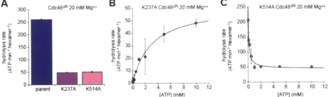
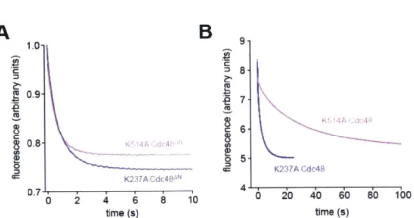
Effects of nucleotide-binding mutations
In other AAA+ hexamers, ATP binding can be weakened substantially by Lys->Ala mutations in the Walker-A motifs (Wang et al., 2003). I constructed these Walker-A mutations in the D1 ring (K237A) or the D2 ring (K514A) of Cdc48AN. Both the K237A and K514A mutations resulted in an -5-fold reduction in the hydrolysis rate of 10 mM ATP compared to the parent enzyme (Fig. 2A). For K237A Cdc48AN, the hydrolysis rate followed Michaelis-Menten kinetics with a KM of -3 mM, which I assume represents the interaction of ATP with the unmutated D2 ring (Fig. 2B). For K514A Cdc48AN, by
contrast, the hydrolysis rate decreased hyperbolically with ATP concentration until reaching a plateau (Fig. 2C). This surprising behavior can be rationalized if the K514A mutation destabilizes the hexamer, a smaller oligomer has higher ATPase activity than the hexamer, and increasing ATP stabilizes the hexamer. Monomers would be inactive as the active site spans the interface between subunits, and thus it seems likely that dimers or trimers of K514A Cdc48AN have higher ATPase activity then the hexamer. The half-maximal decrease in ATPase activity occurred at an ATP concentration of 80 nM, which is likely to represent the interaction of ATP with the unmutated D1 active sites.
A 3 C481N 20 nM M B 60 K237A Cdc48IN 20 mM Mg++ C 250 K514A Cdc48- 20 mM Mg+
250 50 200 200 40 150 150- 30 100 20 100 50 10. S 0~ 0 0
parent K237A K514A 0 0 2 4 12
[ATP] (mM) [ATPI (mM)
Figure 2. Effects of ATP-binding mutations in the Walker-A motifs of the D1 and D2
rings. (A) K237A and K514A Cdc48AN hydrolyze 10 mM ATP at a rate approximately one-fifth of the Cdc48AN parental rate. Values are means (N=2) SEM. (B) ATP
dependence of hydrolysis by the K237A Cdc48AN mutant. The line is a fit to the Michaelis-Menten equation (rate = Vmax/(1 + KM/[ATP]) with a fitted KM of -3 0.6 mM
and a Vmax of 62 5 min- ATP hexamer'. Values are means (N=2) SD. (C)
Increasing ATP decreases the rate of hydrolysis by K514A Cdc48AN, suggesting that hydrolysis at low ATP concentrations results from a dimer or trimer (see text). The line is a fit to a hyperbolic function. Values are means (N=2) SD.
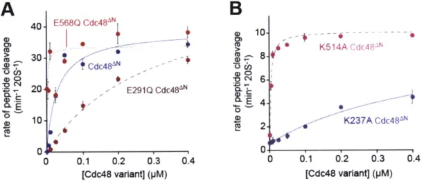
Allosteric interactions
Each ring of Cdc48 contains six active sites for ATP hydrolysis. In principle, therefore, ATP binding and hydrolysis in different subunits of either the D1 or the D2 rings could occur with positive cooperativity (Hill constant > 1), no cooperativity (Hill constant = 1), or negative cooperativity (Hill constant < 1). To test these possibilities, I measured initial
rates of hydrolysis by Cdc48AN and the Walker-B variants over a range of ATP concentrations and fitted the resulting curves to the Hill form of the Michaelis-Menten equation (Fig. 3A). ATP hydrolysis by wild-type Cdc48AN showed strong positive cooperativity (Hill constant 3.6 0.6) and an apparent KM of -80 nM. The E291Q
Cdc48AN enzyme had an apparent KM of -60 nM and Hill constant (0.84 0.16)
consistent with no cooperativity or weak negative cooperativity.2 The E568Q Cdc48AN variant showed either weak positive cooperativity or no cooperativity (Hill = 1.33 0.27) and had an apparent KM of -55 nM. Thus, robust and positively cooperative ATP hydrolysis in Cdc48AN requires wild-type active sites in both the D1 and the D2 rings. This result suggests that ring-ring communication is important for robust Cdc48 function.
300-250- Cdc48 200 150-100 50' E568Q Cdc48" 0 0.25 0.5 0.75 1.0 1.25 [ATP] (mM) 200-CdC48"lO 150- 100- 50-0.001 0.01 0.1 1 10 [ATP] (mM)
B
D1-D2 linkerC
Ta Sc Mm HOE
20 150-100 Cdc48.MftlO S 50. 0 0.001 0.01 0.1 1 10 [ATP] (mM) GSEGGTSGAT Cdc48 SIEPSSLREVMVEVPNVHWDDIGGLEDV Cdc4S NSNPSALRETVVESVNVTWDDVGLDEI Cdc48 QSNPSALRETVVBVPQVTWEDIGGLEDV Cdc48 QSNPSALRETVVEVPQVTWEDIGGLEDVF
250 Cdc48-200 cdc48 cdc4S L rms10 x 150-e k 100-S 50. 0.Il 4 4 A 012 A Hill constant less than one can indicate true negative cooperativity (binding to initial sites makes
binding to subsequent sites weaker) or indicate that there are two sets of non-interacting binding sites with different affinities.