HAL Id: hal-02898283
https://hal-amu.archives-ouvertes.fr/hal-02898283
Submitted on 13 Jul 2020HAL is a multi-disciplinary open access archive for the deposit and dissemination of sci-entific research documents, whether they are pub-lished or not. The documents may come from teaching and research institutions in France or abroad, or from public or private research centers.
L’archive ouverte pluridisciplinaire HAL, est destinée au dépôt et à la diffusion de documents scientifiques de niveau recherche, publiés ou non, émanant des établissements d’enseignement et de recherche français ou étrangers, des laboratoires publics ou privés.
Distributed under a Creative Commons Attribution| 4.0 International License
Blood Transcriptomic Signature: Evidences for
Erythrocyte Alteration, Immune/Inflammatory
Dysregulation, and Brain Dysfunction
Sandrine Cabantous, Belco Poudiougou, Aurélie Bergon, Abdoulaye Barry,
Aboubacar Oumar, Abdoulaye Traore, Christophe Chevillard, Ogobara
Doumbo, Alain Dessein, Sandrine Marquet
To cite this version:
Sandrine Cabantous, Belco Poudiougou, Aurélie Bergon, Abdoulaye Barry, Aboubacar Oumar, et al.. Understanding Human Cerebral Malaria through a Blood Transcriptomic Signature: Evidences for Erythrocyte Alteration, Immune/Inflammatory Dysregulation, and Brain Dysfunction. Mediators of Inflammation, Hindawi Publishing Corporation, 2020, 2020, pp.1-15. �10.1155/2020/3280689�. �hal-02898283�
Research Article
Understanding Human Cerebral Malaria through a Blood
Transcriptomic Signature: Evidences for Erythrocyte Alteration,
Immune/Inflammatory Dysregulation, and Brain Dysfunction
Sandrine Cabantous,
1,2Belco Poudiougou,
3†Aurélie Bergon,
4Abdoulaye Barry,
5Aboubacar A. Oumar,
6Abdoulaye M. Traore,
3Christophe Chevillard
,
4Ogobara Doumbo,
3†Alain Dessein,
1,7and Sandrine Marquet
1,4,8 1Aix Marseille Univ, INSERM, UMR906, GIMP, Labex ParaFrap, 13005 Marseille, France 2Aix Marseille Univ, INSERM, INRAE, C2VN, Marseille, France3Malaria Research and Training Center, Department of Epidemiology of Parasitic Disease, Faculty of Medicine, USTTB,
BP 1805 Bamako, Mali
4Aix Marseille Univ, INSERM, TAGC UMR U1090, 163 Av de Luminy, 13288 Marseille, CEDEX 9, France 5Pediatric Wards, Gabriel Toure Hospital, Bamako, Mali
6Centre des Oeuvres Universitaires, University of Bamako, BP 1805 Bamako, Mali 7BILHI Genetics, Marseille, France
8CNRS, Marseille, France †Deceased
Correspondence should be addressed to Sandrine Marquet; sandrine.marquet@univ-amu.fr Received 7 February 2020; Accepted 8 May 2020; Published 22 June 2020
Academic Editor: Mirella Giovarelli
Copyright © 2020 Sandrine Cabantous et al. This is an open access article distributed under the Creative Commons Attribution License, which permits unrestricted use, distribution, and reproduction in any medium, provided the original work is properly cited. Background. Cerebral malaria (CM), a reversible encephalopathy affecting young children, is a medical emergency requiring rapid clinical assessment and treatment. However, understanding of the genes/proteins and the biological pathways involved in the disease outcome is still limited. Methods. We have performed a whole transcriptomic analysis of blood samples from Malian children with CM or uncomplicated malaria (UM). Hierarchical clustering and pathway, network, and upstream regulator analyses were performed to explore differentially expressed genes (DEGs). We validated gene expression for 8 genes using real-time quantitative PCR (RT-qPCR). Plasma levels were measured for IP-10/CXCL10 and IL-18. Results. A blood RNA signature including 538 DEGs (∣FC ∣ ≥2:0, adjusted P value ≤ 0.01) allowed to discriminate between CM and UM. Ingenuity Pathway Analysis (IPA) and Kyoto Encyclopedia of Genes and Genomes (KEGG) revealed novel genes and biological pathways related to immune/inflammatory responses, erythrocyte alteration, and neurodegenerative disorders. Gene expressions of CXCL10, IL12RB2, IL18BP, IL2RA, AXIN2, and NET were significantly lower in CM whereas ARG1 and SLC6A9 were higher in CM compared to UM. Plasma protein levels of IP-10/CXCL10 were significantly lower in CM than in UM while levels of IL-18 were higher. Interestingly, among children with CM, those who died from a complication of malaria tended to have higher concentrations of IP-10/CXCL10 and IFN-γ than those who recovered. Conclusions. This study identified some new factors and mechanisms that play crucial roles in CM and characterized their respective biological pathways as well as some upstream regulators.
1. Introduction
Malaria remains a major health problem worldwide with about half of the world’s population at risk of infection, accounting for 3.2 billion people and resulting in
approxi-mately 219 million of cases annually (WHO, 2018). Although, most malaria cases are uncomplicated, a fraction of them (1%-3%), mostly young children, progress to severe disease, a life-threatening form which is characterized with higher mortality than UM. Approximately 435,000 people died of
malaria in 2017, 91% of whom were residents of sub-Saharan Africa. Therefore, there is an urgent need for reliable diagno-sis and efficacious treatment.
Cerebral malaria (CM) is one of the most severe forms of disease with a complex etiology. CM is a neurological com-plication due to Plasmodium falciparum infection that is caused in part by the sequestration of parasitized red blood cells in the microvessels of the brain, leukocyte adhesion to the microvasculature, cytotoxic lymphocyte activation, and an unregulated inflammatory response [1, 2]. Some key mediators of this inflammatory process including TNF-α, IL-6, IL-1β, IL-12, and IFN-γ have been involved in CM [3–6]. In addition, several polymorphisms have been associ-ated with susceptibility/resistance to CM [7]. These polymor-phisms are localized in genes having a role in three biological functions, the red blood cell physiology, the cytoadhesion of the infected red blood cells, and the immune response. But despite the advances and insights gained from genetic and immunological studies, our mechanistic understanding of physiological and molecular changes in human CM is still limited. Therefore, there is a need to make further progress in the understanding of pathogenic mechanisms and in the identification of molecular biomarkers to help for a better and faster medical care which would not only decrease the fatality rate but also reduce health-care costs and economic burden of the disease. As the molecular processes driving complex disease usually affect sets of genes acting in concert, one strategy is to use high throughput“omics” technologies to investigate in a pathological context several thousands of genes simultaneously. Hence, transcriptomic analysis is a powerful tool that can now be used to realize this previously unattainable goal in the study of complex infectious diseases. Recently, we carried out a study of the transcriptome of CM and UM children, from which we explored only some path-ways and genes related to neurodegenerative diseases in order to validate our experimental approach [8]. Herein, we hypoth-esize that a whole gene expression profiling from peripheral blood may be robust enough to identify a specific molecular RNA signature of CM subjects allowing to elucidate the sig-naling pathways, gene interaction networks, and upstream regulators playing critical roles in the pathogenesis of CM. We also sought to identify new potential biomarkers for CM.
2. Materials and Methods
2.1. Ethics Review and Approval. This study was approved by the local ethics committees of the Faculty of Medicine, Pharmacy, and Odonto-Stomatology of the University of Bamako and by French ethics committees, including those of INSERM, the Comité de Protection des Personnes (pro-tocol 212 CO2), the Ministère de l’Enseignement Supérieur et de la Recherche (protocol DC-2011-1426), and the Com-mission Nationale de l’Informatique et des Libertés (proto-col 1564177). All experimental methods were performed in accordance with the Declaration of Helsinki. Written informed consent was obtained from all parents.
2.2. Patients. CM was defined according to the World Health Organization criteria as a state of unarousable coma with a
Blantyre coma scale score of<2, a hematocrit of >16%, and parasitemia with asexual stages of P. falciparum. Meningitis was ruled out by lumbar puncture. Subjects with uncompli-cated malaria (UM) had a thick bloodfilm positive for P. fal-ciparum, a Blantyre coma scale score of 5, and a hematocrit of >26%. The children with UM had never developed CM. Malian children were recruited through the Pediatrics Department of Gabriel Toure Hospital in Bamako, as part of a larger prospective field study. All blood samples were collected immediately after diagnosis on admission to hospi-tal, and before treatment of the children.
2.3. RNA Extraction. A total of 13 patients with CM (male to female ratio, 7 : 6; mean age (±SD), 6:2 ± 3:8 years) and 12 patients with UM matched for age and sex (male to female ratio, 6 : 6; mean age (±SD), 6:5 ± 3:6 years) were selected for gene expression analysis; no patients died in these two groups. Among these samples, 7 CM and 8 UM were used for microarray studies. The samples were selected according to a very strict criteria and homogeneous phenotypes to obtain sufficient statistical power despite a limited number of samples. Peripheral blood mononuclear cells (PBMCs) were isolated within 1 hour after sample collection. Total RNA was extracted with TRIzol reagent (Life Technologies) according to the manufacturer’s instructions but with an additional purification step performed with the RNA Clean and Concentrator kit (Zymo Research). High-quality RNA samples with integrity values of>8 (Agilent 2100 Bioanaly-zer, Agilent technologies) were used as criteria to select sub-jects for microarray analysis (7 CM and 8 UM).
2.4. Whole-Transcriptome Analysis. The whole-transcriptome analysis was performed by using the Agilent technology, as previously described [8]. Each microarray contained about 22,700 genes based on RefSeq and 7,419 large intergenic non-coding RNAs. Fold-changes (FC) andP values were calculated for each gene, using the GeneSpring Software (Agilent). The moderated t test and the Benjamini-Hochberg correction for multiple tests were used to obtain the adjustedP values. Genes were considered significantly differentially expressed if presented an absolute FC between groups greater than 2.0 (∣FC ∣ ≥2:0) and adjusted P value ≤ 0.05).
2.5. Bioinformatics and Statistical Analysis. Hierarchical clus-tering was carried out with the TMEV software 4.9.0 to show the distinguishable transcript expression patterns among samples using the average-linkage hierarchical clustering (HCL) with Pearson’s correlation coefficient. The hierarchi-cal clustering heatmap included the most significant probes (∣FC ∣ ≥2:0) and adjusted P value ≤ 0.01 after the Benjamini-Hochberg correction. Canonical pathways, net-works analysis, and upstream regulator analysis were per-formed with Ingenuity Pathway Analysis (IPA, Qiagen) [9]. Significant DEGs (∣FC ∣ ≥2:0 and adjusted P value ≤ 0.05) were used for functional annotation by the IPA tool. This analysis allows us to determine the relevant biological path-ways and other associations connecting the differentially expressed transcripts. Canonical pathways significant to the input data set were identified from the IPA library of
canonical pathways based upon 2 parameters, (1) the ratio of the number of genes within the data set mapping to the path-way divided by the total number of genes mapping to the canonical pathway and (2) aP value (calculated based upon Fischer’s exact test) determining the probability that each biofunction assigned to that dataset, and the canonical path-way is not due to chance alone. Canonical pathpath-ways are the idealized or generalized pathways that represent common properties of a particular signaling module or pathway. Net-works were generated based on an algorithmically generated score based on connectivity between genes. A numerical value (Z-score) was used to rank networks according to how relevant they were to genes presented within the data set. Upstream regulators are the genes that affects the expres-sion of numerous other genes. Network analysis reveals interactions between molecules in datasets presented as a graph. In addition, we also identified the significantly enriched KEGG pathways of DEGs (adjustedP value ≤ 0.05 and∣FC ∣ ≥2:0) using DAVID 6.8.
2.6. Quantitative Reverse Transcription Polymerase Chain Reaction (RT-qPCR). We reverse-transcribed 400 ng of total RNA using the High-Capacity cDNA Archive kit (Applied Biosystems, Thermo Fisher Scientific) according to the man-ufacturer’s protocol. The expression of selected genes was analyzed by TaqMan PCR, on an ABI 7900 real-time PCR thermocycler (Life Technologies), according to the manufac-turer’s instructions. The relative fold change obtained by qPCR of the candidate genes was determined using the 2-ΔΔCt method after normalization with GAPDH.
2.7. Protein Quantification. The quantitative determination of IP-10/CXCL10 and IL-18 in the plasma of children was performed by ELISA (BD Biosciences and Abcys, respec-tively) according to the manufacturer’s instructions. A total of 99 children was included corresponding to 17 fatal CM, 39 nonfatal CM, and 41 UM. The 17 children died several hours after their admission to the hospital. Unmatched groups were compared by the Mann-WhitneyU test, with P value ≤ 0.05 considered to be significant. For the analysis of correlation between IP-10/CXCL10 and IFN-γ, the Spear-man correlation coefficient test was used. Both tests were conducted by the use of SPSS (SPSS, version 10.1).
3. Results
3.1. Blood Transcriptomic Signature in CM. A total of 7 CM and 8 UM subjects with very good RNA quality were used to perform gene expression profiling analysis. This analysis revealed that there were 1,366 significantly differentially expressed probes between CM and UM with an absolute fold change ∣FC ∣ ≥2:0 and adjusted P value ≤ 0.05 after the Benjamini-Hochberg correction. Accordingly, we identi-fied 1,044 upregulated and 322 downregulated probes between CM and UM. A hierarchical clustering heatmap (Figure 1(a)) demonstrated the specific expression of the 538 most signifi-cant probes (∣FC ∣ ≥2:0 and adjusted P value ≤ 0.01 after the Benjamini-Hochberg correction) and revealed the degree of separation between CM and UM subjects. Of these probes,
432 were upregulated and 106 were downregulated. This hierarchical clustering allowed us to get an RNA signature that discriminates between CM and UM (Figure 1(a)). Inter-estingly, among the differentially expressed genes (DEGs) that make up this signature, some had never been previously involved in malaria pathogenesis and/or severity. Notably, the upregulated genes GPR88, EPB42, GPNMB, S100P, GMPR, CRYAA, OSBP2, SLC6A9, MSR1, and SMOX as well as the downregulated genes SAMD12, SPON1, IL12RB2, NET1, AXIN2, IL18BP, TNFRSF25, and TRAP1 have attracted our attention because of their biological function (Figure 1(a), Table 1). Of note that among the genes included in this signature, some have already been involved in the development of clinical forms of malaria, and either encode for proteins of erythrocytic surface membrane ANK1 and GYPC (Table 1) or for inflammatory/immune components CXCL10, CCL2, CXCR2, VWA1, CD177, IL1R2, ARG1, IL2RA, and CXCL2 (Figure 1(a), Table 1).
3.2. Canonical Pathway Enrichment Analysis of DEGs. In order to gain a greater insight into specific pathophysiologi-cal process, enriched pathway and functional classification analyses of DEGs were performed using IPA. This analysis was conducted with the DEGs exhibited ∣FC ∣ ≥2 and adjustedP value ≤ 0.05 after the Benjamini-Hochberg correc-tion. A total of 24 canonical pathways were significantly altered (Figure 1(b)). Interestingly, the top 4 canonical path-ways were the granulocyte adhesion and diapedesis with 12% (20/167) and 2% (4/167) genes upregulated and downregu-lated, respectively, in CM, the arginine degradation (33% up and 17% down), the LXR/RXR activation (11% up, 2% down), and the agranulocyte adhesion and diapedesis (10% up and 3% down). Additional significant pathways were also of particular interest such as IL-8 signaling (6thranked path-ways in terms of significant), the neuroprotective role of THOP1 in Alzheimer’s disease (13th), IL-10 signaling (17th), and role of IL-17A in Arthritis (18th) because of their involvement in brain disorders and inflammatory/immune response. Of the significant genes that overlap with the above-listed canonical pathways, the great majority (146 versus 51) showed higher expression in the patients who developed CM as compared to those who had UM. Among these genes, some were previously shown to play a role in severe malaria, specially CXCL10, ARG1, ATP2B4, and MMP9. MMP9, ARG1, and CXCL10 were common to at least 3 of the 24 significant canonical pathways. Of these, MMP9 and ARG1 were upregulated in CM while surpris-ingly CXCL10 was downregulated.
3.3. KEGG Pathway Enrichment Analysis of DEGs. A KEGG pathway enrichment analysis based on the 1,366 significant probes with ∣FC ∣ ≥2 (of these 994 with genes ID) revealed that the“malaria” pathway (hsa05144) was the first most sig-nificant. Among the top 7 pathways, six have been previously identified by others using transcriptome analysis in malaria [10, 11], including malaria (hsa05144), cytokine-cytokine receptor interaction (hsa04060), TNF signaling pathways (hsa04668), chemokine signaling pathways (hsa04062), Sal-monella infection (hsa05132), and cell cycle (hsa04110).
MSR1 CXCL2 (2) IL1R2 CXCR2 CD177, ARG1 (2)⁎ CRYAA, SMOX SLC6A9⁎ ANK1 VWA1 EPB42 OSBP2, GMPR ANK1 GYPC, GPR88 IL2RA⁎, CCL2 TRAP1 SAMD12, TNFRSF25 AXIN2⁎, SPON1, TNFRSF25 CXCL10 (2)⁎ NET1⁎ S100P, GPNMB –1.3485115 –0.6742557 –0.0
CM1 CM2 CM3 CM4 CM6 CM7 CM8 UM1 UM5 UM7 UM2 UM3 UM4 UM6 UM8
–2.0 0.0 2.0
(a)
Importantly, the novel identified pathway was a “NOD-like receptor signaling pathway” (has04621) which highlights the key role of the inflammasome in the CM development.
3.4. Interaction Network Analysis. To discover potential novel regulatory networks and causal relationship associated with our DEGs, we have built and explored transcriptional
100 167 6 121 177 8 196 124 4 4 4 206 6 112 45 7 1 69 69 9 9 9 127 10 23 Percentage 90 80 70 60 50 40 30 20 10 0
Granulocyte adhesion and diapedesis
Arginine degradation VI (Arginase 2 pathway)
LXR/RXR activation
Agranulocyte adhesion and diapedesis
Airway pathology in chronic obstructive pulmonary disease
IL-8 signaling
Atherosclerosis signaling 𝛼-tocopherol degradation Melatonin degration II
Arginine degradation I (arginase pathway)
Osteoarthritis pathway
Urea cycle
Neuroprotective role of THOP1 in Alzheimer’s disease
Pyrimidine ribonucleotides de novo biosynthesis
Phosphatidylcholine Biosynthesis I
Histamine Biosynthesis
IL-10 signaling
Role of IL-17A in Arhtritis
Pathogenesis of multiple sclerosis
Citrulline biosynthesis
Iron homeostasis signaling pathway
Heme biosynthesis II Calcium transport I
Pyrimidine deoxyribonucleotides de novo biosynthesis I
4.0
No overlap with dataset
–log (p–value) 3.0 3.5 2.5 2.0 1.5 1.0 0.5 0.0 Upregulated No change Downregulated (b)
Figure 1: (a) Heatmap for differentially expressed genes between cerebral malaria (CM) and uncomplicated malaria (UM) children. Hierarchical clustering of microarrays was obtained using Pearson’s correlation on probes with a fold change greater than two and adjustedP value ≤ 0.01. The red color represents high expression, while the green color represents low expression. The blue and purple bars at the top represent the CM and UM children, respectively. Some interesting candidate genes are indicated, in red for the new players of CM pathophysiology, in black for the genes previously described in malaria. (b) Stacked bar chart displaying the top canonical pathways found to be differentially represented in comparing gene expression in CM and UM children absolute fold change ≥2, adjusted
P value ≤ 0.05. The total number of genes in each pathway is displayed above each bar. Pathways are ranked by statistical significance; the
orange dot indicate the -log (P value). The percentage of dysregulated genes is indicated for each pathway, downregulated genes are in green, and the upregulated genes are in red. The ratio of the numbers of DEGs to the total number of genes in the pathways ranged from 9% to 100%.
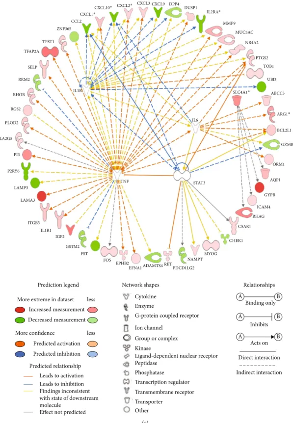
networks using IPA. We obtained 5 interactive gene networks to be significantly associated with CM (IPA score > 20): (1) connective tissue and development and function, tissue morphology, and connective tissue disorders (score = 36) (Figure 2(a)), (2) cell death and survival, connective tissue dis-orders, and hematological disease (score = 36) (Figure 2(b)), (3) digestive system development and function, organismal development (score = 30), (4) cellular movement, inflamma-tory response, and cell-to-cell signaling and interactions (score = 23), and (5) immunological and dermatological
dis-ease and conditions and inflammatory disdis-ease (score = 23). The top 2 network diagrams indicated that most of DEGs are apparently involved in the host response and erythrocyte development and function. Among genes of network 1, only one were found to be downregulated (CCL2) and 28 genes were upregulated (Figure 2(a)). Collectively, the group of upregulated genes of the network 2 is predominated by genes encoding for the red blood cell proteins with SLC4A1 as hub gene whereas the group of downregulated genes with IL-4 as hub gene was involved in the immune response (Figure 2(b)).
Table 1: Significant genes differentiating between CM and UM (∣FC ∣ >2 and adjusted P ≤ 0:01).
Probe name Gene symbol (gene name) Regulation status Absolute FC New key players of cerebral malaria
A_33_P3380462 GPR88 (G protein-coupled receptor 88) Up 9.1
A_23_P140675 EPB42 (protein 4.2, erythrocytic) Up 6.1
A_23_P134426 GPNMB (glycoprotein NMB) Up 6
A_23_P58266 S100P (S100 calcium-binding protein P) Up 4.6 A_24_P277657 GMPR (guanosine monophosphate Reductase) Up 4.1
A_33_P3323559 CRYAA (crystallin, alpha-A) Up 4
A_23_P321935 OSBP2 (oxysterol-binding protein 2) Up 4
A_33_P3326225 SAMD12 (sterile alpha motif domain-containing protein 12) Down 3.6
A_32_P133072 SPON1 (spondin 1) Down 3.5
A_33_P3402615 SLC6A9∗(solute carrier family 6, member 9) Up 3.4 A_23_P72077 IL12RB2∗(interleukin 12 receptor beta 2) Down 3.1 A_23_P392942 MSR1 (macrophage scavenger receptor 1) Up 2.9
A_23_P102731 SMOX (spermine oxidase) Up 2.6
A_23_P83931 NET1∗(Neuroepithelial cell transforming gene1) Down 2.1
A_24_P298027 AXIN2∗(axis inhibitor 2) Down 2.1
A_33_P3228322 IL18BP∗(interleukin 18 binding protein) Down 2.1 A_33_P3234530
TNFRSF25 (tumor necrosis factor receptor superfamily, member 25) Down 2
A_23_P126844 2.2
A_23_P3849 TRAP1 (tumor necrosis factor receptor-associated protein 1) Down 2 Previously involved in malaria
A_33_P3343175
CXCL10∗(chemokine, CXC motif, ligand 10) Down 7.7
A_24_P303091 7.8
A_23_P89431 CCL2 (chemokine, CC motif, ligand 2) Down 7
A_33_P3416668 VWA1 (Von Willebrand factor A domain-containing protein 1) Up 6.6
A_21_P0011751 CD177 (CD177 antigen) Up 5.8
A_24_P63019 IL1R2 (interleukin 1 receptor, type II) Up 5.2 A_33_P3352382 ARG1∗(arginase 1) Up 4.6 A_33_P3319967 4.5 A_33_P3376321 ANK1 (ankyrin 1) Up 3.2 A_23_P216108 3.5
A_23_P127288 IL2RA∗(interleukin 2 receptor, alpha) Down 3
A_24_P139901 GYPC (glycophorin C) Up 2.9
A_33_P3214550 CXCR2 (chemokine, CXC motif, receptor 2) Up 2.6 A_24_P257416
CXCL2 (chemokine, CXC motif, ligand 2) Up 2.1
A_23_P315364 2.5
Partial list of DEGs included in the RNA signature. Interesting candidate genes identified here as new players of human CM (at the top of the table) or previously involved in malaria (at the bottom of the table). Absolute FC values are shown. The eight genes selected for qPCR validation are indicated by an asterisk (∗).
Relationships TNS1⁎ PLIN2 HMGA2 SREBF1 LMNA MYOG PPARGC1A P38 MARK WNT4 NT5C1A AMPK CCL2 ERK1/2 CXCR2 GPSM1 ITLN1 ADAM9 RET BAG1⁎ PGF HSPA1A/HSPA1B IL1 MUC5AC SLC2A1 CHMP3 KCNH2 DUSP1 LDL APOE CD72 LDL–cholesterol NPC1L1 ADRB3 CXCL1⁎ PCSK9 Network shapes Cytokine Enzyme
G-protein coupled receptor Ion channel
Group or complex Kinase
Ligand-dependent nuclear receptor Peptidase Phosphatase Transcription regulator Transmembrane receptor Transporter Other Prediction legend
More extreme in dataset Increased measurement Decreased measurement More confidence less less Predicted activation Predicted inhibition Predicted relationship Leads to activation Leads to inhibition Findings inconsistent with state of downstream molecule
Effect not predicted
Binding only A B Inhibits A B Acts on Direct interaction Indirect interaction A B (a) Figure 2: Continued.
IL18BP IL 12 (complex) RNA polymerase II FOS TCR SIAH2 GZMB IFI6⁎ DDIT4 IRF4 LAMP3 CCR3 HERC6 GYPE GYPA⁎ GATA1 GYPB RHCE/RHD RHAG CA2 SLC4A1⁎ AQP1 ICAM4 KLF1 EPB42 C5AR1 IL4 PLOD2 IL12RB2 Gm–csf CD3 SPTA1 CSN1S1 Interferon alpha IL2RA⁎ Network shapes Cytokine Enzyme
G-protein coupled receptor Ion channel
Group or complex Kinase
Ligand-dependent nuclear receptor Peptidase Phosphatase Transcription regulator Transmembrane receptor Transporter Other Prediction legend
More extreme in dataset Increased measurement Decreased measurement More confidence less less Predicted activation Predicted inhibition Predicted relationship Leads to activation Leads to inhibition Findings inconsistent with state of downstream molecule
Effect not predicted
(b)
LAMP3 LAMA3 ITGB3 IL1R1 IGF2 GSTM2 FST FOS EPHB2
EFNA1ADAMTS4 RET
TNF STAT3 IL6 SLC4A1⁎ IL1B PDCD1LG2 NAMPT MYOG CHEK1 C5AR1 RHAG ICAM4 GYPB AQP1 ORM1 GZMB BCL2L1 ARG1⁎ ABCC3 UBD TOB1 PTGS2 NR4A2 MUC5AC MMP9 IL2RA⁎ DUSP1 DPP4 CXCL9 CXCL2⁎ CXCL10⁎ CXCL1⁎ CCL2 ZNF365 TPST1 TFAP2A SELP RRM2 RHOB RGS2 PLOD2 PLA2G5 PI3 P2RY6 CXCL3 Relationships Network shapes Cytokine Enzyme
G-protein coupled receptor Ion channel
Group or complex Kinase
Ligand-dependent nuclear receptor Peptidase Phosphatase Transcription regulator Transmembrane receptor Transporter Other Prediction legend
More extreme in dataset Increased measurement Decreased measurement More confidence less less Predicted activation Predicted inhibition Predicted relationship Leads to activation Leads to inhibition Findings inconsistent with state of downstream molecule
Effect not predicted
Binding only A B Inhibits A B Acts on Direct interaction Indirect interaction A B (c)
Figure 2: Gene network and upstream analysis by Ingenuity Pathway Analysis (IPA; Qiagen Inc.). Gene network highlighting the candidate genes and their interaction with the genes presented as nodes and relationship between two indicated as a line. Upstream regulator analysis allows the identification of transcriptional regulators and their target genes dysregulated in our dataset. (a) Gene network for connective tissue development and function, tissue morphology, and connective tissue disorders. (b) Gene network for cell death and survival, connective tissue disorders, and hematological disease. (c) The transcriptional regulator TNF was rankedfirst among the upstream regulators, and 38 genes were predicted to be activated or inhibited by TNF. Four additional upstream regulators SLC4A1, IL-1B, STAT3, and IL-6 have been identified.
Interestingly, IL-18BP (interleukin 18-binding protein), known to encode a decoy receptor for IL-18, was downregu-lated. Importantly, this cytokine whose expression is medi-ated by inflammasome has important functions in innate and adaptive immunity.
3.5. Upstream Regulators. Based on IPA,five upstream regu-lators including TNF (P = 1:4 × 10⁻⁷), IL-6 (P = 2:3 × 10⁻⁴), IL-1B (P = 1:4 × 10⁻⁵), STAT3 (P = 5:3 × 10⁻⁵), and SLC4A1 (P = 8:6 × 10⁻⁶) were involved in regulating DEGs in CM (Figure 2(c)). TNF, IL-6, IL-1B, and STAT3 were not dysreg-ulated in our dataset whereas SLC4A1 was upregdysreg-ulated in CM. A total of 55 genes were affected by at least one upstream regulator; of these, 39 and 16 were upregulated and downreg-ulated, respectively.
3.6. Microarray Data Validation by RT-qPCR. A total of eight candidate genes were selected from the RNA signature for qPCR validation. We have deliberately chosen genes with an increase (ARG1 and SLC6A9) or a decrease (IP-10/CXCL10, IL-12RB2, IL-18BP, IL-2RA, AXIN2, and NET1) in gene expression in CM children (Table 1 and Figure 2(b)) by including genes recently shown to be involved in CM. Most of the selected genes are involved in the inflammatory response which plays a critical role in CM as shown here. All the selected genes showed significant changes between CM and UM consistent with those observed by microarrays (Figure 3).
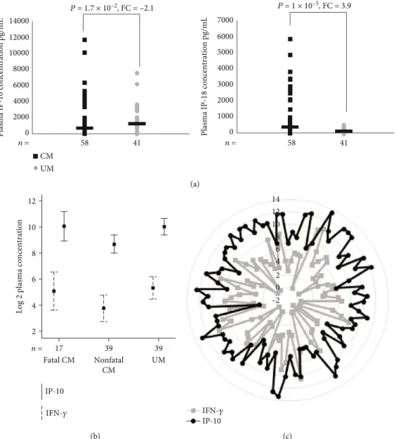
3.7. Plasma Concentration of IP-10/CXCL10 and IL-18. Sur-prisingly, we observed a lower transcription level of IP-10/CXCL10 in CM compared to UM at the admission to the hospital both by microarray and RT-qPCR whereas most studies suggested CXCL10 as risk factor in severe disease. To determine whether the protein level was consistent with the transcript level, we measured plasma IP-10/CXCL10 con-centration (Figure 4(a)). In CM, the median plasma IP-10/CXCL10 concentration was significantly lower than that in UM (583 and 1,250 pg/mL, respectively; P = 1:7 × 10⁻²). Among the children with CM, those who died of the compli-cation of malaria tended to have higher IP-10 concentration than those who recovered (962 versus 440 pg/mL;P = 5:5 × 10⁻²) (Figure 4(b)). The median IP-10 concentration was
1,250 pg/mL in the children with UM and was 440 pg/mL (P = 2 × 10⁻³) in those who recovered from CM. Because IP-10 is a chemokine secreted from a variety of cells in response to interferon-γ (IFN-γ), we tested the correlation between the plasma levels of IP-10 and IFN-γ in a total of 95 children (Figures 4(b) and 4(c)). The analysis revealed a significant positive correlation between IP-10 and IFN-γ plasma levels when it was performed on overall children (Spearman’s correlation coefficient, r = 0:475; P = 1 × 10⁻⁶).
A positive significant correlation between both protein levels was also observed among fatal CM (r = 0:584; P = 1:4 × 10⁻²), CM who recovered (r = 0:492; P = 1 × 10⁻³), and UM (r = 0:402; P = 1:1 × 10⁻²). We also quantified plasma IL-18, a cytokine known to be an inducer of IFN-γ and Th1 response [12]. IL-18 concentrations were significantly higher in children with CM (median, 338 pg/mL) than in UM
(median, 86 pg/mL) (P = 1 × 10⁻⁵) (Figure 4(a)). Interest-ingly, whereas the plasma concentration of IL-18 was higher in children with CM, the gene expression of IL-18BP was lower in these same children.
4. Discussion
Because the interactions between biomolecules play crucial roles in disease development, the understanding of the topol-ogy of biological networks and pathways is likely to be the most powerful strategy tofind molecules that may have bio-logical and clinical applications. However, deciphering the complex regulatory processes of pathophysiological path-ways in human brain remains a challenge due to the inacces-sibility of antemortem tissue. Fortunately, the studies on peripheral blood can provide important mechanistic knowl-edge that may have therapeutic implications [13–15] as shown previously into various infectious and neurological diseases [16–19]. Hence, we speculated that multiple dysreg-ulated pathways may favor CM and that we are able to iden-tify most of them in peripheral blood. We performed gene expression profiling using microarrays of whole-blood sam-ples from CM and UM children, and we showed that our data were robust enough to detect new players of CM.
Wefirst evaluated the transcriptional profiles by hierar-chical clustering and identified a set of genes providing a transcriptomic signature that allows to discriminate between CM and UM children. Even if most of the DEGs were related to expected functions such as erythrocyte alteration, immu-ne/inflammatory response, and brain dysfunction, our analy-sis provided further insight in disease development and identified novel genes never described in malaria pathogene-sis. Secondly, the pathway enrichment analysis revealed that our data were quite robust since some pathways that were dysregulated in previous reports were similarly dysregulated in this study including pathways associated with immune system such as cytokine-cytokine receptor interaction, TNF signaling pathways, and chemokine signaling pathways [10]. Others dysregulated pathways were also consistent with our knowledge of the pathogenesis of CM, as example, the granulocyte adhesion and diapedesis as well as the arginine degradation.
Among the genes that may be involved in the immune response, ARG1 is strongly expressed in CM compared to those with UM which is consistent with previous results. Indeed, previous reports showed that M2-like activation monocyte phenotype was associated with hypoargininemia, NO insufficiency, higher mononuclear cell arginase 1 mRNA, and disease severity in Tanzanian children with P. falciparum malaria [20]. It has been proposed that M2 cells produce arginase 1 that converts arginine to ornithine and urea, caus-ing depletion of arginine and reduction of NO. Absence of NO causes increased endothelial cell adhesion molecule expression and adherence of parasitized red blood cell to endothelium, having as a consequence the blockage of blood flow and distal tissue ischemia. In addition, the increased polarization towards the M2 phenotype and the increased arginase activity may also affect the immune response in malaria by altering the metabolism substrates available to
lymphocytes and other immune cells. In addition, we showed that new key pathways governing lipid metabolism (LXR/RXR activation) and inflammasome (NOD-like receptor signaling pathway) were altered in our study. Interestingly, NOD-like receptors (NLRs) are a family of cytosolic proteins that play an important role in inflammation and immunity, and besides its established role in inflammation, the LXR/RXR system has emerged as a key regulator of cholesterol, fatty acid and glu-cose homeostasis, and neuroprotection [21–23]. The path-way and network analysis allow us to confirm that most of DEGs identified here are involved in the immune/tory response and revealed a crucial role of the inflamma-some. Inflammasome-mediated secretion of 1β and IL-18 is aimed at eliminating the infectious pathogen through the induction of secondary mediators and the recruitment of additional immune cells to the infection site and/or pyrop-tosis of the infected cells. While the inflammasome-mediated response is beneficial for the host, it must be tightly regulated; otherwise, it may be associated with pathology. It is known that IL-18BP is able to bind mature IL-18 and prevents attachment to the IL-18 receptor. Here, we observed higher levels of IL-18 in the plasma of CM children compared to UM and lower expression of IL-18BP in CM suggesting that disease could be associated with an imbalance of IL-18 and IL-18BP resulting in an inappropriate inflammasome activa-tion. Although the level of IL-18 is higher in CM, no signi fi-cant difference in expression of the transcript was observed between the two groups. It has been described that transcript
level and cognate protein level do not necessarily correlate due to the regulation of translation [24]. IL-18/IL-18BP could be considered as a couple with therapeutic potential [25]. This hypothesis is consistent with recent findings showing that IL-33, which can reduce inflammasome activation and IL-1β production in microglia, is downregulated in the brain during fatal ECM (experimental CM) [26]. The authors dem-onstrate that manipulation of the IL-33-NLRP3 axis may be an effective therapy to suppress neuroinflammation and improve the efficacy of antimalarial drug treatment of CM. In addition, specific mutations in inflammasome genes result-ing in constitutive or inappropriate activation of the inflam-masome have been associated with neurological diseases. Hence, inflammasome has emerging recently as a pathogenic and therapeutic target in neurological diseases [27–29], and we believe that its activation could also have important conse-quences in CM. Another gene linked to inflammation, AXIN2 is shown to be less expressed in CM and acts as a negative regulator of Wnt/β-catenin signaling pathway that plays a critical role in many aspects of cell differentiation and immune cell function [30]. Indeed Wnt/β-catenin signaling has been shown to play an important role in the expression of several inflammatory molecules during infections [31], and aberrant signaling has been reported in neurodegenera-tive disorders such as Alzheimer’s and Parkinson’s diseases.
Interestingly, the 5 upstream regulators TNF, IL-6, IL-1B, STAT3, and SLC4A1 that we have identified here are all mol-ecules that have already been involved in malaria, and some
CXCL10 P = 3.6 × 10⁻⁵, FC = –9 0 1 2 3 4 5 6 7 8 0 1 2 3 Rela ti ve le ve l o f g ene exp ressio n Rela ti ve le ve l o f g ene exp ressio n 0 0.5 1 1.5 2 2.5 3 3.5 4 0 1 2 3 0 0.5 1 1.5 2 2.5 3 3.5 4 0 1 2 3 0 1 2 3 4 5 6 7 0 1 2 3 0 2 4 6 8 10 12 14 16 18 20 0 1 2 3 0 0.5 1 1.5 2 2.5 3 3.5 0 1 2 3 0 0.2 0.4 0.6 0.8 1 1.2 1.4 1.6 1.8 2 0 1 2 3 0 5 10 15 20 25 0 1 2 3 CM UM IL12RB2 P = 1.2 × 10⁻³, FC = –4.1 IL18BP P = 1.2 × 10⁻³, FC = –2.2 IL2RA P = 3.9 × 10⁻³, FC = –2.8 ARG1 P = 3.7 × 10⁻⁵, FC = 8.5 AXIN2 P = 1.6 × 10⁻², FC = –2.4 NET1 P = 3 × 10⁻³, FC = –1.7 SLC6A9 P = 5.8 × 10⁻⁴, FC = 2.2
Figure 3: Real-time polymerase chain reaction-based validation of messenger RNA levels for 8 significantly dysregulated genes between CM and UM. Samples from 13 children with cerebral malaria (CM) and 12 with uncomplicated malaria (UM) were analyzed. Relative expression levels were calculated from 2-ΔΔCtvalues. Values for children with CM are represented by black squares, and values for children with UM are represented by gray circles. The horizontal lines indicate median values. We used the Mann-WhitneyU test to compare the results for the CM and UM groups.
of them are considered as key players in a diverse mechanism of early innate host defense which are critical for the outcome of Plasmodium infection. Our study provided further evi-dence of their role and allowed the identification of second-ary mediators of CM. A total of 55 molecules were shown to be dysregulated by these upstream regulators and then may favor disease development. Although TNF was not dif-ferentially expressed here, TNF has been identified by others as a risk factor for the development of severe forms [4, 32– 34]. However, its mode of action and causal regulatory vari-ants were not clearly identified because genetic studies showed contradictory results across study populations [7, 35, 36]. Here, it is suggested that TNF could play a key role in pathogenic mechanisms via its regulatory effect on many other molecules. IP-10/CXCL0 is part of these effector mole-cules. Surprisingly, we found that the IP-10/CXCL10 gene
expression and protein levels were higher in UM than in those who recovered from CM, suggesting protective effect, and that these levels were higher in fatal CM compared to CM survivors in favor to a pathogenic role. Consistently with our findings, previous studies showed that IP-10/CXCL10 levels were elevated in fatal CM and were tightly associated with CM mortality [37, 38]. In addition, there is growing evi-dence that IP-10/CXCL10 plays a role in both infectious and noninfectious causes of CNS neuronal injury, dementia, and inhibition of angiogenesis. IP-10 is an IFN-γ-induced che-mokine with chemotactic activity for activated Th1 lympho-cytes [38] which is quite consistent with the correlation between IFN-γ and IP-10 that we observed in our patients. Our results therefore suggested a complex regulation of this gene and a dual role of IP-10 (protective and aggravating) as it has been shown for IFN-γ in both malaria [39] and
CM UM
P = 1.7 × 10⁻², FC = –2.1 P = 1 × 10⁻⁵, FC = 3.9
14000
Plasma IP-10 concentration pg/mL
12000 10000 8000 6000 4000 2000 0 7000
Plasma IP-18 concentration pg/mL
6000 5000 4000 3000 2000 1000 0 58 41 n = n = 58 41 (a) 12 10
Log 2 plasma concentration
8 6 4 2 17 n = Fatal CM Nonfatal CM UM 39 39 IP-10 IFN-𝛾 (b) 14 12 10 8 6 4 2 0 –2 IFN-𝛾 IP-10 (c)
Figure 4: (a) Plasma IP-10 and IL-18 concentrations in children with CM (n = 58, black squares) and UM (n = 41, gray circles). The nonparametric Mann-WhitneyU test was used to assess differences. (b) Plasma IP-10 and IFN-γ concentrations in children with fatal CM and nonfatal CM and UM. The levels are represented in log. (c) Plasma IP-10 and IFN-γ concentrations for each child with CM or UM. The levels are represented in log. We used Spearman’s correlation coefficient.
Chagas disease [40]. This regulation can be partly achieved by the upstream regulators identified here, TNF, 1B, IL-6, and STAT3. Overall, we can assume that high levels of CXCL10 may cause vascular injury resulting in breakdown in the blood-brain barrier (BBB) which may lead to accumu-lation of leukocytes that induced local hyperinflammation and to a lethal neuropathological syndrome. On the basis of all these data, we can hypothesize that subjects infected by P. falciparum must develop an effective immune response by producing chemokines such as IP-10/CXCL10 and inflammatory cytokines such as IFN-γ which can control the infection. Unfortunately, this immune response cannot be properly regulated in some people, and an excessive inflammatory response can cause tissue damages and death. Thus, such excessive production on arrival at the hospital seems to be a diagnosis of poor clinical course. In addition, the kinetics of production of the molecules involved in the inflammatory response and determining in the clinical outcome of the patient as suggested recently for the IFN-γ production is essential [41]. However, it remains to be estab-lished how this chemokine might contribute to protect against CM.
Finally, this analysis supported again that pathways and genes having a role in the neurodegenerative disorders were dysregulated in CM as we have previously pointed out [8] and which has been confirmed by others [11]. Here, we highlighted the role of 3 genes (GPR88, GPNMB, and GMPR) showing higher expression in CM (FC > 4) and that could be interesting key players of CM since higher expres-sion of them are also associated to Parkinson’s and Alzhei-mer’s diseases. These two neurodegenerative disorders share with CM both some clinical symptoms such as memory loss or vasoconstriction and also some pathophysiological features such as a modification of the blood-brain barrier and an increase in the expression of ICAM-1 [42, 43]. The GPR88 is part of the top gene with a fold increase of 9.1. This gene encodes a brain-specific G protein-coupled receptor that plays a role in dopaminergic function. This cell surface receptor which is involved in the recognition of iRBCs (infected red blood cells) [44] is also emerging as a novel drug target for central nervous system disorders including schizo-phrenia, Parkinson’s disease (PD), and anxiety [45]. Another new key player of CM was the glycoprotein nonmetastatic melanoma protein B (GPNMB), a glycoprotein observed upon tissue damage and inflammation and associated with astrocytes, microglia, and macrophages. It has been identified as a novel Alzheimer’s disease- (AD-) related factor in both transgenic mice and sporadic AD patients by expression pro-filing [46]. In addition, gene variations in GPNMB were also linked with PD risk and with significant increased expression of GPNMB [47]. Finally, the GMPR gene, which encodes protein GMPR1, was upregulated in CM as observed in AD cases and which exhibited a gradual increase with AD pro-gression. Importantly, the increased expression of GMPR makes the product of GMPR (GMPR1) a potential therapeu-tic target [48].
Finally, several studies on gene expression analysis using microarrays showed promising results even by using a small number of samples [10, 16, 49]. Here, despite the limited
number of subjects studied for the transcriptome analysis, we obtained sufficient statistical power to identify blood tran-scriptomic signature which distinguish CM and UM. Obvi-ously, additional studies carried out on a larger number of subjects, and using RNAseq technology would be very useful to confirm the role of some genes identified here.
In conclusion, our study provides fundamental knowl-edge on the molecular profile characteristic of CM. In this work, we have identified a new set of genes as key signaling and regulatory molecules involved in the pathophysiology of CM. We show that transcriptional profiling in whole blood is powerful enough to allow a comprehensive analysis of host physiology during CM and the identification of biomarkers that may be potential therapeutic targets.
Data Availability
The data used to support thefindings of the study are avail-able from the corresponding author upon request.
Conflicts of Interest
The authors do not have commercial or other associations that might pose a conflict of interest.
Acknowledgments
This work was supported by the French Research Ministry, by the Institut National de la Santé et de la Recherche Médi-cale (INSERM), by the European Union (IC18-CT98 0373), and by the ParaFrap“French Parasitology Alliance for Health Care” (ANR-11-LABX-0024-01). This work has not been presented previously at a scientific meeting. We thank Pr Edecio Cunha-Neto for the helpful advice. The authors thank all the children and their parents for their participation in this study. This work is dedicated in memory of Ogobara Doumbo and Belco Poudiougou.
References
[1] L. H. Miller, D. I. Baruch, K. Marsh, and O. K. Doumbo,“The pathogenic basis of malaria,” Nature, vol. 415, no. 6872, pp. 673–679, 2002.
[2] L. H. Miller, H. C. Ackerman, X. Z. Su, and T. E. Wellems, “Malaria biology and disease pathogenesis: insights for new treatments,” Nature Medicine, vol. 19, no. 2, pp. 156–167, 2013.
[3] M. E. Molyneux, H. Engelmann, T. E. Taylor et al., “Circulat-ing plasma receptors for tumour necrosis factor in Malawian children with severe falciparum malaria,” Cytokine, vol. 5, no. 6, pp. 604–609, 1993.
[4] D. Kwiatkowski, I. Sambou, P. Twumasi et al.,“TNF concen-tration in fatal cerebral, non-fatal cerebral, and uncomplicated Plasmodium falciparum malaria,” Lancet, vol. 336, no. 8725, pp. 1201–1204, 1990.
[5] N. H. Hunt and G. E. Grau, “Cytokines: accelerators and brakes in the pathogenesis of cerebral malaria,” Trends in Immunology, vol. 24, no. 9, pp. 491–499, 2003.
[6] W. L. Mandala, C. L. Msefula, E. N. Gondwe, M. T. Drayson, M. E. Molyneux, and C. A. MacLennan,“Cytokine Profiles in
Malawian Children Presenting with Uncomplicated Malaria, Severe Malarial Anemia, and Cerebral Malaria,” Clinical and Vaccine Immunology, vol. 24, no. 4, 2017.
[7] S. Marquet, “Overview of human genetic susceptibility to malaria: From parasitemia control to severe disease,” Infection, Genetics and Evolution, vol. 66, pp. 399–409, 2018.
[8] S. Cabantous, O. Doumbo, B. Poudiougou et al., “Gene Expression Analysis Reveals Genes Common to Cerebral Malaria and Neurodegenerative Disorders,” The Journal of Infectious Diseases, vol. 216, no. 6, pp. 771–775, 2017. [9] A. Krämer, J. Green, J. PollardJr, and S. Tugendreich,“Causal
analysis approaches in Ingenuity Pathway Analysis,” Bioinfor-matics, vol. 30, no. 4, pp. 523–530, 2014.
[10] R. S. Sobota, A. Dara, J. E. Manning et al.,“Expression of com-plement and toll-like receptor pathway genes is associated with malaria severity in Mali: a pilot case control study,” Malaria Journal, vol. 15, no. 1, 2016.
[11] S. Nallandhighal, G. S. Park, Y. Y. Ho, R. O. Opoka, C. C. John, and T. M. Tran, “Whole-Blood Transcriptional Signatures Composed of Erythropoietic and NRF2-Regulated Genes Dif-fer Between Cerebral Malaria and Severe Malarial Anemia,” The Journal of Infectious Diseases, vol. 219, no. 1, pp. 154– 164, 2019.
[12] F. Biet, C. Locht, and L. Kremer, “Immunoregulatory func-tions of interleukin 18 and its role in defense against bacterial pathogens,” Journal of Molecular Medicine (Berlin, Germany), vol. 80, no. 3, pp. 147–162, 2002.
[13] B. Zhang, C. Gaiteri, L. G. Bodea et al.,“Integrated Systems Approach Identifies Genetic Nodes and Networks in Late-Onset Alzheimer's Disease,” Cell, vol. 153, no. 3, pp. 707– 720, 2013.
[14] S. Mohr and C. C. Liew, “The peripheral-blood tran-scriptome: new insights into disease and risk assessment,” Trends in Molecular Medicine, vol. 13, no. 10, pp. 422–432, 2007.
[15] C. C. Liew, J. Ma, H. C. Tang, R. Zheng, and A. A. Dempsey, “The peripheral blood transcriptome dynamically reflects sys-tem wide biology: a potential diagnostic tool,” The Journal of Laboratory and Clinical Medicine, vol. 147, no. 3, pp. 126– 132, 2006.
[16] P. Montaldo, M. Kaforou, G. Pollara et al.,“Whole Blood Gene Expression Reveals Specific Transcriptome Changes in Neona-tal Encephalopathy,” Neonatology, vol. 115, no. 1, pp. 68–76, 2019.
[17] R. Shamir, C. Klein, D. Amar et al.,“Analysis of blood-based gene expression in idiopathic Parkinson disease,” Neurology, vol. 89, no. 16, pp. 1676–1683, 2017.
[18] Y. Xu, Y. Yao Shugart, G. Wang et al.,“Altered expression of mRNA profiles in blood of early-onset schizophrenia,” Scien-tific Reports, vol. 6, no. 1, article 16767, 2016.
[19] I. Nikolayeva, P. Bost, I. Casademont et al.,“A blood RNA sig-nature detecting severe disease in young dengue patients at hospital arrival,” The Journal of Infectious Diseases, vol. 217, no. 11, pp. 1690–1698, 2018.
[20] J. B. Weinberg, A. D. Volkheimer, M. P. Rubach et al., “Mono-cyte polarization in children with falciparum malaria: relation-ship to nitric oxide insufficiency and disease severity,” Scientific Reports, vol. 6, no. 1, article 29151, 2016.
[21] M. Baranowski,“Biological role of liver X receptors,” Journal of Physiology and Pharmacology, vol. 59, Supplement 7, pp. 31–55, 2008.
[22] B. A. Laffitte, L. C. Chao, J. Li et al., “Activation of liver X receptor improves glucose tolerance through coordinate regu-lation of glucose metabolism in liver and adipose tissue,” Pro-ceedings of the National Academy of Sciences of the United States of America, vol. 100, no. 9, pp. 5419–5424, 2003. [23] G. Cermenati, F. Abbiati, S. Cermenati et al.,
“Diabetes-induced myelin abnormalities are associated with an altered lipid pattern: protective effects of LXR activation,” Journal of Lipid Research, vol. 53, no. 2, pp. 300–310, 2012.
[24] C. P. Moritz, T. Mühlhaus, S. Tenzer, T. Schulenborg, and E. Friauf, “Poor transcript-protein correlation in the brain: negatively correlating gene products reveal neuronal polarity as a potential cause,” Journal of Neurochemistry, vol. 149, no. 5, pp. 582–604, 2019.
[25] H. Mühl and M. Bachmann, “IL-18/IL-18BP and IL-22/IL-22BP: two interrelated couples with therapeutic potential,” Cellular Signalling, vol. 63, article 109388, 2019.
[26] P. Strangward, M. J. Haley, M. G. Albornoz et al.,“Targeting the IL33-NLRP3 axis improves therapy for experimental cere-bral malaria,” Proceedings of the National Academy of Sciences of the United States of America, vol. 115, no. 28, pp. 7404–7409, 2018.
[27] M. K. Mamik and C. Power,“Inflammasomes in neurological diseases: emerging pathogenic and therapeutic concepts,” Brain, vol. 140, no. 9, pp. 2273–2285, 2017.
[28] S. Voet, S. Srinivasan, M. Lamkanfi, and G. Loo, “Inflamma-somes in neuroinflammatory and neurodegenerative diseases,” EMBO Molecular Medicine, vol. 11, no. 6, 2019.
[29] J. G. Walsh, D. A. Muruve, and C. Power,“Inflammasomes in the CNS,” Nature Reviews. Neuroscience, vol. 15, no. 2, pp. 84– 97, 2014.
[30] A. Suryawanshi, R. K. Tadagavadi, D. Swafford, and S. Manicassamy,“Modulation of inflammatory responses by Wnt/β-catenin signaling in dendritic cells: a novel immuno-therapy target for autoimmunity and cancer,” Frontiers in Immunology, vol. 7, 2016.
[31] O. Silva-García, J. J. Valdez-Alarcón, and V. M. Baizabal-Aguirre,“The Wnt/β-catenin signaling pathway controls the inflammatory response in infections caused by pathogenic bacteria,” Mediators of Inflammation, vol. 2014, Article ID 310183, 7 pages, 2014.
[32] G. E. Grau, T. E. Taylor, M. E. Molyneux et al.,“Tumor necro-sis factor and disease severity in children with falciparum malaria,” The New England Journal of Medicine, vol. 320, no. 24, pp. 1586–1591, 1989.
[33] W. McGuire, A. V. S. Hill, C. E. M. Allsopp, B. M. Greenwood, and D. Kwiatkowski,“Variation in the TNF-α promoter region associated with susceptibility to cerebral malaria,” Nature, vol. 371, no. 6497, pp. 508–511, 1994.
[34] S. A. Olaniyan, O. K. Amodu, A. A. Bakare, M. Troye-Blom-berg, O. O. Omotade, and K. A. Rockett,“Tumour necrosis factor alpha promoter polymorphism, TNF-238 is associated with severe clinical outcome of falciparum malaria in Ibadan Southwest Nigeria,” Acta Tropica, vol. 161, pp. 62–67, 2016. [35] S. Cabantous, O. Doumbo, S. Ranque et al.,“Alleles 308A and
238A in the tumor necrosis factor alpha gene promoter do not increase the risk of severe malaria in children with Plasmo-dium falciparum infection in Mali,” Infection and Immunity, vol. 74, no. 12, pp. 7040–7042, 2006.
[36] T. G. Clark, M. Diakite, S. Auburn et al.,“Tumor necrosis fac-tor and lymphotoxin-alpha polymorphisms and severe
malaria in African populations,” The Journal of Infectious Diseases, vol. 199, no. 4, pp. 569–575, 2009.
[37] N. O. Wilson, V. Jain, C. E. Roberts et al., “CXCL4 and CXCL10 predict risk of fatal cerebral malaria,” Disease Markers, vol. 30, no. 1, pp. 39–49, 2011.
[38] V. Jain, H. B. Armah, J. E. Tongren et al.,“Plasma IP-10, apo-ptotic and angiogenic factors associated with fatal cerebral malaria in India,” Malaria Journal, vol. 7, no. 1, 2008. [39] S. Cabantous, B. Poudiougou, A. Traore et al.,“Evidence that
interferon-gamma plays a protective role during cerebral malaria,” Journal of Infectious Diseases, vol. 192, no. 5, pp. 854–860, 2005.
[40] C. Chevillard, J. P. S. Nunes, A. F. Frade et al.,“Disease toler-ance and pathogen resisttoler-ance genes may underlie Trypano-soma cruzi Persistence and Differential Progression to Chagas Disease Cardiomyopathy,” Frontiers in Immunology, vol. 9, 2018.
[41] T. King and T. Lamb,“Interferon-γ: the Jekyll and Hyde of malaria,” PLoS Pathogens, vol. 11, no. 10, article e1005118, 2015.
[42] B. S. Desai, A. J. Monahan, P. M. Carvey, and B. Hendey, “Blood-brain barrier pathology in Alzheimer's and Parkinson's disease: implications for drug therapy,” Cell Transplantation, vol. 16, no. 3, pp. 285–299, 2017.
[43] M. T. Heneka and M. K. O'Banion,“Inflammatory processes in Alzheimer's disease,” Journal of Neuroimmunology, vol. 184, no. 1-2, pp. 69–91, 2007.
[44] M. A. Terkawi, R. Takano, and K. Kato, “Differential Gene Expression Profile of Human Neutrophils Cultured with Plas-modium falciparum-Parasitized Erythrocytes,” Journal of Immunology Research, vol. 2018, Article ID 6709424, 8 pages, 2018.
[45] N. Ye, B. Li, Q. Mao et al., “Orphan receptor GPR88 as an emerging neurotherapeutic target,” ACS Chemical Neurosci-ence, vol. 10, no. 1, pp. 190–200, 2018.
[46] M. Hüttenrauch, I. Ogorek, H. Klafki et al., “Glycoprotein NMB: a novel Alzheimer's disease associated marker expressed in a subset of activated microglia,” Acta Neuropathologica Communications, vol. 6, no. 1, p. 108, 2018.
[47] M. N. Murthy, C. Blauwendraat, UKBEC et al., “Increased brain expression of GPNMB is associated with genome wide significant risk for Parkinson's disease on chromosome 7p15.3,” Neurogenetics, vol. 18, no. 3, pp. 121–133, 2017. [48] H. Liu, K. Luo, and D. Luo, “Guanosine monophosphate
reductase 1 is a potential therapeutic target for Alzheimer's disease,” Scientific Reports, vol. 8, no. 1, 2018.
[49] A. Thiam, M. Sanka, R. Ndiaye Diallo et al.,“Gene expression profiling in blood from cerebral malaria patients and mild malaria patients living in Senegal,” BMC Medical Genomics, vol. 12, no. 1, p. 148, 2019.