Health sciences/Anatomy/Gastrointestinal system/Microbiota [URI /692/698/2741/2135]
Health sciences/Diseases/Gastrointestinal diseases/Liver diseases/Non-alcoholic fatty liver disease / [URI /692/699/1503/1607/2750]
Gut microbiota and human NAFLD: disentangling microbial signatures from metabolic disorders
Judith Aron-Wisnewsky‡1,3, Chloé Vigliotti‡1,2, Julia Witjes4, Phuong Le1,2, Adriaan G. Holleboom 4, Joanne Verheij5, Max Nieuwdorp4,6 and Karine Clément*1,3
1
Sorbonne Université, INSERM, UMRS U1269, Nutriomics research unit, Paris, France
2
Institute of Cardiometabolism and Nutrition, Integromics team, Paris, France 3
Assistante Publique Hôpitaux de Paris, Nutrition department, Pitié-Salpêtrière hospital, CRNH Ile de France, Paris
4
Amsterdam UMC, location AMC, dept of Vascular Medicine, University of Amsterdam, Amsterdam, the Netherlands
5
Amsterdam UMC, location AMC, Dept of Pathology, University of Amsterdam, Amsterdam, the Netherlands
6
Amsterdam UMC, location VUMC, dept of Internal Medicine, Free University, Amsterdam, the Netherlands
‡
co-first authors
Abstract
Gut microbiota dysbiosis has been repeatedly observed in obesity and type 2 diabetes mellitus (T2DM), two metabolic diseases strongly intertwined with nonalcoholic fatty liver disease (NAFLD). Animal studies have demonstrated a potential causal role of gut microbiota in NAFLD. Human studies have started to describe microbiota alterations in NAFLD and have found a few consistent microbiome signatures discriminating healthy individuals from NAFLD, nonalcoholic-steatohepatitis or cirrhosis. However, patients with NAFLD often present with obesity and/or insulin resistance and T2DM, and these metabolic confounding factors for dysbiosis have not always been taken into account. Patients with different NAFLD severity stages often present with heterogeneous lesions and variable demographic characteristics (including age, sex and ethnicity), which are known to affect the gut microbiome and have been overlooked in most studies. Finally, multiple gut microbiome sequencing tools and NAFLD diagnostic methods have been used across studies that could account for discrepant microbiome signatures. This Review provides a broad insight into microbiome signatures for human NAFLD and explores issues with disentangling these signatures from underlying metabolic disorders. More advanced metagenomics studies, as well as multi-omics studies using system biology approaches, are needed to improve microbiome biomarkers.
[H1] Introduction
The gut microbiota (that is the microbial community within the gastrointestal tract) has critical physiological roles in host digestion, immunity and metabolism1,2. Initially only studied by culture-based methods, the characterization of the gut microbiota3 has deepened with the rapid development of high-throughput sequencing technology
(shotgun sequencing or pyrosequencing). Constructed gut microbiota reference gene catalogues4,5 have further enabled the determination of the composition of the gut microbiota and prediction of microbiome functions6. In addition to these technological advances, decreasing costs and reduced analytical turnaround due to bioinformatic pipeline development have enabled increasingly accessible and efficient microbiome studies. Thus, knowledge on microbiome characteristics in common diseases, especially metabolic diseases7, has substantially increased in the past 15 years.
The need for microbiome characterization in metabolic diseases was initially stimulated by pioneering studies using germ-free mice and gut microbiota transfer, which reported the contribution of gut microbiota to weight gain and metabolic alterations8,9. Studies using conventional mice receiving lipopolysaccharide (LPS, a major component of the Gram-negative bacterial outer membrane) infusions also provided evidence of the role of gut microbiota in metabolic injuries and its influence on insulin resistance10. Since these initial studies, microbiome signatures linked to obesity11–13 and type 2 diabetes mellitus (T2DM)14,15 and associated complications were discovered, raising the concept of human gut microbiota dysbiosis (that is,alteration in microbiota composition and functional capacities with modification of microbiome signatures16) in metabolic diseases (reviewed elsewhere17). Currently, these findings are being pursued to develop microbiota-based therapeutics such as probiotics18, prebiotics19, synbiotics20, and faecal microbiota transplantation [G] [Au: Please do not delete these marks, they are to flag the glossary terms to our Production team] (FMT) 21,22 to improve metabolic health and personalized patient care.
Nonalcoholic fatty liver disease [G] (NAFLD), and the more advanced stage nonalcoholic steatohepatitis [G] (NASH)23, are common comorbidities of obesity and
T2DM with an increasing burden for society24. NAFLD-related liver failure has become the second leading cause of liver transplantation in the Western world 25. As liver biopsy is the diagnostic gold standard for NAFLD and NASH, and it is an invasive, inconvenient and impractical tool in a public health setting26, the complete understanding of the complex pathophysiology of these diseases remains limited. Moreover, although mouse models of NAFLD and NASH are helpful, they are not optimal27 and can limit the translation of results to clinical research27. As obesity, T2DM, and NAFLD–NASH are linked clinically and pathophysiologically, exploring the gut microbiome seems to be a relevant approach to gain a better understanding of NAFLD and NASH. Although this level of characterization of NAFLD and NASH is markedly less than that for obesity and diabetes, there is a rapidly growing body of evidence exploring the contribution of the gut microbiome to NAFLD physiopathogenesis28–30 using high-throughput sequencing in cohorts of individuals spanning the NAFLD–NASH disease spectrum. As there is a large overlap between NAFLD and metabolic disorders in respect to the disease spectrum and contributing factors, some metagenomic signatures of NAFLD might be shared with those already observed in obesity and T2DM. Thus, deciphering signatures specific to liver alterations would be most useful for future NAFLD diagnostic biomarkers. We herein review the gut microbial and gut microbial-derived metabolite signatures associated with NAFLD development and progression focusing on their relationship with disease progression in human. We specifically focused on which microbial signatures are specific to liver injury versus those common to other metabolic diseases and the putative methodological biases that could explain divergent results across the literature.
[H1] NAFLD and related liver fibrosis
NAFLD is defined as the pathological accumulation of lipid droplets in >5% of hepatocytes23. This disease can progress towards NASH, which is diagnosed by liver biopsy and the histological examination of the degree of steatosis [G] , inflammation and hepatocyte ballooning23,31. NASH can also present with liver fibrosis [G] 23,31,32, which is the main prognostic lesion for disease progression33,34, eventually leading to cirrhosis35 [G] and/or hepatocellular carcinoma36–38 and other liver-related complications among which include ascites, hepatic encephalopathy and portal hypertension39. NAFLD is highly prevalent and has become the most common cause of chronic liver disease in the Western world affecting up to 40% of the general population40 and reaching sometimes 90%41,42 in obese populations worldwide43. NAFLD is closely associated with overweight or obesity and metabolic disorders such as insulin resistance, hypertension and T2DM, and is even recognized as the hepatic component of metabolic syndrome44,45 (Box 1). NAFLD and metabolic syndrome both increase the risk of cardiovascular diseases and T2DM46; therefore, NAFLD and metabolic syndrome probably have similar risk profiles47.
For research purposes, several scores or algorithms based upon histological evaluation of liver biopsy samples have been developed to enable patient classifications in epidemiological studies. For example, the NALFD Activity Score (NAS) is a scoring system calculated from the semi-quantitative evaluation of steatosis, lobular inflammation and hepatocyte ballooning48. Although accurate in low (<3) or high (>5) values to exclude or diagnose NASH, respectively, NAS scoring is often inaccurate within the intermediate values (scores 3–4)31
. As a consequence, European guidelines recommend to only use NAS for disease severity evaluation once the diagnosis has been made23. A newer diagnostic algorithm, the Steatosis
Activity and Fibrosis (SAF) score, which includes the semi-quantitative scoring of these factors, to enable the classification of patients as no NAFLD, NAFLD or NASH, has demonstrated improved performance compared with NAS, in particular within the intermediate values of the NAS (scores 3-4)31. Indeed, compared with NAS, the SAF score emphasizes the importance of activity, the main culprit of NASH, and therefore provides more accurate and comprehensive histological description. As such, it is now qualified as a true diagnostic score in the European guidelines23. Despite the utility of these scoring approaches, they rely on liver biopsy, which has drawbacks such as sampling error, inter-individual variations in pathologist reading and the risk of complications, of which the most worrisome is internal bleeding.
As performing liver biopsies26 on all patients with NAFLD49 is unfeasible for disease screening, diagnosis or examining progression in both routine care and research, noninvasive diagnostic methods using plasma samples50, ultrasonography51, MRI 52 or liver elastography (including both transient and magnetic resonance53–55) have been developed56–58, and offer good diagnostic performance for liver fibrosis53,54. (Box 2). These methods have been widely used for early disease detection (steatosis), disease severity assessment, identification of patients needing a liver biopsy for confirmatory diagnosis (patients with divergent results obtained upon two noninvasive tests53,59,60) and for assessment of disease progression (fibrosis). Despite their obvious benefit compared with liver biopsy, these noninvasive tools are also hampered by several limitations (summarized in Box 2)58. They are, in general, not sensitive enough to evaluate the complete spectrum of NAFLD histological lesions45 and lack validity to be used for routine diagnosis (reviewed elsewhere54,57,61 ). Transient elastography can be seen as an exception, however, as it was validated against liver biopsy with good area under the receiver operator
characteristic (AUROC) values ranging from 0.70 to 0.89 for both steatosis and fibrosis in a large population composed of 450 patients with the complete spectrum of NAFLD fibrosis stages62.
Despite these noninvasive tests, liver biopsy remains the gold standard for NAFLD and NASH diagnosis. Thus, new biology-based, inexpensive, easily accessible, highly sensitive and specific prognostic and diagnostic biomarkers are urgently needed. As the gut microbiota might have a pathophysiological role in NAFLD development, the use of noninvasive microbiota-related biomarkers from stool (microbiota signatures) and/or blood sampling (metabolic or microbiota-derived signatures) could be an interesting alternative to currently developed noninvasive tests or could be considered as a complementary approach.
[H1] NAFLD and gut microbiota
Mouse studies and faecal transplant experiments have provided evidence of a causal role of gut microbiota in NAFLD development. First, cohousing experiments with mice prone to developing NASH due to genetic modifications in the inflammasome pathway and healthy wild-type mice demonstrate that microbiota sharing through coprophagia leads to wild-type mice developing liver steatosis and inflammation63. Also, direct FMT (from weight-matched obese mice with or without steatosis to germ-free recipients) replicates some NAFLD alterations64. These liver alterations include increased hepatic triglyceride content and augmented expression of hepatic genes involved in lipid uptake, lipogenesis, fatty acid catabolism and very low-density lipoprotein export64. These phenotypes were traced to gut microbiota composition differences between weight-matched mice with or without steatosis with steatotic mice displaying an increase in two bacterial species (Lachnospiraceae
bacterium 609 and a relative of Barnesiella intestinihominis)64. Although mouse models might seem to be a solution to explore the microbiota and liver disease, mice experiments present many limitations to extrapolating information to humans. As reviewed in length65, mouse models do not develop the complete spectrum of histological lesions observed in human NAFLD (that is, hepatocyte ballooning or cirrhosis), nor is it always associated with overweight and/or insulin resistance as in human NAFLD. Whereas some mouse models (choline-deficient mice) can ultimately develop the same final histological alterations as those observed in humans, the pathophysiology completely differs between mice and humans, since the former usually lose weight 65. Additionally, the microbiota of mice and humans differs substantially66: in terms of composition (the vast majority of genera found in mice are absent in humans) and of dominant genera as well as specific genus and species abundance. Finally, mice and humans display major digestive tract architecture differences, which also influences gut microbiota composition66. These limitations make the evaluation of the role of gut microbiota within NAFLD in mouse models a challenge. One solution to circumvent this hurdle is using FMT from diseased patients to germ-free mice in an attempt to reproduce the patients’ hepatic phenotype. Indeed, FMT from humans with NASH to germ-free mice leads to the transmission of some NASH features among which hepatic steatosis and inflammation, which are exacerbated during high-fat diet (HFD) feeding67. However, germ-free mice have an immature immune system68 and immunity and/or inflammation balance is extremely important in metabolic disease development10. As conventional animals have a developed immune system and also allow engraftment of donor microbiota, use of conventional mouse models for FMT studies might be an alternative solution68 for studying the role of the microbiota in rodent models. Notably,
faecal transfer from obese women with hepatic steatosis to conventional mice fed a chow diet, induces increased hepatic triglyceride content within 14 days69. Despite some of these limitations, evidence from rodent studies collectively strengthen the idea that the gut microbiota contributes to NAFLD development.
Several hypotheses have provided mechanistic insights into the pathways of how the gut microbiota might contribute to NAFLD development and progression to NASH, reviewed in detail elsewhere28,70. In brief, they include increased intestinal permeability that leads to LPS release to the host, which can trigger tissue and systemic inflammation, and the action of microbially-produced metabolites (including trimethylamine N-oxide (TMAO), choline or ethanol) and bile acid signaling, which can also affect immunity28,70,71. On the basis of these hypotheses, human studies have compared the gut microbiota composition between patients with NAFLD, NASH, NAFLD-cirrhosis, and healthy liver as controls to discover gut microbiota or microbiota-related metabolite signatures to be used as noninvasive diagnostic tools. We will hereafter focus on microbiota signatures observed in steatosis, NASH or NAFLD-cirrhosis in humans (mainly adults but also review some literature concerning paediatrics)70,72. Notably, gut dysbiosis occurs in obesity and T2DM17. We discuss signatures that are also seen during those metabolic diseases.
[H1] NAFLD gut microbiome signatures
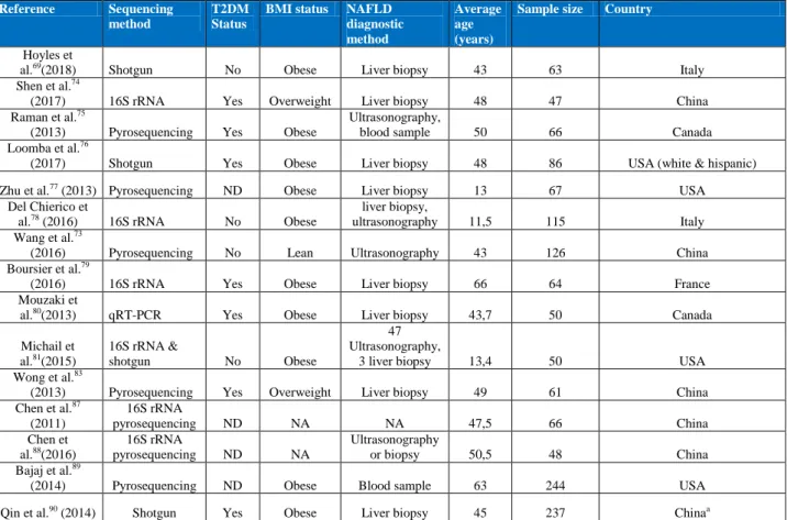
Owing to heterogeneity in the literature, we have focused on concordant results across studies and describe bacterial signatures associated with the different stages of liver disease severity in humans. We summarize results according to taxonomic levels (bacterial phylum, family, genus and species) that are associated with different NAFLD progression stages (steatosis, NASH), NAFLD-fibrosis, and
cirrhosis and show that some bacterial signatures overlap with those described in obesity or T2DM (Figure1 & 2).
[H3] Simple steatosis to NASH signatures. Comparing patients with NAFLD to
healthy individuals as controls69,73, a consistent altered signature is observed at the level of phylum (increased Proteobacteria69,74–76), family (increased Enterobacteriaceae74,77 and decreased Rikenellaceae77,78 and Ruminoccaceae 73–75), and genera (increased Escherichia69,77, Dorea75,78, Peptoniphilus77,78 and decreased Anaerosporobacter55,73, Coprococcus69,73,77, Eubacterium69,77, Faecalibacterium55,77
and Prevotella69,79). Although these initial results suggest a measurable dichotomy in microbial signatures between individuals with hepatic steatosis and controls, there are, however, large discrepancies found across studies with divergent results for phylum, family, genus and species69,73–81, as described in detail in Table 1 and 2.
Similarly to NAFLD, when comparing patients with NASH to healthy individuals as controls, using either liver biopsy42,69,74,76–81 or noninvasive biomarkers73,75,78,81, some concordant microbial signatures are observed, which also overlap with NAFLD signatures: phylum (increased Proteobacteria74–77), family (increased Enterobacteriaceae74,77 and decreased Ruminococcaceae73–75,77,82 and Rikenellaceae77,78) and genera (increased Dorea75,78 and decreased
Faecalibacterium77,82,83, Coprococcus73,77,82, Anaerosporobacter73,83). However, similar to results observed for steatosis, the abundance of some bacteria69,72–81,83 , display opposite trends across the literature as shown in Table 1 and 2.
[H3] From NAFLD-fibrosis to NASH-cirrhosis. Few studies have focused
microbial composition as a function of fibrosis progression. Nevertheless, concordant signatures are observed and detailed in Table 1 and 2. When compared with patients with advanced fibrosis, individuals with less severe liver alterations or healthy individuals as controls display decreased abundance of Gram-negative bacteria, decreased Fusobacteria phylum, increased Enterobacteriaceae family, (Bacteroides, Ruminococcus, and Shigella genera74,79 and by contrast increased76
Gram-positive bacteria, Firmicutes phylum, Prevotellaceae family and Prevotella genus.
One caution in interpreting the findings is that these studies used various experimental designs each comparing different stages of fibrosis severity (that is, comparison of patients with mild to moderate fibrosis (F0-F2) versus advanced fibrosis (F3-F4), or cirrhosis)76, whereas others compared patients with no to little fibrosis (F0–F1) to patients with moderate to advanced fibrosis (F≥2)74
. Finally, another study compared patients with moderate to advanced fibrosis (F≥2) with patients having mild fibrosis (F0–F1) with or without NASH79
. These differences in experimental design could explain discrepancies found in the proposed microbial signatures (Table 1 and 2). For example, Bacteroides vulgatus and Escherichia coli are the most abundant species in advanced fibrosis (F3-F4)76. B. vulgatus abundance is also increased from mild–moderate fibrosis to advanced fibrosis76
. Interestingly, the B. vulgatus signature is a common observation associated with metabolic alterations as it is also increased with increasing BMI, specifically in severe obesity, which is characterized by decreased microbial gene richness84. Furthermore, B. vulgatus abundance increases with increasing haemoglobin A1c (Hba1c) levels 84. B. vulgatus is also associated with insulin resistance85 and is decreased in obese women receiving prebiotics (inulin-type fructans), a treatment
that improves insulin sensitivity19. Likewise, E. coli has been shown to be increased in patients with T2DM86. These examples illustrate overlapping observations between bacterial signatures linked to NAFLD-fibrosis or NAFLD and those linked with metabolic disorders as obesity and diabetes (Figures 1 and 2). Although models have been proposed to use the microbiome as a reservoir for diagnostic signatures of NAFLD-fibrosis76, further confirmation in independent cohorts and across geographical regions are necessary to assess their clinical relevance.
In patients with cirrhosis (some of whom had ‘pure’ NASH-related cirrhosis, whereas others were of different aetiology such as viral hepatitis, as detailed later), metagenomic signatures are relatively consistent across studies76,87–90, confirming the importance of oral microbes invading the intestine in this disease. Taxa including
Prevotella88,90, Veillonella73,90 and Streptococcus87,90,91, being part of the oral cavity
bacterial ecosystem, seem to discriminate between patients with cirrhosis and healthy individuals. Likewise, whereas some signatures appear consistent when comparing patients with cirrhosis to healthy individuals as controls (decreased abundance of Lachnospiraceae87,89, Veillonella4,88, Prevotella4,88 and increased abundance of Enterobacteriaceae74,87,89,92), others display contradictory trends across studies76,87,88. In general, microbial signatures of cirrhosis are related to a drastic shift in taxa composition leading to an increase of pathogenic taxa and a decrease in taxa proposed to be metabolically beneficial89 (Table 1 and 2). A functional consequence of this taxa shift might be increased endotoxaemia. Indeed, it was demonstrated in mice that during HFD-induced NAFLD, there was a concomitant increase in LPS levels and changes in gut microbiota composition93,94. Furthermore, exacerbation of NAFLD into NASH was also associated with increased LPS levels94., and bacterial production of antibacterial peptides has been proposed to help maintain
intestinal barrier integrity89. Thus, combined effects of increased endotoxaemia, reduced butyrate production and reduced bile acid production (discussed further later) could worsen cirrhosis progression89. Another feature also associated with other common diseases is the reduction of levels of Faecalibacterium prausnitzii95 in
cirrhosis90,91. Indeed, F. prausnitzii, known to have anti-inflammatory properties and to be abundant in healthy conditions11,12, is reduced in abundance in a number of diseases, including intestinal disorders (inflammatory bowel disease96 or irritable bowel syndrome97), obesity11,12 and diabetes15,98.
However, the evaluation of gut microbiota contribution in liver disease progression (from steatosis to NASH, and NASH-cirrhosis) is limited and bacterial markers are frequently identified in one study, yet not confirmed in independent cohorts. As the origin of liver disease is heterogeneous by nature, most studies in cirrhosis have included patients with different aetiologies including hepatitis B87,90, biliary disease-related cirhosis88,90, alcohol-related cirrhosis87,90, NASH or a combination of different diseases. Patients were also included at different stages of disease severity with compensated cirrhosis [G] or decompensated cirrhosis [G]89,90. These differences could collectively explain why associated metagenomic signatures of liver disease progression are not frequently replicated. Nevertheless, a study exploring well-characterized patients with non-NAFLD, NAFLD without advanced fibrosis or NAFLD-cirrhosis provides a potentially promising diagnostic signature, which includes 27 bacterial features and 3 demographic characteristics (BMI, age and gender). This signature is able to robustly identify NAFLD-cirrhosis and was further confirmed in an independent cohort with an AUROC of 0.9291, which remained accurate after adjustment for T2DM. Although promising, this signature will need to be further confirmed in different cohorts including various ethnicities and
individuals from different geographical regions. Furthermore, to use these microbial signatures in clinical practice, validated assays will have to be developed to enable easy and reproducible diagnosis.
Although the literature provides initial information regarding gut bacterial groups as promising signatures of different stages of liver disease progression, whether the microbiota is a causal factor, which interacts with the complex pathophysiological processes driving disease from mild fibrosis to severe fibrosis74,76,79 and eventually cirrhosis still needs to be demonstrated.
[H1] Gut-derived metabolites and pathways
Studies have evaluated the metabolomic signatures associated with NAFLD or NAFLD-fibrosis and have been extensively reviewed99. Among these signatures are molecules produced by bacterial communities (Figure 3) such as LPS100, short-chain fatty acids such as butyrate, propionate and acetate (the balance of which mediates beneficial or detrimental effects on the liver)71 and products derived from bile acid metabolism acting on FXR within the liver or the intestine101–103 . Changes in these metabolites are suspected to have a role in the pathophysiology of liver injuries99. Herein, we choose to focus on novel studies exploring substrates of gut microbiota metabolism or circulating gut microbiota-derived metabolites. Importantly, all of these metabolites have also been demonstrated to be involved in obesity and metabolic alterations, including T2DM. For example, evidence from mice during obesity, LPS is increased and promotes the activation of insulin resistance pathways in tissues93. Although, SCFAs have beneficial effects on metabolic health, they are also involved in energy harvesting and, therefore, potentially contribute to increased weight gain104. Levels of SCFAs have been found to be increased in faecal samples from individuals
who are obese as compared with healthy individuals105. Overall, understanding their specific role in NAFLD physiopathology is complex.
[H3] Choline, betaine and circulating methylamines. Mice fed a
choline-deficient diet106 are recognized as a representative model of NAFLD107,108 and reducing dietary choline leads to both increased liver fat and gut bacteria modifications108. Choline is an essential nutrient and a component of phosphatidylcholine, which is a precursor of acetylcholine (neurotransmitter) found in food. Choline is a substrate that can be oxidized to betaine. HFD-induced NAFLD mice fed a diet with standard levels of choline exhibit a decrease in systemic phosphatidylcholine with increasing severity of NAFLD109. This observation was translated to humans in which patients with an increasing severity of NAFLD display a decreased ratio of betaine:choline110. As demonstrated in mice and humans111,112, dietary choline is metabolized by the gut microbiota into trimethylamine (TMA), which is further metabolized in the liver by the enzyme FMO3 and results in the production of TMAO17. Increased circulating TMAO is proposed as a biomarker of cardiovascular events and kidney dysfunction17,112,113, and the increase of circulating levels TMAO positively correlates with the increase of Deferribacteres and
Tenericutes in the gut in mice113. In humans, elevated levels of TMAO were seen in individuals with prevotella enterotype, and several OTUs were significantly increased in patients with higher concentrations of TMAO [Au: in humans?] 113. Mouse studies
demonstrate that increased NAFLD severity is associated with increased urinary levels of both TMA and TMAO109. These observations can be seen as paradoxical as increased consumption of choline and phosphatidylcholine correlates with increased production of TMA and TMAO111,112 . The pathophysiological explanation by which TMA and/or TMAO has a role in NAFLD development therefore needs further
examination114, but the proposed mechanisms include a reduction of host choline bioavailability due to a switch in microbiota metabolism to methylamine production as well as urinary excretion114. In human studies, TMAO is independently associated with increasing severity of NAFLD when comparing patients with NAFLD to healthy individuals110.
[H3] TMAO and bile acids. Another major function of the gut microbiota is the
deconjugation of primary bile acids into secondary bile acids. Overall, primary bile acids are involved in cholesterol metabolism, facilitate the absorption of dietary fat and fat-soluble molecules, and have a role in regulatory pathways115. Primary and secondary bile acids have endocrine functions and modulate numerous host metabolic pathways through different receptors116. Secondary bile acids are notably preferential ligands of the G protein-coupled bile acid receptor-1 (TGR5) a key actor of energy, glucose and lipid metabolism in the host103. The gut microbiota not only regulates secondary bile acid metabolism but also inhibits the liver synthesis of lipids by alleviating FXR inhibition117. Differences in bile acid pool size and composition has been associated with metabolic diseases118. Thus, gut dysbiosis could influence bile acid pool, composition and homeostasis. Evidence from mice and humans suggests that bile acid bioconversion by the gut microbiota (deconjugation, dehydrogenation and dehydroxylation) is related to NAFLD and NASH progression119, as previously reviewed101. Interestingly, a decrease in bile acids could be associated with NAFLD through TMAO production since TMAO induces a decrease in the total bile acid pool by inhibiting two key enzymes involved in bile acid metabolism: CYP7A1 and CYP27A1110,113,120. In agreement with this hypothesis, patients with advanced cirrhosis exhibit a decreased conversion of bile acids with concomitant modifications
of their microbiota composition, including higher Enterobacteriaceae but lower Lachonospiraceae, Ruminococcaceae and Blautia abundance 121,122.
[H3] 3-(4-hydroxyphenyl) lactate. A study published in 2019 has shown that
3-(4-hydroxyphenyl) lactate is associated with increased severity of NAFLD-fibrosis both in a test and validation cohort, both comprising 156 individuals (one from the Twin and family study, the other from an independent prospective study)123 (Figure
3). Interestingly, 3-(4-hydroxyphenyl) lactate is a gut microbiota-derived product of
aromatic amino acid metabolism. These results are in line with another study performed in patients with different stages of steatosis, which showed decreased microbial gene richness and an alteration in aromatic amino acid and branched-chain amino acid metabolism in steatosis69. This metabolite could be used as non-invasive biomarker of NAFLD, but needs further confirmation.
[H3] Ethanol. Production of ethanol by the gut microbiota could also play a
part in NAFLD physiopathology. In children, the gut microbiota of individuals with NAFLD exhibits increased abundance of ethanol-producing bacteria as compared with those who were obese or healthy children as controls77. In the absence of ethanol consumption, adults with NASH display increased breath ethanol concentrations124, which could be attributed to those with NAFLD producing more gut microbiota-derived ethanol as compared with healthy controls. These results suggest that gut microbiota ethanol production might serve as a liver toxin contributing to the development of NAFLD and its progression towards NASH125. A study performed in mice and further validated in humans, indeed displayed that some bacteria (namely
Klebsiella pneumoniae) were able to produce ethanol from glucose, in the absence of
[H3] Short-chain fatty acids. Short-chain fatty acids (SCFAs), a group comprised of butyrate, acetate and propionate, are locally produced in the colon through microbial fermentation of normally non-digestible complex carbohydrates (dietary fiber)99,127. Their role and mechanism of action in NAFLD development has been extensively reviewed127,128. Microbially produced SCFAs are absorbed primarily through diffusion or co-transport in the colon whereas their intestinal signalling effects are mediated by activation of G-protein-coupled receptors (GPR41 and GPR43)129. SCFAs have been proposed as an important substrate to increase liver triglyceride levels and promote energy storage and weight gain130 as SCFAs are involved in fatty acid synthesis and gluconeogenesis131. Human studies comparing NAFLD, NASH and healthy individuals as controls have observed an increased faecal concentration of SCFAs in patients with NAFLD and/or NASH 132 concomitantly with an increase in abundance of bacterial groups involved in their production. Furthermore, this increased faecal SCFA and microbial signature observed during NASH132 are associated with reduced numbers of resting regulatory T-cells (rTregs) (CD4+CD45RA+CD25+) and higher Th17:rTreg ratio in peripheral blood, which are systemic immunological features previously observed in NASH133 .
Nevertheless, SCFA action is quite complex, as they also can provide metabolic benefits. GRP43 activation by SCFAs reduces pro-inflammatory production and immune cell (T cells)134 infiltration, whereas GRP43-/- mice or germ-free mice with lower levels of SCFA display increased inflammation both at the level of circulating immune cells as well as in the colon , a feature usually seen in NASH135. However, inflammation during NASH development was not assessed in these models and the effect of SCFAs on liver inflammation needs further investigation.
Furthermore, although dietary fibres have been shown to be beneficial for metabolic health10, some interventional studies using soluble fibres have, in contrast, led to increased liver disease in mice with genetically-induced or high fat diet-induced microbial dysbiosis136,137. Thus, although soluble fibres can induce positive metabolic effects, such as those observed on glucose metabolism, the effects of soluble fibre supplementation in NASH mouse models need further exploration before interventional studies in patients. Moreover, due to interindividual variability in the gut microbiota, dietary supplementation of fibre might need to be personalized because of possible different effects in different individuals, which could also be further complicated due to the variations of soluble fibres available. Most importantly, each SCFA exerts specific and somehow different metabolic effects. Thus, assessing their balance both at the fecal and systemic level in patients with NASH and after a dietary intervention would probably decipher more precisely their overall role in NAFLD development, exacerbation or improvement. Finally, it was reported that SCFAs could have a beneficial role in NAFLD through epigenetic modulation via histone deacetylase (HDACs) inhibition. Indeed, it was shown in rats that histone deacetylase (HDACs) inhibition decreased liver gene expression involved in NAFLD mostly lipogenic genes, such as acetyl-CoA carboxylase (Acc), fatty acid synthase (Fasn), and sterol regulatory element binding protein 1c (Srebp1c)138 . This finding provides important mechanistic insights, reviewed elsewhere127,128.
New bacterial metabolites and derived factors will be identified as contributors to liver disease in the future. Thus, it will be essential to determine how these factors, together with changes in LPS, biliary acid metabolism and SCFAs, contribute to NAFLD development and progression. Investigations are now needed to determine
the relevance of these molecules as biomarkers and/or predictors for diagnosis of NAFLD and NASH and its progression.
[H1] Issues in NAFLD metagenomics studies
The number of clinical studies investigating gut microbiota signatures associated with NAFLD and/or NASH or fibrosis is increasing rapidly. However, careful interpretation is needed when reviewing the literature due to the heterogeneous cohorts used across studies with differences in sex, ethnicity, liver disease severity stages, BMI, presence of T2DM, patient populations (paediatric or adult), corpulence, and other associated metabolic diseases. The gut microbiota sequencing technologies used vary among studies, and other critical factors influencing the gut microbiota, such as dietary consumption or drug intake are scarcely measured or under-reported.
[H3] Population variability in demographic characteristics. Although there
is some consistency in the literature for microbiome signatures, there is a noticeable lack of reproduced findings or confirmation in independent cohorts. This heterogeneity might originate from different methodological approaches and from major inter-individual variability among recruited patients regarding particular demographic characteristics. While one study examined only women69, most studies examined adults69,73–76,79,80,83 and children77,78,81 of both sexes. Yet some studies have controlled for sex69,73–75,80,110 and most have also controlled for age69,73– 77,79,90,110
. Nevertheless, it is important to note that there are histological specificities for adolescent and adult NAFLD139,140, as recalled in recent pediatric clinical guidelines 141. For example, in paediatric NASH, the ballooning degeneration, classic zone 3 fibrosis, and parenchymal inflammation often seen in adult NASH are less
common in children NASH140,141. Furthermore, several histological types are found in children: type 1 resembling the adult form, type 2 NASH is mainly characterized as NASH yet with no or minimal ballooning degeneration, and finally a third type with with overlapping features139. Studies were also performed in different geographical regions (North America, Canada75,80 and USA76,81), Asia (China73,83), Europe (France79, Italy and Spain69) probably with various ethnic backgrounds and cultural and food habits. Importantly, ethnicity seems to strongly influence microbial composition even in individuals living in the same geographical area. For example, gut microbial diversity differed substantially in different ethnic groups all living in the Netherlands and ethnicity accounted for a major part of these differences 142. Thus, whereas there are different liver disease risks across ethnicities143, it might translate into differential microbiome-related signatures142. Notably, some studies controlled for dietary habits73,74,77,80,81,110, which could relate, in part, to cultural differences.
[H3] Population variability in corpulence. Obesity, and particularly
abdominal obesity, is a well-known risk factor for NAFLD144, making these diseases strongly inter-dependent. In published reports, although some studies included lean individuals73,74,83, others examined individuals who were overweight74,83 or obese (mixing different classes of obesity)69,75,76,78–81,87. Several studies even further stratify by the severity of obesity classes (class I: BMI 30-35kg/m² 75,77,79), one study in particular focused on severe or morbid obesity (BMI> 35 or BMI >40 69), and it has been demonstrated that gut microbiota dysbiosis is exacerbated with increasing obesity severity11,84.
Metagenomic studies clearly demonstrate relationships between corpulence and gut microbiome changes. Microbial gene richness, for example, strongly
decreases with increasing BMI11,12,84. Furthermore, although individuals with obesity generally share common bacterial signatures and modified functional properties, some signatures differ across the obesity spectrum11, with peculiar signatures only found in populations with extreme BMIs (above 40kg/m²)84. On the basis of these findings, microbiome-related signatures in studies comparing patients with NASH with different degrees of obesity and lean or overweight patients with NAFLD as controls80 or patients with different classes of obesity and NASH and healthy lean individual as controls74 might be overestimated and mostly related to the degree of obesity145. The link between closely associated metabolic disorders, such as obesity and NAFLD is likely to be more complex than compositional shifts in bacterial composition alone. Thus, it remains difficult to conclude whether these signatures are solely related to liver alterations, BMI or both. In one study, Shen et al.74 did not find any statistically significant shift in gut microbiota, when comparing patients with NAFLD stratified by BMI. Patient groups were, however, of small size (n=47) and the study had limited power to definitively conclude the absence of NAFLD–NASH specific microbial signatures according to corpulence. By contrast, Wang et al. focused their analytical comparisons between NAFLD and healthy individuals with normal and comparable BMI73. They demonstrate a microbiome signature of patients with NAFLD independently of corpulence differences. Finally, to limit bias owing to differences in corpulence, studies exploring NAFLD microbiome signature have adjusted their statistical analysis on BMI (for example, according to the studies, the preformed partial Spearman’s rank-based correlation (pSRC) coefficients adjusted on BMI, or linear regression adjusted for BMI, multivariate models, analysis of covariance (ANCOVA) or finally logistic regression analysis)69,73,74,76,79,80,82,89,110.
Despite the described interactions between corpulence and liver disorders (from steatosis to cirrhosis considering individuals who are lean or obese), Enterobacteriaceae is consistently increased in both individuals with NAFLD and those who are obese in numerous studies74,77,87,89,146,147. Decreased microbial gene richness is found both in individuals with obesity11,12 and patients with NAFLD in some studies74,91, but also in lean individuals with NAFLD73. More importantly, BMI is a proxy of obesity and future studies should extend phenotyping to other measures related to body fat amount and distribution (for example, abdominal versus gynoid distribution) [G] to examine the interplay between fat distribution, different stages of liver alterations and microbiome alterations. Indeed, microbial gene richness is negatively correlated with increasing visceral fat deposition84.
[H3] Population variability in metabolic diseases and related treatments.
Another major confounder lies in obesity-associated metabolic comorbidities, which are also involved in NAFLD physiopathology. Diabetes is a major risk factor involved in NAFLD development 148 and its presence strongly exacerbates NAFLD to overt NASH, including forms associated with mild and advanced fibrosis149. Metabolic syndrome15 or T2DM14,15,85 per se are known to be associated with microbial signatures. Thus, it might be tricky to disentangle microbial signatures from NAFLD and linked metabolic disorders, such as diabetes. Four NAFLD studies74–76,79 included patients with T2DM and two other studies evaluated the signature and predicted function of the gut microbiome of patients with T2DM without known NAFLD14,15. Despite the difference in geographical demography (European women with diabetes15 and Chinese adults with diabetes14,15), these studies showed one consensus finding: a statistically significant reduction in abundance of
butyrate-producing bacteria in T2DM. However, they did not examine whether patients with T2DM had NAFLD; extrapolating from epidemiological evidence, most probably, those patients had both NAFLD and T2DM. Clostridia73,150 and Lactobaccillus15,75 are two common species signatures found in both T2DM and NAFLD (increased abundance of Lactobacillus and decreased abundances of Clostridia in both patients with NAFLD and those with T2DM compared with healthy groups). At a predicted functional level, both patients with T2DM and those with NAFLD display consistent decrease in butyrate-producing bacteria15,73,77. E. coli is consistently enriched in studies exploring either solely cirrhosis or solely diabetes 14,90. By contrast, the
Roseburia genus shows opposite trends across studies15,73,75,77 (Table 2). However, one study included only six individuals with T2DM among their NAFLD cohort75, potentially explaining these discrepant results since Roseburia is also known to be
decreased in T2DM15. Importantly, some studies looking for a NAFLD microbial signature actually controlled their analysis for the presence of T2DM69,73,76,79,90 to limit this bias. For example, microorganisms signature remained associated with NAFLD after proper adjustments for T2DM but results were not always replicated in all studies (for example, Propionibacterium acnes69, Bacteroides fragilis69, Anaerosporobacter73, Enterobacteriaceae79) In studies including patients without T2DM, only one controlled for insulin resistance82.
Medication use is another critical feature influencing microbiome signature variability among individuals151,152. Studies exploring microbiome signatures of NAFLD74–76,79,80 included patients with T2DM whereas others excluded them73,77,78,81,83, but most do not clearly state the list of current medications that study participants are taking. Only three studies evaluating microbial signature during NAFLD have controlled for medication use69,73,76. Metformin is the first line of
pharmaceutical therapy prescribed in T2DM and is commonly used in NAFLD since 50–75% of NAFLD patients have T2DM according to population and ethnicities153,154.The effect of metformin on the gut microbiome is well-documented86,155,156. Metformin increases the abundance of Akkermanisa
muciniphila155,156, a bacteria associated with improved insulin sensitivity in mice and humans18,157,158. Furthermore, treating mice on a HFD with metformin recapitulates the improvement in insulin sensitivity and switches the microbiota composition towards that of mice on chow diet156. Patients with T2DM taking metformin display a specific microbiome signature with an increase in Escherichia species86, which could explain, in part, the increased E. coli found in patients with NAFLD-fibrosis69,76,77 . Owing to increased risk of cardiovascular events, statins are frequently prescribed to patients with T2DM and NAFLD–NASH and could strongly influence the gut microbiome159,160. A study in mice observed a reduction in microbial diversity upon statin treatment, a modification in bile acid pools and a reduction of SCFA producing bacteria which was also confirmed in humans160. Nevertheless, those findings need further confirmation in larger-scaled studies. Proton pump inhibitors, which are frequently given to patients with cirrhosis, have been shown to switch microbiome composition towards an increased abundance of oral bacteria in the gut microbiota 161, thus the ‘oral microbiome’ signature observed in cirrhosis could well be related to drug intake rather than disease. It remains critical in microbiome studies examining patients with NAFLD or NASH to collect information on diabetes history and drug intake. Although some studies have controlled for medication, no study has yet controlled for each of the above potential biases when looking for a microbial signature of NAFLD.
[H3] Variability in liver injury diagnostic methods. Another source of variability in
the reviewed studies is the different grades and stages and heterogeneity of liver disease alterations (steatosis, NASH and fibrosis)75,79,81. Even if the use of the SAF score seems to improve inter-observer variability, the inter-individual pathologist variation when examining biopsy samples is acknowledged in NAFLD diagnosis162,163. As liver lesions are closely intertwined, deciphering the specific microbial signatures of each histological lesion is challenging. Studies have focused on different stages of disease progression. One study focused on the steatosis state69, three on NASH77,81,83, and seven investigated the NAFLD spectrum (from steatosis to NASH)73–76,78–80. Two reports focused on different fibrosis stage74,79 and three on cirrhosis87,89,90, whereas one study investigated a larger disease spectrum of fibrosis to cirrhosis76. However, in the latter study, patients with fibrosis probably displayed concomitant lesions of NASH and/or steatosis. As most studies do not focus on the same stages of disease progression, it is rather challenging to underline concordant microbial signatures.
The diagnostic method used to classify NAFLD lesions is also an important factor to consider when examining these studies. Liver biopsy, the most reliable diagnostic method and currently considered as the gold standard, was used in nine studies69,74,76,77,79,80,83,88,90. However, others used less reliable and noninvasive tools such as ultrasonography, MRI or blood tests73,75,78,81 (such as liver enzymes (alanine aminotransferase, aspartate aminotransferase and γ-glutamyl transferase)73,75,78,81, and also other metabolism-related biomarkers (levels of fasting serum glucose73,78, insulin78, triglyceride78 or complete lipid profile73), or liver-related biomarkers (albumin and platelet count 73) (Box 2). These noninvasive tools are designed to specifically characterize one histological aspect, it might well be that these patients displayed
heterogeneous lesions (steatosis and some degree of fibrosis) that were not investigated. Furthermore, the degree of lesions severity is variably expressed. For example, some studies considered simple steatosis78, whereas other took into account the steatosis score and discriminated steatosis severity from S0 to S369. These aspects are critical and could partly explain disparities found in clinical studies exploring microbiome signatures of NAFLD and liver disease progression. In addition to placing a priority on using liver biopsy when possible, diagnostic tools and the interpretation of their results used to assess liver alterations should be taken into consideration when comparing across the literature.
[H3] Bias due to circadian rhythm. Another overlooked factor in the literature
examining gut microbiota and NAFLD is the contribution of circadian rhythm. Circadian rhythm is a well-known and pivotal regulator of liver metabolic pathways, which are altered in NAFLD development164. For example, jetlag (seen as a perturbation of the circadian clock) is associated with the worsening of metabolic alterations (that is, the hallmarks of NAFLD), which show further perturbations during obesity165. Animal studies and experimental models have shown that feeding time also influences the circadian rhythm and, subsequently, host physiology166. Some human population cohorts confirmed those findings167. Studies performed in different mouse models (genetic invalidation of clock genes, antibiotic treated or conventional mice with or without modification of the light–dark phases) have shown that the gut microbiota displays rhythmic oscillations in the colon during the day in terms of proliferation, composition, functions and metabolite production168–170 and depend upon function of the host circadian clock. These oscillations (that is, existence of time-of-day-specific profiles of microbiota functionality) were demonstrated to be
controlled by host feeding intake. Interestingly, modifying feeding times results in a shift of cycling bacteria. Moreover, timed feeding can restore the loss of fluctuations in circadian clock deficient mice169 . Perturbations of the circadian rhythm are also associated with metabolic impairment and microbiota dysbiosis, both in experimental models as well as in humans169. On the other hand, the gut microbiota and its circadian oscillations also influence the rhythmic expression of host intestinal and liver genes that are not known to be involved in the circadian clock168. Additionally, these gene expression patterns are modified in the absence of gut microbiota as genes from several metabolic pathways lose their oscillatory patterns, whereas genes from other metabolic pathways gain rhythmicity with the lack of microbiota in animal models168. As such, germ-free mice submitted to light–dark cycle display impaired circadian hepatic gene expression168, demonstrating the role of gut microbiota in the circadian clock effects. Thus, gut microbiota changes could influence the rhythmicity of several host metabolic pathways contributing to NAFLD physiopathology. This aspect could be due to microbiota functions rather than composition as gut microbiota-produced metabolites from the diet can also modulate liver clock gene expression, observed when treating hepatic organoids with different SCFA170. Furthermore, in clock gene knockout models, modulating feeding time can rescue abrogated host gene expression oscillation170. These observations highlight the combined contribution of both circadian clock and diet acting on gut microbiota and host physiology. Whether these results obtained in mice models are relevant in human needs further investigation. This aspect also raises the question if the methodology of studies examining gut microbiota signatures in NAFLD need to consider patients’ circadian clock phenotype and food intake rhythm, which could help explain some discrepancies in gut microbial signatures observed across studies.
[H3] Sequencing methods. Another important point to be considered when
comparing different results across the literature is the methods used to sequence and analyze the gut microbiota. Furthermore, sampling (home faeces auto-collection use different devices that have not all been evaluated, one complete bowel movement stool or a sample of that material, mucosal or luminal microbiota in studies including surgery sampling, faeces versus caecal or other parts of the intestine in mouse experiments) itself as well as the specific steps of analysis, including steps before sequencing (i.e. sample concentration, lysis, purification and extraction), also need to be harmonized to enable comparisons between studies as reviewed herein171. Studies used a variety of methods such as quantitative PCR80, 16S ribosomal RNA (rRNA) sequencing 74,78,79, or shotgun sequencing [G] 69,76,90. It has been previously shown that Sanger, Roche 454, or Illumina-based 16S rRNA or shotgun sequencing can lead to different results172. The results from 16S rRNA sequencing are less granular and accurate than those from whole shotgun sequencing, because the 16S approach sequences a single region of the bacterial genome whereas shotgun sequences the complete genome173. Species prediction is not optimal using 16S rRNA sequencing171, and the results derived from pyrosequencing frequently lack numerous species because of the choice of tagged-primers174. Shotgun sequencing produces extended information regarding read sequences, since it can sequence and amplify the complete genome. Shotgun sequencing, therefore, produces more information as it also includes unknown metagenomic species, which potentially lead to increased discovery potential. Metagenomic sequencing enables the prediction of functional potential based on gene and species annotations.
In addition to the sequencing technology, the bioinformatic pipelines used to analyze sequencing data also contributes to the variability of results. Taxonomic analyses in 16S rRNA sequencing is easier due to established standard pipelines such as QIIME175 and Mothur176 and functional profiling in 16S rRNA is predominantly done with PICRUST177. However, until now, few studies precisely describe the microbial species that could be used as indicators of liver disease status76,90 and even fewer provide clues for the discovery of new species. Although microbiota catalogues are currently quite exhaustive, the variability of sequencing method and sequencing depth might influence findings. The use of whole metagenome sequencing can provide this information, but requires pipeline harmonization and dedicated bioinformatics expertise178.
The lack of power in statistical analysis is another issue regarding the reliability of microbial biomarkers that can be used with confidence. Uncertainty about the absence or presence of certain aspects of NAFLD and unbalanced population distributions might affect statistical results. Obtaining a validated healthy and liver-biopsied group is difficult owing to obvious ethical reasons. Indeed, in other fields such as oncology, ethical concerns have already been raised about research biopsy and their potential risk to harm participants as well as the adequacy of voluntary informed consent 179,180.Thus, this leads to a lack of statistical power and phenotypic uncertainty because of limitations in non-invasive testing. In addition to finding microbial abundance differences between groups, P values are often used and interpreted as whether the abundance difference is statistically significant or not. However, P values alone do not provide reliable results. Some studies perform only P value tests without False Discovery Rate (FDR) correction77,79,80,83,87,89. By contrast, nine studies used FDR or Bonferroni corrections69,73–76,78,81,88,90. For example, in
Mexican children with obesity, B. plebeius is negatively correlated with BMI percentile, but this finding is no longer statistically significant after FDR correction181. FDR should be mandatory when exploring metagenomics signatures. Importantly though, Falony et al. estimated that a sample size of approximately 1,700 individuals would be needed to adequately assess the relationship between obesity, NAFLD and microbiota composition in a cohort study when correcting for age, gender, ethnicity and other variables151, which questions the feasibility to actually achieve such a trial.
[H3] Multi-omics approaches. As a complement to gut metagenomics, additional
‘omics’ data and systems biology approaches are a good method to further confirm microbial signature for a specific disease. By using both metagenomics and metabolomics182 or lipidomics183,184 , the gut microbiome compositional and functional signatures can be characterized and further linked with concentrations or production of gut-derived metabolites in blood, urine or faeces. An additional approach could also be to combine metagenomics to metatranscriptomics data, bringing some clarity to the gut microbiota genes specifically activated and providing further functional insights. Such approaches have already been used in other metabolic diseases (such as T2DM or obesity84,185) and are emerging in the field of NAFLD69. The use of approaches combing multi-omics techniques, coupled with computational science, will probably enable better insights regarding microbiota contribution in the pathophysiological pathways involved in these metabolic diseases and help stratify patients based on their multi-omics profiles. However, it does not preclude researchers from designing correct clinical trials in particular choosing carefully the best control groups.
[H1] Conclusions
Clinical studies have revealed gut microbiome signatures in NAFLD, NAFLD-fibrosis and cirrhosis, which could serve as future noninvasive diagnostic biomarkers for liver disease diagnosis or evolution prognosis. Nevertheless, the relevance of identified signatures needs to be further examined in longitudinal studies where physicians can prospectively examine the deterioration of liver status. This strategy was conducted for gut microbiota-derived factors in Crohn’s disease186 and in the cardiovascular field111. However, discrepancies are nevertheless observed across studies in NAFLD, which might originate from their large heterogeneity in terms of microbiome sequencing method, bioinformatic pipelines, liver diagnostic method and disease severity spectrum, as well as clinical and demographic characteristics. Obesity and T2DM are associated with strong microbiota dysbiosis and identified liver disease microbiome signatures are often biased by the additional presence of obesity and/or diabetes status, which are not always accounted for. There is, therefore, an urgent need for more investigations with strong study designs (Box 3). Studies should account for confounding metabolic disorders, such as T2DM and obesity, population background, medication and dietary intake. An option could also be to additionally examine identified-signatures in well-selected and clinically harmonized cohorts of patients with either T2DM or obesity. However, considering some potential confounding factors, such as circadian rhythm, might prove to be a complicated task for both researchers and patients. Future studies should also consider investigating gut microbiome signatures in a two-step manner; that is discovery and validation cohorts using varied and documented ethnic backgrounds and include patients with a biopsy-proven NAFLD and/or NASH diagnosis. Where possible, repeated exploration with a second longitudinal study incorporating liver biopsy should also be considered
to confirm clinical evolution. Overall, whether optimal study design is feasible remains an open question and in any case should be considered in the context of large-scale research consortia. Moreover, the understandable reluctance from physicians and patients, as well as ethical committees, to repeat liver biopsy is also a limitation. Finally, advanced methods in predicting microbiome signature (such as deep learning combined with multi-omics approach) are needed in this field. Exploring the whole microbial ecosystem in which the interaction of microorganisms could be as important as the abundance of single or multiple family, genus or species is a priority. Similarly, the importance of microbiome-altering factors, such as phages or viruses and fungi is worth examining. Combining microbiome signatures with systemic microbial-derived metabolites could help in the future to diagnose patients with liver alterations in routine care. The potential establishment of reliable biomarkers will determine how future NAFLD–NASH treatments modulate these signatures to develop biomarkers enabling follow-up for therapeutic response.
Highlighted references :
- Bedossa, P. et al. Histopathological algorithm and scoring system for evaluation of liver lesions in morbidly obese patients. Hepatology 56, 1751–1759 (2012). - This work proposes a novel algorithm to classify patients with no
NAFLD, NAFLD or overt NASH that is more robust than previous ones, and since its development has been used in many studies.
-
- Hoyles, L. et al. Molecular phenomics and metagenomics of hepatic steatosis in non-diabetic obese women. Nature Medicine 24, 1070–1080 (2018).
- This work reveals molecular networks linking the gut microbiome (using
metagenomic) and the host phenome (hepatic transcriptome as well as urine and plasma metabolome) to hepatic steatosis.
- Shen, F. et al. Gut microbiota dysbiosis in patients with non-alcoholic fatty liver disease. HBPD INT 16, 375–381 (2017).
- This work is one of the first to use the Hiseq 2000 platform to sequence
the microbiome and microbiota discover signature of NAFLD (biopsy-proven) as compared to healthy controls in a Chinese cohort.
-
- Loomba, R. et al. Gut microbiome based metagenomic signature for non-invasive detection of advanced fibrosis in human nonalcoholic fatty liver disease. Cell metabolism 25, 1054-1062.e5 (2017).
- This work offers a first microbiota signature of NAFLD-related fibrosis
severity using whole-genome shotgun to sequence microbiome in patients with biopsy-proven NASH and fibrosis.
- Del Chierico, F. et al. Gut microbiota profiling of pediatric nonalcoholic fatty liver disease and obese patients unveiled by an integrated meta-omics-based approach. Hepatology 65, 451–464 (2017).
- This work offers a microbial signature of NAFLD–NASH in children and
uses several control groups (one obese without NAFLD and one healthy control group).
- Bajaj, J. S. et al. Altered profile of human gut microbiome is associated with cirrhosis and its complications. J. Hepatol. 60, 940–947 (2014).
- This work addresses the gut microbial signature of patients with
cirrhosis compared with healthy individuls, then addresses whether this signature is stable over time in compensated cirrhosis as well as further asseses the changes in patients undergoing decompensated cirrhosis.
- Qin, N. et al. Alterations of the human gut microbiome in liver cirrhosis. Nature
513, 59–64 (2014).
This work was the first study to offer a microbial signature of liver cirrhosis in adults, comparing 98 patients to 83 healthy individuals using quantitative metagenomics.
- Wu, H. et al. Metformin alters the gut microbiome of individuals with treatment-naive type 2 diabetes, contributing to the therapeutic effects of the drug. Nat.
Med. 23, 850–858 (2017).
- This work was the first study to decipher the effect of metformin on gut
microbiota signature in a randomized control trial, including individuals with drug-naïve T2DM, using metagenomic analysis and gut stimulator experiments with faeces transfer in germ-free mice.
1. Fouhy, F., Ross, R. P., Fitzgerald, G. F., Stanton, C. & Cotter, P. D. Composition of the early intestinal microbiota: knowledge, knowledge gaps and the use of high-throughput sequencing to address these gaps. Gut Microbes 3, 203–220 (2012).
2. Prakash, S., Tomaro-Duchesneau, C., Saha, S. & Cantor, A. The gut microbiota and human health with an emphasis on the use of microencapsulated bacterial cells. J. Biomed. Biotechnol. 2011, 981214 (2011).
3. Fraher, M. H., O’Toole, P. W. & Quigley, E. M. M. Techniques used to characterize the gut microbiota: a guide for the clinician. Nat Rev Gastroenterol Hepatol 9, 312–322 (2012). 4. Qin, J. et al. A human gut microbial gene catalogue established by metagenomic sequencing.
Nature 464, 59–65 (2010).
5. Li, J. et al. An integrated catalog of reference genes in the human gut microbiome. Nat. Biotechnol. 32, 834–841 (2014).
6. Karlsson, F., Tremaroli, V., Nielsen, J. & Bäckhed, F. Assessing the human gut microbiota in metabolic diseases. Diabetes 62, 3341–3349 (2013).
7. Lynch, S. V. & Pedersen, O. The Human Intestinal Microbiome in Health and Disease. N. Engl. J. Med. 375, 2369–2379 (2016).
8. Bäckhed, F. et al. The gut microbiota as an environmental factor that regulates fat storage. Proc. Natl. Acad. Sci. U.S.A. 101, 15718–15723 (2004).
9. Ridaura, V. K. et al. Gut microbiota from twins discordant for obesity modulate metabolism in mice. Science 341, 1241214 (2013).
10. Cani, P. D. et al. Changes in gut microbiota control metabolic endotoxemia-induced
inflammation in high-fat diet-induced obesity and diabetes in mice. Diabetes 57, 1470–1481 (2008).
11. Le Chatelier, E. et al. Richness of human gut microbiome correlates with metabolic markers. Nature 500, 541 (2013).