692
TRANSACTIONS OF THE ROYAL SOCIETY OF TROPICAL. MEDICINE AND HYGIENE, VOL. 76, No. 5, 1982
Comparative
morphometric
analysis of bloodstream
and lymph forms
of Trypanosoma
(T.) brucei brucei grown
in vitro and in vivo
H. HECKER AND R. BRUN
Swiss Tropical Institute, Socinstrasse 57, CH40.51 Base& Switzerland
Summary
The fine structure of bloodstream forms of Typa- nosoma brucei cultivated in vitro, and of trypanosomes from lymph and blood of mammalian hosts, was compared morphometrically. The cell volume, quan- titative parameters of the mitochondrion and of glycosomes were mainly investigated. A Coulter Channelyzer was used for the first time to measure the mean cell volume of living parasites. In vitro, the monomorphic trypanosomes between the feeder layer cells showed lower values for mitochondrial para- meters than the slightly pleomorphic forms from the supernatant medium. Trypanosomes in culture were very similar morphologically to forms from lymph nodes of rats. Despite some morphometric differences between cultivated blood stream forms and those grown in viva, the similarity of both populations was clear. Both populations, however, differed significant- ly from stages found in the vector or from procyclic culture forms.
Introduction
Bloodstream forms of pleomorphic, tsetse fly trans- missible stocks of Typanosoma (T.) brucei (including human pathogenic stocks) can be grown continuously in co-cultivation with fibroblast-like feeder layer cells in Minimum Essential Medium supplemented with 15% serum (BRUN et aE., 1981). These investigators showed that several feeder layer/serum combinations supported continuous growth of bloodstream forms with retention of infectivity for mammalian hosts as well as for the tsetse fly (SCHONI et al., 1982). The surface coat, another characteristic feature of the mammalian stage, could also be demonstrated. Two different trypanosome populations were described from these cultures: one in the supernatant medium exhibiting slight pleomorphism and a second mono- morphic one intercellularly between feeder layer cells resembling slender bloodstream forms.
The present study aimed to analyse quantitatively the ultrastructure of in vitro cultivated bloodstream forms in comparison to bloodstream and lymph forms from mammalian hosts and to find out if the difference observed between the two culture popula- tions could be confirmed by stereological methods. In addition, we were interested to see how direct measurement of the cell volume, using a Coulter Channelyzer would compare to the indirect method previously employed, which is based on the measure- ment of nuclear volume from smears and the stereo- logically assessed nuclear-cytoplasmic volume ratio (HECKER et al., 1972).
Materials and Methods Typamsome stock
Trypanosoma brucei brucei STIB 247, a pleomorphic stock-which is readily transmitted by tsetse flies,-was used for all exneriments. This stock was isolated in the Serengeti Rational Park from a hartebeest (Alce- laphus buselaphus) and cryopreserved in liquid N2 after one rat passage.
Culture system
Bloodstream forms were grown in a system consist- ing of a feeder layer in Minimum Essential Medium (MEM) supplemented with 15% inactivated rabbit serum. The fibroblast-like cell line MEF was isolated from whole embryos of a vole, Microtus montanus.
Isolation of the cell line, composition of the culture medium and maintenance of cultures were described elsewhere (BRUN et aE., 1981). The rabbit serum was prepared in our laboratory from blood of New Zealand White rabbits.
Cultures were initiated by adding metacyclic forms obtained from infected tsetse flies (G&s& m. morsituns) by salivation, to wells of 24-well tissue culture clusters containing a confluent MEF feeder layer and I.0 ml medium. The cultures were incu- bated at 36.X in 4% COI-96% air in a humidified incubator.
Isolation of typanosomes from culture
Trypanosomes growing in the supernatant medium were pelleted by centrifugation for 7 min at 1000 g, resuspended in PSG buffer, pH 8, and passed through a DEAE cellulose column (LANHAM, 1968). For the isolation of trypanosomes growing intercellularly the feeder layer had to be mechanically detached and the cells separated from one another by passing them several times through a Pasteur pipette. The tissue culture cells were allowed to settle and the super- natant, containing trypanosomes, was centrifuged for 7 min at 1000 g. The pellet was resuspended in PSG and passed again through a DEAE cellulose column. The trypanosome suspension was subsequently used to make smears, for size distribution analysis and for electron microscopy.
Isolation of typanosomes from laboratory animals
The mice used were female Swiss ICR (26-28 g) and the rats were female white SIV (130 gj. Blood: stream forms from mice and rats were isolated from titrated blood taken from the heart of an ether anaesthetized animal. The blood was diluted with an eaual volume of MEM and centrifueed for 15 min at 46 g to separate blood cells from trypanosomes. The supernatant was removed to another tube and the
H. HECKBR AND R. BRUN 693
trypanosomes pelleted by centrifugation for 7 min at 1000 e. The oellet was resusnended in PSG and the trypaiosome a suspension pas’sed through a DEAE cellulose column.
cytoplasmic volume ratio the mean cytoplasmic volume as well as the mean cell volume could be calculated (BURRI & HECKER, 1979).
Lymph forms were isolated from lymph nodes of infected rats. The lymph nodes were triturated in PSG with a loose-fitting teflon nestle. Cells and fragments of the lymph nodes were’ allowed to settle. The supernatant containing trypanosomes was further processed as above.
Size distribution analysis and determination of the mean cell volume
Size distribution analysis was carried out with a Coulter Counter ZBI (with a 100 ym orifice and the settings A = I = 112) in combination with a Coulter Channelyzer C-1000, setting the base channel threshold to 5 and the window width to 50 with EDIT on. For each channel the channel volume was multiplied by the number of trypanosomes, resulting in the total volume of the cells classified in the respective channel. From these numbers the mean cell volume (Vc) + standard error (SE) of all trypano- somes in channels 4 to 61 could be calculated. About 8 x lo4 cells were measured from each trypanosome population.
Light microscopy
Smears were prepared using approx. 10’ cells per sample. The trypanosomes were pelleted (7 min at 1000 g) in 2% bovine serum albumin in MEM and smears prepared from the pellet. They were fixed either with methanol for 20 min or with 1% formalin in PSG for one hour and stained with Giemsa.
In addition to the direct measurement of the mean cell volume with the Coulter Channelyzer an indirect method was used for comparison. The nuclear volume was determined from Giemsa stained smears. Assum- ing the nuclei to be prolate ellipsoids, the long and short half axes of 50 to 100 nuclear profiles were measured per sample and the mean nuclear volume was calculated. From this value and the nuclear-
Electron microscopy
Trypanosomes isolated from culture, or from blood or lymph nodes of laboratory animals were centri- fuged for 7 min at 1000 g and the pellets resuspended in a small volume of 2% BSA in MEM. This suspension was filled into capillary tubes, one end of which was sealed with plasticine, and centrifuged for 10 min at 1000 g. The resulting pellets were fixed for 90 min in 2.5% glutaraldehyde-O*l M cacodylate buffer (pH=7.3) at 4°C. They were then left over- night in cold 0.2M cacodylate buffer with 5% sucrose and post-fixed for two hours in 2% Os04-0.2 M cacodylate buffer at 4°C. The samples were block stained in 70% acetone containing 2% uranyl- acetate, dehydrated in acetone and propyleneoxide, and embedded in epon.
Morphomety
The mean ouantitative composition of the trvpano- somes was evaluated using stereological principles (BURRI & HECKER. 1979: WEIBEL. 1979). For each sample 80 to 120 micrographs were used at a final magnification of - 58000 corresponding to about 420 urn of randomly sectioned trypanosomes. The volume of the flagellum was added to the cytoplasmic volume since the Coulter Channelyzer measured the total cell volume including the flagellums.
The following cellular parameters were investi- gated: the nuclear-cytoplasmic volume ratio (Vnu/ Vcy) and the volume density of the nucleus with respect to the cell volume (Vv,,,J. Other parameters were expressed with respect to the cytoplasmic v9lume: the volume densities of the mitochondrion Vvmi,cr), of glycosomes (Vvs&, of lipid inclu- sions (Vvti,sy) and the flagellum (V~Q~); the surface densities of the outer (SV~~,~~) and of the inner mitochondrial membranes (SvGi,cvj.
From these parameters the following secondary
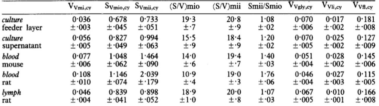
Table I-Relative morphometric parameters (means k standard errors) of T. brucei STIB 247
vvmi,cY SVmio,cy SVmii,cy (SN)miO (SN)mii SmiiSrnio Vvai,,, V~ii,~~ Vvfl~~
culture 0.036 O-678 0.733 19.3 20.8 1.08 0.070 0.017 0.181
feeder layer IL .003 f -045 z!I *05 1 f.7 +*9 +.02 f .006 f.002 +*008
culture O-056 0.827 0.994 15.5 18.4 1.20 0.070 0.025 O-127
supernatant f -005 k-049 k-063 +.9 +.9 f.02 k-005 + .002 + -009
blood o-077 1.048 1.464 14.0 19.4 1.40 o-05 1 0.028 0.145
mouse f .006 + .062 + .090 5.6 +*7 f *03 5.004 +*002 k.006
blood 0.108 1.146 2.039 10.9 19.0 1.76 0.046 0.027 0.115
rat +a010 + *074 +*179 +*4 f.3 f-06 -t *004 f *003 f -005
lymph 0.046 0.839 O-898 18.9 20-o 1.07 0.067 0.010 0.166
rat + so04 k.041 + *052 +1*0 +*8 f so3 k -005 t.001 xk -008
hni,cy - volume density of mitochondrion (ym3/um3), Svmip,q = membrane @m2/um3), Sv,,,i cY =
surface density of mitochondrial outer surface density of mitochondrial inner membrane, (S/V)mio = ratio of the surface area of the outer membrane to the mitochondrial volume (um2/unr3), (S/V)mii = ratio of the surface area of the inner membrane to the mitochondrial volume, Smii/Smio = ratio of the surface area of the inner to .the outer mitochondrial membrane (um2/unrr), Vv volume density of glycosomes, Vvii,q = volume density of lipid inclusions, V~fl,~ = volume density o f” iY cY = the flagellum, cy = with respect to cytoplasmic volume, flagellum included.
694 MORPHOMETRY OF T. brucei GROWN in vitro AND in vivo
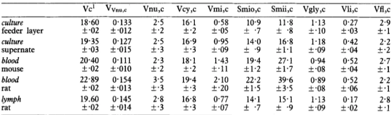
Table II-Morphometric parameters (means + standard errors) of T. brucei STIB 247
vc’ vvnu,c Vnu,c Vcy,c Vmi,c Smio,c Smii,c Vgly,c Vli,c Vtl,c
culture 18.60 0.133 2.5 16.1 0.58 10.9 11.8 1.13 0.27 2.9
feeder layer rt.02 k.012 +*2 k.2 f.05 Zk.7 IL.8 k.10 k.03 +*1
culture 19.35 0.127 2.5 16.9 0.95 14.0 16.8 l-18 0.42 2.2 supernate f -03 k-015 k-3 t-3 t-09 + -9 kl.1 f -09 k.04 k.2 blood 20.40 0.111 2.3 18.1 1.43 19.4 27.1 0.94 0.52 2.7 mouse k.02 Zk.010 Zk.2 k.2 L.11 kl.2 kl.7 t-08 k-04 f-1 blood 22.89 0.154 3.5 19.4 2.10 22.2 39.6 0.89 0.52 2.2 rat k-02 k.013 +*3 k.3 k-20 fl.5 f3.5 rk *08 +*06 f.1 lrmph 19.60 0.145 2.8 16.8 0.77 14.1 15.1 1.13 O-17 2.8 rat +*02 k-014 +*3 k-3 k-07 + -7 f -9 f -09 f-02 f.1
tdetermined with a Coulter Channelyzer
Vc = absolute cell volume (um3), ‘Vnu,c = volume density of nucleus (um3/ym3), Vnu,c = absolute volume of nucleus, Vcy,c = absolute volume of cytoplasm, Vmi c
surface area of mitochondrial outer membrane (urn ), Smii,c = absolute surface area of mitochondrial inner ?2 = absolute volume of mitochondrion, Smio,c = absolute membrane, Vgly,c = absolute volume of glycosomes, Vli,c = absolute volume of lipid inclusions, Vfl,c = absolute volume of flagellum. c = with respect to cell volume, flagellum included. -
Table III-Direct and indirect determination of rial volume, (S/V)mii, and the ratio of the surface
mean ceil volume, flagellum included (3~ standard areas of the inner to the outer mitochondrial mem-
errors) of T. brucei STIB 247 branes, SmiiiSmio.
vc vc’ VP
culture 18-60 26.1 22.4
feeder layer t-02 k2.7 k2.4
culture 19.35 26.8 21.5
supernate k-03 k3.4 k3.2
Vc = values from Coulter Channelyzer (direct measurement), Vcf = values derived from the nuclear volume of formalin fixed trypanosomes and from the nuclear cytoplasmic volume ratio, Vc” = values de- rived from the nuclear volume of methanol fixed trypanosomes .
From the absolute cell. volume (Vc) and the volume density of the nucleus (VvnuJ, the absolute volume of the nucleus (Vnu,c), and in a second step the volume of the cytoplasm (Vcy,c), could be calculated. Using Vcy, c and relative cellular parameters absolute values per cell could be obtained: Vmi,c = absolute volume of the mitochondrion, Vgly,c =of the glyco- somes, Vli,c = of the lipid inclusions, Vfl,c = of the flagellum; Smio = absolute surface area of the mitochondrial outer membrane, and Smii = of the mitochondrial inner membrane.
Parameters were calculated-as means + SE. Re- sults were compared statistically using Student’s t-test with a significance limit1 of 2P~O.05.
Table IV-Pleomorphism and mean cell volume of the T. brucei STIB 247 populations examined
Results culture feeder layer culture supernate blood mouse blood rat lymph rat pleomorphism % Vcell'
Pm3 slender intermediate stumpy
18.6 82 18 0
19.35 58 36 6
20.4 68 30 2
22.9 17 36 47
19.6 37 63 0
The morphometric parameters are listed in Tables I to III. Only the functionally more relevant parameters are presented below.
Mean cell volume (Vc)
‘determined with a Coulter Channelyzer
The mean cell volume of trypanosome populations was determined for the first time with a Coulter Channelyzer (Fig. 1). Vc was represented by about 8 x lo4 cells for each population, and therefore even slight differences between mean values were statisti- cally significant (Table II). The highest Vc was measured for the bloodstream population from the rat which consisted of 17% slender forms and almost 50% stumpy forms (Table IV). The bloodstream popula- tion from the mouse with 68% slender and hardly any stumpy forms gave a significantly lower value. In general, Vc of bloodstream forms was higher than Vc of culture or lymph node forms.
parameters were derived: the ratio of the surface area of the mitochondrial outer membrane to the mitochondrial volume, (S/V)mio, the ratio of the surface area of *he inner membrane to the mitochond-
In addition to the direct determination of Vc with the Coulter Channelyzer, an indirect method (BURRI & HECKER, 1979) was employed using the nuclear volume as measured from smears and the nuclear cytoplasmic volume ratio. This indirect method gave higher values than the Channelyzer (Table III).
H. HECKER AND R. BRUN 695
ABC,D E
10 20 30 40
Cell volume 1rrn-Y
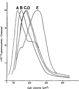
Fig. 1. Size distribution curves of T. brucei STIB 247 populations grown in vitro and in viva A = cultore, supernatant population; B = culture, feeder layer population; C = rat lymph; D = mouse blood; E = rat blood.
However, the values obtained from methanol fixed smears were much closer to the Channelyzer results than were those obtained from formalin fixed smears.
Mizochondrion
The mean volume density, Vvmi,cy (Table I, Fig. 2) and the mean absolute volume of the mitochon-
“Vrni,q I
CF CS BM BR LR
Fig. 2. Volume density of the mitochondrion (mean i standard error) of T. brucei STIB 247 populations grown in vim and in viva CF = culture, population between feeder layer,cells; CS = culture, supernatant population; BM = blood of mouse, BR = blood of rat; LR = lymph of rat.
drion, Vmi,c (Table II) were significantly higher in the rat blood (many stumpy forms, Table IV) as compared to the trypanosomes from mouse blood (mainly slender forms). The values for culture forms and forms from lymph nodes were significantly lower than those for bloodstream forms. With respect to culture forms, the pleomorphic forms in the super- natant medium contained more mitochondrion than the monomorphic trypanosomes located in the feeder layer. The surface densities and absolute surface areas of the outer and inner mitochondrial membranes were in accordance with the values of the mitochondrial volume.
The ratio of the surface of the outer mitochondrial membrane and the mitochondrial volume (S/V)mio, (Table I) reflects to a certain extent the mean diameter and size of the organelle: the smaller this ratio the larger the mitochondrial profiles. The largest mitochondrion was found in the sample from rat blood, significantly smaller ones in mouse blood and in the supernatant of the culture. Trypanosomes isolated from the feeder layer and from lymph nodes contained the smallest mitochondrion. The mor- phometrically assessed ratio of the surface area of the inner membrane to the mitochondrial volume, (S/ V)mii, (Table I) is assumed to represent the ratio of enzymes of the respiratory chain to enzymes of the tricarboxylic acid cycle. This parameter proved to be similar for all the populations examined.
Glycosomes
Glycosomes are microbody-like, membrane bound- ed organelles which sometimes contain crystalloid inclusions (B~HRINGER & HECKER, 1975). Glycolytic enzymes which are involved in the trypanosomes’ extramitochondrial respiration have been localized within this organelle (OPPERDOES & BORST, 1977). “VglpCY O.lO- 0.05. 0.01. I
I
T T :I. 1; . ..I. ,;j I:,,: ..I ‘;;‘;. i ;: : . . . ’ . . .’ . . . . :. .. ‘.. .:’ . . . ; l.. ., : :j . . : ; . : .I :. :’ ..:: ;. . . . ,. : : . . . :; : : , ,. : :: CF CS BM BR LRFig. 3. Volume density of the glycosomes of T. brucei STIB 247 populations grown in vitro and in Volvo. For explanations see Fig. 2.
696 MORPHOMETRY OF T. brucei GROWN in vitro AND in vivo
Volume densities, VvEtv,cy (Table I, Fig. 3) and absolute volumes of glycosomes, Vgly,c, did not differ significantly between culture forms and forms from lymph nodes, although bloodstream forms showed significantly lower values.
Lipid inclusions
Lipid inclusions were present in the form of strongly osmiophilic particles, but triglyceride-like inclusions could also be-found (B~HRINGER & HECK-
ER, 1975). Their total volume density, Vvti.cy, (Table I) and their absolute volume, Vli,c (Table II) were highest in bloodstream forms. Trypanosomes from culture showed a lower lipid content, especially the population between the feeder layer cells. The lowest lipid content was measured in lymph node forms.
Discussion
This study aimed to find out if bloodstream forms of T. brucei grown in vitro are similar to forms from the mammalian host with respect to their ultrastruc- tural composition. The main cellular parameters used for this comparison were the cell volume, the mitochondrion and the glycosomes. BRUN et al.
(198 1) have shown that two trypanosome populations exist in a bloodstream form culture: one, which appears monomorphic and slender, between the feeder layer cells and a second one, slightly pleomor- phic, in the supernatant medium. In the present investigation this observed difference was also ex- pressed by the morphometric parameters of the mitochondrion, indicating that the two populations may also physiologically be slightly different. The few stumpy forms found in the supernatant were probably causing the difference in the mitochondrial values. This is, in addition to the light microscopical findings another indication that the stumpy forms in culture may be considered as ‘real’ stumpy forms.
The quantitative ultrastructural composition of trypanosomes from lymph nodes was very similar to the one of culture forms. Closer in all features than to that of the bloodstream forms from a mammalian host. These findings suggest that the trypanosomes in culture more probably represent tissue/lymph forms than simply bloodstream forms. In a previous inves- tigation on lymph forms from the rat similar mor- phometric data were obtained (TANNER et al., 1980). There were slight but statistically significant mor- phometric differences between bloodstream forms cultivated in vitro and those grown in vivo. These ultrastructural differences are presumably an express- ion of physiological dissimilarities. between the try- panosome populations. This is not surprising because the culture system is a very approximate substitution for the mammalian host, and adaptations to the conditions in vitro have to be expected. On the other hand we found distinct differences between our morphometric results for bloodstream forms grown in vitro and in vivo, compared to previously published, data for procyclic forms of the various parasite stages in the vector. Procyclic culture forms are more than twice as large in volume, and all the mitochondrial parameters showed two to three times highes values (GHIOTTO et al., 1979). Comparison with forms in the midgut and the salivary glands of tsetse flies also revealed lame differences (B~HRINGER~ & HECKER, 1975; ‘HECK~R 1980). Because different trypanosome
stocks were used in the investigations cited above, direct comparison with our results has to be made with caution.
The morphometrical examination of cultivated bloodstream forms clearly demonstrated that the trypanosomes in culture were morphologically very similar to forms from a mammalian host. The presence of a surface coat on cultivated bloodstream forms and their infectivity for mammalian hosts as
well as for the vector Glossina m. morsitans have been demonstrated earlier (BRUN et al., 1981; SCH~NI et al.. 19821. There are also strong similarities in res$ration (HILL et al., 1978; HILL, 1980), and, so far, no difference has been found between blood- stream forms from culture and from a mammalian host. Desoite sliaht mornhometrical differences be- tween in “vitro and in vbo bloodstream forms, the cultivated forms may really be regarded as mamma- lian stages. With some precautions they can be used for in vitro investigations replacing bloodstream forms from a mammalian host.
The mean cell volume of trypanosome populations was determined for the first time directly with a Coulter Channelyzer. The values were -within a narrow range (18.6 to 22.9um3) but still differed significantly from one another due to the large cell samples investigated. The size distribution curves (Fig. 1) showed the dissimilarities between samoles much more clearly than did their mean volumes. The two trypanosome populations found in culture pro- duced different size distribution curves although their mean cell volumes were closely similar. The mono- morphic trypanosomes in the feeder layer gave one sharp peak, whereas the supernatant forms produced a curve with a peak and a shoulder which most probably represent the stumpy forms present in this sample. The curve for the bloodstream forms from the rat with almost 50% stumpy forms showed a broader peak shifted to the right. This indicated that the stumpy forms have a larger cell volume than the slender forms. However, the volume increase from slender to stumpy forms was less than 50%. In earlier investigations u&g other T. brucei stocks an increase of the cell volume of about 2-S times from slender to stumpy forms has been reported (HECKER, 1980,
review). These volumes were determined by an indirect method (BURRI -& HECKER, 1979) which was also used in this work. For the two trypanosome populations in culture the cell volumes were deter- mined with the Coulter Channelyzer and by the indirect method. For the latter method methanol and formalin fixed smears were used for the estimation of the mean nuclear volume. It became obvious that the fixation had a crucial influence on the nuclear volume. The values obtained from methanol fixed smears were closer to the directly measured cell volume than the ones obtained from formalin fixed preparations. The fact that the fixation of the smears had a significant effect on the preservation of the nucleus and thus on the estimation of the cell volume, suggests that the indirect method should be used with caution. For two of the stocks previously used to determine the cell volume indirectly, a direct measurement with the Coulter Channelyzer was done for slender and stumpy populations. We found that stumpy forms were slightly (but not more than 50%) larger in volume than slender forms; for one stock the volume of
H. HECKER AND R. BRUN 697 slender and stumpy forms was almost identical
(results not published). It therefore must be assumed that the indirect method overestimates the nuclear volume of stumpy forms.
Acknowledgements
The present investigation was supported in part by the Swiss National Foundation for Scientific Research, Grant Nr. 3.346-0.78. The authors gratefully acknowledge critical discussion of the manuscript by Dr. Leo Jenni and the skillful technical assistance by Erika Flury, Maja Hofmann and Margrit Schonenberger.
References
Bohringer, S. & Hecker, H. (1975). Quantitative ultras- tructural investigations of the life cycle of Ttypanosoma brucei. A morphometric analysis. Journal of Protozoology, 22, 463-467.
Brun, R., Jenni, L., Schonenberger, M. & Schell, K.-F. (1981). In vitro cultivation of bloodstream forms of Typanosoma brucei, T. rhodesiense and T. gambiense. Journal of Protozoology, 28, 470-479.
Burri, P. H. & Hecker, H. (1979). Stereological study of trypanosoma, a small protozoon. In: Stereological Methods, Volume 1. Weibel, E. R. (Editor). London: Academic Press, pp. 343-348.
Ghiotto, V., Brun, R., Jenni, L. & Hecker, H. (1979). Trwanosoma brucei: mornhometric changes and loss of infectivity during transformation of bloodstream forms to procyclic culture forms in vitro. Experimental Parasitol- ogy, 48, 447-456.
Hecker, H. (1980). Application of morphometry to pathogenic trypanosomes (Protozoa, Mastigophora). Pathology Research and Practice, 166, 203-217.
Hecker, H., Burri, P. H., Steiger, R. F. & Geigy, R. (1972). Morphometric data on the ultrastructure of the pleomor- phic bloodforms of Typanosoma brucei. Acta Tropica, 29, 182-188.
Hill, G. C. (1980). Biochemical studies using Typanosoma rhodesiense cultured-infective trypomastigotes. In: The host invader interplay. Van den Bossche, H. (Editor). Amsterdam: Elsevier/North Holland Biomedical Press. pp. 555-566.
Hill, G. C., Shimer, S., Caughey, B & Sauer, S. (1978). Growth of infective forms of Typanosoma (T.) brucei on buffalo lung and Chinese hamster lung tissue culture cells. Acta Tropica, 35, 201-207.
Lanharn, S. M. (1968). Separation of trypanosomes from blood of infected rats and mice by anion-exchangers. Nature, 218, 1273-1274.
Opperdoes, F. R. & Borst, P. (1977). Localization of nine glycolytic enzymes in a microbody-like organelle in Typanosoma brucei: The glycosome. FEBS Letters, 80, 360-364.
Schoni, R., Jenni, L. & Brun, R. (1982). Cyclical transmis- sion of in vitro cultivated bloodstream forms and procyclic trypomastigotes of Typanosoma brucei brucei by Glossina morsitans mwsitans. Zeitschrift ftir Parasiten- kunde, (in press).
Tanner, M., Jenni, L., Hecker, H. & Brun, R. (1980). Characterization of Typanosoma brucei isolated from lymph nodes of rats. Parasitology, 80, 383-391. Weibel, E. R. (1979). Stereological Methods, Volume 1,
London: Academic Press.