1726 Cardiovascular Research 1993;27: 1726- I734
Short review
Sodiudcalcium exchanger in heart muscle:
molecular biology, cellular function, and its special
role in excitation-contraction coupling
Dan Schulze, Paulo Kofuii, Robert Hadley, Mark
S Kirby, Robert S Kieval,
Andrea Doering, Ernst
he NdCa exchanger was first identified by Reuter and Seitzl and Baker et a / * in 1968 and 1969. TheT
functional importance of this transporter in many tissues has become clear as detailed investigations into its cellular, biophysical, and molecular properties have been completed. Historically there have been four epochs in our understanding of the N d C a exchanger in the heart. ( 1 ) Its identification as a transport entity in 1968.’ (2) The quantitative linkage between intracellular sodium con- centrations and force production.3-” These findings had certainly been anticipated by earlier work by Niedergerke and coworkers.”-’‘ (3) Quantitative estimation of the stoichiometry in functioning heart cells of 3 Na to (4) The molecular characterisation of the N d C a We are now actively examining how the N d C a exchanger works and the relation between its structure and its cellular function. This new work is accompanied by an exciting resurgence of the second epoch: the role of the NdCa exchanger in excitation-contraction coupling.1 ca.7 15-17
Functional importance of the NdCa exchanger: role of the NdCa exchanger in controlling resting calcium The NdCa exchanger serves as the principal calcium extrusion mechanism in heart muscle.” I‘ 23
’‘
In the steadystate the entry of calcium into the cell via “leak” pathways, calcium channels, and calcium permeable channels must be balanced by the extrusion of calcium. Any time-average increase in calcium entry will eventually come into balance as the time-average extrusion of calcium, principally by the NdCa exchanger, increases. The extrustion of calcium by the NdCa exchanger increases as intracellular calcium is increased. Thus the cost of this regulation is an increase in cytosolic calcium, [Ca’’li. Increasing [Ca2+], is associated with a very much larger accumulation of calcium in the sarcoplasmic reticulum. There is, consequently, an enormous amplification of the effect of an increase in time-average calcium influx if one examines sarcoplasmic reticular
Niggli, and
W J Lederer
function. This feature was well described by Eisner and coworkers in the early 1980s‘ ” and supported by recent
work.’6 Mullins also noted the power-function dependence of tension on intracellular sodium in heart muscle when he examined the published data.” Because of the speed of pumping by the Ca-ATPase in the sarcoplasmic reticulum and its great capacity,” ” ” even very brief changes in
calcium influx can produce large changes in force. There is an additional new part played by the N d C a exchanger that has only recently been identified: as direct activator of the sarcoplasmic reticular calcium release channels.3G3’ This is a somewhat contentious issue at present. It is thus appro- priate to discuss two related questions that are important in excitation-contraction coupling: ( I ) Where are the NdCa exchanger proteins located? (2) What functional experiments lead us to believe that the spatial organisation of the sarco- lemma1 calcium sources and sarcoplasmic reticular calcium release channels is very important?
Localisation of the NdCa exchanger in heart muscle Immunofluorescence localisation of the NdCa exchanger has recently been carried out in guinea pig and rat heart cells.”
’‘
Whereas the two reports do not agree entirely with the distribution, they do agree that the T tubular system has an abundance of NdCa exchanger proteins. Our report3‘ provides positive evidence that the NdCa exchanger proteins are found in great amounts of the exterior sarcolemmal surfaces and on the intercalated discs of the heart cells. By this account, then, the NdCa exchanger proteins are found everywhere on the sarcolemma. This seems to be appropriate as this spatial arrangement would provide efficient extrusion from all regions of the interior. The presence of many N d C a exchanger proteins in the “exterior” sarcolemmal membrane (by contrast with the T tubular sarcolemmal membrane) is also supported by the finding that copious amounts of N d C a exchanger are noted in experiments by others and by us using the giant patch method.”4 Blebs formed from the putative exterior membranes can be voltage University of Maryland School of Medicine, 660 W Redwood St, Baltimore, MD 21201, USA - Department of Microbiology and Immunology: D Schulze; Department of Pharmacology and Experimental Therapeutics: P Kofuji; Department of Physiology: R Hadley, M S Kirby, R Kieval, A Doering, W J Lederer; University of Bern, Bern, Switzerland - Department of Physiology: E Niggli. Correspondence to Dr Lederer.Molecular biology of NdCa exchanger in heart muscle 1727
clamped with a suction pipette and NdCa exchanger current can readily be measured. This provides functional evidence in support of the distribution of the NdCa exchanger made by Kieval eta/.” It is worth noting by way of caution, however, that no one has yet shown which components of the sarco- lemma1 membrane system actually give rise to the blebs.
The report by Frank er aIT3 differs from our findings, principally because they have argued that there is a much greater density of NdCa exchangers on the T tubular membranes than on other sarcolemmal membranes and have suggested that this finding may be functionally important.33 Our work, done in parallel and independently, finds no such distinctions. Indeed, our results present evidence of the very clear distribution of the NdCa exchanger to the non- transverse tubular sarcolemmal membranes as well as to the T tubular membranes. Specifically, we find abundant NdCa exchanger protein on the exterior (non-transverse tubular) sarcolemmal membrane and in the intercalated disc mem- brane.34 Some of the differences found between the studies of Frank et a13’ and those of Kieval er al” may arise from the specific fixation methods used, from the slight differ- ences in the preparation of cells, or from the differences in primary antibodies. Both groups have been working together
to resolve the differences. Functionally the advantages of preferentially localising the NdCa exchanger to the T tubules (over other sarcolemmal membranes) are unclear. Instead, our findings, which lack resolution below about 0.4 IJ.. suggest that the NdCa exchanger protein is fairly
uniformly distributed over d l of the sarcolemmal surfaces.
The apparently greater density of NdCa exchanger protein in the intercalated disc regions presumably reflects the highly infolded nature of these regions rather than greater density per square micron of membrane surface area. Although it is not clear exactly how the NdCa exchangers located on the intercalated disc regions of the sarcolemma contribute in any unique manner to calcium regulation, their presence at the very least argues against the preferential targeting of this transport system to the T tubules. Possibly, with the exceptions of regions of the intercalated disc that contain tight junctions of packed arrays of gap junctions, much of the exterior surface of the intercalated disc has access to the extracellular space. For that reason, it presents a large surface available for the NdCa exchanger to use in the regulation of [Ca2+li. Future studies will investigate whether or not anchoring proteins may be linked to the NdCa exchanger to link its position to that of other proteins or other cellular structures.
Role of the NdCa exchanger in excitation-contraction coupling
There are two explicit roles that the NdCa exchanger may have in excitation-contraction coupling in the heart. (1) The NdCa exchanger regulates calcium content of the sarco- plasmic reticulum by regulating the resting [Ca”], level (see previous and later comments). (2) The NdCa exchanger can directly contribute to the calcium that enters the myocardial cells during the upstroke and plateau phases of the action potential. During the action potential the NdCa exchanger can contribute directly to the calcium that triggers the calcium induced calcium release process (discussed later) and can extrude calcium at different rates during the plateau phase of the action potential.” 4s-s’ The NdCa
exchanger turnover rate is clearly voltage dependent, dependent on the Na+ and Ca” gradients and many other factors (for example, pH).
There is considerable evidence, both direct and indirect, that the principal contribution of the NdCa exchanger to the excitation-contraction coupling process is through its regulation of the resting [Ca”], level. The importance arises for two reasons. ( 1 ) The calcium content of the sarcoplasmic reticulum seems to have a profound effect on the amount of calcium that is released when the sarcoplasmic reticulum is triggered to release calcium (by calcium influx across the sarcolemma; see earlier discussion). (2) The sensitivity of the sarcoplasmic reticular calcium release process to triggering calcium is due in part to the amount of calcium that resides within the sarcoplasmic reticulum. There are two lines of evidence. One line comes from experiments in normal heart cells and this is reviewed later. The second argument arises by comparing normal heart cells with calcium overloaded heart cells.52 O’Neill and coworkers found that in normal heart cells local increases in intracellular calcium by flash photolysis of caged calcium caused only local contractions but did not produce a propagating wave of increased [Ca”],.” By contrast, in cells that had been subjected to calcium overload, a similar flash produced a global response, activating a propagating wave of increased [Ca”], (our unpublished observations). Importantly, wave propa- gation must also depend on the level of saturation of CaZt binding sites in the cytoplasm. For example, as resting [Ca”], rises, the cytosolic binding sites become more nearly saturated and the effective buffering power of the cell for calcium decreases. A given number of calcium ions will raise [Ca”], more as the buffering power of the cell for calcium declines. In regard to the issue of the propagating wave of increased [Ca”], during the early phase of calcium overload, however, we have found that resting [Ca”], increases only slightly (tens of nM), based both on force measurements and direct measurements of [Ca”], with fluorescent indicators. This slight increase in [Ca”],, not enough to change the buffering power of the cytoplasmic calcium binding sites significantly, is more than adequate to produce a significant increase in the sarcoplasmic reticular calcium content and is thought by us to be only a secondary factor.
The experiments in normal heart cells that suggest that the “sensitivity” and possibly the “gain” of the sarcoplasmic reticular calcium release process depends on sarcoplasmic reticular calcium content were first published in 198753-55 and have led to several continuing controversies. The termin- ology can be rather confusing. Sensitivity refers to the level of calcium needed to trigger sarcoplasmic reticular calcium release. Gain refers to the amount of calcium released for a given triggering signal. Because the elementary events involved in sarcoplasmic reticular calcium release are not seen with sufficient temporal or spatial resolution with our present methodology, “gain” is used to describe the properties of the elementary events whereas “apparent gain” refers to the results of experiments actually carried out.
The two essential findings were that the sensitivity of the [Ca?+], transient measured with fura-2 salt injected into heart cells depended on the activation protocol and not on the peak Icd or the integrated Ica. For example, depolarisations of the heart cell from -55 mV to voltage around -40 mV produced a small [Ca”], transient but virtually no Ic,. This result indicates that very little calcium flux can trigger significant sarcoplasmic reticular calcium release, suggesting that the gain and the sensitivity of the triggering mechanism are high. On the other hand, with depolarisations from -55 to -28 mV the [Ca”], transient was graded with different durations of depolarisation. This suggests that there is little positive feed- back in the system, a finding consistent with low gain in the
1728 Schulze, Kofuji, Hurlley, Kirby, Kievul, Doering, Niggli, Lederer
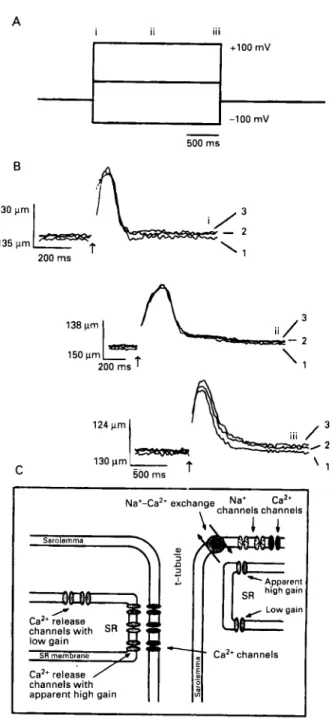
system. Importantly, it was possible to truncate the [Ca”], transient by early repolarisations. This result also suggests that the system does not have much positive feedback or that the system can operate at low gain (where release is proportional to the trigger). Whereas a nearly maximal calcium transient was produced with a depolarisation to 0 or +I0 mV, a large integrated Ica was associated with that [Ca’+Ii transient. By contrast with this finding, a nearly maximal [Ca2+Ii transient was seen on repolurisurion from +I00 mV (to -55 mV) when the integrated Ica tail current was quite small. These experiments suggested to us that a simple calcium induced calcium release mechanism with constant sensitivity and fixed gain was inconsistent with the findings. One possibility was that sarcolemmal voltage influences calcium induced calcium release. The other possibility was that some other factor was responsible for the findings. Figure 1 shows the experiment designed to examine the issue. Flash photolysis experiments were carried out in voltage clamped heart cells.” Control experiments had shown that most of the twitch produced by the photorelease of caged calcium arose from calcium induced calcium release and not directly from the uncaging of the calcium. The experiments show that voltage across the sarcolemmal membrane does not significantly influence calcium induced calcium release in intact cells. Further experiments showed that the apparent gain of the CICR system was low when it was activated by flash photolysis of caged calcium. Figure 1 also shows the “solution” to the problem. The microanatomy of the sarcolemmal-sarcoplasmic reticulum region can bestow apparent high gain on some elements of the system while the actual release process operates at low gain. The spatial arrangement requires close approximation of the sarcolemmal and sarcoplasmic reticular membranes for at least some of the sarcolemmal calcium sources (for example, calcium channels) and some of the sarcoplasmic reticular calcium release channels. This result sets the stage for the results of Leblanc and Hume.”’ These workers found that when they blocked Ic;,. they could still produce a depolarisation activated [Ca”], transient as long as the sodium current remained intact. They argued that the NdCa exchanger was activated by the influx of sodium and that it consequently brought in calcium from the extracellular space as it extruded the sodium that had entered on depolarisation. For such a system to work, a “fuzzy space” was proposed that was, in effect, a subsarcolemmal region of the cell that had slowed diffusion.3‘b3’
’*
Figure 2 shows a diagram of the hypothesised region.” Interestingly, the requirements of the experiments of Leblanc and Hume” and those of Niggli and Ledere9’ are remarkably similar. There are many experi- ments now being carried out by diverse groups examining the importance of the spatial organisation of the heart cell with respect to excitation-contraction coupling. A major experimental challenge must be continually considered in these experiments: how does the sarcoplasmic reticular calcium load affect the results? As noted, the sarcoplasmic reticular calcium content seems to be an important factor in regulating the sensitivity and the gain of the calcium release mechanism and calcium induced calcium release.Molecular operation and biophysics of the NdCa exchanger
The NdCa exchanger in a normal heart cell produces only a small calcium efflux as it just balances the time averaged calcium influx.” At a resting [Ca”], level, its overall turnover rate is limited by the very low [Ca?+], of about 100-200 nM.
... A + l o 0 rnV -100 rnV 500 rns 138 prn 150 prn - 2 ‘1 200 rns 130prnl
t
C 500 rns7
Na+-Ca2+ exchange,
,channejs Nat c h y n e l s Ca2tchannels with SR
channels with apparent high gain
‘ 1
Figure 1 Voltage dependence of calcium induced calcium release
in guinea pig heurt muscle cells. ( A ) From a holding potential
of 4 0 nil! the membrane pcitential was changed to -100 ml! 0 m y or +I00 mV,for 2 s. Flashes of UV light to release caged calcium (DM-nitrophen) were triggered at times indicated ( i ) , ( i i ) ,
or (iii). ( B ) Twitches were activated bv the flash that occurred at the same time NS the voltage change ( i ) , in the steady state 500 ms after
the voltage step ( i i ) , or upon return to the holding potential ( i i i ) .
Nickel ( 5 mM) wus applied to block the NdCu exchanger and Icv
(2), und ( 3 ) show three results in the same cell. ( C ) Proposed interaction of calcium channels, sodium channels, and N d C a exchange with the sarcoplasmic reticular calcium release channels.
A close association in a space with restricted diffusion yields
release channels with an apparently high gain (positive feedback)
because they are exposed to a high [Ca’+], during the time of
calcium influx and release whereas release channels further away froni the sarcolemrnu exhibit their intrinsic low gain. From Niggli
and Lederer, I 99e7 with permission.
At these concentrations of [Ca’+],, the rate limiting step is calcium binding to the exchanger at the intracellular site. If calcium is suddenly increased, then the turnover rate suddenly rises. We use DM-nitrophen as the “calcium caging” agent and load it into the cell of interest through the
- - - I
lntracellular space
Figure 2 Scliemutic diagram to illustrate the relation tkur rnay
exist between the sodium channels. the NdCu exchanger, surco- lemma1 calcium channels, sarcoplasmic culcium releme channels, and the hymheticul ‘3fiCz:y s p a c ~ ” . The j i i q .space is u very thin region of slowed diffusion. From Lederer et a1 ‘ I with permission.
Moleculur biology of NdCu exchanger in heart muscle
L
A
5. C
voltage clamp ~ i p e t t e . ~ ’ s9-hZ A flash of UV light causes the photolysis of the DM nitrophen and a step increase in
intracellular calcium is achieved. Within 200 p,s the inward NdCa exchanger current is seen under normal conditions. It decays as intracellular calcium falls. All experiments are done under conditions to remove the sarcoplasmic reticulum
as a calcium buffer (ryanodine is applied) and to minimise movement (2,3-BDM is applied). If the NdCa exchanger is blocked by cold, by nickel, or by other agents, there is still
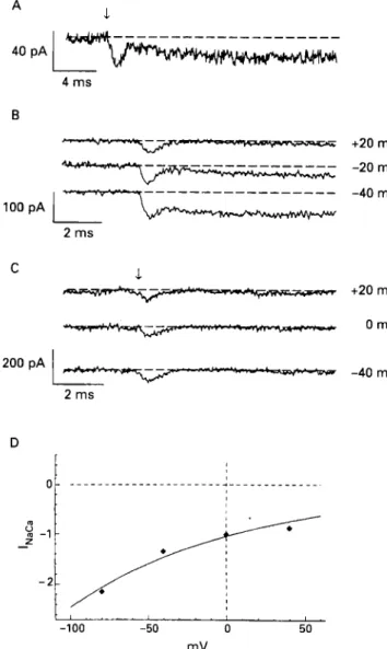
an inward current that can be seen. Figure 3C shows this. The current is an inward current transient. The baseline current is close to zero before the photorelease of calcium. As rapidly as calcium increases, there is an increase in inward current. Despite a maintained level of [Ca”], (because all extrusion systems are blocked), the current rapidly decays back to zero.
An inward current can arise from the inward movement of
a positive charge or the outward movement of a negative charge. The observed current transient suggests that either a
current source is gated for a brief period or that a fixed number of mobile but “tethered” charges move during a
molecular rearrangement. Once moved to a new con- figuration, a steady state is established and no further move- ment of charge takes place. Both mechanisms account for a
current transient. The first is similar to an inactivating ionic current whereas the second is similar to a gating current.
We have attributed the observed current transient in the experiments shown in fig 3 to a one time rearrangement of
the NdCa exchanger protein as it carries intracellular calcium out of the cell. Because the Na translocation step is blocked by the experimental conditions, the exchanger cannot (over the course of these experiments) return to cycle again. Thus the amount of the integrated current transient is limited by the number of NdCa exchanger proteins in the cell membrane and the state of the exchanger. By state we mean whether the exchange protein is capable of binding to an intracellular calcium ion and transporting it to the outside. If all of the NdCa exchanger proteins were in an intra- cellular calcium receptive state, then the integrated current transient would be proportional to the actual number of protein molecules in the sarcolemmal membrane. If all of the
rCCCClllhP.PC.C;-- 200 p A
1
I r 2 m s D I729 +20 mV -20 mV -40 mV +20 mV 0 mV -40 mV I -100 -50 0 50 mVFigure 3 Conforinatinn c u r r t ~ i s activated by a step
increase in intracellular calcium even when the N d C a exchanger
is blocked. ( A ) Cold (12°C) lurgelv blocks the N a K a current but does not uffect the I,,,,,,. Holding potential is 0 t n K In this and other panels, the urrow indicutes the time of the Jlash thut uctivares the
photolysis of caged culcium. ( B ) Voltuge dependence of the cold
treated N d C a exchanger curretit (20°C). ( C ) Ni” ( 4 mM) is used
to block the NdCu exchanger complete!\: The only current seen
with the step increase of caged calcium is lc.c,,,t. (D) Voltage
dependence o j the Na/Ca exchanger current. The observed voltuge
dependence was 114 niVJor each e-fold change of current. From
Niggli and Lederer, 1991“1 with permission.
NdCa exchanger proteins were in an extracellular state, so
that none could bind calcium on photolysis, then there should be no current transient.
In the case of our experiments, the state of the NdCa exchanger is relatively stable when the exchanger is blocked, since full cycling is clearly inhibited. We noted that removing extracellular sodium was also an equally effective means of blocking the NdCa exchanger current while leaving the current transients shown in fig 3C. Thus we have hypothesised that the component of the cycle that is blocked by the manoeuvres noted is the sodium translocation step. Clearly step increases in calcium can produce a current transient. An interesting and important finding was that, when the NdCa exchanger is not quite fully blocked, there is a secondary component of exchanger current that is maintained. This component occurs along with the earlier
1730 Schulze, Kofuji, Hudley, Kirby, Kievul, Doering, Niggli, Lederer
current transient mentioned previously. It represents a steady current component that seems to be summed with the current transient. We hypothesise that this modulatable component arises because the Na translocation component is partly functional (see also fig 5).
The voltage dependencies of the fully functioning NdCa exchanger and the modulatable component are the same, whereas the voltage dependence of the calcium activated early transient is much less. These findings suggest to us that when the rate of intracellular calcium binding is fixed as would occur with a steady level of [Ca"],, then the Na translocation step is rate limiting and voltage sensitive. Consequently the voltage dependence of the NdCa exchanger would provide information about the charge on the sodium bound form of the NdCa exchanger. If this is true, then the voltage dependence shown in fig 3D is 114 mV for each e-fold change in current. This very weakly voltage sensitive response suggests that less than a single charge is sensing the voltage across the membrane in this rate limiting translocation step. Specifically, these results suggest that +0.44 charges may be sensing the membrane voltage and moving through the full voltage field during the Na translocation step. If this were true then -0.56 charges would be moving through the voltage field during the calcium translocation step. This would explain why we find an inward current transient that is activated by the step increase in [Ca'+Ii. We see the inward current transient because a fraction of the NdCa exchangers are in the intracellular calcium receptive state before the step increase in [Ca"],. With the step increase in calcium, calcium binds to the NdCa exchanger protein and moves out of the cell, with each exchanger contributing an outward movement of -0.56 charges.
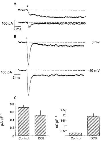
Interestingly, when dichlorobenzamil is added to the pipette, it increases the amount of transient current activated by a step increase in [Ca"], produced by photolysis of DM-nitrophen. Figure 4 shows this result. Figure 4 also shows that the intracellular application of dichlorobenzamil only slightly inhibits the NdCa exchanger current but it very significantly increases the integrated charge moved during the [Ca"], activated current transient.
These data provide much useful information about how the NdCa exchanger functions. The voltage dependence of the exchanger suggests that -2.56 charges reside on each "naked" exchanger. The maximum Iconf, when integrated, provides information on the number of exchanges (lower limit) that move after photolysis and this permits us to provide a lower limit on the exchanger density (250.~-'). Knowing the maximum size of the NdCa exchanger current and the density permits us to estimate the turnover rate of the exchanger (about 2500.s-').
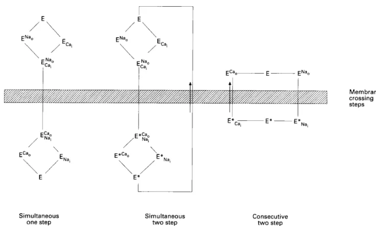
All of the data taken together provide compelling evidence that a simultaneous one step NdCa exchanger model is inadequate to explain how the NdCa exchanger works (see fig 5 ) . This model would not permit calcium activated
conformation current or Icon+ The consecutive two step model is preferred over the simultaneous two step model because self exchange can occur when the counter ion is not present. Thus the data presented here and in other publications strongly support a consecutive two step exchanger model.
Molecular biology of the NdCa exchanger The NdCa exchanger was cloned from dog heart in
1990" and this event was a major finding for all cellular physiologists working on heart muscle. Hydrophobicity
2.5 2
5
1.5 7 IY '
0.5 Control DCBFigure 4 Effects of 3'.4'-dichlorobenzamil (DCB) on the N d C a current and on lco,,j. ( A ) Control current (upper trace) and current after the addition of I00 pM extracellular DCB. The effect is similar to that of other N d C a exchanger blockers with respect to the N d C a exchanger current, but I,,,,,, wac not significantly changed. ( B ) Tvpical currents when I mM DCB was included in the patch clamp pipette. ( C ) Magnitude of N d C a exchanger current (left) and I(#2nf (right) in the presence and absence of DCB in the patch clamp pipette. From Niggli and Lederer, 199IM' with permission.
plots suggest that five membrane spanning regions are followed by a large intracellular loop, which is followed by six membrane spanning regions. The N d C a exchanger has been cloned subsequently in human heart,I9
'"
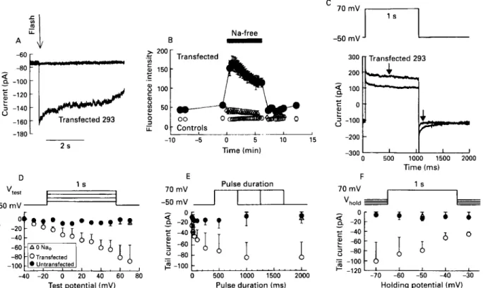
cow heart,'* and rabbit kidney.2' The sequence and function (so far) has remained similar across species and tissues. The retinal rod has a completely different NdCa exchanger, one identified as an Na-Ca-K exchanger because of the important part played by potassium in the transport process.64 Figure 6 shows a scan of the northern blot from human tissues and reveals that the principal peak found at 7.2 kb is seen in all tissues examined, suggesting that within the resolution of this method the N d C a exchanger is similar. It is interesting to note that there are three tissues with a second peak at 1.7 kb in heart, brain and kidney. The function of this smaller piece is not known.When the full length cDNA is inserted into an expression vector, electrical and transport function can be seen. When expressed in a human embryonic kidney cell line (293 cells), the exchanger responds to step increases in [Ca2+], (fig 7A), to reduction in extracellular sodium (fig 7B), and to voltage clamp pulses (figs 7C, D, E, F). The stage is thus set to further characterise where the modulatory sites of the N d C a exchanger are and how exactly the modulation comes about.
Molecular biology qf NdCu exchanger in heart rnuscle 1731 Simultaneous one step Simultaneous t w o step Membrane crossing steps Consecutive two step
Figure 5 Three simplijied diagrams of exchanger models. Leji: simultaneous one step model. This model does not permit partial reaction
membrane currents and hence is incompatible with our data. Middle and left: simultaneous two step untl corisecictitv twro step models are compatible with our datir. The simultaneous two step model does not, however, permit self exchange in the uhsence of the other cations. The consecutive two step model is ,favoured as the simplest model computible with the duta. From Nigqli rind Leilerer. 1991"' with permission.
Distance
Figure 6 Scan o f a northern blot from human tissues. The 7.2 kh
peak is the principul transcript from the human N d C a exchanger. Traces can be identified by examining the 7.2 kh peak and are in the order (greatest to least peak height): heurt, brain, lung, placenta. kidney. liver. pancreas. and skeletal muscle. The normalised (by p-actin) amount of materiul places the tissues in order,from the most N a K a exchanger transcript to the least: heart; brain: kidney: lung: pancreus; placetito: skeletal muscle, and liver. From Kqfirji et al, 1992'' with permission.
Modulation of the NdCa exchanger
We examined how intracellular pH may affect the NdCa exchanger. Use of the giant patch method permitted us to change the intracellular solutions and to assess how they affect the NdCa exchanger. Figure 8 shows how the NdCa exchanger is activated by intracellular sodium. The outward current is activated by increases in intracellular sodium. The current is half maximally activated by intracellular sodium of about 20 mM. Importantly, however, the NdCa exchanger is largely blocked by acidifying the intracellular pH from 7.4 to 6.4 (fig 8, lower curve, empty symbols). Figure 9 shows the effect of different pH levels at a fixed intracellular
sodium concentration of 60 mM. The normal intracellular pH (about 7.2) is sufficiently acidic to inhibit about half of the NdCa exchanger transport. This means that the NdCa exchanger is very sensitive t o the actions of P H , . ~ ~ Proteo- lysis by intracellular chymotrypsin, however, significantly removes this regulation? j5 37
'*
6s This result suggests thatthe proton inhibition arises in part from a part of the protein that is accessible to proteolytic attack. The best guess for such
a target site is the large intracellular loop. It is this loop that contains sites for regulation by [Ca"],, "a'],, and ATP,.
Summary
The NdCa exchanger has been examined with respect to its molecular biology, its cellular function, and its role in
excitation-contraction coupling. The NdCa exchanger plays
a central part in excitation-contraction coupling, setting the level of sarcoplasmic reticular calcium and contributing to the triggering of sarcoplasmic reticular calcium release. Functional biophysical studies with isolated single cells and caged calcium provide evidence that the NdCa exchanger works as a two step sequential transporter. In the heart there are about 250 exchangers.p-', operating at a turnover rate of up to about 2 5 0 0 ~ ' , with the exchanger carrying -2.56 charges under normal conditions. The NdCa exchanger has been recently cloned from diverse mammalian species and several tissues and is largely conserved. It is clear, however, that the function of the NdCa exchanger is different in the different tissues. Thus work is in progress in several laboratories, including ours, to determine how the NdCa exchanger achieves its tissue specific function. Several modulatory motifs have been seen in studies of the exchanger that may explain some of the tissue specific
1732
1.2
Schulze, Kofuji, Hadley, Kirby, Kieval, Doering, Niggli, Lederer
- A -60 r * = -120 3 -140 u -160 Transfected 293 -180 L 2 s B
-
Na-free.!
1
Transfected 5 150 .-p
100 I 1-...E
o Controls -10 -5 0 5 10 15 lime (min);",
100-
D .. I s "rest-
E 7o mVI
Puls; duraiion,
-50 mV -50mV = -100 -40 -20 0 20 40 60 80 0 500 1000 1500 2000'
-lZo -70 -60 -50 -40 -30 -300'
0 500 1000 1500 2000 F Time (ms)'"v:
d
L
%
Test potential (mV) Pulse duration (ms) Holding potential (mV)
Figure 7 Transfected human enrbtyonic kidney cells (293 cell line) show full N d C a exchanger function in fir11 length transcript from P9-4
probe. ( A ) Compurison of current activated by a step increase in [Ca2+], activated by ,flash photolysis of DM-nitrophen in the absence and prrsence of nickel. ( B ) Confocal image quantitative fluorescence of 293 cells loaded with Juo-3 to nieusure intracellulur calcium und determine h m it chntiged with an exposure to sodium free extracellular solutions. ( C ) Membrane curretit change after voltage dependent changes in intrucellulur sodium and calcium. Tail currents are plotted in panels D-F: ( D ) Dependence qf tail current on level of depolarisation. ( E ) Dependence of tuil current on duration of depolarisation. ( F ) Dependence of tail current on repolarisation level. I n all cases (D. E. and F ) tuil currents respond appropriately to voltage protocols in the transferted cells. GeneBank accession number f o r human N d C a e.rchanger clone: M96368. From Kofuji et a1 1992" with permission.
1.41
T
0 20 40 60 80 100
"a+] (mmol.litre-')
Figure 8 Sodiuni dependence of outward N d C a exchanger current. The filled circles show the steady state current activated by the given sodium concentration at pH 7.2 and the empw circles show the current activated at p H 6.4. All currents are normalised to the current amplitude measured at 6 0 mM Na'. From Doering and Lederer, I 99340 with permission.
differences. Interestingly the modulation of the NdCa exchanger (for example, by protons, sodium, calcium, ATP, calmodulin) seems to arise from interactions with the intra- cellular loop.
This work has been supported by NIH grants to D S and W J L, the Maryland and American Heart Association, and the Swiss Science Foundation to EN.
Key terms: NdCa exchanger; excitation-contraction coupling, heart. Received 18 May 1993.
o After chyrnotrypsin digestion)
0.0' I I I I 1 I
6.0. 6.5 7.0 7.5 8.0 8.5 9.0
5.0,
4.0 PH
Figure 9 Dependence on pH of the N d C a exchanger current
before und ufter exposure to a-chvmottypsin. The filled circles show the steady .state currents activated by 6 0 mM sodiutn at p H vatying from 4.0 to 9.0. Current values measured at pH 6.0, 5.0,
and 4.0 are averaged and plotted at 6.0. Currents in different patches were normalised to the current measured at pH 7.2 but the maximum current value is expressed as 1.0 f o r ease of compurison with the p H dependence ajier a-chvmotrypsin digestion. The e m p h circles show the current activated by 6 0 mM sodium after the membrane patch huJ been exposed to I mg.ml-' a-chymottypsin for one minute. Whereas the untreated patch is highly sensitive to pH in the range of 7.2, the digested putch is re1ativel.v insensitive. From Doering and Lederer, 1993" with permission.
Molecular biology of NdCa exchanger in heart muscle 1733
1 Reuter H, Seitz N. The dependence of calcium efflux from cardiac muscle on temperature and external ion composition. J Physiol
(kind) 1968;195:45 1-70.
2 Baker PF, Blaustein MP, Hodgkin AL, Steinhardt RA. The influence of calcium on sodium efflux in squid axons. J Physiol
(Lond) 1969;200:43 1-58.
3 Eisner DA, Lederer WJ. The electrogenic sodium-calcium exchange. In: Allen TJA, Noble D, Reuter H, eds. Sodium-
calcium exchange. Oxford: Oxford University Press, 1989:
4 Lederer WJ, Vaughan-Jones RD, Eisner DA, Sheu S-S, Cannell MB. The regulation of tension in heart muscle by intra- cellular sodium. In: Cardiac muscle: the regulation oj‘ excitation
and contraction. London: Academic Press, 1988.
5 Eisner DA, Lederer WJ. Na-Ca exchange: stoichiometry and electrogenicity. Am J Phvsiol I985248:C 189-C202.
6 Eisner DA, Lederer WJ, Vaughan Jones RD. The quantitative relationshio between twitch tension and intracellular sodium
178-207.
activity in’sheep cardiac Purkinje fibres. J Physiol (Lond) 1984; 35525 1-66.
7 Eisner DA. Lederer WJ. Vaughan-Jones RD. The control of tonic tension by membrane potential and intracellular sodium activity in the sheep cardiac Purkinje fibre. J Phvsiol ( k i n d ) 1983; 3 3 5 7 2 3 4 3 .
8 Eisner DA, Lederer WJ. The relationship between sodium pump activity and twitch tension in cardiac Purkinje fibres. J Physiol
(Lond) 1980;303:475-94.
9 Eisner DA, Lederer WJ. Inotropic and arrhythmogenic effects of potassium-depleted solutions on mammalian cardiac muscle.
J Phvsiol (Lond) 1979;294:255-77.
10 Deitmer JW, Ellis D. The intracellular sodium activity of cardiac Purkinje fibres during inhibition and re-activation of the Na-K pump. J Physiol (Land) 1978;284:241-59.
11 Deitmer JW, Ellis D. Changes in the intracellular sodium activity of sheep heart Purkinje fibres produced by calcium and other divalent cations. J Physiol (Londj I978;277:437-53.
12 Luttgau HC, Niedergerke R. The antagonism between Ca and Na ions on the frog’s heart. J Physiol (Lond) 1958;143:48C-505. 13 Niedergerke R, Orkand RK. The dependence of the action
potential of the frog’s heart on the external and intracellular sodium concentration. J Physiol (Lond) 1966;184:3 12-34. 14 Gadsby DC, Niedergerke R, Page S. Do intracellular concen-
trations of potassium or sodium regulate the strength of the heart beat? Nature 197 1;232:65 1-3.
15 Bridge JHB, Smolley JR, Spitzer KW. The relationship between charge movements associated with Ica and I,,.,, in cardiac myocytes. Science I990;248:37&8.
16 Crespo LM, Grantham CJ, Cannell MB. Kinetics, stoichiometry and role of the Na-Ca exchange mechanism in isolated cardiac myocytes. Nature 1990;345:618-21.
17 Reeves JP, Hale CC. The stoichiometry of the cardiac sodium- calcium exchange system. I Biol Chem 1984;259:7733-9.
18 Aceto JF, Condrescu M, Kroupis C, et a / . Cloning and expression of the bovine cardiac sodium-calcium exchanger. Arch Biochem
Biophys 1992;298:553-60.
19 Kofuji P, Hadley RW, Kieval RS. Lederer WJ, Schulze DH. Expression of the Na-Ca exchanger in diverse tissues: a study usine the cloned human cardiac Na-Ca exchanger. Am J Phvsiol
199f;263:C1241-9.
I
20 Komuro I. Wennineer KE. Philioson KD. Izumo S. Molecular cloning and characterization of‘ the human cardiac Na+/Ca’+ exchanger cDNA. Proc Natl Acad Sci USA 1992;89:4769-73. 21 Reilly RF, Shugrue CA. cDNA cloning of a renal Na+-Ca2+
exchanger. Am J Phvsiol 1992;262:FI 105-9.
22 Nicoll DA, Longoni S, Philipson KD. Molecular cloning and functional expression of the cardiac sarcolemmal Na”’-Ca” exchanger. Science 1990:250:562-5.
23 Bers DM, Bridge JHB, Spitzer KW. Intracellular Ca” transients during rapid cooling contractures in guinea-pig ventricular myocytes. J Ptiysiol (Lond) 1989;417:537-53.
24 Bridge JH, Spitzer KW, Ershler PR. Relaxation of isolated ventricular cardiomyocytes by a voltage-dependent process.
Science 1988;241:823-5.
25 Eisner DA, Lederer WJ, Vaughan-Jones RD. The dependence of sodium pumping and tension on intracellular sodium activity in voltage-clamped sheep Purkinje fibres. I Physiol (Lond) 198 I ; 26 Frampton JE, Orchard CH, Boyett MR. Diastolic, systolic and sarcoplasmic reticulum [Ca”] during inotropic interventions in isolated rat myocytes. I Physiol (Lond) 1991;437:35 1-75. 27 Mullins LJ. An electrogenic saga: Consequences of sodium-
calcium exchange in cardiac muscle. In: Blaustein MP, Lieberman M, eds. Elecrrogenic transport: fundamental principles
and physiological implications. New York: Raven Press, 1984:
317: 163-87.
16 1-79.
28 Lederer WJ, Berlin JR, Cohen NM, Hadley RW, Bers DM, Cannell MB. Excitation-contraction coupling in heart cells: roles of the sodium-calcium exchange, the calcium current and the sarcoplasmic reticulum. Ann NY Acad Sci 1990;588: 190-206. 29 Bers DM, Bridge JHB. Relaxation of rabbit ventricular muscle by
Na-Ca exchange and sarcoplasmic reticulum calcium pump: ryanodine and voltage sensitivity. Circ Res 1989;65:33442. 30 Lederer WJ, Niggli E, Hadley RW. Sodium-calcium exchange
(technical comment). Science 1991;251: 137&1.
31 Lederer WJ, Niggli E, Hadley RW. Sodium-calcium exchange in excitable cells: fuzzy space. Science 1990;248:283.
32 Leblanc N, Hume JR. Sodium current-induced release of calcium from cardiac sarcoplasmic reticulum [see comments]. Science
I990;248:372-6.
33 Frank JS, Mottino G , Reid D, Molday RS, Philipson KD. Distribution of the Na+-Ca” exchange protein in mammalian cardiac myocytes: An immunofluorescence and immunocolloidal gold-labeling study. J Cell Biol 1992;117:33745.
34 Kieval RS, Bloch RJ. Lindenmayer GE. Ambesi A. Lederer WJ. Immunofluorescence localization of the Na-Ca exchanger in heart cells. Ani J Physiol 1992;263:C545-50.
35 Hilgemann DW, Matsuoka S, Nagel GA. Collins A. Steady-state and dynamic properties of cardiac sodium-calcium exchange. Sodium-dependent inactivation. J Geri Physiol 1992;100:905-32. 36 Hilgemdnn DW, Collins A. Mechanism of cardiac Na+-Ca’+ exchange current stimulation by MgATP: Possible involvement of aminophospholipid translocase. J Physiol (Land) 1992;454:59-82. 37 Hilgemann DW, Collins A, Matsuoka S. Steady-state and dynamic properties of cardiac sodium-calcium exchange. Secondary modulation by cytoplasmic calcium and ATP. J Gen Physiol 1992; 100:933-61.
38 Matsuoka S, Hilgemann DW. Steady-state and dynamic properties of cardiac sodium-calcium exchange. Ion and voltage depen- dencies of the transport cycle. J Geri Physiol 1992100: 963-100 I .
39 Doering AE. Lederer WJ. Cytoplasmic acidity inhibits sodium- calcium exchange in cardiac cells. (Abstract) Biophys J 1991;59: 544A.
40 Doering AE, Lederer WJ. The mechanism by which cytoplasmic protons inhibit the sodium-calcium exchanger in guinea-pig heart cells. J Physiol (Londj 1993;466:481-99.
41 Doering AE. Lederer WJ. Voltage-dependent block of the Na-Ca exchanger in heart muscle examined using giant excised patches from guinea pig cardiac myocytes. Ann NY Arad Sci 1991;639: 172-6.
42 Hilgemann DW. Regulation and deregulation of cardiac Na(+)- Ca2+ exchange in giant excised sarcolemmal membrane patches.
Nature 1990;344:242-5.
43 Hilgemdnn DW, Nicoll DA, Philipson KD. Charge movement during Na’ translocation by native and cloned cardiac Na+/Ca” exchanger. Nature I991 ;352:7 15-8
44 Hilgemann DW. Giant excised cardiac sarcolemmal membrane patches: sodium and sodium-calcium exchange currents. P’iigers
Arch 1989;415:247-9.
45 O’Neill SC, Valdeolmillos M, Lamont C, Donoso P, Eisner DA. The contribution of Na-Ca exchange to relaxation in mammalian cardiac muscle. Ann NY Acad Sci 1991;639:444-52.
46 Wier WG. Sodium-calcium exchange in intact cardiac cells: exchange currents and intracellular calcium transients. Ann NY
Acad Sci 1991;639:36674.
47 Eisner DA. The Wellcome prize lecture. Intracellular sodium in cardiac muscle: effects on contraction. Exp Physiol I990;75: 437-57.
48 Beuckelmann DJ, Wier WG. Sodium-calcium exchange in guinea- pig cardiac cells: Exchange current and changes in intracellular Ca”. J Physiol (Lond) 1989;414:499-520.
49 Mullins LJ. Role of Na-Ca exchange in heart. In: Sperelakis N, ed. Physiology and pathophysiology of the heart. Boston: KIuwer Academic Publishers, 1989:241-5 I .
SO Orchard CH, Lakatta EG. Intracellular calcium transients and developed tension in rat heart muscle: A mechanism for the negative interval-strength relationship. J Gen Physiol 1985;86: 637-5 I .
5 I Mullins LJ. The generation of electric currents in cardiac tibers by N d C a exchange. Am J Phvsiol 1979;236:C103-10.
52 O’Neill SC, Mill JG, Eisner DA. Local activation of contraction in isolated rat ventricular myocytes. Am J Phvsiol 1990;258:
53 Cannell MB, Berlin JR, Lederer WJ. Does voltage affect excitation-contraction coupling in the heart? Science 1989246: 1640.
54 Lederer WJ, Berlin JR. Cohen NM, Nichols CG, Smith GL, Cannell MB. Calcium current and excitation-contraction coupling in heart. In: Rosen MR, Palti Y, eds. Lethal (irrhyfhmias resitlting
from myocardial ischetnin and infarction. Boston: KIuwer
Academic Publishers, 1989. C1165-8..
1734 Schulze, Kofuji, Hutlley. Kirby, Kievul, Doering. Niggli, Lederer
55 Lederer WJ, Cannell MB, Cohen NM. Berlin JR. Excitation- contraction coupling in heart muscle. M<J/ Ci4/ Biochem 1 989;89:
1 15-9.
56 Cannell MB. Berlin JR. Lederer WJ. Effect of membrane potential changes on the calcium transient in single rat cardiac muscle cells.
Science 1987;238: 14 19-23.
57 Niggli E. Lederer WJ. Voltage-independent calcium release in heart muscle. Si.ierrce 1990;250:565-8.
58 Levesque PC, Leblanc N. Hume JR. Role of reverse-mode Na+-Ca” exchange in excitation-contraction coupling in the heart.
Ann NY Aciid Sci 199 I ;639:386-97.
59 McCray JA. Fidler-Lim N. Ellis-Davies CCR, Kaplan JH. Rate of release of Ca” following laser photolysis of the DM-nitrophen-Ca” complex. Biochemistry 199231:
60 Niggli E. Lederer WJ. Molecular operations of the sodium- calcium exchanger revealed by conformation currents. N c i f i i r ~
I99 I ;349:62 14.
8856-6 I .
61 Kaplan JH. Special topic: caged compounds in cellular physiology. Anriu Rev PhV.si<~/ 1990;52:853-5.
62 Kaplan JH. Somlyo AP. Flash photolysis of caged compounds: new tools for cellular physiology. Trerids Neifrosci 1989:12:54-9
lpublished erratum appears in Trrrids N r u m s c i 1989;12: 1581.
63 Kaplan JH, Ellis-Davies CCR. Photolabile chelators for the rapid photorelease of divalent cations. Proc Ncrtl Actid Sci USA 198X: 64 Reillnder H, Achilles A, Friedel U, Maul G, Lottspeich F, Cook NJ. Primary structure and functional expression of the Na/Ca, K-exchanger from bovine rod photoreceptors. EMRO J I992:Il:
1689-95.
65 Philipson KD, Bersohn MM, Nishimoto AY. Effects of pH on Na+-Ca’+ exchange in canine cardiac sarcolemnial vesicles. Circ
Rrs I9 82;50:287-Y 3.
66 Collins A. Somlyo AV, Hilgeniann DW. The giant cardia,c membrane patch method: Stimulation of outward Na+-Ca-’ exchange current by MgATP. J Pliysiol (Lond) 1992:454:27-57. 85:657 1-75.