A monoclonal antibody against Echinococcus multilocularis
Em2 antigen
P. DEPLAZES andB. GOTTSTEIN*
Institute of Parasitology, University of Zurich, Winterthurerstrasse 266a, CH-8057 Zurich, Switzerland (Received 7 November 1990 ; revised 25 January 1991 ; accepted 25 January 1991)
SUMMARY
A monoclonal antibody (MAb G i l ) species-specific to the Em2 antigen of Echinococcus multilocularis was generated for (i) further biological characterization of the Em2 antigen, (ii) easy affinity-purification of Em2 antigen for immuno-diagnostic and immunological investigations and (iii) development of a sandwich-ELISA for the detection of Em2 antigen in diagnostic samples and thus species-specific identification of is. multilocularis metacestode material. The MAb Gl 1 was used in an antibody sandwich-ELISA to detect soluble Em2 antigen with a methodical sensitivity of 80 ng E. multilocularis antigen/ml of solution. MAb G i l specifically detected Em2 antigen in all of 15 is. multilocularis-iso\ates originating from various geographical areas and in none of other helminth isolates (e.g. Echinococcus granulosus, E. vogeli, and others). Further biological analysis by FITC-labelled MAb G i l demonstrated unique binding activity to the laminated layer of the metacestode. Also, oncospheres were binding FITC-labelled MAb G i l on an outer layer synthesized during cul-tivation in vitro for 13 days after hatching. Application of the MAb G i l antibody sandwich-ELISA for investigation of solubilized oncospheres confirmed the in vitro synthesis of Em2 antigen by oncospheres on day 13 p.i. Adult stages (somatic antigens) and freshly hatched oncospheres were always MAb G i l negative. Solid-phase MAb G i l was used for puri-fication of the corresponding Em2 antigen by affinity chromatography. A preliminary serological evaluation of the Em2(Gl 1) antigen by ELISA revealed identical immunodiagnostic characteristics, compared to Em2 obtained by classical means, thus suggesting the presented method for future isolation of large-scale Em2 antigen.
Key words: Echinococcus multilocularis, Em2 antigen, monoclonal antibody, affinity chromatography, Em2-sandwich-ELISA.
INTRODUCTION
Alveolar echinococcosis of humans is caused by infection with the metacestode (larval stage) of Echinococcus multilocularis. Early serological detec-tion and treatment of persons with alveolar echino-coccosis may reduce mortality (Schantz & Gottstein, 1986). By the time the disease becomes clinically manifest, the lesions have often progressed so that surgical complete resection is impossible. In a previous study we showed that a purified antigenic polypeptide (antigen Em2) from E. multilocularis demonstrated immunodiagnostic characteristics suit-able to detect by ELSA early cases of alveolar echinococcosis among large groups of persons living in endemic areas (Gottstein, Schantz & Wilson, 1985; Gottstein et al. 1987). This was attributed to the operating characteristics (diagnostic sensitivity 94 °0; specificity 100 °0 for non-Echinococcus
re-actions, 94 °0 with respect to Echinococcus granulosus
infections) defining the Em2-ELISA (Gottstein, 1985); the test furthermore allowed a reliable * Reprint requests to Dr B. Gottstein, Institute of Parasitology, University of Zurich, Winterthurerstrasse 266a, CH-8057 Zurich, Switzerland.
This publication is dedicated to Professor J. Eckert on the occasion of his 60th birthdav.
discrimination of human cases of alveolar form cystic echinococcosis (Gottstein, Eckert & Fey, 1983; Gottstein et al. 1986). Field-application of the Em2-ELISA in the frame of sero-epidemiological screening of human populations resulted in the first published finding of spontaneously 'died-out' or ' aborted' E. multilocularis lesions in human patients (Rausch et al. 1987). These patients maintained a high anti-Em2 antibody concentration, despite the fact that only non-viable calcified parasite residues remained as lesions in the liver. Interestingly, anti-Em2 antibody concentrations dropped to negative rapidly after surgical resection of the liver lesion in question. The Em2-ELISA is now being established as a W H O reference immunodiagnostic test for alveolar echinococcosis (WHO/CDS/VPH/88-78). The Em2-ELISA was recently also investigated for assessing adult-stage E. multilocularis infections in definitive hosts (foxes), resulting in the hypothesis that the presence of anti-Em2 immunoglobulins rather reflected a limited post-oncospheral parasite development in the definitive host than the actual presence of intestinal E. multilocularis worms, thus indicating a metacestode-stage specificity of the antigen (Gottstein et al. 1991).
The present report describes the preparation of a species-specific anti-Em2 (E. multilocularis) mono-clonal antibody for (i) further biological
character-ization of the Em2 antigen, (ii) easy purification of Em2 antigen for immunodiagnostic and immuno-logical investigations and (iii) development and application of a sandwich-ELISA for the specific detection of Em2 antigen (and thus E. multilocularis) in diagnostic samples.
MATERIALS AND METHODS
Experimental design
Species-specific monoclonal antibodies (MAb) were generated against Echinococcus multilocularis with the following strategy, (i) Primary screening with ELISA of MAb with antigens reflecting species-specificity and genus-species-specificity, including cross-reactivity and non-specificity. Primary selection of MAb was based on the specific reaction with Em2 antigen (Gottstein, 1985). (ii) Characterization of Em2-binding MAb with the following techniques and aims. Sandwich-ELISA using solid-phase MAb and a dot-ELISA were performed using different kinds of parasite and control antigens. Both assays demonstrated Em2 specificity in relation to stage-and species-specificity. Further demonstration of stage-specificity was by direct immunofluorescence with native E. multilocularis parasite material of different stages and FITC-labelled MAb. (iii) A direct comparative analysis was made by ELISA of Em2-antigen purified with solid-phase MAb (anti-gen Em2(Gll)) versus Em2 anti(anti-gen (Gottstein, 1985), a purified recombinant II/3-10 antigen known to be not related immunologically to Em2 antigen (Muller et al. 1989) and a crude E. multilocularis somatic antigen. The investigation was done with characteristic and defined sera from human patients with alveolar echinococcosis.
Antigens
Metacestode tissue of a Swiss Echinococcus multi-locularis isolate (CH24) maintained in jirds (Meriones unguiculatus) by intraperitoneal transplantation (Eckert & Pohlenz, 1976) was homogenized in a Polytron PCU-2 blender and frozen/thawed 3 times using liquid nitrogen and a + 3 7 °C water-bath. Subsequently the material was ultrasonicated (60 s, 40 W, 8 0 % pulse) and sedimented at 10000^ for 30 min at + 4 ° C . The supernatant fraction con-taining somatic antigens was used to immunize mice for monoclonal antibody production. An equiva-lently processed antigen but originating from a transplant in cotton rats (Sigmodon hispidus) was used for a first step MAb screening and pre-selection in ELISA. Purification of the species-specific E. multilocularis Em2 antigen was done exactly as described previously (Gottstein, 1985), this Em2 antigen was used in parallel for primary MAb screening. Somatic antigens were derived from the following Echinococcus materials for further
sero-logical characterization of MAb (using ELISA, dot-ELISA and sandwich-dot-ELISA): (i) E. multilocularis metacestodes from other isolates than that listed above (number of isolates in parentheses) originating from Switzerland (8), France (3), Alaska (1), Germany (1) and Japan (2); (ii) gravid adult stage E. multilocularis tapeworms recovered from the mucosa of the small intestine from a naturally infected Swiss fox, washed 3 times with PBS prior to further processing; (iii) E. granulosus protoscoleces from hydatid cysts dissected from the lungs of naturally infected Swiss cattle; (iv) E. vogeli meta-cestodes from experimentally infected jirds (gift from Dr R. L. Rausch, University of Washington, Seattle). All Echinococcus somatic antigens were prepared as described in the paragraph above. E. granulosus hydatid cyst fluid was collected from fertile lung or liver cysts of the following host species (origin, number of isolates): Cattle (Switzerland, 13); horse (Switzerland, 2); sheep (Sardinia, 1); pig (Poland, 2); camel (Egypt, 1); human (Switzerland, 5). Non- Echinococcus somatic antigens were of following origin (origin, number of isolates): Taenia solium cysticerci (Mexico, 1; South Africa, 1); Cysticerus bovis (T. saginata) (Switzerland, 2); T. crassiceps metacestode (from experimentally in-fected BALB/c mice, 1); Cysticercus tenuicollis (T. hydatigena) (Switzerland, 1); Mesocestoides corti metacestode (from experimentally infected BALB/c mice, 1); mature adult stages from Toxocara canis, Fasciola hepatica, Dicrocoelium dendriticum; control tissues from muscles and liver of human, murine and bovine origin. All these additional somatic antigens were prepared exactly as described above. For obtaining excretory/secretory antigens (E/S-Ag) from E. multilocularis, a metacestode (CH 24 isolate) cell suspension (obtained by homogenization of metacestode tissue in a Tenbroek's homogenizer) was cultivated for 10 days in D M E M (Gibco) with 100 U penicillin G and 100 /ig streptomycin per ml without foetal calf serum. Collected culture medium was dialysed and concentrated using an Amicon ultrafiltration unit and a YM-10 membrane. Purifi-cation of the recombinant E. multilocularis II/3-10 antigen was exactly as described by Muller et al. (1989).
All protein concentrations were assessed by the Bio-Rad protein assay and with bovine albumin as a standard.
Hatching of eggs and activation of oncospheres for in vitro cultivation
E. multilocularis eggs were isolated under bio-hazard conditions from adult-stage tapeworms recovered from the small intestine of naturally infected necrop-sied foxes. Basically the same procedure as described by Deplazes & Eckert (1988) for T. hydatigena was employed for isolation of viable E. multilocularis eggs
and subsequent oncosphere activation and in vitro cultivation. Embryophore disruption was done in 1 % sodium hypochlorite solution (1 % active chlor-ine concentration, pH 11) for 5 min at room temperature. Oncospheres were washed twice in PBS and once in 0-025 M HC1 (in physiological saline solution) by sedimentation at 600 £ for 10 min. Activation of the oncospheres was performed with a solution of 1 % pancreatin (Fluka), 1 5 % dog bile and 1 % NaHCO3 in distilled water (sterilized by
0-25 jiva pore size filtration) for 15-30 min at + 37 °C with vigorous shaking once every 5 min. Activating solution was removed by washing with D M E M (Flow). Activated oncospheres were cultivated in 30 ml plastic tissue-culture flasks (Falcon) contain-ing a monolayer of Swiss mouse embryo (3T3) cells (Flow) in 10 ml of tissue-culture medium (DMEM with 100 U penicillin G and 100 fig streptomycin (KC Biological) per ml with 10 % foetal calf serum). The culture medium was replaced on days 5, 9 and 13 of cultivation. The whole process was visually monitored in a Leitz Labovert FS microscope.
Generation of monoclonal antibody {MAb)
Female BALB/c mice aged 6 weeks were immunized by s.c. injection of 50 firm of somatic metacestode antigen of E. multilocularis (prepared as described above) emulsified in complete Freund's adjuvant (Difco). Three weeks later the procedure was repeated but with incomplete Freund's adjuvant. Ten days after the first boost the same amount of antigen diluted in 200 fi\ of PBS was injected intraperitoneally. Three to four days after this last boost, spleen cells of mice were fused with AG8 x 63 myeloma cells (provided by Dr A. Metzler, Institute of Virology, University of Zurich) using a 50 % polyethylene glycol (PEG) 1500 (Bioproducts) sol-ution in serum free D M E M (Gibco). Fusion and cell-culture procedures were carried out as described by De St. Groth & Scheidegger (1980).
Ten to 15 days after fusion the supernatant fractions from wells containing hybridomas were screened by ELISA with somatic E. multilocularis metacestode antigen (cotton rat isolate) and Em2 antigen. Supernatants positive in primary screening were subsequently tested in ELISA with somatic E. granulosus protoscoleces, T. hydatigena and T. solium metacestode antigen. Hybridomas selected for species-specific anti-Em2 antibody synthesis (and others) were recloned 3 times by limiting dilution and preamplified in 30 ml plastic tissue-culture flasks (Falcon) for generation of ascites-producing tumours in BALB/c mice. Pristane (1 ml/animal) was injected i.p. into the animals 15 days and 3 days prior to i.p. injection of lOxlO6 hybridoma cells. Ascites fluid was harvested after necropsy 2-3 weeks later and stored frozen at
- 2 0 °C.
Purification and isotyping of MAb
Anti-Em2 (and other) MAb were partially purified from ascites by precipitation with 50 % saturated ammonium sulphate (Fleischmann, Pain & Porter,
1962).
MAb isotypes were determined by immuno-diffusion ('mouse monoclonal typing kit', The Binding Site Ltd).
ELISA for MAb
The ELISA used for screening and selection of MAb (from supernatants of hybridoma cell-cultures) was carried out as previously described (Gottstein, Eckert & Fey, 1983) with the following modi-fications. The polystyrene surface of MicroELISA plates (Nunc-Immuno Plate MaxiSorp, No. 4-39454A) was coated with antigen (5 fig protein antigen/ml) in 100/*l of 0 1 M NaHCO3/Na2CO3,
pH 9-6 ( + 0-02% NaN3) at 4 °C overnight in a
humid chamber. The wells were then washed 3 times with P B S + 0-3% Tween 20 (PBS/Tween) and incubated with the same buffer for 20 min. Super-natants were added undiluted to each well and incubated for 2 h at 37 °C. After 3 washes (PBS/ Tween) 100/d of sheep anti-mouse IgG-IgM-IgA-alkaline phosphatase conjugate (The Binding Site Ltd) was incubated in a 1: 500 dilution (PBS/Tween) for 2 h at 37 °C. Further procedures for visualization of the serological reaction corresponded to those described previously (Gottstein et al. 1983).
Antigen detection by sandwich-ELISA
The polystyrene surface of MicroELISA plates was coated with 5 /ig (/well) of partially purified anti-Em2 MAb G i l and control MAb G10/4 (Giardia lamblia-specific (Aggarwal, Merritt & Nash, 1989), generously provided by Dr T . E. Nash, L P D , N I A I D , N I H , Bethesda) as described above. Somatic- and E/S-antigens of various helminth species and control tissues were examined for the presence of epitopes binding uniquely to MAb G i l at a concentration of 50 fig/m\ PBS-Tween. MAb G i l coupled to alkaline phosphatase from calf intestine (Grade I, Boehringer Ltd) according to a standard method (Engvall & Perlmann, 1972), was used as conjugate (1:200 dilution in PBS-Tween for 1 h at room temperature) for detecting primary antibody—antigen reactions. The conjugate was stored as a 50% glycerol dilution at —20 °C.
Dot-ELISA
Soluble antigen preparations were spotted as 3 fi\ drops (0-5 mg protein/ml PBS) onto strips of nitrocellulose (BA 85, Schleicher & Schuell). Strips
were air-dried at room temperature for 2 h, washed 3 times with PBS-Tween, the last wash solution was left for 30 min. Strips were then incubated with alkaline phosphatase-conjugated M A b G l l (1:500 in PBS-Tween) for 2 h at room temperature. Visualization of antigen-MAb G i l (alkaline phosphatase-labelled) complexes was performed as described by Dao (1985).
Direct immunofluorescence assay
The presence of antigens reactive to MAb was also determined using direct immunofluorescence. For that purpose anti-.E. multilocularis M A b G l l and control MAb G10/4 were both conjugated to fluor-escein isothiocyanate according to standard pro-cedures. Freshly collected (from naturally or ex-perimentally infected animals) adult-stage E. multi-locularis tapeworms, E. multimulti-locularis metacestodes (protoscoleces and germinal layer including lamin-ated layer) and E. multilocularis oncospheres after hatching and E. multilocularis oncospheres after 13 days of in vitro cultivation were all washed 3 times in PBS prior to incubation (at 4 °C) with F I T C -labelled M A b G l l or control MAb G10/4 (1:20 final dilutions) for 30 min. After repeated washes (3 times with PBS) the fluorescence was monitored with an Olympus BH-2 microscope and photo-graphed onto Kodak Ektachrome EPY-50 film.
Affinity purification of antigens
Partially purified ascites (MAb G i l and control MAb G10/4) was coupled to CNBr-activated Seph-arose 4B according to the manufacturer's instruc-tions (Pharmacia Fine Chemicals). Affinity chrom-atography was performed as described previously by Baumann & Gottstein (1987). Briefly, somatic meta-cestode antigen (0'5 mg) from E. multilocularis (CH10 isolate) was incubated with the antibody-Sepharose (5 ml of slurry, gel previously equilibrated with PBS containing 0-5 M NaCl) for 2 h at room temperature by slowly rotating the chromatography column. After washing the gel thoroughly with PBS (0-5 M NaCl) the bound protein was eluted with 5 M MgCl2. The eluate was dialysed against PBS and
concentrated by ultrafiltration as described above, storage was at —80 °C until use. The eluate obtained from solid-phase G i l MAb was designated E m 2 ( G l l ) .
Sera
The sera used for analytical and comparative investi-gations of different E. multilocularis antigens were obtained from 30 patients with clinically, parasito-logically or histoparasito-logically proven alveolar echino-coccosis (Echinococcus multilocularis). The group was statistically matched by sex and age. All sera were
subjectively pre-selected from previous studies ac-cording to their known and quantitatively varying reactivity with Em2-antigen. The data resulting from the present study thus do not reflect operating characteristics of antigens such as diagnostic sen-sitivity.
ELISA for human serum antibodies
All sera listed above were simultaneously tested in parallel runs with ELISA using E. multilocularis somatic antigen (CH10 isolate), Em2 antigen (Gott-stein, 1985), E m 2 ( G l l ) and recombinant E. multi-locularis II/3-10 antigen (Muller et al. 1989). The latter antigen was included as a control E. multi-locularis antigen not related to Em2. The ELISA technique employed corresponded to that described previously (Muller et al. 1989).
RESULTS
Generation of hybridomas and selection of MAb Five fusions were performed. Supernatants (cell culture medium taken from growing hybridoma cells) were tested in ELISA with somatic antigen from E. multilocularis metacestodes (cotton rat isolate). All in all, 26 supernatants were considered antibody-positive (Ai at least >10 times the
negative control value). Subsequently, positive supernatants were tested in ELISA with Em2 antigen (Gottstein, 1985), and also with E. granulosus hydatid cyst fluid (bovine isolate) and somatic antigens derived from T. hydatigena and T. saginata metacestodes. One single cell line out of the 26 positive showed a strong reaction with all antigens investigated and thus was disposed as a non-specific MAb-producing cell line. All other 25 cell lines showed strong reactivity to Em2 antigen in ELISA, and 10 from them were also positive with E. granulosus hydatid cyst fluid antigen but negative with T. hydatigena and T. saginata metacestode antigen.
One MAb ( G i l , IgGj isotype) which reacted uniquely with crude E. multilocularis metacestodes and purified Em2-antigen (therefore assumed to be E. multilocularis species-specific) was selected for further cloning and production of ascites fluid.
MAb Gil binding properties in sandwich-ELISA and dot-ELISA
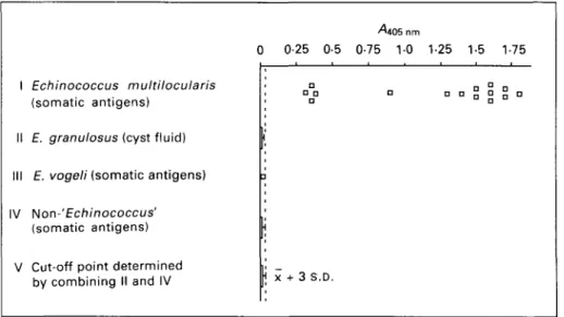
A solid-phase M A b G l l sandwich-ELISA was used as a first step to establish the operating characteristics such as specificity (Fig. 1) and methodical sensitivity (Fig. 2).
Specificity of the assay was tested by investigating 15 isolates of E. multilocularis versus 24 isolates of E. granulosus, 1 isolate of E. vogeli and various
non-1 II III IV V 0 Echinococcus multilocularis (somatic antigens)
E. granulosus (cyst fluid) E. vogeli (somatic antigens)
Non-' Echinococcus' (somatic antigens) Cut-off point determined by combining II and IV
^405 nm
0-25 0-5 0-75 10 1-25 1-5 1-75 ! n°Q n „ „ » jj ° n
| X + 3 S.D.
Fig. 1. Specificity of MAb G i l sandwich-ELISA with antigens tested at a concentration of 50 /ig protein/ml. /, Echinococcus multilocularis, 15 isolates originating from Switzerland, France, Alaska, Germany, Japan and Canada. / / , E. granulosus hydatid cyst fluids from bovine (n = 13), equine (n = 2), porcine (n = 2), ovine (n = 1), camel (n = 1) and human (n = 5) origin. / / / , E. vogeli somatic metacestode antigen. IV, Non-Echinococcus somatic antigens were from Taenia solium cysticerci (n = 2), T. saginata cysticerci (n = 3), T. crassiceps metacestodes (n = 1), T. hydatigena metacestodes (n = 2), Mesocestoides corti metacestodes (n = 1). Adult-stage somatic antigens were from Toxocara canis,
Fasciola hepatica and Dicrocoelium dendriticum, control extracts from muscle and liver of human, mice and cattle
origin. V, The cut-off threshold determining the lower resolving limit was calculated on the basis of yi405nm values
from / / and IV.
1-6-1-4 1-2-1 - 0 • 0-8 0-6 0-4 0-2 0 — • • - • • - • • - • • - • -a—a—Q—p--800 200 50 12-5 3-1 0-8 0-2 005 001
Antigen in test solution (^g/ml)
Fig. 2. Titration of ( • ) Echinococcus multilocularis somatic antigen, ( • ) E. granulosus hydatid cyst fluid antigen and ( • ) Taenia solium cysticercus somatic antigen by MAb G i l sandwich-ELISA. E. multilocularis somatic antigen was also controlled by irrelevant MAb G10/4 sandwich-ELISA (O). The lower resolving limit corresponds to that shown in Fig. 1.
Echinococcus helminth and control antigens (listed in detail in Fig. 1). The assay was controlled by simultaneous testing on solid-phase irrelevant G10/4 MAb. The respective control values (never exceeding 0-02 Aiob nm for all non-£. multilocularis
antigens and 0-20 AM6 nm for all E. multilocularis
antigens) were individually subtracted from the value obtained with the specific solid-phase MAb G i l . The data show that all E. multilocularis isolates were clearly detected by the present sand-wich-ELISA, and specificity was 100% due to absolutely no binding activity with any antigens derived from other Echinococcus species, other helminths or control tissues. All antigen concen-trations had been equivalently adjusted to 50 /ig protein/ml after optimizing test parameters and determining methodical sensitivity (see Fig. 2). Arbitrarily, we selected a positive/negative threshold characterized by a cut-off ^4405nm-value set at
JC + 3S.D. from all non-Zs. multilocularis isolates. The methodical sensitivity was determined by plotting titration values obtained with primary solid-phase MAb G i l and values from solid-phase irrelevant control MAb G10/4, including sequentially hom-ologous E. multilocularis metacestode antigen and MAb Gil-alkaline phosphatase conjugate in this reaction (Fig. 2). Based on this experiment the final methodical sensitivity was depicted as a concen-tration of 80 ng E. multilocularis protein/ml test solution. For demonstration of insignificant back-ground-reactivity the figure includes also E. granu-losus and T. solium antigens tested on solid-phase MAb G i l , data of the corresponding control re-actions with G10/4 are not shown as values never exceeded 0-02v4405nm. Testing titrated E.
multi-locularis antigen with solid-phase control MAb G10/4 showed some non-specific reaction at concentrations larger than 13 fig protein/ml. These
^ " * V « ^ Methods Antigens ^ ^ ^
Dot-ELISA
1 Echinococcus multilocularis
Metacestode somatic antigen Metacestode E/S-antigen Adult (gravid) somatic antigen Oncosphere somatic antigen
Oncosphere (cultivated 13 days
in vitro) somatic antigen
II Purified E. multilocularis antigens Em2
Em2(G11) %
Em 11/3-10
III Control antigens
Mouse liver somatic antigen Mouse serum Calf serum Culture medium E. granulosus protoscolex somatic antigen Sandwich ELISA Control MAb MAb G11 G10/4 A 405 nm 0-887 0-117 0-811 0023 0002 0004 0002 0004 0-710 0050 0-785 0051 0-516 0008 0002 0002 0002 0007 0006 0006 0001 0002 0004 0016 0002 0006
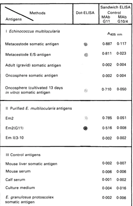
Fig. 3. Specificity of dot-ELISA and sandwich-MAb G i l - and control MAb G10/4-ELISA.
values, reflecting minor non-specificity of E. multi-locularis antigen components (they were never ob-served with any other antigens tested), were subse-quently corrected by subtraction, similar to the corrections performed in Fig. 1,
Analysis of stage-specificity by sandwich-ELISA and dot-ELISA (Fig. 3 /) revealed that only antigens derived from the metacestode provided epitodes with MAb G i l - b i n d i n g properties. No reactivity of MAb G i l was observed with antigens from adult-stage tapeworms and with oncospheres freshly hatched from E. multilocularis eggs. MAb G i l , however, was able to react with antigen derived from oncospheres cultivated in vitro for 13 days. For further analytical characterization of the antigen with MAb G i l - b i n d i n g epitope(s), the previously purified Em2 antigen (Gottstein, 1985) and the recombinant II/3-10-antigen (Muller et al. 1989), known to be not related to Em2, were included in the
investigation. The respective results shown in Fig. 3 //confirm the Em2 specificity of MAb G l 1 or, in other words, proving that the Em2 antigen carries the MAb Gil-binding epitope. As expected, the recombinant 11/3-10 antigen, as a negative control, exhibited no binding activity to MAb G i l . In parallel to the sandwich-ELISA, the corresponding immunological reactions were tested by dot-ELISA with the same antigens but in solid-phase. Sero-logical results were qualitatively identical to those obtained in sandwich-ELISA.
All control antigens (Fig. 3 / / / ) showed no binding properties in the MAb G i l - and control MAb G10/4-sandwich-ELISA.
A - - • • • , - • - . ;
' ' 50 Mm
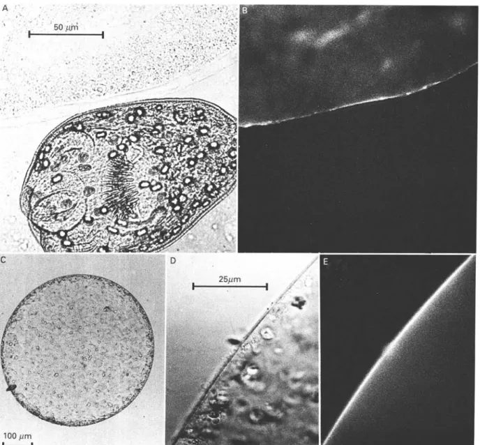
Fig. 4. Direct immunofluorescence analysis of the localization of the MAb Gilbinding Em2 antigen. (A) MAb G l l -FITC-labelled native Echinococcus multilocularis tissue under normal light. (B) The same as in (A) but under fluorescence. (C and D) MAb Gl 1-FITC-labelled native oncosphere, in vitro cultivated for 13 days, under normal light. (D) Magnified section of (C). (E) The same as (D) but under fluorescence.
Characterization of MAb Gil binding properties by fluorescence microscopy
Fluorescence microscopy analysis showed that from native E. multilocularis metacestode tissues only the laminated layer was stained by MAb G i l , with a relatively strong surface fluorescence (Fig. 4). Native protoscoleces did not appear to bind MAb G i l , nor did adult-stage E. multilocularis tapeworms or onco-spheres freshly hatched from E. multilocularis eggs (data for the latter two not shown). Oncospheres cultivated in vitro developed into spherical organisms with a surrounding thin layer. This layer demon-strated specific binding of MAb G i l at day 13 of in vitro cultivation.
Control reactions performed with irrelevant MAb G10/4 revealed that all parasite tissues
investi-gated did not non-specifically bind irrelevant mouse IgG.
Overall, the fluorescence microscopy analysis was in perfect agreement with the results from sandwich-and dot-ELISA.
ELISA with Em2(Gll)-antigen
In order to determine the immunodiagnostic characteristics of the affinity-purified E m 2 ( G l l ) antigen, an analysis was performed by ELISA in comparison with other E. multilocularis antigens listed below and in Fig. 5. The results of investi-gating sera from human patients with alveolar echinococcosis clearly revealed similar operating characteristics for E m 2 ( G l l ) and Em2 (Gottstein, 1985) antigens, demonstrated by the relatively high
0604 02 n -r = • G BB 0-87 • I D °° P" A D D G <= 1 2 -O) 1 1-0-o | 08 -o Z 06 -E ui E 04 -c O ^ 0 2 -0 -, r=0-23 H • D • D a G • G D • B 20 -CD O ) C ID o 15 05 -0 -0-2 -0-4 -0-6 ^405 nm Em2(G11) antigen
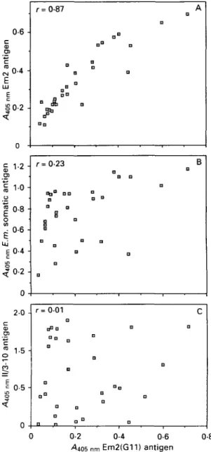
Fig. 5. Direct comparative analysis by ELISA of Em2 antigen purified with solid-phase MAb (antigen Em2(Gll)) versus (A) Em2 antigen (Gottstein, 1985), (B) a crude Echinococcus multilocularis somatic antigen and (C) a purified recombinant 11/3-10 antigen known to be unrelated immunologically to Em2 antigen (Muller
et al. 1989); the investigation was done with defined sera
from 30 human patients with alveolar echinococcosis (see Material and Methods section).
correlation coefficient (r = 0-87). Crude E.
multi-locularis somatic antigen (CH10 isolate) also
ex-hibited some minor degree of correlation with the Em2(Gll) antigen (r = 023), whereas the recom-binant II/3-10 antigen (Muller et al. 1989) demon-strated no statistical correlation with the Em2(Gll) antigen (r = 0-01).
DISCUSSION
In this paper we describe a murine monoclonal antibody (designated MAb G i l ) that specifically reacts with an epitope present on the previously
characterized Em2-antigen of E. multilocularis meta-cestodes (Gottstein, 1985). The reactivity of this antibody was investigated by sandwich-ELISA and dot-ELISA with various isolates of E. multilocularis metacestodes for demonstrating that the epitope corresponding to MAb Gil was ubiquitously pre-sented by all isolates. This observation confirmed directly the demonstration of conservation of the Em2 antigen previously assessed by indirect anti-Em2 antibody detection in human patients with alveolar echinococcosis, originating from geographi-cally dispersed endemic areas (Gottstein et al. 1986). Direct proof for the localization of the epitope recognized by MAb Gil on the Em2 antigen was found by investigating affinity-purified (according to Gottstein (1985)) Em2 antigen in sandwich- and dot-ELISA. In both test systems, MAb Gil showed binding activity to the Em2 antigen, whereas a negative control antigen (recombinant II/3-10 anti-gen, known to be not related to the Em2 antigen) remained negative as expected. Indirect evidence for Em2 identity was also obtained by a comparative investigation of sera from human patients with alveolar echinococcosis in ELISA with different solid-phase antigens: Em2(Gll) versus Em2 (Gott-stein, 1985) demonstrated statistically a good cor-relation. Some degree of correlation occurred also with somatic E. multilocularis metacestode antigen. This was to be expected because of the relatively high proportion of Em2 antigen observed previously in somatic extracts (Gottstein et al. 1983; Gottstein, 1985).
Of considerable interest was the observed meta-cestode stage-specificity of the E. multilocularis epitope binding MAb G i l and being localized on the Em2 antigen. According to direct immunofluorescence analysis, the molecule with MAb G l l -binding activity appeared to be accumulated in the laminated layer either adjacent to the germinal layer or to oncospheres developed in vitro for 13 days. The laminated layer is known to remain for extremely long times in infected host tissue, even after spontaneous 'dying-out' of the larval parasite (Rausch et al. 1987). The remarkably long per-sistence of anti-Em2 antibodies in serum from patients with such aborted lesions hence becomes explained, as well as the very rapid decrease of anti-Em2 antibody concentrations (finally becoming negative) observed after complete surgical resection of such dead lesions (Lanier et al. 1987; Gottstein
et al. 1989).
Structures of the native germinal layer did, in general, not bind MAb Gil in the direct immuno-fluorescence analysis, although some individual, not further defined cells appeared fluorescent (data not shown). The acellular nature of the MAb G i l -positive laminated layer still requires clarification of (i) the localization of the site of production of the epitope in question, (ii) the nature of the respective
synthesizing parasite cells and (iii) the exact bio-chemical properties of the molecule(s) carrying the MAb Gl 1-binding epitope. Such experiments are presently under investigation.
In conclusion, the development of a species-specific monoclonal antibody against an epitope of the Em2 antigen proved useful for further biological characterization and purification of the Em2 antigen. Furthermore, the application of MAb G i l in a sandwich-ELISA demonstrated also its potential value for the diagnostic identification of E. multi-locularis metacestodes and simultaneously for its discrimination from various other cestode, helmin-thic or bacterial cell extracts.
We wish to thank Professor J. Eckert for the fruitful discussions and suggestions concerning the experiments and the manuscript. The authors would also like to express gratitude to Dr M. Aubert (Nancy, France) and Dr P. Jacquier (Zurich) for providing some of the parasite material. The work was supported by the Swiss National Science Foundation (grant no. 3-29651.90).
R E F E R E N C E S
AGGARWAL, A., MERRITT, J. W. & NASH, T. E. ( 1 9 8 9 ) .
Cysteine-rich variant surface proteins of Giardia
lamblia. Molecular and Biochemical Parasitology 23,
3 9 ^ 8 .
BAUMANN, D. & GOTTSTEIN, B. (1987). A double-antibody
sandwich ELISA for the detection of Entamoeba
histolytica antigen in stool samples of humans. Tropical Medicine and Parasitology 38, 81-5.
DAO, L. (1985). An improved method of antigen detection on nitrocellulose: in situ staining of alkaline phosphatase conjugated antibody. Journal of
Immunological Methods 82, 225-31.
DE ST. GROTH, F. s. & SCHEIDEGGER, D. (1980). Production
of monoclonal antibodies: strategy and tactics. Journal
of Immunological Methods 35, 1—21.
DEPLAZES, p. & ECKERT, j . (1988). Massengewinnung und
Lagerung von Taenia hydatigena-Eiern sowie Isolierung lebensfahiger Onkospharen. Schweizer
Archiv fur Tierheilkunde 130, 307-20.
ECKERT, j . & POHLENZ, j . (1976). Zur Wirkung von
Mebendazol auf Metazestoden von Mesocestoides corti und Echinococcus multilocularis. Tropical Medicine and
Parasitology 27, 247-62.
ENGVALL, E. & PERLMANN, P. (1972). Enzyme-linked
immunosorbent assay, ELISA III. Journal of
Immunology 109, 120-35.
FLEISCHMANN, J. B., PAIN, R. H. & PORTER, R. R. ( 1 9 6 2 ) .
Reduction of y-globulin. Archives of Biochemistry and
Biophysics 1 (Suppl.), S174-S180.
GOTTSTEIN, B. (1985). Purification and characterization of a specific antigen from Echinococcus multilocularis.
Parasite Immunology 7, 201—12.
GOTTSTEIN, B., ECKERT, J. & FEY, H. (1983). Serological
differentiation between Echinococcus granulosus and
E. multilocularis infections in man. Parasitology Research 69, 347-56.
GOTTSTEIN, B., SCHANTZ, P. M. & WILSON, J. F. ( 1 9 8 5 ) .
Serological screening for Echinococcus multilocularis infections with ELISA. Lancet i, 1097-8.
GOTTSTEIN, B., SCHANTZ, P. M., TODOROV, T., SAIMOT, A. G.
& JACQUIER, p. (1986). An international study on the serological differential diagnosis of human cystic and alveolar echinococcosis. Bulletin of the World Health
Organization 64, 101—5.
GOTTSTEIN, B., LENGELER, C , BACHMANN, P . , HAGEMANN, P., KOCHER, P . , BROSSARD, M., WITASSEK, F. & ECKERT, J.
(1987). Sero-epidemiological survey for alveolar echinococcosis (by Em2-ELISA) of blood donors in an endemic area of Switzerland. Transactions of the
Royal Society of Tropical Medicine and Hygiene 81,
960-4.
GOTTSTEIN, B., TSCHUDI, K., ECKERT, J. & AMMANN, R.
(1989). Em2-ELISA for the follow-up of alveolar echinococcosis after complete surgical resection of liver lesions. Transactions of the Royal Society of
Tropical Medicine and Hygiene 83, 389—93. GOTTSTEIN, B., DEPLAZES, P. J., ECKERT, J., MULLER, E.,
SCHOTT, E., HELLE, O., BOUJON, P . , WOLFF, K., WANDELER, A., SCHWIETE, U. & MOEGLE, H. ( 1 9 9 1 ) .
Serological (Em2-ELISA) and parasitological examinations of fox populations for Echinococcus
multilocularis infections. Journal of Veterinary Medicine, Series B (in the Press).
LANIER, A. P . , TRUJILLO, D. E., SCHANTZ, P. M., WILSON, J. F., GOTTSTEIN, B. & MCMAHON, B. J. ( 1 9 8 7 ) .
Comparison of serologic tests for the diagnosis and follow-up of alveolar hydatid disease. American
Journal of Tropical Medicine and Hygiene 37, 609—15. MULLER, N., GOTTSTEIN, B., VOGEL, M., FLURY, K. &
SEEBECK, T. (1989). Application of a recombinant
Echinococcus multilocularis antigen in an ELISA for
diagnosis of human alveolar echinococcosis. Molecular
and Biochemical Parasitology 36, 151-60.
RAUSCH, R. L, WILSON, J. F., SCHANTZ, P. M. & MCMAHON,
B. j . (1987). Spontaneous death of Echinococcus
multilocularis: Cases diagnosed serologically (by
Em2-ELISA) and clinical significance. American Journal of
Tropical Medicine and Hygiene 36, 576-85.
SCHANTZ, P. M. & GOTTSTEIN, B. (1986). Echinococcosis
(Hydatidosis). In Immunodiagnosis of Parasitic
Diseases, vol. 1 (ed. Walls, K. W. & Schantz, P. M),