Echinococcus multilocularis: molecular and
immunochemical characterization of diagnostic antigen
II/3-10
R. FELLEISEN and B. GOTTSTEIN*
Institute of Parasitology, University of Berne, Ldnggasse-Strasse 122, 3001 Berne, Switzerland (Received 9 December 1992; accepted 1 March 1993)
SUMMARY
A recombinant Echinococcus multilocularis antigen (II/3-10), which had previously been shown to exhibit immuno-diagnostic characteristics highly specific for human alveolar echinococcosis, and the corresponding native parasite antigen, were further characterized with immunochemical and molecular biological methods. Immunoblot analysis using a polyclonal antiserum raised in rabbits against the recombinant protein, and subsequent Northern hybridization analysis, revealed that the native antigen was expressed by E. multilocularis at the adult as well as at the metacestode stage. In metacestodes, the antigen was shown by using indirect immunofluorescence and the same antiserum to be localized within the germinal layer and membrane structures of developing protoscolices. Electrophoretic analyses revealed remarkable differences in the apparent molecular weight of the antigen under reducing and non-reducing conditions. In further immunoblot analyses, anti-II/3-10 antibodies identified the corresponding epitopes on bands with identical MT in all E.
multilocularis isolates investigated (European, Asian and North American). By Southern hybridization analyses of the
respective gene, phylogenetically related sequences were shown to be present in other helminth species such as E.
granulosus and several Taenia spp. In the same respect, immunoblotting revealed that anti-II/3-10 antibodies reacted
with antigens of different Mr from various E. granulosus isolates and some other cestode species, indicating the presence of shared and thus potentially cross-reacting epitopes. T h e relevance of these findings for the immunodiagnostic per-formance of the recombinant antigen is discussed.
Key words: Echinococcus multilocularis, Taeniid cestodes, immunodiagnosis, recombinant antigen.
INTRODUCTION
Alveolar echinococcosis is a zoonotic disease of man caused by infection with the larval stage (metacestode) of Echinococcus multilocularis. The parasite is found in its natural life-cycle only on the Northern hemisphere, where endemic areas include mainly central Europe, Canada and North America, China, Japan and others. Disease is attributed to the tumour-like invasive growth of the parasite located primarily in the liver of the patients. By the time the disease becomes clinically manifest, the lesions caused by the metacestodes have often reached an extension too large for complete surgical resection. Therefore, early serological detection of alveolar echinococcosis already in the stage of small hepatic lesions and subsequent treatment of patients are important means to reduce mortality.
A reliable immunodiagnosis of alveolar echino-coccosis depends upon the availability of sufficient amounts of well-defined species-specific and diagnostically sensitive parasite antigens. In two * Reprint requests to: Professor Dr B. Gottstein, Institute of Parasitology, University of Berne, Langgasse-Strasse 122, CH-3012 Berne/Switzerland.
former publications we summarized the identific-ation and cloning of the gene fragment coding for the immunoreactive recombinant E. multilocularis anti-gen 11/3 (Vogel et al. 1988) and the expression in bacteria of its subfragment II/3-10 carrying the relevant epitopes (Miiller et al. 1989). Immuno-logical evaluation of the recombinant antigen 11/3-10 revealed a high diagnostic sensitivity and specificity (Gottstein et al. 1993) indicating a high potential value of this antigen for immunodiagnosis. Besides the well-established diagnostic parameters of antigen II/3-10 only little was known about its biological nature. Therefore the present paper describes the characterization of the recombinant antigens II/3 and II/3-10 and the corresponding native parasite antigen with molecular biological and immunochemical methods.
MATERIALS AND METHODS
Parasites
Echinococcus multilocularis metacestode tissues were obtained from experimentally infected gerbils (Meriones unguiculatus), or C57BL/6J mice (parasite isolates corresponded in the main to those described previously by Gottstein & Mowatt (1991)) or from Parasitology (1993), 107, 335-342 Copyright © 1993 Cambridge University Press
336
Table 1. Documentation on the parasite isolates used for immunoblotting, Southern and Northern hybridization analyses, respectively
Isolate code CH6 CH22 KF5 (clone) F4 Al CDN1 SLI144 F 4 Eg-h Eg-c Eg-s Eg-p Eg-d T c Th T s a T s o T t Me M e Parasite species Echinococcus multilocularis Echinococcus granulosus Taenia crassiceps T. hydatigena T. saginata T. solium T. taeniaeformis Moniezia expansa Mesocestoides corti Parasite stage Metacestode Metacestode Metacestode Metacestode Metacestode Metacestode Metacestode Adult Metacestode Metacestode Metacestode Metacestode Metacestode Larval Adult Adult Larval Adult Adult Larval Geographical origin Switzerland Switzerland France France Austria Canada Alaska France Switzerland Switzerland Switzerland Switzerland Switzerland Egypt/Sudan Switzerland Switzerland South Africa Switzerland Switzerland Switzerland Reference (remarks) Gottstein & Mowatt (1991) Gottstein & Mowatt (1991) Gottstein et al. (1992) Gottstein & Mowatt (1991)
—
Gottstein & Mowatt (1991) Gottstein & Mowatt (1991)
—
Horse liver cyst Cattle lung cyst Sheep lung cyst Pig liver cyst Donkey liver cyst
Host origin Gerbil Gerbil C57BL/6J Gerbil C57BL/6J Gerbil Microtus D o g Horse Cattle Sheep Pig Donkey BALB/c D o g Human Human Cat Cattle BALB/c
naturally infected rodents caught on St Lawrence Island/Alaska (for details see Table 1). Adult stages of E. multilocularis were obtained from experimen-tally infected dogs after necropsy of the animals 24 days post-infection (p.i.) (dogs were perorally infected each with 125 g fertile metacestode tissue of the E. multilocularis isolate F4). Worms were recovered by incubating the resected small intestine in phosphate-buffered saline (PBS) at 37 °C for 1 h under slight agitation. Detached worms were aspirated from the sediment and washed 4 times in Hank's balanced salt solution (HBSS) at 37 °C prior to freezing at — 80 °C. E. granulosus cyst walls and protoscolices were isolated from naturally infected horses, cows, pigs or sheep (all Swiss isolates). Other adult or metacestode-stage cestodes and other helminths were obtained as described previously (Gottstein & Mowatt, 1991). An overview list of all parasite materials used in this paper is provided in Table 1.
Reagents used for recombinant DNA techniques Restriction endonucleases and E. coli DNA-Polymerase I (Kornberg polymerase and Klenow fragment) were obtained from Boehringer, Mannheim, FRG. T4 DNA ligase was purchased from New England Biolabs, Beverly, MA, USA. Sequenase 2'0-DNA sequencing kit and gene-clean DNA purification kit were supplied by United States Biochemical Corporation, Cleveland, OH, USA. Ultrapure reagents for RNA-Isolation were pro-vided by Gibco BRL, Basel, CH.
Radioactive materials
Radioactive nucleotides [a32P]dCTP and [a32P]dATP for labelling of nucleic acids and [a3oS]dATP for sequence analyses were obtained from Dupon NEN, Regensdorf, CH.
Nucleic acids
Ultrapure deoxynucleotides and pd(N)6 random
primers for labelling of DNA probes were purchased from Pharmacia, Diibendorf, CH. DNA molecular weight standard 1 kbp ladder and RNA 024-9-5 kb ladder markers were obtained from New England Biolabs, Beverly, MA, USA, and from Gibco BRL, Basel, CH, respectively.
Recombinant DNA methods
All recombinant DNA methods, unless otherwise stated, were performed as described by Sambrook, Fritsch & Maniatis (1989).
Purification of nucleic acids
Genomic DNA from different parasite species was purified using the 'DNA extraction kit' obtained from Stratagene, Heidelberg, FRG, according to the manufacturer's protocols.
For the purification of total RNA a modification of the method described by Glisin, Crkvenjakov & Byus (1974) and Ullrich et al. (1977) was used.
Either 07 g of packed adult parasites or 3 g of metacestode tissue, respectively, were homogenized in 5 volumes of 4 M guanidinium-thiocyanate con-taining 100 mM Tris-HCl (pH 7-5) and 1% 2-mercaptoethanol. After homogenization, sodium lauryl sarcosinate was added to a final concentration of 0-5 %. The samples were layered onto a cushion of 5-7 M CsCl/lOmM EDTA, pH 7-5 (ethylene-dinitrilo-tetraacetic acid) and ultracentrifuged for 24 h in a Beckman SW41 swinging bucket rotor at 32000 rpm. The pelleted total RNA was redissolved in diethyl pyrocarbonate (DEPC)-treated water.
Subcloning and sequencing
For determination of the nucleotide sequence of the cDNA inserts of clone II/3 and II/3-10, these were subcloned into the unique EcoRl site of plasmid vector pBluescript KS+. Sequencing was performed with Sequenase 2-0 (United States Biochemical Corporation, Cleveland, OH, USA) and [35S]dATP following the manufacturer's protocol. Sequencing primers KS and SK located adjacent to the poly-linker region were used. DNA sequences were processed and derived amino acid sequences were compared to the SWISS-PROT sequence database (release 21) using the HUSAR computer program set (Heidelberg Unix Sequence Analysis Resources) of the German Cancer Research Center (DKFZ, Heidelberg, FRG).
Southern and Northern hybridizations
For Southern analysis, 20 fig aliquots of genomic DNA of different parasite species and isolates were digested with the appropriate restriction enzyme and electrophoresed through 0-8 % agarose gels. RNA analysis was performed by separating 8 fig of total RNA on a 1'2% formaldehyde agarose gel (Lehrach et al. 1977). The samples were transferred to Nytran Nylon membranes (Schleicher & Schiill, Dassel, FRG) following the manufacturer's protocols. For preparation of a specific probe, the cDNA insert coding for II/3 was isolated and radiolabelled to high specific activity by random hexamer priming/ extension reaction in the presence of [<*32P]dCTP or [a32P]dATP (Feinberg & Vogelstein, 1983, 1984). Hybridization was performed in 50% formamide/ 6 x SSPE/0-5 % SDS (sodium dodecyl sulphate) and 50% formamide/2-5 x Denhardt's reagent/5 x SSPE/0-1% SDS for Southern and Northern analysis, respectively. Following incubation for 12 h at 42 °C the filters were washed for 30 min with 6 x SSPE/0-1% SDS at room temperature, for 60 min at 37 °C with 1 x SSPE/0-1 % SDS, and for 60 min at 65 °C with 0-1 x SSPE/1 % SDS, if not otherwise stated. ( l x S S P E : 0-18 M NaCl/10mM NaPO4, pH 7-7/1 mM EDTA.)
SDS-polyacrylamide gel electrophoresis (SDS-PAGE) and immunoblotting
Parasite extracts were prepared from the respective parasite tissues as described previously (Gottstein, 1991). Aliquots of the protein extracts were mixed with an equal volume of 2 x sample buffer (100 mM Tris-HCl, pH 6-8/4% SDS/10% glycerol/0-1 % bromophenol blue) containing 100 mM dithiothreitol (DTT) or not depending on the experiment. Samples were boiled for 5 min and separated by SDS—PAGE according to Laemmli (1970) using 4-20% pre-cast gradient gels (Bio-Rad, Glattbrugg, CH). Transfer to nitrocellulose was performed by the Western-blot technique (Towbin, Staehelin & Gordon, 1979). To accomplish the immunoblotting procedure, the filters were saturated with 3 % bovine serum albumin (BSA) in PBS/01 % Tween. Antisera were incubated with the filters at 200-fold dilution in PBS/0-1 % Tween overnight at 4 °C. Bound antibodies were detected by anti-species antibodies conjugated to horseradish peroxidase (see below). The peroxidase-dependent colour reaction was carried out by incubating filters in 100 ml of staining solution (1 x PBS/1-5 mM 3,3'-diamino-benzidine tetrahydrochloride/0-003 % H2O2). Reagents used for immunological studies
Goat anti-rabbit IgG peroxidase-conjugate was purchased from Sigma, Buchs, CH (Cat. No. A 0545). The corresponding fluorescein-isothiocyanate (FITC)-conjugate was from Southern Bio-technology, Birmingham, AL, USA (Cat. 4010-02). Polyclonal hyperimmune serum directed against II/3-10 was obtained by immunization of rabbits with 100/ig II/3-10 antigen (purified according to the method of Miiller et al. 1989) per injection, using RIBI® adjuvant and the immunization protocol provided by the manufacturer (Ribi Immunochem. Res., Hamilton, MO, USA).
Immunofluorescence microscopy
The direct immunofluorescence staining of the native II/3-10 antigen was performed on cryo-sections of hepatic liver lesions (8 fim thickness) from a rodent (Microtus oeconomus) naturally infected with E. multilocularis originating from St Lawrence Island, Alaska (this isolate was subsequently main-tained in gerbils and designated as isolate SLI 144). Sections on glass slides were fixed in acetone, air dried and washed 3 times with PBS. One hundred fi\ of an incubation solution (Gottstein & Nash, 1991) containing 5 /A of hyperimmune rabbit serum or 5 fi\ of pre-immune rabbit negative control serum was dropped on the section and incubated for 30 min at 37 °C in a humid chamber. Slides were viewed in an Olympus BH-2 microscope and photographed on Kodak Ektachrome 64T film.
R. Felleisen and B. Gottstein
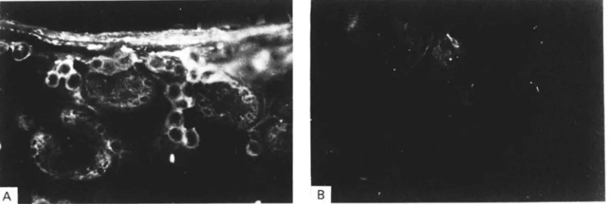
Fig. 1. Immunofluorescence analyses of cryosections from Echinococcus multilocularis metacestode tissue (liver lesion from a naturally infected rodent, see Materials and Methods section) using (A) rabbit hyperimmune serum against recombinant II/3-10 antigen. (B) Pre-immune negative control serum tested on a section of the same morphological area as in (A).
RESULTS
Immunofluorescence studies
In order to identify the localization of the I I / 3 antigen in situ, immunofluorescence studies using a polyclonal antiserum raised in rabbits against the affinity-purified recombinant II/3-10 antigen were performed. While no reaction above background was found with the pre-immune control serum (Fig. IB), an intensive fluorescence was observed with the undifferentiated germinal layer and in the periphery of the individual cell conglomerates inside the protoscolices (Fig. 1A). Consequently the 11/3 antigen seemed to be associated with membrane structures. Remarkably, no fluorescence was observed on the syncytial layer from the tegument of the developing protoscolices.
Stage specificity of the II j 3 antigen
For the identification of the native I I / 3 antigen in parasite extracts, immunoblot analyses using the same rabbit hyperimmune serum were performed. Under reducing S D S - P A G E running conditions, anti-II/3-10 antibodies identified 2 molecules in metacestode extracts with apparent molecular weights of 65 kDa and 52 kDa (Fig. 2).
Additionally, we were able to detect bands with identical molecular weights in extracts of adult parasites suggesting expression of the 11/3 antigen in both developmental stages. The protein of 52 kDa potentially could be interpreted as a processing or degradation product of the 65 kDa protein.
In contrast, marked differences in the apparent molecular weight of the I I / 3 antigen were observed under non-reducing S D S - P A G E running con-ditions (Fig. 2). A band of about 200 kDa was detectable in metacestodes as well as in the adult parasites. Speculatively, the reason for these differences could be explained by oligomerization of the I I / 3 antigen under native conditions or by a close
1 2 3 1 2 3 I I I I I I I 200 kDa •97-4 -66-2 - 4 5 0 - 3 1 0 - 21-5 - 14-4 A B
Fig. 2. Detection of Echinococcus multilocularis antigens with anti-II/3-10-specific hyperimmune serum raised in rabbits. Reducing SDS—PAGE running conditions (A) and non-reducing SDS—PAGE running conditions (B) were performed. Extracts of E. multilocularis metacestodes (Lanes 1), adults (Lanes 2), and extracts of Escherichia coli expressing recombinant 11/3-10 antigen (Lanes 3) were analysed.
association with other proteins. But this point deserves clarification by further investigations.
Species specificity of the 11/3 antigen
Investigation of potential variability of the I I / 3 antigen within E. multilocularis isolates originating from geographically different areas was done by immunoblotting. No intra-species variation could be observed with respect to the I I / 3 antigen; all isolates studied showed the same banding pattern (data not shown).
In the enzyme-linked immunosorbent assay (ELISA) with the affinity-purified recombinant II/3-10 antigen, a weak immunological cross-re-action was observed with a few sera from patients with hydatidosis and cysticercosis (Gottstein et al. 1993). Therefore, extracts of E. granulosus metacestodes isolated from different intermediate
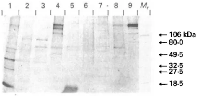
1 2 3 4 5 6 7 - 8 9 Mr I I I I I I I I I I I • -106 kDa - 8 0 0 -49-5 -32-5 -27-5 -18-5 Fig. 3. Immunoblot with extracts of taeniid cestodes with II/3-10-specific hyperimmune serum. Eg-s (Lane 1), Eg-c (Lane 2), Eg-h (Lane 3), Eg-p (Lane 4), Th (Lane 5), Tso (Lane 6), Tsa (Lane 7), Me (Lane 8), and CH6 (Lane 9). Molecular weight markers (Afr) are
indicated. (For explanation of abbreviations refer to Table 1.) 1 2 - 3 I I I I I -9-5 kb -7-5 -4-4 -2-4 -1-4 -0-24
Fig. 4. Northern hybridization with radioactively labelled insert of clone II/3 as a probe. Total RNA isolated from Echinococcus multilocularis adults (EmAd, Lane 1) and metacestodes (KF5, Lane 2) and from E. granulosus metacestodes (Eg-c, Lane 3) was analysed. (For explanation of abbreviations refer to Table 1).
hosts and of different cestode species were analysed by immunoblotting with the anti-II/3-10 specific hyperimmune serum (Fig. 3). Bands of different molecular weights could be detected with the E. granulosus isolates and other cestode species reflecting the presence of cross-reacting epitopes. Northern and Southern hybridizations
Further investigations with regard to these findings were performed at the level of nucleic acid. In Northern hybridizations using the radioactively labelled insert of clone 11/3 as a probe, a single band of about 2-1 kb was found in total RNA of E. multilocularis metacestodes and adult parasites (Fig. 4). It is interesting to note that a transcript of the same size was observed in total RNA isolated from
E. granulosus metacestodes but with a lower intensity of the signal. In immunoprecipitation following in vitro translation of total RNA in a cell-free rabbit reticulocyte extract, a single protein of about 65 kDa was precipitated from the translation mixture by the anti-II/3-10 antiserum both in metacestodes and adult parasites; a protein of 52 kDa was not observed (data not shown).
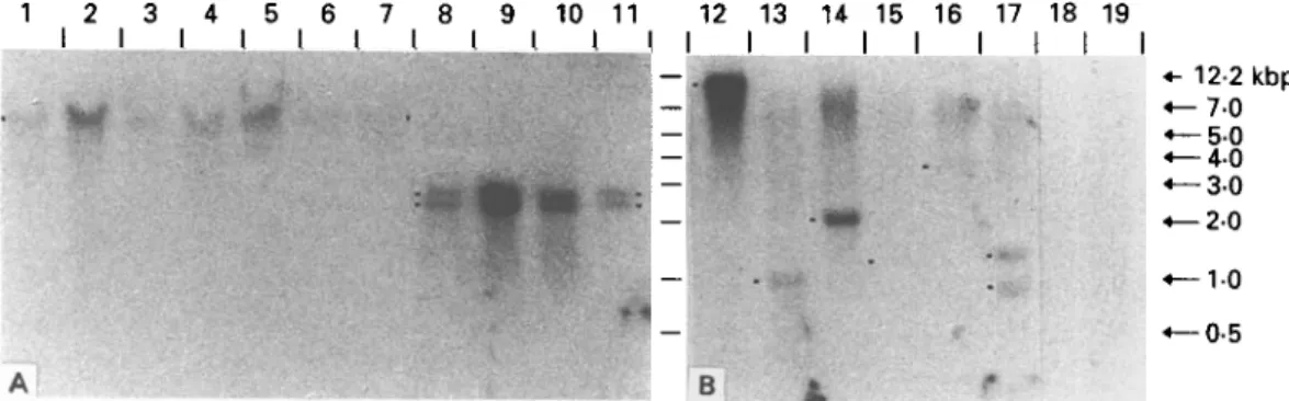
A uniform banding pattern could be observed in Southern hybridization with genomic DNA digested with EcoRl of several E. multilocularis isolates originating from geographically different areas. A band of about 5-5 kbp could be detected with the 11/3 specific probe in all isolates studied (Fig. 5).
Consistent with the results of Northern analysis, cross-hybridizing bands were found with E. granulosus genomic DNA. All E. granulosus isolates studied, originating from different intermediate hosts, showed an identical banding pattern, differing from E. multilocularis with respect to a double band of about 2-5 and 2-8 kbp, respectively (Fig. 5).
With DNA of several other taeniid cestodes, no bands hybridizing with the II/3 specific probe were detectable under stringent washing conditions (final wash 65 °C, 0-1 x SSPE/1 % SDS), presumably due to decreased sequence homology to the E. multi-locularis gene fragment (data not shown). In contrast, using conditions of low stringency (final wash room temperature, 1 x SSPE, 0-1 % SDS), cross-hybridization was clearly detectable with genomic DNA of Taenia crassiceps, T. taeniaeformis, and T. saginata (Fig. 6; to obtain a clearcut banding pattern, the enzyme BamHI was chosen for digestion of the genomic DNAs). Furthermore, weak signals were observed with T. hydatigena and T. solium, indicating the widespread distribution of the 11/3 gene or related sequences in the family Taeniidae. No signal was detectable with Moniezia expansa and
Mesocestoides corti.
Sequences of clones 11/3 and 11/3-10
The clone 11/3 and its derivative 11/3-10 carried cDNA inserts of 1 kbp and 0-6 kbp, respectively (Miiller et al. 1989). Each insert was isolated, subcloned into pBluescript KS+, and its respective nucleotide sequence determined. Both strands were completely sequenced. The primary DNA sequences and the derived amino acid sequences of the two clones in question are shown in Fig. 6.
An open reading frame extends over most of the nucleotide sequence of clone 11/3. An amber stopcodon (TAG) at position 886 is followed by a 3' non-coding region of 190 bp and a poly-A tail of 28 A-residues. The underlined ATTAAA sequence element found 21 bp upstream of the poly-A tail presumably serves as a polyadenylation signal.
The sequence of the cDNA of truncated clone 11/3-10, which was generated by sonication of the
R. Felleisen and B. Gottstein 340 1 2 3 4 5 6 7 8 9 10 11 12 13 H 15 16 17 18 19
<—10 •—0-5 A B 4 f
Fig. 5. Southern analyses of genomic DNA with radioactively labelled insert of clone II/3 as a probe. (A) Digestion with EcoRl and conditions of high stringency (Lanes 1-11); (B) restriction with BamUl and low stringency (Lanes 12-19). CH6 (Lane 1), CH22 (Lane 2), Al (Lane 3), F4 (Lane 4), KF5 (Lane 5), CND1 (Lane 6) and SLI144 (Lane 7); isolates Eg-c (Lane 8), Eg-h (Lane 9), Eg-p (Lane 10) and Eg-d (Lane 11); CH6 (Lane 12), Tc (Lane 13), Tt (Lane 14), Th (Lane IS), Tso (Lane 16), Tsa (Lane 17), Me (Lane 18) and Me (Lane 19). (For explanation of abbreviations refer to Table 1.)
iGMTTCCGGtriTTTTTTTTTC-n'GGTGGAAAAATCCAAGATTAACflAGCGCATTTTGGCATTGTGTAC'rggCAAC 7 5 P F S ' F L V E K S K l K r K & f L A k C f G K CATG AGCTCTACATGCGTAGAAGAAAGTCAGACTCTATTGAGGTGCAACAGATGAAGATTCAGGCCAAGGAGGAA 1 5 0 H E I . Y M R R R K S D S I E V Q Q M K I Q A K E E CGTGAATTGftAGGAGGCTGAGAGACAACGCCTGAAGGAGGAACGATTGCAACGITA5'GGAAAATGAACAGAAACTG 2 2 5 R E L K E A E R Q R L K E E R L Q R M . E N E O K L CGGGAGCTTCGfGCTCAAATGGTCCAAAAGGAGTCtGACTTAGCGGATAreAAGAAtAAGGCAtCTGCCTATGftG 3 0 0 R E L R A Q M V E K E S D L A D M K N K A S A Y E AGTAAGATTGCGGAGCTGGAGATGCTGCTACAGCAGGAGCGACATGCGCGTSAGAGTCTTCAGAAGAGCCAAGAC 3 7 5 S K 1 A E I E M L I . Q Q E R H A R E S I . Q K S Q D
AAAC TGGCGGAGATGAACAGAAAGCTGAAGGAGGAG ACTGCGGCATCAGCCGAAGAGCGCGACCGTCTGAXGGCC 4 5 0
K L A E M N R K . L K E E T A A S A E E R D R L M A CACCG.TGACGAAGTGCAACGCGAAGrTGAGGCTCAGAAGGTCGCCATGGCCAAGAAGGAAGCTGAAAAGGCTCAG 5 2 5 Q R D E V Q R E V E A Q K V A M A K K E A E K A Q GCTGAAGCTGAGCTTCGCAGMlTGCGTGAGAAACACGAtGCAAACCACAAGTCCCAGGTCAATGGCAGTGGTGAC 6 0 0 A E A E L R R M S E K H O A K H K S Q V N G S G D GCTGCTTCGCAGGATGATGAAAGTGAAGCCAAGGAACTTGAGGTGATACCAAATGTGAGGCGGACGGAGGAATCG 6 7 5 A A S Q D D E S E A K E L E V I P N V R R T E E S AGGGTGACGGCCGTCTCTAAGAATGAGACGCTCCAGACGAAGCTGGCCAACCTCAAAATGGAGTTGAGCTCGACA 7 5 0 R V T A V S K N E T L Q T K L A N L K M E L S S T CGCGATCAGTCGAAAATGCGCGACATTGATCGTCGTCATGAGTACAATGTGCGGGAGGGTAATGACAAGTACAAG 8 2 5 R D Q S K M R D I D R R H E Y N V R E G N D K Y K ACACTGCGCAACATTCGCAAGGGCAACACCATGTGTCGTGTTGAACAGTTTGAGTCGATGTAGAAATGTTACAGT 9 0 0 T L R N I R K G N T M C R V E Q F E S M * TGTCTTCATTCCCCTCATCTTTCTGCAATTTTGGACCCTCTATCACTACGGTTACTTCCTCAATCATGCTGCTAC 9 7 5 AGTGCTATCCAACATTCCATTTTTATTCTCGCGATGCACACCTGTCTTTTCATTCTGCCCCTTCTTATCTAACGT 1 0 5 0 CCACTTTMIAA&CTGCTATCGTACGCTAAAAAAAAAAAAAAAAAAAAAAAAAAAfllCCGGAATTCl 1 1 1 5
Fig. 6. Nucleotide and deduced aminoacid sequences oi Echinococcus multilocularis cDNA-clone 11/3. Adaptor sequences containing EcoRl sites used for cloning are boxed. Presumable polyadenylation signal ATTAAA is underlined. Sequence of truncated clone II/3-10 is shaded in grey. Last nucleotide (G at position 574) of II/3-10 indicated by an arrow head is followed by adaptor sequences (not shown).
cDNA-insert encoding antigen II/3 and subsequent sistent with the size observed for the corresponding cloning of the shortened fragment, is located in the recombinant antigens (Vogel et al. 1988; Miiller et 5'-terminal region of clone II/3 ending at position al. 1989).
575. The coding capacities of the two cDNAs are 255 In order to establish a possible function for the and 188 aminoacids, potentially yielding proteins of antigen, the deduced amino acid sequence of clone 29-5 kDa and 21-5 kDa, respectively. This is con- II/3 was compared to the sequences of the
SWISS-PROT data base. It proved to be identical to the sequence of the E. multilocularis antigen Em 10 recently published by Frosch et al. (1991), representing about 52 % of its C-terminal sequence. While only minor nucleotide changes at the ulti-mate 5'-end in the coding region of the II/3-cDNA were found, that presumably could be due to cloning artefacts ( T T T . T T T . T T T instead of G A G . T T T . T C T ; Fig. 6), extensive differences concerning the 3-non-coding region of clone 11/3 compared to the sequence published by Frosch et al. (1991) were observed. The 11/3 clone ends prior to the last 27 nucleotides of the non-coding region showing no homology to the corresponding sequence published for EmlO. This raises the possibility of cDNA clones resulting from transcripts of different copies of the respective gene.
DISCUSSION
In two former studies the identification of an immunodiagnostic recombinant E. multilocularis antigen I I / 3 (Vogel et al. 1988) and the expression in bacteria of its subfragment II/3-10 carrying the relevant epitopes (Muller et al. 1989) have been described. The recombinant antigen allowed de-tection of anti-Zs. multilocularis antibodies in human patients with high diagnostic sensitivity and specificity (Gottstein et al. 1993). Although the immunodiagnostic performance of the antigen had been studied already in detail and its suitability for immunodiagnosis of alveolar echinococcosis is well established, almost nothing was known about its biological nature. But, generally, characterization of antigens used for immunological diagnosis exceeding diagnostic evaluation is desirable. The purpose of the present study therefore was the analysis of the recombinant antigen and the corresponding native parasite antigen with immunological and molecular-biological methods.
Nucleotide sequence analyses of the cDNAs coding for 11/3 and II/3-10 revealed the identity of their sequences with a section of the sequence published for antigen EmlO, an E. multilocularis-antigen which was described by Frosch et al. in 1991. The clone 11/3 represented about 5 2 % of its C-terminal end, clone II/3-10 was located at the 5' end of clone I I / 3 .
In immunoblot anti-II/3-10 antibodies identified two molecules in metacestode extracts with apparent molecular weights of 65 kDa and 52 kDa. Two bands with about the same molecular weights have been observed in metacestode extracts using an antiserum specific for antigen EmlO (65 and 55 kDa, respectively; Frosch et al. 1991).
Based on the nucleotide sequence analysis and the fact that anti-II/3-10 (as shown in our study) and EmlO-specific antiserum (Frosch et al. 1991) detected molecules in the same molecular weight
range, it seems reasonable to assume that both antigens are identical although the complete se-quence of the gene represented by clone I I / 3 is still required for confirmation.
Immunoblot analysis using a II/3-10-specific antiserum raised in rabbits against the affinity-purified recombinant antigen and Northern hybridization analysis with a specific probe revealed expression of the antigen in metacestodes as well as in adult parasites. The level of expression seems to be similar in both developmental stages. This novel finding could reflect an important function of the antigen, independent of the developmental stage.
All isolates of E. multilocularis originating from geographically different areas showed the same banding pattern in immunoblot with the specific hyperimmune serum. Clone II/3-10 was originally isolated from a cDNA library constructed from metacestode tissue of a Swiss isolate (CH-14; Vogel et al. 1988). Cross-reaction of II/3-10 anti-bodies with antigens from parasite material of different geographical origin was consistent with the reactivity of patient sera from the corresponding regions with the recombinant II/3-10 antigen (Muller et al. 1989).
Immunoblot analyses with the II/3-10 anti-serum clearly demonstrated the presence of cross-reacting epitopes in E. granulosus and several Taenia species. These findings contributed to the expla-nation for the weak immunological cross-reaction observed with a few sera from patients with hydatidosis and cysticercosis in the ELISA with recombinant II/3-10 antigen (Gottstein et al. 1993). Nevertheless, immunological reactivity of sera from patients with respective diseases seemed to be directed mainly against epitopes differing from those recognized by hyperimmune antibodies, thus finally resulting in an appropriate specificity of the recombinant II/3-10 antigen for the immuno-diagnosis of alveolar echinococcosis.
The results concerning the immunological characterization of the II/3-antigen could be con-firmed by experiments at the nucleic acid level. Southern hybridization showed that DNA sequences hybridizing with a II/3-specific probe were present in E. granulosus and several Taenia species. While the I I / 3 related sequences in E. granulosus seemed to be relatively conserved compared to the II/3-gene of E. multilocularis, homology of the corresponding sequences in the different Taenia species was less prominent.
Frosch et al. (1991) demonstrated the EmlO antigen by immunoblot analysis in E. multilocularis but not in E. granulosus. Therefore they suggested a possible role for this antigen in infiltrative tumour-like growth of E. multilocularis metacestodes. In addition to E. multilocularis we were able to detect the 11/3 antigen in E. granulosus and some Taenia species which do not demonstrate chronic invasive
342
growth characteristics such as observed in alveolar echinococcosis. Provided that both antigens are identical, the novel findings presented in this publication are in contradiction. Therefore experi-ments are now underway to isolate and analyse the complete II/3-gene from E. multilocularis and from E. granulosus for comparative characterization.
We thank Mrs I. Tanner for her expert technical as-sistance. The authors are indebted to Dr R. L. Rausch (Seattle), Dr J. Eckert (Zurich), Dr M. Aubert (Nancy), Dr H. Auer (Vienna) for the supply of parasite materials and Dr I. Benz (ZMBH, Heidelberg) for the processing of sequence data. This work was supported by a grant obtained from the Swiss National Science Foundation (No. 31-29651.90).
REFERENCES
FEINBERG, A. p. & VOGELSTEIN, B. (1983). A technique for radiolabeling DNA restriction endonuclease fragments to high specific activity. Analytical Biochemistry 132, 6-13.
FEINBERG, A. p. & VOGELSTEIN, B. (1984). Addendum: A technique for radiolabelling DNA restriction endonuclease fragments to high specific activity.
Analytical Biochemistry 137, 266.
FROSCH, P . M . , FROSCH, M . , PFISTER, T . , SCHAAD, V. & BITTER-SUERMANN, D. (1991). Cloning and
characterization of an immunodominant major surface antigen of Echinococcus multilocularis. Molecular and
Biochemical Parasitology 48, 121-30.
GLISIN, V., CRKVENJAKOV, R. & BYUS, C. ( 1 9 7 4 ) . Ribonucleic acid isolated by cesium chloride centrifugation. Biochemistry 13, 2633.
GOTTSTEIN, B. (1991). Echinococcus multilocularis: antigenic variance between different parasite isolates.
Parasitology Research 77, 359—61.
GOTTSTEIN, B. & NASH, T. E. (1991). Antigenic variation in
Giardia lamblia: infection of congenitally athymic
nude and scid mice. Parasite Immunology 13, 649—59.
GOTTSTEIN, B., DEPLAZES, P. & AUBERT, M. ( 1 9 9 2 ) .
Echinococcus multilocularis: immunological study on
the ' Em2-positive' laminated layer during in vitro and
in vivo post-oncospheral and larval development. Parasitology Research 78, 291-7.
GOTTSTEIN, B., JACQUIER, P . , BRESSON-HADNI, S. & ECKERT, J. (1993). Improved primary immunodiagnosis of alveolar echinococcosis in humans by an enzyme-linked immunosorbent assay using the Em2plus
-antigen. Journal of Clinical Microbiology 31, 373-6. GOTTSTEIN, B. & MOWATT, M. R. (1991). Sequencing and
characterization of an Echinococcus multilocularis DNA probe and its use in the polymerase chain reaction.
Molecular and Biochemical Parasitology 44, 183-94.
LAEMMLI, u. K. (1970). Cleavage of structural proteins during the assembly of the head of the bacteriophage T4. Nature, London 227, 680-5.
LEHRACH, H., DIAMOND, D., WOZNEY, J. M. & BOETKER, H. (1977). RNA molecular weight determinations by gel electrophoresis under denaturing conditions, a critical reexamination. Biochemistry 16, 4743-51.
MULLER, N . , GOTTSTEIN, B., VOGEL, M., FLURY, K. & SEEBECK, T. (1989). Application of a recombinant
Echinococcus multilocularis antigen in an
enzyme-linked immunosorbent assay for immunodiagnosis of human alveolar echinococcosis. Molecular and
Biochemical Parasitology 36, 151-60.
SAMBROOK, ] . , FRITSCH, E. F. & MANIATIS, T. ( 1 9 8 9 ) .
Molecular Cloning: a Laboratory Manual, 2nd edn.
Cold Spring Harbor Laboratory Press, Cold Spring Harbor, NY.
TOWBIN, H., STAEHELIN, T. & GORDON, J. ( 1 9 7 9 ) . Electrophoretic transfer of proteins from
polyacrylamide gels to nitrocellulose sheets: procedure and some applications. Proceedings of the National
Academy of Sciences, USA 76, 4 3 5 0 ^ .
ULLRICH, A., SHINE, J., CHIRGWIN, J., PICTET, R., TISCHER, E., RUTTER, w. j . & GOODMAN, H. M. (1977). Rat insulin genes: construction of plasmids containing the coding sequences. Science 196, 1313.
VOGEL, M., GOTTSTEIN, B., MULLER, N. & SEEBECK, T. (1988). Production of a recombinant antigen of
Echinococcus multilocularis with high
immunodiagnostic sensitivity and specificity.