Hormone Therapy Failure in
Human Prostate Cancer:
Analysis by Complementary
DNA and Tissue Microarrays
Lukas Bubendorf, Meelis Kolmer,
Juha Kononen, Pasi Koivisto, Spyro
Mousses, Yidong Chen, Eija
Mahlama¨ki, Peter Schraml, Holger
Moch, Niels Willi, Abdel G.
Elkahloun, Thomas G. Pretlow,
Thomas C. Gasser, Michael J.
Mihatsch, Guido Sauter, Olli-P.
Kallioniemi
Background: The molecular
mecha-nisms underlying the progression of
prostate cancer during hormonal
therapy have remained poorly
under-stood. In this study, we developed a
new strategy for the identification of
differentially expressed genes in
hor-mone-refractory human prostate
can-cer by use of a combination of
comple-mentary DNA (cDNA) and tissue
microarray technologies. Methods:
Dif-ferences in gene expression between
hormone-refractory CWR22R prostate
cancer xenografts (human prostate
cancer transplanted into nude mice)
and a xenograft of the parental,
hor-mone-sensitive CWR22 strain were
analyzed by use of cDNA microarray
technology. To validate the data from
cDNA microarrays on clinical prostate
cancer specimens, a tissue microarray
of specimens from 26 prostates with
benign prostatic hyperplasia, 208
pri-mary prostate cancers, and 30
hor-mone-refractory local recurrences was
constructed and used for
immuno-histochemical detection of protein
ex-pression. Results: Among 5184 genes
surveyed with cDNA microarray
tech-nology, expression of 37 (0.7%) was
increased more than twofold in the
hormone-refractory CWR22R
xeno-grafts compared with the CWR22
xe-nograft; expression of 135 (2.6%) genes
was reduced by more than 50%. The
genes encoding insulin-like growth
factor-binding protein 2 (IGFBP2)
and 27-kd heat-shock protein (HSP27)
were among the most consistently
over-expressed genes in the CWR22R
tu-mors. Immunohistochemical analysis
of tissue microarrays demonstrated
high expression of IGFBP2 protein in
100% of the hormone-refractory
clini-cal tumors, in 36% of the primary
tumors, and in 0% of the benign
pros-tatic specimens (two-sided P = .0001).
Overexpression of HSP27 protein was
demonstrated in 31% of the
hormone-refractory tumors, in 5% of the
pri-mary tumors, and in 0% of the benign
prostatic specimens (two-sided P =
.0001). Conclusions: The combination
of cDNA and tissue microarray
tech-nologies enables rapid identification
of genes associated with progression
of prostate cancer to the
hormone-refractory state and may facilitate
analysis of the role of the encoded gene
products in the pathogenesis of human
prostate cancer. [J Natl Cancer Inst
1999;91:1758–64]
Despite the widespread use of
prostate-specific antigen screening for early
detec-tion, prostate cancer remains the second
leading cause of cancer-related death
among men in western countries (1).
Metastatic, hormone-refractory prostate
cancer is the end-stage, lethal form of the
disease. Defining the molecular
mecha-nisms underlying the transition of an
an-drogen-responsive prostate cancer to a
hormone-refractory prostate cancer
repre-sents both an intriguing biologic question
and a critical clinical problem (2). It is
important to better understand the
bio-logic basis of prostate cancer progression,
since no effective therapies exist for
end-stage, hormone-refractory disease.
There are several in vitro and in vivo
models for the study of
hormone-refractory prostate cancer. For example,
numerous hormone-independent strains
of the LNCaP human prostate cancer cell
Affiliations of authors: L. Bubendorf, M. Kolmer,
J. Kononen, S. Mousses, Y. Chen, O.-P. Kallioni-emi, Cancer Genetics Branch, National Human Ge-nome Research Institute, Bethesda, MD; P. Koivisto, E. Mahlama¨ki, Laboratory of Cancer Ge-netics, Tampere University Hospital, Finland; P. Schraml, H. Moch, N. Willi, M. J. Mihatsch, G. Sauter (Institute of Pathology), T. C. Gasser (Uro-logic Clinics), University of Basel, Switzerland; A. G. Elkahloun, Research Genetics, Inc., Huntsville, AL; T. G. Pretlow, Institute of Pathology, Case Western Reserve University, Cleveland, OH.
Correspondence to: Olli-P. Kallioniemi, M.D.,
Ph.D., National Institutes of Health, 49 Convent Dr., MSC 4470, Rm. 4A24, Bethesda, MD 20892-4470 (e-mail: okalli@nhgri.nih.gov).
See “Notes” following “References.”
line have been developed (3). Several
hor-mone-refractory xenograft model systems
also exist. Human xenografts are
con-structed by the introduction of human
prostate tissue or cells into
immunodefi-cient mice where they can be serially
transplanted. For example, the CWR22
xenograft tumor grows in nude mice and
recurs as hormone-refractory disease after
castration of the mice (4). The availability
of such model systems will become
in-creasingly powerful, as high-throughput
genomic technologies, such as large-scale
parallel gene expression analysis with
complementary DNA (cDNA)
microar-rays or serial analysis of gene expression
(5,6), become more widely available. The
quantity of information obtained from
the analysis of the expression of
thou-sands of genes at once creates unique
opportunities for research but also poses
substantial challenges. For example,
which of the hundreds of differentially
expressed genes identified in large-scale
gene expression surveys are important
primary events and which are
down-stream or secondary changes?
Further-more, are novel genes discovered from
experimental model systems of cancer
progression also involved in the cancer
progression of human patients? By use of
traditional methods in molecular
pathol-ogy, substantial work is required to
ana-lyze the frequency of involvement or the
clinical significance of just a single gene
or protein. We recently developed a tissue
microarray-based technology for
high-throughput molecular analyses of human
cancer (7). This tumor tissue microarray
(“tissue chip”) technique is based on the
arraying of cylindrical biopsy specimens
from hundreds of different tumors into a
single paraffin block. Consecutive
sec-tions of this tissue microarray block can
then be used for the analysis of multiple
molecular alterations at the DNA, RNA,
and protein levels in hundreds of tumors
per experiment.
In this study, we combined the cDNA
and tissue microarray technologies to
identify molecular alterations associated
with the progression of human prostate
cancer. First, the CWR22/CWR22R
hu-man prostate cancer xenograft model (4)
was used to screen for differential
mes-senger RNA (mRNA) expression of more
than 5000 genes between
hormone-refractory and hormone-responsive
pros-tate cancers. Two consistently
overex-pressed genes, insulin-like growth
factor-binding protein 2 (IGFBP2) and the 27-kd
heat-shock protein (HSP27), were then
validated to be involved in clinical
pros-tate cancer progression on the basis of
im-munohistochemical analysis of the
en-coded proteins in a prostate cancer tissue
microarray containing 264 clinical
speci-mens from various stages of tumor
pro-gression.
M
ATERIALS ANDM
ETHODSXenograft tumors. CWR22 is a serially trans-plantable, human prostate cancer that was derived from a Gleason score 9 primary prostate cancer with osseous metastasis (8). CWR22 is highly responsive to androgen deprivation, with marked tumor regres-sion after castration (4). About half of the treated animals develop recurrent tumors (CWR22R) over a time from a few weeks to several months. CWR22R is not dependent on androgen and is able to grow in castrated animals (4). Nude mice were housed and cared for as described earlier (8,9). Their care was in accord with institutional guidelines. Fresh-frozen human prostate xenograft tissues (one sample from CWR22 and four independent hormone-refractory CWR22R strains) were obtained.
Comparative genomic hybridization. Compara-tive genomic hybridization was used to characterize the tumor progression in this model system and was carried out essentially as described previously (10), with some modifications. In brief, tumor (test) and normal male (reference) DNAs were labeled by nick translation incorporating either SpectrumGreen or SpectrumRed deoxyuridine diphosphates (Vysis Inc., Downers Grove, IL). Labeled DNAs were hy-bridized to denatured normal peripheral blood met-aphase slides. After acquisition of digital images on wavelengths matching the 4 ⬘,6-diamidino-phenylindole, SpectrumGreen and SpectrumRed emissions, green-to-red-ratio profiles were quanti-tated with Quips XL program (Vysis Inc.). Green and red intensities were normalized so that the av-erage green-to-red ratio in each metaphase was set to 1.0. Chromosomal regions where ratios exceeded 1.2 were considered as gained, and those regions where the ratio was less than 0.8 was considered as lost.
cDNA microarrays. RNA was prepared from CWR22/CWR22R xenografts as described by Chir-gwin et al. (11), with minor modifications. mRNA was purified with the use of oligo(dT) selection with DynaBeads (Dynamic Analysis Inc., Huntsville, AL) according to the manufacturer’s instructions. Two different cDNA microarray formats were used (Clontech Laboratories, Inc. [Palo Alto, CA], and Research Genetics, Inc. [Huntsville, AL]). The Atlas human cDNA expression array from Clontech Labo-ratories, Inc. contains 588 duplicate spots on a single membrane, each representing 8–10 ng of cDNA of known and sequence-verified genes. These arrays were hybridized with [32P]deoxycytidine
triphos-phate (dCTP)-labeled cDNA probes prepared from 2 g of polyadenylic acid–RNA. In addition, we used cDNA array filters from Research Genetics, Inc. (Prostate array, version I), with transcripts known to be expressed in the prostate on the basis of ex-pressed sequence tag (EST) sequences found in nor-mal or nor-malignant cDNA libraries. These filters con-tained 5184 spots (each with 5 ng of cDNA) of
known genes (n⳱ 1960) or expressed sequence tags (ESTs; n⳱ 3224), which were not sequence veri-fied. These arrays were hybridized with [33
P]dCTP-labeled cDNAs derived from 50g of total RNA. After overnight hybridization at 68 °C in Ex-pressHyb solution (Clontech Laboratories, Inc.), the filters were washed and exposed to a high-resolution screen (Molecular Dynamics, Sunnyvale, CA) for 3 days and scanned on a Storm PhosphorImager® (Molecular Dynamics). The spot intensities reflect-ing gene expression levels on the Atlas human cDNA array filter were quantified with Image-Quant® software (Molecular Dynamics), and those on the Research Genetics prostate-specific filter were quantified with a custom software (Dearray software: Y. Chen). The normalization of the spot intensities within an experiment (CWR22R versus CWR22) was done on the basis of the average of the intensities of all spots. The gene expression profiles of the CWR22Rs were compared with the gene ex-pression profile of CWR22. To define genes/ESTs as underexpressed or overexpressed, an at least two-fold expression difference was required. In addition, visual confirmation of all differentially expressed spots on filters was performed. The gray-scale im-ages were pseudocolored (red for hormone refrac-tory and green for hormone responsive) and overlaid for better visualization of the relative expression in-tensities with Adobe Photoshop software (Adobe Systems Inc., San Jose, CA).
Reverse transcription–polymerase chain reac-tion (RT–PCR). cDNA was prepared by reverse transcriptase reaction by use of oligo(dT) primer (Research Genetics, Inc.). PCR was carried out with specific primers for the IGFBP2 (Gene Bank #M35410) and HSP27 gene (Gene Bank #M54079) at an annealing temperature of 55 °C for 27 cycles generating 391-base-pair (bp) and 260-bp products, respectively. Aliquots of the reaction products were subjected to electrophoresis on a 2% agarose gel and visualized by staining with ethidium bromide. Am-plification of the human asparagine synthetase gene by use of specific primers was used as a control.
Prostate tissue microarray. Formalin-fixed and paraffin-embedded tumor and benign control speci-mens were obtained from the archives of the Insti-tutes for Pathology, University of Basel (Switzer-land) and the Tampere University Hospital (Finland). All sections of tumors and controls were reviewed by one pathologist (L. Bubendorf). Tumor grading was performed according to the method of Gleason (12). The specimens included 208 primary prostate cancers, 30 transurethral resection speci-mens from locally recurrent hormone-refractory cancers operated on from 1976 through 1997, and 26 transurethral resections for benign prostatic hyper-plasia as benign controls. The group of primary (non-hormone-refractory) prostate cancers consisted of 56 incidentally detected tumors in transurethral resections for presumed benign prostatic hyperplasia (stage T1a or b), 137 radical prostatectomy speci-mens from patients with clinically localized disease (stage T2), and specimens from 15 patients with locally extensive disease (stage T3 or T4) (13). More than one sample per tumor specimen was arrayed in 34 of the 238 patients. In these cases, the sample with the strongest immunohistochemical staining was chosen for the immunohistochemical classifica-tion. The array also included 114 autopsy specimens from hormone-refractory metastatic prostate
can-cers. These were excluded from this analysis, since immunohistochemistry is often unreliable in tissues from routine autopsies because of protein degrada-tion. The prostate tissue microarray was constructed as previously described (7). In brief, core tissue bi-opsy specimens (diameter, 0.6 mm) were taken from the least differentiated regions of individual paraf-fin-embedded prostate tumors (donor blocks) and precisely arrayed into a new recipient paraffin block (35 × 20 mm) with a custom-built precision instru-ment (Beecher Instruinstru-ments, Silver Spring, MD). Af-ter the block construction was completed, 5-m sec-tions were cut with a microtome by use of an adhesive-coated tape sectioning system (Instrumed-ics, Hackensack, NJ) to support the adhesion of the array elements. The presence of tumor tissue on the arrayed samples was verified on an hematoxylin– eosin-stained section.
Immunohistochemistry. Antigen retrieval was performed by treatment in a pressure cooker for 5 minutes. Standard indirect immunoperoxidase pro-cedures were used for immunohistochemistry (ABC-Elite; Vector Laboratories, Inc., Burlingame, CA). A goat polyclonal antibody, C-18 (1 : 1000; Santa Cruz Biotechnology, Inc., Santa Cruz, CA) was used for detection of IGFBP2. HSP27 protein was detected by use of a monoclonal mouse anti-body HSP27 (1 : 100; BioGenex Laboratories, San Ramon, CA). The reactions were visualized by di-aminobenzidine as a chromogen. The primary anti-bodies were omitted for negative staining controls. The intensity of the cytoplasmic IGFBP2 and HSP27 staining was classified into four groups (negative, weak, intermediate, and strong staining). The number of tumors that could be analyzed for IGFBP2 and HSP27 expression differed slightly from each other because of loss of representative prostate cancer tissue on consecutive sections of some punch samples.
Statistical analysis. Contingency table analysis was used to analyze the relationship between immu-nohistochemical staining, grade, and stage (total chi-squared test). All P values were two-sided.
R
ESULTSAnalysis of Chromosomal Alterations
by Comparative Genomic
Hybridization
The hormone-sensitive CWR22
xeno-graft contained five chromosomal
aberra-tions, including gain of 1q, gain of whole
chromosomes 7, 8, and 12, and loss of 2q.
The same five aberrations were also
pre-sent in the hormone-refractory CWR22R
xenograft, indicating that the recurrent
tu-mor was a clonal derivative of the
pri-mary CWR22. In addition, the CWR22R
showed a gain of chromosome 14q, which
was not present in the primary CWR22
(data not shown).
cDNA Microarray Analysis of Gene
Expression Changes
cDNA microarray experiments were
first performed with a nylon filter-based
588 clone array (Clontech Laboratories,
Inc.). This analysis revealed 10
overex-pressed and 14 underexoverex-pressed genes in at
least two or more of the four
hormone-refractory CWR22R xenografts as
com-pared with the hormone-responsive
CWR22 xenograft (Table 1). Among
these, HSP27 was substantially
overex-pressed in three of the four CWR22R
strains (median ratio, 2.6) and IGFBP2
in all four CWR22Rs (median ratio,
2.6). Two other members of the
insulin-like growth factor (IGF) pathway—
insulin-receptor and IGF-II—were also
markedly overexpressed in two of the
four CWR22R xenografts. RT–PCR
analysis confirmed the finding that the
ex-pression of IGFBP2 and HSP27 was
in-creased in hormone-refractory CWR22R
strains as compared with
hormone-sensitive CWR22 strains (Fig. 1).
In addition to these consistently
differ-entially regulated genes in two or more
xenograft specimens, 47 genes were
over-expressed and 89 genes were
underex-pressed in only one of the four
hormone-refractory CWR22R xenografts.
To further explore the differential gene
expression patterns in hormone-refractory
prostate cancer, we analyzed the same
tu-mors with a much larger cDNA
microar-ray (5184 spots, Research Genetics, Inc.)
containing a comprehensive collection of
genes and ESTs found to be expressed in
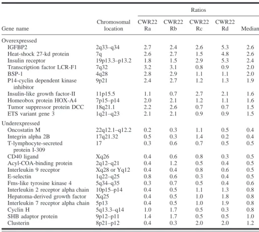
Table 1. Most consistently overexpressed and underexpressed genes in the complementary DNA microarray experiments and the ratios of gene expression in hormone-refractory human prostate cancer xenografts (CWR22Ra–d) compared with gene expression in a xenograft of the hormone-sensitive
strain CWR22 Gene name Chromosomal location Ratios CWR22 Ra CWR22 Rb CWR22 Rc CWR22 Rd Median Overexpressed IGFBP2 2q33–q34 2.7 2.4 2.6 5.3 2.6 Heat-shock 27-kd protein 7q 2.6 2.7 1.5 4.8 2.6 Insulin receptor 19p13.3–p13.2 1.8 1.5 2.9 5.3 2.4 Transcription factor LCR-F1 7q32 3.2 3.1 0.8 0.9 2.0 BSP-1 4q28 2.8 2.9 1.1 1.1 2.0
P14-cyclin dependent kinase inhibitor
9p21 2.4 2.7 1.2 1.3 1.9
Insulin-like growth factor-II 11p15.5 1.1 0.7 2.7 2.1 1.6 Homeobox protein HOX-A4 7p15–p14 2.0 2.1 1.2 1.1 1.6 Tumor suppressor protein DCC 18q21.1 2.2 2.6 0.7 0.7 1.5 ETS variant gene 3 1q21–q23 2.1 2.1 0.9 0.9 1.5 Underexpressed Oncostatin M 22q12.1–q12.2 0.2 0.3 1.1 0.5 0.4 Integrin alpha 2B 17q21.32 0.5 0.3 1.4 0.2 0.4 T-lymphocyte-secreted protein I-309 17 0.3 0.6 0.7 0.5 0.5 CD40 ligand Xq26 0.4 0.6 0.8 0.3 0.5 Acyl-COA-binding protein 2q12–q21 0.4 1.2 0.5 0.4 0.5 Interleukin 9 receptor Xq28 or Yq12 0.4 0.4 0.8 0.6 0.5
E-selectin 1q22–q25 0.8 0.6 0.3 0.4 0.5
Fms-like tyrosine kinase 4 5q34–q35 0.3 0.7 0.5 0.4 0.6 Interleukin 2 receptor alpha chain 10p15–p14 0.4 0.5 1.1 1.3 0.8 Hepatoma-derived growth factor Xq25 0.4 0.5 1.0 1.8 0.8 Interleukin 7 receptor alpha chain 5p13 0.4 0.5 1.0 1.9 0.8
Cyclin H 5q13.3–q14 1.0 1.7 0.5 0.3 0.8
SHB adaptor protein 9p12–p11 1.4 1.7 0.5 0.5 1.0
Clusterin 8p21–p12 0.4 0.3 2.0 2.0 1.2
*IGFBP2⳱ insulin-like growth factor-binding protein 2; LCR-F1 ⳱ locus control region F1; BSP-1 ⳱ transforming growth factor- signaling protein-1; DCC ⳱ deleted in colorectal carcinoma; ETS ⳱ E-twenty-six specific; SHB⳱ src homology B.
Fig. 1. Reverse transcription–polymerase chain re-action analysis of insulin-like growth factor-binding protein 2 (IGFBP2), 27-kd heat-shock protein (HSP27), and asparagine synthetase (internal con-trol) expression in one hormone-responsive (CWR22) and in four hormone-refractory prostate cancer xenografts (CWR22Ra–d).
cDNA libraries from normal or malignant
prostate. Altogether, 172 overexpressed
or underexpressed genes or ESTs
(ap-proximately 3%) in at least three of the
four hormone-refractory derivatives were
discovered as compared with the
un-treated, hormone-sensitive human
pros-tate cancer xenograft. Thirty-seven
tran-scripts (0.7%) were substantially (ratio
>2) elevated and 135 (2.6%) were
under-expressed (ratio <0.5) in the CWR22R
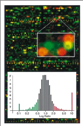
xe-nografts. A pseudocolored overlay of one
CWR22/CWR22R comparison and the
corresponding ratio distribution are
shown in Fig. 2.
Histology and Immunohistochemistry
To evaluate whether the gene
expres-sion changes seen in the
hormone-refractory CWR22R tumors reflected
molecular changes involved in tumor
pro-gression in patients with prostate cancer,
we created a tissue microarray to analyze
the expression of two overexpressed
genes, IGFBP2 and HSP27, at the protein
level in 238 different human prostate
can-cers and in 26 benign prostate tissues. The
total number of evaluable specimens
on the tissue microarray was 264 for the
IGFBP2 and 258 for the HSP27
immuno-staining.
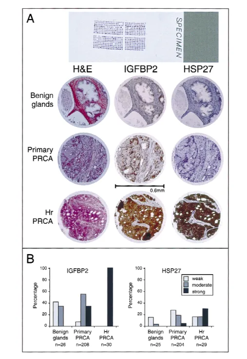
In these arrayed clinical specimens, a
strong association was seen between
in-creased IGFBP2 and HSP27 protein
ex-pression and the progression of prostate
cancer to hormone-refractory disease
(Fig. 3). A strong cytoplasmic IGFBP2
staining was present in all of the 30
lo-cally recurrent, hormone-refractory
pros-tate cancers, in 74 (36%) of the 208
primary tumors, and in none of the 26
benign prostate specimens (Fig. 3; P
⳱
.0001, two-sided). HSP27 was strongly
expressed in nine (31%) of 29
recur-rent tumors, in 11 (5%) of 204 primary
tumors, but never in the secretory
tate epithelial cells of 25 benign
pros-tatic hyperplasia specimens (Fig. 3; P
⳱
.0001, two-sided). There was no
statisti-cally significant association between
IG-FBP2 or HSP27 expression and tumor
grade or T stage in the primary tumors
(data not shown). A subgroup of 36
patients had received primary
neoadju-vant endocrine therapy before radical
prostatectomy, but their IGFBP2 and
HSP27 expression data were similar to
those of the untreated patients (data not
shown).
D
ISCUSSIONThe transition from a
hormone-sensitive human prostate cancer to a
hor-mone-refractory recurrent strain in the
CWR22 xenograft model system
re-sembles the clinical progression of human
prostate cancer (4). As shown in this
study by comparative genomic
hybridiza-tion, there was a close clonal genetic
re-lationship between the primary and
recur-rent xenograft tumors. Furthermore, many
of the alterations seen by comparative
ge-nomic hybridization in this model system,
such as gains of chromosome 7 and 8, are
similar to those commonly found in
clini-cal specimens from patients with prostate
cancer. The cDNA microarray technology
allows rapid, large-scale screening of
ex-pression of hundreds or thousands of
genes in a single experiment (5). Here, up
to 170 genes (3.3%) were identified to be
differentially expressed between the
pri-mary and recurrent (hormone-sensitive
and hormone-refractory) xenograft
tu-mors. This high number of differentially
expressed genes illustrates the complex
molecular basis of prostate cancer
pro-gression. The regrowth of the
hormone-refractory tumor during androgen
depri-vation therapy may necessitate a complex
reprogramming of multiple key
regula-tory mechanisms involving cell growth,
apoptosis (i.e., programmed cell death),
and other signaling pathways. It will be
important to identify the molecular
mechanisms that contribute to the
devel-opment of recurrent tumors and to
exam-ine if some of the signaling pathways
in-volved would provide starting points for
the development of novel diagnostic or
therapeutic approaches for patients with
advanced, hormone-refractory prostate
cancers.
The translation of gene-expression
findings from model systems to human
patients with cancer presents several
chal-lenges. First, although this xenograft
model system displayed phenotypic
prop-erties resembling human prostate cancer
progression, it remains important to
vali-date whether the same alterations of gene
expression and the same signaling
path-ways contribute to the disease progression
in human cancer patients. Second, to
uti-lize the cDNA microarray data for the
de-velopment of improved diagnostic or
therapeutic approaches, it remains
criti-cally important not only to screen for
ex-pression of many different genes but also
to screen many different tumor tissues
and establish an accurate frequency of
in-Fig. 2. Hybridization of the pros-tate complementary DNA microar-ray containing 5184 genes (Re-search Genetics, Inc.). A color image overlay of the CWR22 hy-bridization (green) and CWR22R recurrent xenograft (red) is shown. Spots with more red color repre-sent transcripts overexpressed in the hormone-refractory tumor in comparison to the primary tumor, yellow spots indicate genes that were equally abundant, and green spots indicate underexpressed genes in CWR22R. Genes that were not expressed in either of the two tissues appear in the black background color. Inset (histo-gram) shows a normal frequency distribution of the log10intensity
r a t i o s f o r C W R 2 2 R v e r s u s CWR22 for all of the 5184 spots on the microarray. The ratios are displayed on the x-axis, and the relative frequency of genes with the given ratios is indicated on the
y-axis. Ratios of genes that have a
twofold or higher expression in the recurrent than in the primary xeno-graft tumor are shown as red bars and those with a 50% or more re-duction as green bars.
volvement of these genes in different
stages of the prostate cancer progression.
A substantial amount of work is required
to fully explore the role of just a single
gene in cancer. Before performing
full-length cDNA cloning, functional
analy-ses, and other tedious experiments, one
would have to prioritize the long list of
potential target genes that always emerges
from cDNA microarray experiments and
to perform large-scale studies of clinical
specimens. In this study, we first took
ad-vantage of the fact that the pattern of gene
expression in the recurrent xenograft
tu-mors was different from one animal to
another. Therefore, we decided to first
concentrate on those genes that were
dif-ferentially expressed in two or more
re-current xenograft tissues. One would
ex-pect that such genes are more likely to be
associated with hormone therapy failure,
whereas genes that are only
overex-pressed in one case may be important
only for that particular tumor. The
deci-sive step was the evaluation of gene
ex-pression patterns in clinical specimens by
use of our newly developed tissue
micro-array technology.
Evaluation of all candidate genes
emerging from the present cDNA
micro-array experiments in a large series of
un-cultured clinical tumors would take years
if traditional methods were used.
Further-more, after a few hundred genes had been
analyzed, one would run out of the
avail-able tumor tissues. Tissue microarray
technology substantially facilitates the
translation of basic research findings to
clinical applications (7) and makes it
pos-sible to perform in situ analysis of
hun-dreds of tumors either at the DNA, the
RNA, or the protein level. This study was
done with immunocytochemical
tech-niques, but expression analyses of newly
identified genes could also be analyzed by
mRNA in situ hybridization when
anti-bodies are not available. Such a strategy
allows one to quickly validate and further
explore in a large number of clinical
specimens the in vivo significance of
can-didate genes discovered with the cDNA
microarrays. Only minute amounts of
tis-sues are required to make the tissue
mi-croarray blocks, causing minimal damage
to the original tumor blocks. Since one
can generate multiple replicate tissue
mi-croarray blocks, each of which can be
sec-tioned 200–300 times, one could easily
generate thousands of tissue microarray
sections from the same set of clinical
tu-mor material. Each section can be utilized
for the analysis of a different molecular
marker.
The small size of the samples makes
tissue microarrays a powerful screening
tool. However, the small tissue samples
may not always be representative of the
whole tumor and, therefore, the
preva-lence of a molecular alteration in a tissue
microarray analysis may be
underesti-mated. However, sampling bias may not
be a serious concern if the tumor areas are
carefully selected for punching. In our
previous studies (7,14), we found a high
concordance between gene-amplification
frequencies on tissue microarrays when
compared with the data from the
litera-ture. The representativeness of tissue
mi-croarray data could be improved by
in-Fig. 3. A) Hematoxylin–eosin and immunohistochemical staining of insulin-like growth factor-binding protein 2 (IGFBP2) and 27-kd heat-shock protein (HSP27) on the prostate cancer tissue microarray (original magnification ×200). Benign prostate glands show no immunoreactivity. Primary untreated prostate cancer (PRCA) demonstrates weak immunostaining of IGFBP2 but no immunoreactivity of HSP27. In contrast, hormone-refractory prostate cancer with local recurrence (Hr PRCA) shows strong expression of both IGFBP2 and HSP27. B) Frequency distribution of expression of IGFBP2 and HSP27 during progression to hormone-refractory prostate cancer as measured by immunohistochemistry on a prostate cancer tissue mi-croarray.
cluding several samples from different
sites of a tumor on each array.
Further-more, comparisons of the involvement of
one gene against another on the same
ar-ray or comparisons of one molecular
al-teration between two different stages of
tumor progression will generate relative
frequency estimates that are not biased by
the sampling method. Nevertheless, tissue
microarray technology should be
re-garded as a rapid, high-throughput tool to
survey many different genes and markers
to identify those that are most promising
for clinical applications. These would
then have to be tested on conventional
tissue specimens before clinical
applica-tion.
The tissue microarray results validated
that overexpression of IGFBP2 may be an
important event in hormone-refractory
prostate cancer, not only in the CWR22
xenograft model system but also in
pa-tients who had developed a recurrent
tu-mor during androgen deprivation therapy.
This finding is in agreement with recent
experimental and clinical studies (15–19)
indicating that the IGF system may be a
key growth regulatory pathway in
pros-tate cancer. IGFBP2 is a member of the
IGF growth factor system, which involves
two growth factors (IGF-I and IGF-II),
two IGF receptors (type I and II), seven
IGF-binding proteins (IGFBP1–7), as
well as IGFBP proteinases (16,20). IGF-I
stimulates growth and inhibits apoptosis
in normal and transformed epithelial cells
(21–24). High plasma levels of IGF-I
were recently shown to be associated with
increased risk of getting prostate cancer
(17). Moreover, IGF-I has been shown to
enhance androgen receptor-mediated
gene transcription in the prostate cancer
cell lines DU 145 (after cotransfection
with an androgen-inducible reporter gene
and an androgen receptor expression
vec-tor) and LNCaP in the absence of
andro-gen, suggesting that IGF-I may drive the
androgen-signaling pathway in
hormone-refractory prostate cancer (25). IGFBPs
can enhance or inhibit the bioactivity of
IGFs (IGF-I and IGF-II) by modulating
the availability of free IGFs for their
re-ceptors (26,27). IGFBP2 has also been
suggested to be an enhancer of IGF-I
function (22). It can be speculated that
overexpression of IGFBP2 promotes
sur-vival and androgen-independent growth
of prostate cancer by increasing the
bio-availability of IGFs. Members of the same
pathway (IGF-II and insulin receptor)
were also overexpressed in some of the
hormone-refractory xenograft tissues.
However, IGFBP2 was systematically
and most highly overexpressed,
suggest-ing that it may perhaps have a central role
in modulating the IGF signaling in
hor-mone-refractory prostate cancer.
Alter-ations of IGFBP2 may also play a role in
the development and progression of other
tumor types, such as breast, colorectal,
and ovarian cancers (28–30).
Overexpres-sion of IGFBP2 has also been observed in
cell lines established from several solid
tumors (31,32).
The overexpression of HSP27 in about
one third of hormone-refractory prostate
cancers but in only 5% of primary tumors
is intriguing in light of the fact that
HSP27 has been shown to increase
resis-tance to apoptosis induced by several
drugs such as doxorubicin (33–36).
Blockage of apoptosis may be an
impor-tant feature of hormone-refractory
pros-tate cancer and has been associated with
the differential expression of the Bcl-2
gene family (37–39). It was recently
sug-gested that HSP27 and Bcl-2 act at
differ-ent levels to prevdiffer-ent apoptosis in
immor-talized embryo fibroblasts, depending on
the type of apoptotic stimulus (40). The
role of HSP27 as a predictor of patient
outcome or response to therapy has
re-ceived attention in breast cancer (41–44),
but it has not been extensively studied in
prostate cancer. In one study (45),
vari-able HSP27 immunostaining was found in
13 prostate tumors derived from
transure-thral resection specimens, but no
informa-tion about the hormonal treatment status
was provided. Another study (46) did not
find HSP27 immunoreactivity in radical
prostatectomy specimens from patients
with clinically localized disease. On the
basis of this study, HSP27 expression is
unlikely to play a major role in primary
prostate cancer but may be important in
hormone therapy failure.
In summary, we describe a new
strat-egy based on the combination of cDNA
and tissue microarray technologies to
ex-plore the molecular basis of human
pros-tate cancer progression. Our results
indi-cate that multiple gene expression
changes may contribute to prostate cancer
progression and hormonal therapy failure
and that at least some of the mechanisms
involved in the CWR22 xenograft model
system may be similar to those
contribut-ing to therapy failure and
hormone-refractory prostate cancer growth in
pa-tients. We detected an association
between increased expression of IGFBP2
and HSP27 and the hormone therapy
fail-ure in both the xenograft model system
and in patients’ specimens. Further
stud-ies are needed to evaluate these molecules
as well as dozens of other differentially
expressed genes as diagnostic or
thera-peutic targets for hormone-refractory
prostate cancer.
R
EFERENCES(1) Wingo PA, Ries LA, Giovino GA, Miller DS,
Rosenberg HM, Shopland DR, et al. Annual report to the nation on the status of cancer, 1973–1996, with a special section on lung can-cer and tobacco smoking. J Natl Cancan-cer Inst 1999;91:675–90.
(2) Konety BR, Getzenberg RH. Novel therapies
for advanced prostate cancer. Semin Urol On-col 1997;15:33–42.
(3) Wu HC, Hsieh JT, Gleave ME, Brown NM,
Pathak S, Chung LW. Derivation of androgen-independent human LNCaP prostatic cancer cell sublines: role of bone stromal cells. Int J Cancer 1994;57:406–12.
(4) Nagabhushan M, Miller CM, Pretlow TP,
Gia-conia JM, Edgehouse NL, Schwartz S, et al. CWR22: the first human prostate cancer xeno-graft with strongly androgen-dependent and re-lapsed strains both in vivo and in soft agar. Cancer Res 1996;56:3042–6.
(5) DeRisi J, Penland L, Brown PO, Bittner ML,
Meltzer PS, Ray M, et al. Use of a cDNA mi-croarray to analyse gene expression patterns in human cancer. Nat Genet 1996;14:457–60.
(6) Velculescu VE, Zhang L, Vogelstein B,
Kinz-ler KW. Serial analysis of gene expression. Science 1995;270:484–7.
(7) Kononen J, Bubendorf L, Kallioniemi A,
Bar-lund M, Schraml P, Leighton S, et al. Tissue microarrays for high-throughput molecular profiling of tumor specimens. Nat Med 1998; 4:844–7.
(8) Wainstein MA, He F, Robinson D, Kung HJ,
Schwartz S, Giaconia JM, et al. CWR22: an-drogen-dependent xenograft model derived from a primary human prostatic carcinoma. Cancer Res 1994;54:6049–52.
(9) Pretlow TG, Wolman SR, Micale MA, Pelley
RJ, Kursh ED, Resnick MI, et al. Xenografts of primary human prostatic carcinoma. J Natl Cancer Inst 1993;85:394–8.
(10) Tirkkonen M, Tanner M, Karhu R, Kallioniemi
A, Isola J, Kallioniemi OP. Molecular cytoge-netics of primary breast cancer by CGH. Genes Chromosomes Cancer 1998;21:177–84.
(11) Chirgwin JM, Przybyla AE, MacDonald RJ,
Rutter WJ. Isolation of biologically active ri-bonucleic acid from sources enriched in ribo-nuclease. Biochemistry 1979;18:5294–9.
(12) Gleason DF. Histologic grading and clinical
staging of prostatic carcinoma. In: Tannen-baum M, editor. Urologic pathology: the pros-tate. Philadelphia (PA): Lea & Febiger; 1977. p. 171–8.
(13) International Union Against Cancer.
Classifi-cation of malignant tumours. 5th ed. New York (NY): Wiley-Liss; 1997.
Bissig H, Nocito A, et al. Tissue microarrays for gene amplification surveys in many differ-ent tumor types. Clin Cancer Res 1999;5: 1966–75.
(15) Lee AV, Hilsenbeck SG, Yee D. IGF system
components as prognostic markers in breast cancer. Breast Cancer Res Treat 1998;47: 295–302.
(16) Nunn SE, Gibson TB, Rajah R, Cohen P.
Regulation of prostate cell growth by the insu-lin-like growth factor binding proteins and their proteases. Endocrine 1997;7:115–8.
(17) Chan JM, Stampfer MJ, Giovannucci E, Gann
PH, Ma J, Wilkinson P, et al. Plasma insulin-like growth factor-I and prostate cancer risk: a prospective study. Science 1998;279:563–6.
(18) Hankinson SE, Willett WC, Colditz GA,
Hunter DJ, Michaud DS, Deroo B, et al. Cir-culating concentrations of insulin-like growth factor-I and risk of breast cancer. Lancet 1998; 351:1393–6.
(19) Burfeind P, Chernicky CL, Rininsland F, Ilan
J, Ilan J. Antisense RNA to the type I insulin-like growth factor receptor suppresses tumor growth and prevents invasion by rat prostate cancer cells in vivo. Proc Natl Acad Sci U S A 1996;93:7263–8.
(20) Baxter RC, Binoux MA, Clemmons DR,
Conover CA, Drop SL, Holly JM, et al. Rec-ommendations for nomenclature of the insulin-like growth factor binding protein superfamily. Endocrinology 1998;139:4036.
(21) Harrington EA, Bennett MR, Fanidi A, Evan
GI. c-Myc-induced apoptosis in fibroblasts is inhibited by specific cytokines. EMBO J 1994; 13:3286–95.
(22) Cohen P, Peehl DM, Lamson G, Rosenfeld
RG. Insulin-like growth factors (IGFs), IGF re-ceptors, and IGF-binding proteins in primary cultures of prostate epithelial cells. J Clin En-docrinol Metab 1991;73:401–7.
(23) Cohen P, Peehl DM, Baker B, Liu F, Hintz RL,
Rosenfeld RG. Insulin-like growth factor axis abnormalities in prostatic stromal cells from patients with benign prostatic hyperplasia. J Clin Endocrinol Metab 1994;79:1410–5.
(24) Rajah R, Valentinis B, Cohen P.
Insullike growth factor (IGF)-binding prote3 in-duces apoptosis and mediates the effects of transforming growth factor-beta1 on pro-grammed cell death through a p53- and IGF-independent mechanism. J Biol Chem 1997; 272:12181–8.
(25) Culig Z, Hobisch A, Cronauer MV, Radmayr
C, Trapman J, Hittmair A, et al. Androgen re-ceptor activation in prostatic tumor cell lines by insulin-like growth factor-I, keratinocyte growth factor, and epidermal growth factor. Cancer Res 1994;54:5474–8.
(26) Jones JI, Clemmons DR. Insulin-like growth
factors and their binding proteins: biological actions. Endocr Rev 1995;16:3–34.
(27) Kelley KM, Oh Y, Gargosky SE, Gucev Z,
Matsumoto T, Hwa V, et al. Insulin-like growth factor-binding proteins (IGFBPs) and their regulatory dynamics. Int J Biochem Cell Biol 1996;28:619–37.
(28) Helle SI, Jonat W, Giurescu M, Ekse D, Holly
JM, Lonning PE. Influence of treatment with onapristone on the IGF-system in breast cancer patients. J Steroid Biochem Mol Biol 1998;66: 159–63.
(29) el Atiq F, Garrouste F, Remacle-Bonnet M,
Sastre B, Pommier G. Alterations in serum lev-els of like growth factors and insulin-like growth-factor-binding proteins in patients with colorectal cancer. Int J Cancer 1994;57: 491–7.
(30) Kanety H, Kattan M, Goldberg I, Kopolovic J,
Ravia J, Menczer J, et al. Increased insulin-like growth factor binding protein-2 (IGFBP-2) gene expression and protein production lead to high IGFBP-2 content in malignant ovarian cyst fluid. Br J Cancer 1996;73:1069–73.
(31) Reeve JG, Morgan J, Schwander J, Bleehen
NM. Role for membrane and secreted insulin-like growth factor-binding protein-2 in the regulation of insulin-like growth factor action in lung tumors. Cancer Res 1993;53:4680–5.
(32) Menouny M, Binoux M, Babajko S. IGFBP-2
expression in a human cell line is associated with increased IGFBP-3 proteolysis, decreased IGFBP-1 expression and increased tumorige-nicity. Int J Cancer 1998;77:874–9.
(33) Samali A, Cotter TG. Heat shock proteins
in-crease resistance to apoptosis. Exp Cell Res 1996;223:163–70.
(34) Mehlen P, Hickey E, Weber LA, Arrigo AP.
Large unphosphorylated aggregates as the ac-tive form of hsp27 which controls intracellular reactive oxygen species and glutathione levels and generates a protection against TNFalpha in NIH-3T3-ras cells. Biochem Biophys Res Commun 1997;241:187–92.
(35) Oesterreich S, Weng CN, Qiu M, Hilsenbeck
SG, Osborne CK, Fuqua SA. The small heat shock protein hsp27 is correlated with growth and drug resistance in human breast cancer cell lines. Cancer Res 1993;53:4443–8.
(36) Huot J, Houle F, Spitz DR, Landry J. HSP27
phosphorylation-mediated resistance against actin fragmentation and cell death induced by oxidative stress. Cancer Res 1996;56:273–9.
(37) Koivisto P, Visakorpi T, Rantala I, Isola J.
In-creased cell proliferation activity and de-creased cell death are associated with the emer-gence of hormone-refractory recurrent prostate cancer. J Pathol 1997;183:51–6.
(38) McDonnell TJ, Troncoso P, Brisbay SM,
Logothetis C, Chung LW, Hsieh JT, et al. Ex-pression of the protooncogene bcl-2 in the prostate and its association with emergence of androgen-independent prostate cancer. Cancer Res 1992;52:6940–4.
(39) Raffo AJ, Perlman H, Chen MW, Day ML,
Streitman JS, Buttyan R. Overexpression of bcl-2 protects prostate cancer cells from apop-tosis in vitro and confers resistance to androgen depletion in vivo. Cancer Res 1995;55: 4438–45.
(40) Guenal I, Sidoti-de Fraisse C, Gaumer S,
Mig-notte B. Bcl-2 and Hsp27 act at different levels to suppress programmed cell death. Oncogene 1997;15:347–60.
(41) Love S, King RJ. A 27 kDa heat shock protein
that has anomalous prognostic powers in early and advanced breast cancer. Br J Cancer 1994; 69:743–8.
(42) Conroy SE, Sasieni PD, Amin V, Wang DY,
Smith P, Fentiman IS, et al. Antibodies to heat-shock protein 27 are associated with improved survival in patients with breast cancer. Br J Cancer 1998;77:1875–9.
(43) Vargas-Roig LM, Fanelli MA, Lopez LA,
Gago FE, Tello O, Aznar JC, et al. Heat shock proteins and cell proliferation in human breast cancer biopsy samples. Cancer Detect Prev 1997;21:441–51.
(44) Vargas-Roig LM, Gago FE, Tello O, Aznar JC,
Ciocca DR. Heat shock protein expression and drug resistance in breast cancer patients treated with induction chemotherapy. Int J Cancer 1998;79:468–75.
(45) Thomas SA, Brown IL, Hollins GW, Hocken
A, Kirk D, King RJ, et al. Detection and dis-tribution of heat shock proteins 27 and 90 in human benign and malignant prostatic tissue. Br J Urol 1996;77:367–72.
(46) Storm FK, Mahvi DM, Gilchrist KW. Hsp-27
has no diagnostic or prognostic significance in prostate or bladder cancers. Urology 1993;42: 379–82.
N
OTESPresent address: M. Kolmer, National Public
Health Institute, Department of Human Molecular Genetics, Helsinki, Finland.
Supported by the Swiss National Science Foun-dation (81BS-052807) (to L. Bubendorf); by the Academy of Finland and by the Tampere University Hospital Foundation (to P. Koivisto); and by Public Health Service grant CA57179 from the National Cancer Institute, National Institutes of Health, De-partment of Health and Human Services (to T. G. Pretlow).
We thank Rita Epper, Martina Mirlacher, Martina Storz, and Heidi Oggier, University of Basel (Swit-zerland), for their excellent technical support; Daryl Leja, National Human Genome Research Institute (Bethesda, MD), for his illustration support; and Steve Leighton, Beecher Instruments (Silver Spring, MD), for his help in tissue microarray instrumenta-tion.
Manuscript received March 1, 1999; revised Au-gust 10, 1999; accepted AuAu-gust 18, 1999.