Nephrology
Dialysis
Transplantation
Original Article
Characterization of CD44-mediated hyaluronan binding by renal
tubular epithelial cells
Beat Oertli1, Xiaohong Fan1 and Rudolf P. Wu¨thrich1,2
Physiological Institute1, University of Zu¨rich-Irchel, and Division of Nephrology2, University Hospital, Zu¨rich, Switzerland
Abstract Key words: CD44; cell growth; hyaluronan; tubular
epithelial cells
Background. CD44 is the main receptor for the
extra-cellular polysaccharide hyaluronan (HA). We have recently shown that CD44 is strongly induced on renal
Introduction
tubular epithelial cells ( TEC ) in autoimmune renal injury and that HA accumulates in the renal
interstit-The leukocyte antigen CD44 is a widely distributed ium (Kidney Int 1996; 50: 156–163 and Nephrol Dial
cell surface proteoglycan which functions as a receptor
Transplant 1997; 12: 1344–1353). The functional
signi-for the matrix constituent hyaluronan (HA) [1]. ficance of enhanced tubular CD44 expression and its
Through its expression on non-haematopoietic paren-interaction with HA are not known. The purpose of
chymal cells CD44 is involved in various biological the present study was to characterize renal tubular
processes, including cell migration, metastasis and CD44 expression and CD44-mediated HA binding in
inflammation [2].
vitro and to investigate the growth modulating effects
We have recently shown that CD44 is induced on in response to HA binding by TEC.
proximal tubular epithelium in immune-mediated renal
Methods. RT-PCR analysis, flow cytometry, confocal
injury, including murine lupus nephritis (MRL-Faslpr microscopy and Western blotting were used to examine
model ) [3] and tubulointerstitial disease (CBA/CaH-cell surface and soluble CD44 expression by cultured
kdkd model ) [4]. We also found that HA markedly
TEC, using SV40-transformed mouse cortical tubular
accumulates in the renal interstitium in MRL-Faslpr (MCT ) cells. HA binding characteristics were
exam-mice (unpublished observation) and CBA/CaH-kdkd ined by flow cytometry and effects of HA on TEC cell
mice [4], particularly in areas where tubular epithelial growth by [3H ]thymidine incorporation.
CD44 is upregulated. The functional significance of
Results. By RT-PCR analysis MCT cells expressed
CD44 expression on TEC, its mechanism of interaction predominantly the standard form of CD44 mRNA,
with HA and its role in immune-mediated tubular whereas the expression of variant forms was very weak.
injury are presently not known. We have speculated, Confocal microscopy showed that CD44 was expressed
however, that tubular CD44 could interact with its basolaterally and apically on MCT cells with strong
ligand in vivo and modulate tubular cell function in staining on microvilli. Shedding of CD44 from
response to this ligand interaction [3]. MCT cells could be induced with crosslinking of
anti-Since CD44 is expressed by cultured renal tubular CD44 mAbs or with PMA stimulation. MCT cells
con-epithelial cells ( TEC ) [3] we have used a clonal TEC stitutively bound HA and this binding could be
modu-line to characterize the pattern of CD44 expression lated with anti-CD44 mAbs. Soluble and plate-bound
and its interaction with HA in vitro and to gain HA markedly inhibited MCT cell growth.
insight into the functional significance of CD44
Conclusions. CD44 is a regulated HA receptor on
expression on TEC. Here we show that a defined MCT cells which can be shed into the cellular
environ-TEC line expresses high levels of the standard but not ment. Upon binding of HA, CD44 functions as a
the variant forms of CD44, both apically and baso-growth inhibitory cell surface protein in MCT cells.
laterally. Furthermore, shedding of CD44 from TEC We speculate that the interaction of CD44 with HA
can be induced. CD44 binds HA constitutively and may have important regulatory effects on cell
prolifera-the binding capacity for HA can be modulated tion in tubulointerstitial renal diseases.
with mAbs targeting CD44. We also demonstrate that HA inhibits the growth of TEC through its ligand CD44. Thus, upon interaction with HA renal tubular
Correspondence and offprint requests to: Rudolf P. Wu¨thrich, Division
CD44 functions as a growth inhibiting molecule. We
of Nephrology, University Hospital, Ra¨mistrasse 100, CH-8091
Zu¨rich, Switzerland. speculate that the interaction of CD44 with HA could
cDNA probe encoding for CD44 as described [3]. Blots were
play a role in the regeneration and differentiation of
washed under stringent conditions (final wash in 0.2×SSC,
injured TEC in vivo in various renal diseases with a
1% SDS, 60°C). Following hybridization the blots were
tubulointerstitial component.
exposed to Kodak X-OMAT AR film.
Materials and methods
Flow cytometryReagents Flow cytometry was performed as described [3]. Briefly,
cultured MCT cells were grown to confluence and were then Tissue culture reagents were obtained from Life detached with 5 mM EDTA in HBSS. Cells were washed Technologies (Gaithersburg, MD) and chemicals from once with Ca2+- and Mg2+-free HBSS and were resuspended Sigma (St Louis, MO). Dr J. Lesley (San Diego, CA) in PBS containing 5% FCS. Cells were then incubated with provided us with the IRAWB14.4 hybridoma [5], producing the primary mAb for 1 h at 4°C, were washed twice with an activating anti-CD44 monoclonal antibody (mAb) and PBS/5% FCS and were then incubated with FITC-conjugated with FITC-labelled HA. Purified sodium hyaluronate with goat anti-rat IgG for 1 h at 4°C. After repeated washing a molecular weight of approx. 4–6×10⁵ was obtained from with PBS/5% FCS, cells were fixed with 2% paraformal-Fluka (Buchs, Switzerland). Table 1 lists the target antigen, dehyde. Measurements were performed on an EpicsB Coulter properties and source of the mAbs used in this study. The flow cytometer.
hybridomas for the mAbs IM7.8.1 [6 ], KM114 [7] and KM81 [7] were obtained from the American Type Culture
Collection (ATCC, Rockville, MD). Hybridoma culture Confocal microscopy
supernatants were purified using protein G-Sepharose
CL-6B columns. KM114 was biotinylated according to a The cellular distribution of CD44 on cultured MCT cells was
standard protocol. determined by confocal microscopy. MCT cells were grown
in DMEM/10% FBS medium on coverslips coated with type II collagen until subconfluent. Cells were then fixed in 4%
Cell lines and cell cultures
paraformaldehyde and were blocked with 20 mM glycine and subsequently with 3% milk powder (w/v). For detection An SV40-transformed mouse cortical tubular (MCT ) cell
of intracellular CD44 cells were then permeabilized with line was used to study the expression and functional role of
0.1% saponin (v/v). Rat anti-mouse mAb IM7.8.1 (5 mg/ml ) CD44 [8]. MCT cells were grown in tissue culture dishes in
in 3% milk powder was used to detect CD44. FITC-conjug-DMEM media supplemented with 10% FBS, 10 mM HEPES, ated affinity-purified goat anti-rat IgG (Sigma, St Louis, 100 U/ml penicillin and 100 mg/ml streptomycin.
MO) was used at 1540 as secondary antibody.
CD44 expression was then assessed with a confocal laser scanning microscope (LSM 410, Zeiss, Germany) at
magni-RNA extraction, RT-PCR and Southern blotting for
fications of 630× using the 488 nm line spectrum of a He-Na
CD44
laser source. Optical sections were obtained at focal steps of 28 or 56 nm. Stacks of 51 x-y sections were used to generate Total RNA from cultured MCT cells was extracted as
extended focus projections, x-z sections and 3-D reconstruc-described [9]. Total RNA from murine kidney was isolated
tions, using the Imaris@ software (Bitplane, Zu¨rich, using lysis in guanidinium isothiocyanate and
ultracentrifu-Switzerland ). gation at 35 000 r.p.m. through a CsCl2 gradient overnight
[10]. The RNA was analysed for CD44 expression by RT-PCR as described [3] using a kit (Perkin-Elmer,
Shedding of CD44 from MCT cells
Branchburg, NJ ). For optimal detection of CD44 variant forms we used primers located in the CD44 sequence directly
Shedding in response to crosslinking of anti-CD44 mAb was flanking the variant exon insertion site [11]. The upstream
assessed by flow cytometry. Confluent MCT cells were primer 5∞-ACC CCA GAA GGC TAC ATT TTG C-3∞ and
detached and resuspended in DMEM/10% FBS and were then the downstream primer 5∞-CTC ATA GGA CCA GAA GTT
incubated with mAb IM7.8.1 at 10 mg/ml and crosslinked GTG G-3∞ were used to detect the presence of such variant
using anti-rat IgG for 5 h at 37°C. Adhesion and clumping forms. RT-PCR products were then resolved on 2%
aga-of MCT cells was prevented by shaking the tubes every hour. rose gels.
For detection of remaining cell surface CD44, cells were then To ensure specificity of the amplified fragments we
per-stained with biotinylated KM114 mAb, using a streptavidin-formed Southern blot analysis and hybridization with a
R-phycoerythrin conjugate for detection (Sigma, St Louis, MO). Cells were then analysed by flow cytometry.
Soluble CD44 protein was also assayed in the supernatant Table 1. Characteristics of anti-CD44 monoclonal antibodies of MCT cells by SDS–PAGE and Western blotting. Supernatants from control and from PMA-stimulated MCT (20 mg/ml ) were size fractionated on a 10% SDS–PAGE
Clone Target Properties Source Reference
under reducing and non-reducing conditions and blotted on to nitrocellulose. To detect soluble CD44 the membranes
IM7.8.1 CD44 Neutral ATCC [6 ] were then processed with a chemiluminescence detection KM81 CD44 HA binding blocker ATCC [7]
system ( Western-Star, Tropix, Bedford, MA). Membranes
KM114 CD44 Partial HA binding ATCC [7]
were blocked with casein and were incubated with the
anti-blocker
CD44 mAb IM7.8.1. After washing, blots were incubated
IRAWB14.4 CD44 Induces HA binding J. Lesley [5]
(155000). The 1,2-dioxetane substrate (CDP-Star) was then added to the blots according to the instructions provided from the manufacturer. Membranes were exposed briefly (30 s to 5 min) to Kodak LS film and were then developed.
HA binding to CD44 on MCT cells
The HA binding capacity of MCT cells was assessed by flow cytometry. Confluent cells were detached and resuspended in PBS/5% FCS and incubated with HA-FITC at 15800 for 1 h at 4°C. To control for non-specific binding cells were also incubated with FITC-labelled goat anti-rat IgG. The influence of anti-CD44 mAb on HA-binding was assessed by incubating cells with either purified mAb for 1 h at 4°C before staining with HA-FITC.
Proliferation assays
The influence of soluble HA on proliferation was assessed as follows: MCT cells were grown in 96-well plates (Nunc Intermed, Roskilde, DK ) to confluence with DMEM/10% FBS. Cells were then rested for 24 h in DMEM/1% FBS. HA (500–1000 mg/ml ) was then added and cells were incub-ated at 37°C for the indicincub-ated time. Proliferation was then assessed by [3H ]thymidine incorporation, adding 1 mCi/well and harvesting the cells 18 h later with a Cambridge PHD cell harvester (Cambridge Technology, Cambridge, USA).
(a)
(b)
[3H ]thymidine incorporation was counted with a Kontron Fig. 1. (A) RT-PCR and Southern blot analysis of MCT-and kidney-Betamatic V liquid scintillation counter. derived total RNA for CD44s and CD44v isoforms. Primers flanking The effect of plate-bound HA was investigated as follows: the extracellular insertion site for the variant exons were used to
96-well plates were coated with HA at 500 mg/ml (dissolved detect alternatively spliced transcripts. Lane 1: MCT cells express
in PBS and adjusted to pH 8.5) overnight at 4°C. Plates were mainly standard CD44 (221 bp RT-PCR fragment). Lane 2: Standard and additional variant forms are expressed in the kidney
then washed twice with PBS and blocked for 4 h with 1%
of a 6 month old CBA/CaH-kdkd mouse with tubulointerstitial
(w/v) albumin. Confluent MCT cells were harvested by
tryp-disease. Lane 3: CD44 is mainly expressed as the standard form
sinization and added at 1×10⁴ cells/well, plates were then
(221 bp) in the kidney of a 4-month-old MRL-Faslpr mouse. (B)
incubated in DMEM/10% FBS at 37°C for 48 h. Proliferation Flow cytometric analysis for CD44 cell surface expression by was then assessed as mentioned above, using [3H ]thymidine. MCT cells, using biotinylated anti-CD44 mAb KM114. Shaded area
represents cells stained with control mAb, white area with anti-CD44 mAb. Note that 100% of cells are strongly anti-CD44-positive.
Results
Characterization of CD44 expression by MCT cells
obtained by flow cytometry. Figure 2A shows that RNA was isolated from confluent MCT cells to detect apical microvilli were strongly positive for CD44. The the presence of transcripts encoding for the standard apical expression was confirmed by examining x-z (CD44s) and the alternatively spliced isoforms of CD44 ( Figure 2B, bottom) and y-z sections ( Figure 2B, upper (CD44v) by RT-PCR and Southern blotting. Figure 1A right) which showed membrane bound CD44 around shows that mRNA for CD44s was readily detected in the cell with no preference for apical, basal or lateral MCT cells. Variant forms (CD44v) could only be visu- sides. Sections in the x-y direction indicated that most alized after prolonged exposure of the blots (not of the CD44 protein was membrane bound in shown). In comparison, RT-PCR fragments repres- MCT cells ( Figure 2C, thin arrow), but a cytoplasmic enting CD44v were readily detected in kidney RNA fraction could also be detected in permeabilized cells from CBA/CaH-kdkd mice (positive control ), but not ( Figure 2C, dotted arrow). With the confocal images in kidney RNA from MRL-Faslpr mice with lupus we also obtained evidence for shedding of CD44 nephritis, as described previously [3,4]. by MCT cells. Figure 2D (3D reconstruction) shows Flow cytometry was then used to examine the cell CD44 retraction traces (arrow) and shedded remnants surface expression of the CD44 protein by MCT cells. of cell membranes which are also CD44 positive Figure 1B shows that MCT cells stained intensely with (dotted arrow).
the anti-CD44 mAb KM114 (100% of cells expressing CD44).
Shedding of CD44 by MCT cells
We then used confocal microscopy to examine more
precisely the cellular location of CD44. MCT cells To characterize further the shedding of CD44 by MCT cells we used flow cytometry after crosslinking stained intensely for CD44, confirming the results
Fig. 2. Confocal microscopy of MCT cells stained for CD44. (A) Strong staining for CD44 on apical microvilli. (B) Cross-sections of two
MCT cells. Upper left: x-y section, upper right: y-z section, bottom panel: x-z section. MCT cells express CD44 apically and basolaterally. (C ) MCT cells were permeabilized with 0.1% saponin to detect intracellular CD44. Dotted arrow points to a cell with diffuse intracellular staining sparing the nucleus and more accentuated membrane staining. Thin arrow shows strong rim staining for CD44. (D) 3-D reconstruction of area where cells have retracted (arrow), demonstrating shedded remnants of cell membrane which are CD44 positive (dotted arrow) and CD44 retraction traces.
CD44 with anti-CD44 mAbs. MCT cells were incub- stimulated MCT cell supernatants, demonstrating PMA-induced shedding of CD44 by MCT cells. ated with anti-CD44 mAb IM7.8.1 and bound mAb
was crosslinked with anti-rat IgG. Figure 3A demon-strates that CD44 staining with biotinylated KM114
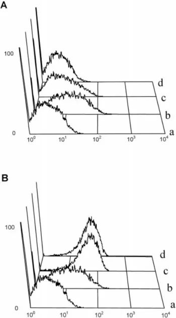
HA-binding capacity of MCT cells is modulated by mAb
was intense (100%) in control cells (thick line). After
against CD44
crosslinking the mean fluorescence intensity decreased
by 55% (thin line). Incubation of MCT cells with To determine whether cell surface CD44 functions as a HA receptor on MCT cells we examined the HA IM7.8.1 alone without crosslinking reduced the mean
fluorescence intensity insignificantly (dotted line). binding capacity using a FITC-labelled HA probe. Incubation with FITC-labelled HA for 1 h showed Thus, crosslinking CD44 with IM7.8.1 and anti-rat
IgG effectively decreased CD44 cell surface staining, moderate HA binding by unstimulated MCT cells ( Figure 4). To exclude non-specific binding for suggesting shedding of CD44 by MCT cells via this
mechanism. HA-FITC, MCT cells were incubated with unlabelled HA at 10-fold excess before staining with HA-FITC. To prove further that CD44 is shed by MCT cells
we then performed SDS–PAGE and Western blotting HA-FITC binding was completely abrogated by this procedure (data not shown).
to detect immunoreactive CD44 in the culture
super-natant of MCT cells after stimulation with the phorbol As previously described, the anti-CD44 mAb KM81 inhibits the HA binding capacity in many cell ester PMA. Cells were grown to confluence and were
then incubated with PMA for 48 h. Figure 3B shows types [7], whereas the activating anti-CD44 mAb IRAWB14.4 is able to induce or increase the HA that a protein around 80 kDa could be detected in the
(a)
(b)
Fig. 3. (A) Shedding of CD44 is induced with crosslinking CD44.
MCT cells were incubated with CD44 mAb IM7.8.1 and anti-rat IgG for 5 h and stained with biotinylated KM114 for remaining CD44. Shaded area: control (staining with secondary reagent); thick black line: control cells stained with biotinylated KM114 alone; dotted line: staining with biotinylated KM114 after incubation with IM7.8.1 without crosslinking; thin black line: staining with biotinyl-ated KM114 after IM7.8.1 incubation with crosslinking. The fluo-rescence shift to the left after crosslinking of CD44 indicates shedding of CD44. (B) Detection of soluble CD44 in the supernatant of MCT cells by Western blotting. MCT cells were stimulated with PMA for 48 h in DMEM/1% FBS and the supernatant was har-vested. Supernatants were run on a 10% SDS–PAGE gel using non-reducing conditions and blotted onto nitrocellulose. Soluble CD44 was detected using the IM7.8.1 mAb. Lane 1: control (48 h); lane 2: MCT cells stimulated with PMA for 48 h; lane 3: protein size marker (Da).
Fig. 4. Flow cytometric analysis for detection of HA binding to
MCT cells. (A) The KM81 mAb inhibits HA binding on MCT cells. Cells were incubated with KM81 before incubation with
FITC-whether HA binding to CD44 on MCT cells could also
labeled HA. (a) control ( FITC-labeled anti-rat IgG only); (b)
be influenced by anti-CD44 mAbs we incubated
HA-FITC only; (c) KM81 at 10 mg/ml followed by HA-FITC; (d)
cells with purified anti-CD44 mAbs before adding KM81 at 50 mg/ml followed by HA-FITC. (B) The IRAWB14.4 HA-FITC. Figure 4A shows that mAb KM81 inhibited induces HA binding on MCT cells. Cells were incubated with
HA binding dose-dependently, displaying complete IRAWB14.4 before incubation with FITC-labeled HA. (a) control; (b) HA-FITC only; (c) IRAWB14.4 at 10 mg/ml followed by
HA binding inhibition at 50 mg/ml. In contrast,
HA-FITC; (d) IRAWB14.4 at 50 mg/ml followed by HA-FITC.
IRAWB14.4 stimulated HA binding markedly at 10 and 50 mg/ml ( Figure 4B). Thus, the modulation
DNA synthesis ([3H ]thymidine incorporation) mark-of HA binding capacity by anti-CD44 mAbs on
edly and in a dose-dependent manner. Furthermore MCT cells was similar to that described for
Figure 5B shows that a significant inhibitory effect of lymphocytes.
HA could also be demonstrated when MCT cells were grown on plate-bound HA (P<0.005, t-test, five
HA inhibits proliferation of MCT cells different experiments).
To gain insight into the functional consequences of HA binding to CD44 on MCT cells we examined the
Discussion
proliferation of these cells in the presence or absence of HA. We used both soluble HA and plate-bound
HA. Figure 5A shows that the addition of soluble HA CD44 is a versatile cell surface glycoprotein with numer-ous structural diversities and functional characteristics at 500 or 1000 mg/ml to confluent MCT cells inhibited
Fig. 5. Soluble and plate-bound HA inhibit the growth of MCT cells. Cells were grown in 96-well plates and proliferation was assessed by
[3H ]thymidine incorporation. (A) Soluble HA when added for 24 h to confluent MCT cells dose-dependently inhibited proliferation (triplicate data from one of three representative experiments). (B) MCT cells grown on HA-coated (500 mg/ml ) plates display reduced proliferation (data are pooled from five different experiments, each individual experiment was done in triplicate, P<0.005).
[2,13,14]. CD44 is expressed by renal proximal tubules It is known that CD44 exists as a soluble molecule in addition to the cell surface form. Cleavage of CD44 in immune-mediated renal injury but not in normal
kidneys [3,4,15,16 ]. Because the in vivo role of enhanced from the cell surface by proteases or insertion of an additional alternatively spliced exon which introduces tubular CD44 expression is not known we have
exam-ined a defexam-ined clonal TEC line (MCT cells) to character- a premature stop codon (thereby creating a low molecular soluble CD44 isoform) have been implicated ize the expression pattern of CD44 and to examine the
functional consequences of HA interaction with CD44 as possible mechanisms responsible for the shedding [17]. Our results demonstrate that MCT cells are
cap-in vitro. Here we show that cultured TEC express CD44
abundantly on the cell surface. CD44 can also be shed able of shedding CD44 after crosslinking or after stimulation by PMA. We can only speculate on the from the cell surface and is found in the supernatant of
cultured MCT cells. We also show that CD44 on functional significance of CD44 shedding from epithe-lial cells such as TEC. Shedding has been described MCT cells functions as a HA receptor whose capacity
to bind its ligand can be modulated with anti-CD44 for various receptors involved in the regulation of the immune system, including adhesion molecules and antibodies. Furthermore we found that the binding of
HA to CD44 inhibits cell proliferation markedly. cytokine receptors. A general function of shedding is the inhibition of interaction between receptor and We chose the clonal cell line MCT to study the
interaction of HA and CD44 on renal tubular epithelial ligand. Katoh et al. have shown that soluble CD44 blocks binding of HA to cell bound CD44 [18]. It is cells because of many advantages compared to other
cell lines or primary culture tubular cells. MCT cells possible that shedding of CD44 from TEC could also regulate the interaction of TEC with HA, thereby display high constitutive expression of CD44s which is
stable and not influenced by culturing conditions, while modulating cell growth and other processes such as cell differentiation.
culture of primary tubular epithelial cells leads to
upregulation of CD44 during the culturing process The ability of CD44 to bind its natural ligand HA differs among cell lines. Three functional states can be (unpublished results). Diseased renal tubular cells
([27], our unpublished results) and MCT cells have an distinguished with respect to HA binding: (i) non-binding; (ii) non-binding but inducible; and (iii) con-apical and basolateral distribution of CD44 while
transfected MDCK cells express CD44 only on the stitutively binding [2]. Monoclonal antibodies against CD44 can be used to modulate the binding for HA in basolateral side [28]. MCT cells therefore reflect more
closely the in vivo situation. Also, renal tubular cell various cells, presumably through changes in con-formation of the CD44 molecule. The KM81 mAb for lines (e.g. C1 [29]) which need an extracellular matrix
constituent to grow were not suitable for our experi- example blocks the binding of HA to CD44, whereas the IRAWB14.4 mAb induces strong HA binding [5,7]. ments because the presence of collagen in the culture
system could have influenced the results. Finally CD44 MCT cells belong to the group of cells which bind HA constitutively. We found that the HA binding capacity on MCT cells is not occupied by endogenous HA, thus
allowing us to assess the interaction of HA and could also be blocked with KM81 and stimulated with IRAWB14.4 in MCT cells. Although little is known CD44 directly.
CD44 expression in MRL-lpr lupus nephritis. Kidney Int 1996;
regarding the physiological stimuli that modulate HA
50: 156–163
binding on TEC and other cell types it is possible that
4. Sibalic V, Fan X, Loffing J, Wu¨thrich RP. Upregulated renal
biologically relevant stimuli such as growth factors or tubular CD44, hyaluronan and osteopontin in kdkd mice with cytokines also influence the binding of HA. interstitial nephritis. Nephrol Dial Transplant 1997; 12:
Using MCT cells as a model system we found that 1344–1353
5. Lesley J, He Q, Miyake K, Hamann A, Hyman R, Kincade
soluble and plate-bound HA inhibit the growth of
PW. Requirements for hyaluronic acid binding by CD44: a role
MCT cells. The role of CD44 and its ligand HA on cell
for the cytoplasmic domain and activation by antibody. J Exp
growth has been investigated in several cell types of Med 1992; 175: 257–266
haematopoietic and non-haematopoietic origin and the 6. Trowbridge IS, Lesley J, Schulte R, Hyman R, Trotter relationship is complex. CD44 functions as a costimula- J. Biochemical characterization and cellular distribution of a polymorphic, murine cell-surface glycoprotein expressed on
tory molecule on T cells [13,19] but can also have a
lymphoid tissues. Immunogenetics 1982; 15: 299–312
negative influence on T cell proliferation [20]. HA may
7. Miyake K, Medina KL, Hayashi S, Ono S, Hamaoka T, Kincade
either stimulate or inhibit endothelial cell and vascular
PW. Monoclonal antibodies to Pgp-1/CD44 block
lympho-smooth muscle cell proliferation, depending on the hemopoiesis in long-term bone marrow cultures. J Exp Med concentration and molecular weight of HA [21–24]. 1990; 171: 477–488
Our data suggest that CD44 could inhibit cell growth 8. Haverty TP, Kelly CJ, Hines WH et al. Characterization of a renal tubular epithelial cell line which secretes the autologous
when interacting with its ligand HA on TEC.
target antigen of autoimmune experimental interstitial nephritis.
Dissociated proliferating cell nuclear antigen (PCNA)
J Cell Biol 1988; 107: 1359–1368
and CD44 expression in epithelial cells of colorectal 9. Wuthrich RP. Vascular cell adhesion molecule-1 ( VCAM-1) neoplasms [25], or the progressive loss of CD44 gene expression in murine lupus nephritis. Kidney Int 1992; 42: expression in invasive bladder cancer [26 ] are other 903–914
10. Chirgwin JM, Przybyla AE, MacDonald RJ, Rutter
examples where epithelial CD44 seems to operate as
WJ. Isolation of biologically active ribonucleic acid from sources
an anti-proliferative molecule. We speculate that CD44
enriched in ribonuclease. Biochemistry 1979; 18: 5294–5299
also has a growth inhibitory role on TEC in vivo. By
11. Hirano H, Screaton GR, Bell MV, Jackson DG, Bell JI, Hodes
limiting the regeneration rate of injured tubules CD44 RJ. CD44 isoform expression mediated by alternative splicing: could participate in the redifferentiation process in tissue-specific regulation in mice. Int Immunol 1994; 6: 49–59
various settings of renal injury. Overshooting of tubu- 12. Lesley J, Hyman R. CD44 can be activated to function as an hyaluronic acid receptor in normal murine T cells. Eur J Immunol
lar epithelial proliferation after injury could thereby
1992; 22: 2719–2723
be prevented.
13. Haynes BF, Telen MJ, Hale LP, Denning SM. CD44—a
molec-In summary we have shown that MCT cells constitu- ule involved in leukocyte adherence and T cell activation. tively express the standard form of CD44 which is Immunol Today 1989; 10: 423–428
shed from the cell surface upon crosslinking of CD44 14. Gu¨nthert U. CD44: a multitude of isoforms with diverse func-tions. Curr Top Microbiol Immunol 1993; 184: 47–63
or after stimulation with PMA. CD44 functions as a
15. Roy-Chaudhury P, Khong TF, Williams JH, Haites NE, Wu B,
receptor for HA on MCT cells and the binding capacity
Simpson JG, Power DA. CD44 in glomerulonephritis:
of CD44 for HA can be modulated by different anti- Expression in human renal biopsies, the Thy 1.1 model, and by CD44 mAbs. The interaction of HA with its receptors cultured mesangial cells. Kidney Int 1996; 50: 272–281 significantly inhibits the proliferation of MCT cells by 16. Nikolic-Paterson DJ, Jun Z, Tesch GH, Lan HY, Foti R, Atkins a mechanism which probably involves the CD44 pro- RC. De novo CD44 expression by proliferating mesangial cells in rat anti-Thy-1 nephritis. J Am Soc Nephrol 1996; 7: 1006–1014
tein. Since CD44 is expressed by injured proximal
17. Yu Q, Toole BP. A new alternatively spliced exon between v9
tubular epithelial cells we postulate that the interaction
and v10 provides a molecular basis for synthesis of soluble
of HA with CD44 could regulate tubular epithelial cell
CD44. J Biol Chem 1996; 271: 20 603–20 607
growth and differentiation in vivo. 18. Katoh S, McCarthy JB, Kincade PW. Characterization of
soluble CD44 in the circulation of mice. Levels are affected by
Acknowledgements. We thank Dr E. Niederer for helping with flow immune activity and tumor growth. J Immunol 1994; 153:
cytometry, Dr M. Ho¨chli for help with the confocal microscopy, Mr 3440–3449
C. Gasser for the graphic illustrations and M. Pfister for helpful 19. Huet S, Groux H, Caillou B, Valentin H, Prieur M, Bernard A. discussions. This study was supported by the Swiss National Science CD44 contributes to T cell activation. J Immunol 1989; 143: Foundation (grant No. 32–40390.94 to R. P. W.), the
Olga-798–801
Mayenfisch Foundation and the Hartmann-Mu¨ller Foundation. B. 20. Rothman BL, Blue ML, Kelley KA, Wunderlich D, Mierz DV, O. is the recipient of a Postgraduate Fellowship from the University
Aune TM. Human T cell activation by OKT3 is inhibited by a of Zu¨rich and is supported by the Swiss National Science Foundation
monoclonal antibody to CD44. J Immunol 1991; 147: 2493–2499 and the Maurice E. Mu¨ller Foundation. XF is the recipient of a
21. West DC, Hampson IN, Arnold F, Kumar S. Angiogenesis Scholarship from the Swiss government. R. P. W. is the recipient of
induced by degradation products of hyaluronic acid. Science a SCORE A Physician Scientist Award from the Swiss National
1985; 228: 1324–1326 Science Foundation.
22. Kumar S, West DC. The effect of hyaluronate and its oligosacch-arides on endothelial cell proliferation and monolayer integrity.
Exp Cell Res 1989; 183: 179–196
References
23. Jain M, He Q, Lee WS, Kashiki S, Foster LC, Tsai JC, Lee ME, Haber E. Role of CD44 in the reaction of vascular smooth 1. Aruffo A, Stamenkovic I, Melnick M, Underhill CB, Seed B.
muscle cells to arterial wall injury. J Clin Invest 1996; 97: 596–603 CD44 is the principal cell surface receptor for hyaluronate. Cell
24. Papakonstantinou E, Karakiulakis G, Roth M, Block LH. 1990; 61: 1303–1313
Platelet-derived growth factor stimulates the secretion of hyalu-2. Lesley J, Hyman R, Kincade PW. CD44 and its interaction with
ronic acid by proliferating human vascular smooth muscle cells. extracellular matrix. Adv Immunol 1993; 54: 271–335
25. Furuta K, Zahurak M, Yang XL, Rosada C, Goodman SN, ment of experimental crescentic glomerulonephritis. Clin Exp
Immunol 1997; 108: 69–77
August JT, Hamilton SR. Relationship between CD44
expres-sion and cell proliferation in epithelium and stroma of colorectal 28. Neame SJ, Isacke CM. Phosphorylation of CD44 in vivo requires both Ser323 and Ser325, but does not regulate membrane neoplasms. Am J Pathol 1996; 149: 1147–1155
26. Sugino T, Gorham H, Yoshida K, Bolodeoku J, Nargund V, localization or cytoskeletal interaction in epithelial cells. EMBO
J 1992; 11: 4733–4738
Cranston D, Goodison S, Tarin D. Progressive loss of CD44
gene expression in invasive bladder cancer. Am J Pathol 1996; 29. Wu¨thrich RP, Glimcher LH, Yui MA, Jevnikar AM, Dumas SE, Kelley VE. MHC class II, antigen presentation and tumor 149: 873–882
27. Jun Z, Hill PA, Lan HY, Foti R, Mu W, Atkins RC, Nikolic- necrosis factor in renal tubular epithelial cells. Kidney Int 1990; 37: 783–792
Paterson DJ. CD44 and hyaluronan expression in the
develop-Received for publication: 22.5.97 Accepted in revised form: 8.10.97

![Fig. 5. Soluble and plate-bound HA inhibit the growth of MCT cells. Cells were grown in 96-well plates and proliferation was assessed by [3H ]thymidine incorporation](https://thumb-eu.123doks.com/thumbv2/123doknet/14925016.663786/6.892.93.573.71.359/soluble-inhibit-growth-cells-proliferation-assessed-thymidine-incorporation.webp)