. . . .
. . . .
An angiotensin II- and NF-kB-dependent
mechanism increases connexin 43 in murine
arteries targeted by renin-dependent hypertension
Florian Alonso
1, Nathalie Krattinger
1, Lucia Mazzolai
1, Alexander Simon
2,
Ge´rard Waeber
1, Paolo Meda
3, and Jacques-Antoine Haefliger
1*
1
Service of Internal Medicine, Laboratory of Experimental Medicine 19-135S, University Hospital, CHUV-1011 Lausanne, Switzerland;2
Department of Physiology, College of Medicine, University of Arizona, Tucson, AZ, USA; and3
Department of Cell Physiology and Metabolism, University of Geneva, School of Medicine, CMU, 1211 Gene`ve 4, Switzerland
Received 2 November 2009; revised 20 January 2010; accepted 25 January 2010; online publish-ahead-of-print 28 January 2010
Time for primary review: 23 days
Aims Connexins (Cxs) play a role in the contractility of the aorta wall. We investigated how connexins of the endothelial cells (ECs; Cx37, Cx40) and smooth muscle cells (SMCs; Cx43, Cx45) of the aorta change during renin-dependent and -independent hypertension.
Methods and results
We subjected both wild-type (WT) mice and mice lacking Cx40 (Cx402/2), to either a two-kidney, one-clip pro-cedure or to N-nitro-L-arginine-methyl-ester treatment, which induce renin-dependent and -independent
hyperten-sion, respectively. All hypertensive mice featured a thickened aortic wall, increased levels of Cx37 and Cx45 in SMC, and of Cx40 in EC (except in Cx402/2 mice). Cx43 was up-regulated, with no effect on its S368 phosphorylation, only in the SMCs of renin-dependent models of hypertension. Blockade of the renin – angiotensin system of Cx402/2 mice normalized blood pressure and prevented both aortic thickening and Cx alterations. Ex vivo exposure of WT aortas, carotids, and mesenteric arteries to physiologically relevant levels of angiotensin II (AngII) increased the levels of Cx43, but not of other Cx. In the aortic SMC line of A7r5 cells, AngII activated kinase-dependent pathways and induced binding of the nuclear factor-kappa B (NF-kB) to the Cx43 gene promoter, increasing Cx43 expression.
Conclusion In both large and small arteries, hypertension differently regulates Cx expression in SMC and EC layers. Cx43 is selec-tively increased in renin-dependent hypertension via an AngII activation of the extracellular signal-regulated kinase and NF-kB pathways.
-Keywords Gap junctions † Cx37 † Cx40 † Cx43 † Cx45 † Aorta † Arteries † Angiotensin II † Renin † NF-kB † Transgenic mice † Hypertension
1. Introduction
The connexin (Cx) channels of gap junctions provide pathways for the communication of both endothelial cells (ECs) and smooth muscle cells (SMCs) along the vascular system,1in patterns which vary in different species and vessels.2,3Gap junctions allow for spreading of signals coor-dinating both vasoconstriction and vasodilation. ECs predominantly express Cx37, Cx40, and some Cx43, whereas SMCs are coupled by Cx43 and Cx45, sometimes with minimal levels of Cx40 and Cx37.1,4 Conditions leading to alterations in the arterial wall and/or associated with altered mechanical stress of ECs and SMCs induce changes of several Cx1,5,6 and, conversely, mice lacking Cx40 or Cx43 feature abnormal regulation of blood pressure.5,7–11
Chronic hypertension results in hypertrophic remodelling of the aortic and carotid walls in humans and animals,12–14as an adaptation to altered hemodynamic.15 These alterations, which result in increased isobaric stiffness16 predominantly in the aorta and proximal elastic arteries15and in cardiac after-load,17also modify vas-cular Cx. However, it is still unclear whether different Cxs similarly change in various forms of hypertension. We have investigated these questions by comparing mice lacking Cx40, a genetic, renin-dependent model of hypertension,7,8,11 with wild-type (WT) mice. Both types of mice were induced to develop a renin-dependent or -independent hypertension by clipping one renal artery (two-kidney, one-clip model; 2K1C)5 and by inhibiting nitric oxide *Corresponding author. Tel: þ41 21 314 09 26; fax: þ41 21 314 08 71, Email: jacques-antoine.haefliger@chuv.ch
synthase (N-nitro-L-arginine methyl ester model; L-NAME),5,7,10,18
respectively.
Our data show that chronic hypertension specifically regulates Cxs in distinct layers of large and small size arteries, by mechanisms that vary for different Cxs. Chronic hypertension up-regulates Cx40 in ECs, and Cx37 and Cx45 in SMCs. In contrast, Cx43 is selectively up-regulated in the SMCs of the renin-dependent models of hyperten-sion, by a hitherto unknown mechanism, which we now demonstrate involves the AngII activation of the ERK and NF-kB pathways.
2. Methods
2.1 Animal experiments
The investigation confirms with the Guide for the Care and Use of Lab-oratory Animals published by US National Institution of Health (NIH Pub-lication No. 85-23, revised 1996), mouse care, surgery, and euthanasia procedures were approved by our institution and the Veterinary Office (Lausanne, Switzerland), and confirms with the guide for the care and use of laboratory animals (University of Lausanne, A5552-01). The gener-ation of Cx402/2 mice and their genotypic characterization have been described.7,11,19Breeding pairs were crossed with control C57BL6 part-ners. Identification of WT and homozygous Cx402/2 mice was per-formed by PCR of genomic DNA.7,11Three- to 4-month-old animals, from litters obtained after at least 10 backcrosses into the C57/BL6 back-ground, were used in all the experiments. WT and Cx402/2mice (5 – 17 mice per group) with a comparable genetic background were compared in each of the following protocols: (i) for the 2K1C model, the left renal artery was clipped with a 0.12 mm internal diameter U-shaped silver clip for 4 weeks;5,7,11(ii) addition to drinking water of either 3 mg/kg/ day candesartan (Cilexetilw
, Astra Zeneca) or 10 mg/kg/day ramipril (Aventis),7addition to drinking water of 80 mg/kg/dayL-NAME (Sigma),
for 3 weeks.
2.2 Measurement of blood pressure
One day before sacrifice, the right carotid artery was penetrated with a catheter connected to a pressure transducer and a computerized data acquisition system,5,7,11,18,20which recorded heart rate and both diastolic and systolic intra-arterial pressure in conscious mice, every 20 s. The system comprised a pressure transducer 156 PC PK 87802 (Honeywell Micro-Switch, Richardson, TX, USA), an HP9000-300 computer Packard, Palo Alto, CA, USA), and a PP3852A scanner (Hewlett-Packard).21All data are shown as the mean of the diastolic and systolic values (MBP). In some experiments, MBP was monitored in 7-day pre-trained mice by a non-invasive computerized tail cuff method (BP-2000, Visitech Systems). The system automatically performs a cuff inflation and deflation cycle and records pulse rate and blood pressure.5,7,11 Records were provided for an average evaluation of blood pressure over a 10-day period. In awake mice, these measurements correlated well with those made by the intra-arterial method (data not shown).
2.3 Measurement of plasma renin
Plasma renin activity (PRA) was measured by a microassay based on angio-tensin I (AngI) trapping by an antibody.5,7The amounts of AngI produced during the incubation period were then determined using a sensitive radioimmunoassay with commercially available125I-angiotensin I (Anawa
Trading SA).5,7
2.4 Measurement of aorta thickness
Euthanized mice were perfused with PBS through the left ventricle and the aortas were excised for cryosectioning. The combined thickness (intima and media) and the internal diameter of the aorta were measured.22
2.5 Ex vivo studies
Fragments of freshly excised aortas, carotids, and superior mesenteric arteries were opened longitudinally in DMEM and exposed to various concentrations of angiotensin II (AngII; Calbiochem), in the presence or absence of either 20 mM candesartan or 20 mM PD98059 (Calbiochem) for the durations given below.
2.6 Cells
A7r5 cells, a line derived from rat embryonic aorta SMCs,23 were obtained from Urs Ruegg (University of Geneva and Lausanne, Switzerland)24,25 and cultured in Dulbecco’s medium. The clone we used in this study expressed the AngII receptor AT1a(see Supplementary
material online, Figure S1).
2.7 Connexin analysis
RNA was analysed by real-time qPCR,5,7,11 using specific primers (see Supplementary material online, Table S1). For en face immunostaining, aortas were fixed, opened longitudinally, and labelled for Cx40, Cx37,2,26and Cx43, using antibodies AB1726 (Chemicon) and AB1738 (Chemicon), diluted 1:50, respectively. For immunolabelling of sections and western blots, the freshly excised vessels were processed as reported.5,27To prepare enriched ECs or SMCs, aortas were longitudin-ally opened in 100 mL PBS, pinned on silicone, and ECs were scraped with a scalpel. The remaining tissue provided enriched SMCs. Samples were homogenized in SDS lysis buffer and run on a 12.5% gel. Membranes were incubated overnight with the antibodies listed above, or antibodies against Cx45 (AB1748, Chemicon), a-tubulin (T5168, Sigma), or b-actin (A5441, Sigma), p-ERK1/2 and ERK1/2 (9101 and 4695, Cell Signaling). To detect phosphorylated Cx43, we used antibodies AB3841 (Chemicon) and sc-22267-r (Santa Cruz), directed against the Ser368 and Ser262 forms, respectively. Immunolabelled proteins were detected by chemilu-minescence, and Cx signals normalized to either tubulin or b-actin levels (given that we could not detect tubulin in purified ECs).11
2.8 Mechanism of Cx43 up-regulation
A7r5 cells were transiently transfected using TRANSFAST (Catalys).28 Reporter plasmids consisted of the luciferase gene fused downstream of 699 or 258 bp fragments of the promoter of the rat Gja1 gene, both of which included a kB-binding site. The following day, cells were incubated for 4 h in the presence or absence of 2 mM AngII, and extracts were prepared for dual-luciferase reporter assay (Promega).28A mutation of the putative nuclear factor-kappa B (NF-kB)-binding site was generated in the Cx43 pro-moter by Quick Site-directed mutagenesis (Stratagene, Europe). The PCR amplification was performed using pGL3-258 and pGL3-699 as templates, and the following forward and reverse primers: 50-TAGTAGGGC
TTGTTTCAGtatATGACCAGGGGGAGGGAGAAG-30 (NF-kB sequence
underlined and mutagenized nucleotides in lowercase), and 50-CTTC
TCCCTCCCCCTGGTCATATACTGAAACAAGCCCTACTA-30.
A7r5 cells were cross-linked and processed for chromatin immunopre-cipitation.28,29 Lysates were immunoprecipitated overnight with anti-bodies against either NF-kB p65 (sc-372 X; Santa Cruz Biotechnology) or NF-kB p50 (sc-1190 X; Santa Cruz). Complexes were eluted, purified, and DNA PCR-amplified to give a 246 bp fragment containing the kB site of the rat Gja1 promoter. For ERK and IkBa western blots, A7r5 cells were processed as described above. To detect NF-kB, nuclear extracts were prepared using an NE-PER kit (Pierce). The following antibodies were used: p-IkBa and IkBa (SC-7977 and SC-203, Santa Cruz), and p-ERK1/2 and ERK1/2 (9101 and 4695, Cell Signaling).
2.9 Statistical analysis
All western blot and qPCR experiments were quantitatively analysed and results are shown as mean þ SEM. One-way analysis of variance (ANOVA) was performed to compare the mean values between
groups, using the post hoc Bonferroni test, as provided by The Statistical Package for the Social Science (SPSS 17.0, Chicago, IL, USA).
3. Results
3.1 Mouse characteristics
Compared with WT, Cx402/2 mice featured the changes previously reported,7,11including increased blood pressure due to elevated cir-culating renin (PRA), and a thickening of the aortic media (see Sup-plementary material online, Table S2). Treatment with candesartan, an antagonist of the AT1 AngII receptor, or ramipril, an inhibitor of the AngII-converting enzyme, did not alter the blood pressure or the aorta thickness of WT mice, in spite of a significant increase in PRA. Both treatments reduced to control the values of blood pressure and aorta thickness of Cx402/2 mice, without altering PRA (see Supplementary material online, Table S2).
The 2K1C treatment increased the blood pressure, PRA, and aorta thickness of WT mice to the levels observed in Cx402/2 mice, which were not affected by this protocol (see Supplementary material online, Table S2).The administration of L-NAME increased
blood pressure by 19% and aorta thickness, but not PRA of WT mice. A smaller (8%) increase in blood pressure was observed in Cx402/2 mice, whose PRA remained significantly higher than that in WT littermates (see Supplementary material online, Table S2). The expression of the AT1a receptor was lower in aortas of
Cx402/2 mice than in those of WT littermates, and, in the latter animals, was increased by the candesartan and ramipril treatments. In WT mice, the 2K1C procedure decreased the levels of the AT1areceptor to the levels of Cx402/2animals, which were not
sig-nificantly affected by this procedure. TheL-NAME treatment did not
alter the expression of the AT1a receptor (see Supplementary
material online, Table S2).
Figure 1 Cx40 and Cx37 increase in the aorta of hypertensive mice. (A) Western blots showed increased Cx40 in renin-dependent and -independent hypertensive WT mice. (B) Cx37 was also higher in the aortas of hypertensive animals. Treatment with candesartan or ramipril reduced the expression of Cx37 in the spontaneously hypertensive Cx402/2 mice. (C ) Western blots demonstrated that Cx37 levels did not change in the aortic intima of either 2K1C orL-NAME hypertensive mice, and were lower in Cx402/2than in WT animals. Treatment with
cande-sartan did not affect Cx37 levels in the endothelium of Cx402/2mice. (D) Cx37 was less abundant in the SMCs than in the ECs of WT controls, whereas it increased in the SMCs of Cx402/2 mice. A Cx37 increase occurred in the media of all hypertensive mice. The levels of SMC Cx37 were also altered after candesartan treatment in Cx402/2 mice. Data represent mean þ SEM of four experiments. *P , 0.05, **P , 0.01 vs. the respective WT animals; 8P , 0.05, 88P , 0.01 vs. WT or Cx402/2untreated or sham-operated animals. (E) En face immunostaining showed that Cx40, but not Cx37, was increased between ECs of hypertensive mice. (F ) Sections confirmed that Cx40 was solely expressed in ECs, whereas Cx37 was detectable in ECs and in the SMCs of hypertensive mice. L, lumen; M, media. Bar represents 30 mm.
3.2 Multiple connexins increase in the
aorta of hypertensive mice
After 4 weeks of hypertension, Cx40 increased in WT mice submitted to either the 2K1C orL-NAME treatment (Figure1A), due to a
tran-scriptional up-regulation (see Supplementary material online, Figure S2A). Immunostaining showed that Cx40 was increased solely between ECs (Figure 1E and F, left panels). At the same time point, Cx37 was also increased in the aortas of all WT mice made hyperten-sive, as well as in hypertensive Cx402/2mice (Figure1B and see Sup-plementary material online, Figure S2B), due to an up-regulation in SMCs (Figure 1C and D). Accordingly, immunostaining and western blot revealed no significant change of Cx37 in ECs, except for the expected2 decrease in Cx402/2 mice (Figure 1C, E and F, right panels). Treatment with either candesartan or ramipril reduced Cx37 expression in Cx402/2, but not WT mice (Figure1B and see Supplementary material online, Figure S2B). The SMC, but not the EC, distribution was altered after both the candesartan (Figure 1C and D) and the ramipril treatments (data not shown). Cx45 was also increased in the aortic SMCs of all hypertensive mice
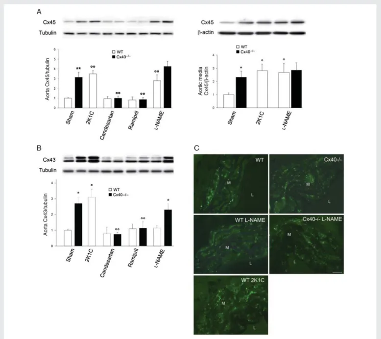
(Figure2A) and was reduced under treatment with either candesartan or ramipril in Cx402/2but not WT mice (Figure2A, left panel, and see Supplementary material online, Figure S2C).
Cx43 increased only in renin-dependent hypertension (Figure2B), due to a transcriptional up-regulation (see Supplementary material online, Figure S2D) which, as judged by its blockade under candesartan or ramipril treatment, required activation of the renin – angiotensin system (RAS) (Figure 2B and see Supplementary material online, Figure S2D). Immunostaining showed that most of Cx43 was expressed by aortic SMCs (Figure2C). Antibodies to Ser262 and Ser368 showed that these changes did not affect the proportion of phosphorylated Cx43 (see Supplementary material online, Figure S3). These results show that chronic hypertension is associated with an up-regulation of all major aortic Cxs, which are differentially regulated by renin-dependent and -inrenin-dependent mechanisms.
3.3 Cx43 is increased by AngII
Given that Cx43 was the only Cx which selectively increased in renin-dependent hypertension, and that AngII is the main RAS Figure 1 (Continued)
activator in this form of hypertension,30,31 we investigated the relationship between these two peptides. By exposing freshly iso-lated aortas of WT mice to AngII, we observed that within 4 h, 10 nM AngII, a concentration that mimics the physiological levels of the vasoconstricting peptide,18,32–35 significantly increased the levels of Cx43. However, the two- to three-fold increase in this protein which was observed in vivo was only reached with micro-molar concentrations (Figures 3A and B). Therefore, we selected a concentration of 2 mM AngII for all other experiments. Western blots showed that within 2 h, 2 mM AngII increased Cx43 in WT SMCs (Figure 3C), and the phosphorylation of ERK1/2 (Figure 3D).
This phosphorylation was not increased following theL-NAME treat-ment (Figure 3E), which also did not alter Cx43 expression (Figure 2B). In the aortas of Cx402/2 mice, AngII induced a smaller and delayed increase in Cx43, and prevented the down-regulation of Cx43 expression which occurred in the absence of the hormone (Figure 3F). Under this condition, the levels of Cx43 decreased in the aorta of Cx402/2 mice to reach control values within 48 h. (Figure 3F), due to rundown of AngII, which is natively high in these animals.7,8 These results show that Cx43 is up-regulated in parallel with the ERK pathway, due to RAS activation.
Figure 2 Cx43 and Cx45 are differently affected by renin-dependent and -independent hypertension. (A) Cx45 increased in all WT mice made hypertensive, and this increase was blocked in Cx402/2mice by RAS inhibitors (left panel). Cx45 was specifically increased in the aortic SMCs of hypertensive mice (right panel). (B) In contrast, Cx43 increased only in aortas of renin-dependent models (Cx402/2 and 2K1C). Data represent
mean þ SEM of four experiments. *P , 0.05, **P , 0.01 vs. the respective WT animals; 8P , 0.05, 88P , 0.01 vs. WT or Cx402/2 untreated or sham-operated animals. (C ) Immunostaining showed Cx43 throughout the media of all aortas. Increased numbers of immunospots were observed in the media of all Cx402/2and hypertensive 2K1C WT mice. M, media; L, lumen. Bar represents 30 mm.
3.4 The AngII-induced regulation is seen
in smaller arteries
Using carotid and mesenteric arteries, which express a Cx pattern analogous to that of the aorta (Figure4A), we also observed increased levels of Cx43 and ERK1/2 phosphorylation in Cx402/2 mice (Figure 4B). When exposed in vitro to AngII, the two types of WT vessels featured an increased expression of Cx43 and phosphoryl-ation of ERK1/2 (Figure4C), as observed in the aorta (Figure3C and D). This change was significantly higher in the small-diameter mesen-teric arteries than in the larger carotids (Figure 4C). These exper-iments show that chronic hypertension similarly targets the aorta and the smallest arteries we could test in mice.
3.5 A7r5 cells represent a convenient
model of aortic SMCs
To further investigate the molecular mechanism of the AngII induction of Cx43, we searched a cell line that would mimic the main features of primary SMCs. We found that A7r5 expressed a Cx pattern alike that of aortic SMCs (Figure5A, C, D) and responded to physiologically rel-evant concentrations of AngII with a similar increase in Cx43 and phosphorylated ERK1/2 (P-ERK1/2) (Figure5A and B). At a 2 mM con-centration, which induced in A7r5 cells a two- to three-fold increase in Cx43 similar to that observed in vivo and in isolated aortas, A7r5 cells also increased the expression of Cx43, but not that of Cx37, Cx40, and Cx45 (Figure 5C and D) and showed a rapid, Figure 3 AngII stimulates Cx43 expression in a dose-dependent manner. (A and B) Western blots of WT aortas exposed for 4 h to AngII showed that the peptide increased the levels of Cx43 and P-ERK1/2, at physiologically nanomolar concentrations, and reproduced the two-fold Cx43 increase observed in vivo, at concentrations .0.1 mM. (C and D) Exposure of WT aortas to 2 mM AngII increased within 2 h Cx43 (C ), and P-ERK1/2 (D). (E) The levels of the P-ERK1/2 increased in aortas of Cx402/2and 2K1C mice, but not in those ofL-NAME-treated mice. (F ) A smaller and delayed effect
was observed with aortas of Cx402/2mice. In the absence of AngII, the levels of Cx43 decreased in this tissue to reach control values within 48 h. Data represent mean þ SEM of three experiments. *P , 0.05, **P , 0.01, 8P , 0.05 vs. respective untreated WT or Cx402/2aortas.§P , 0.05 vs. untreated Cx402/2 aortas at t ¼ 0 h.
AngII-dependent activation of the ERK pathway (Figure 5E). Thus, A7r5 cells reproduced the main Cx and signalling changes observed in native aortas.
3.6 AngII increases Cx43 via activation
of the NF-kB pathway
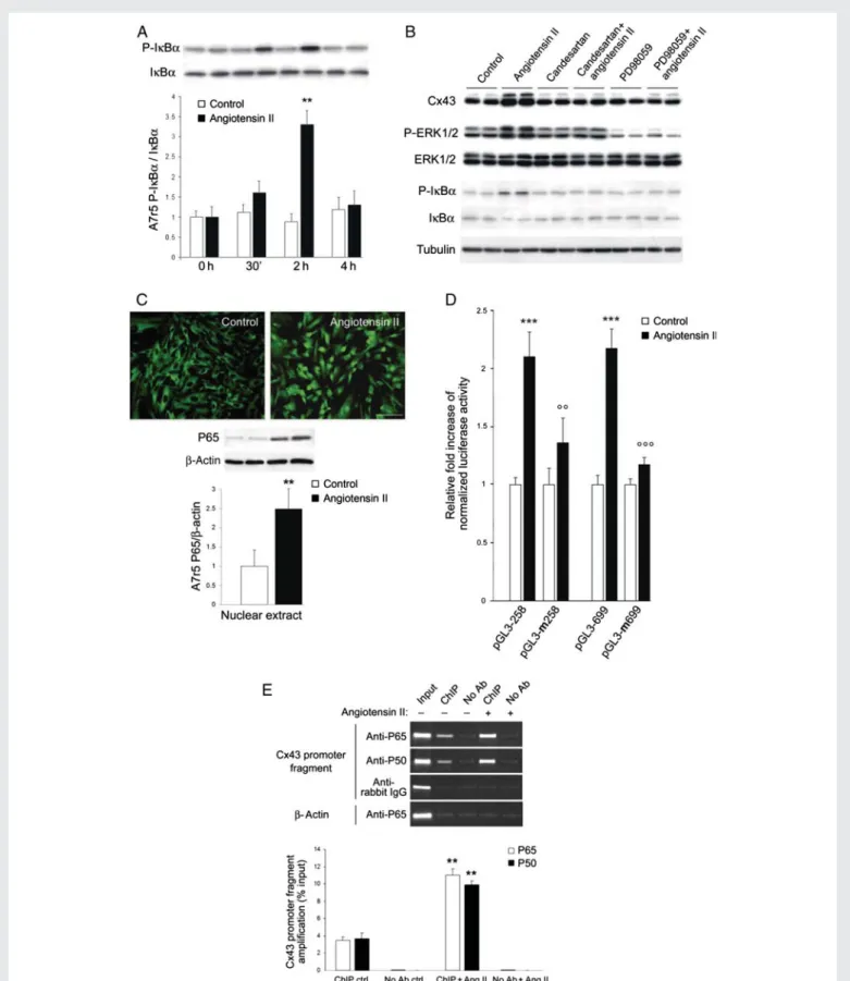
A7r5 cells exposed to AngII increased the proportion of phosphory-lated IkBa36,37(Figure6A and B), with a time course consistent with that of the Cx43 changes (Figure 5A). Immunostaining demonstrated that AngII induced the nuclear translocation of the p65-NF-kB tran-scription factor (Figure 6C). Candesartan, and the inhibitor of the ERK1/2 pathway PD98059, prevented the AngII-dependent induction of Cx43 expression, and ERK1/2 and IkBa phosphorylation (Figure 6B). These results suggest that AngII up-regulates Cx43 via the activation of the ERK and NF-kB pathways.
3.7 NF-kB binds to the promoter of the
Cx43 gene and activates its transcription
We found a canonical kB site between 2312 and 2301 bp of the promoter of the rat Gja1 gene. Therefore, we transfected A7r5 cells with a luciferase cDNA, under control of two fragments of the rat Cx43 promoter, each containing a kB site. After 4 h in the pres-ence of 2 mM AngII, we observed an increase in luciferase activity, whether the reporter gene was driven by the short (258 bp) or long (699 bp) promoter fragment (Figure6D). This increase was pre-vented by mutating the kB site (Figure6D).We incubated chromatin from control and AngII-stimulated A7r5 cells with antibodies against either the P65 or P50 form of NF-kB. Both antibodies immunoprecipitated fragments which, by PCR, yielded a sequence corresponding to 2433 and 2187 bp (relative to the transcription initiation site38) of the Cx43 promoter (Figure6E). After a 2 h exposure to AngII, the recovery of this frag-ment increased in the presence, but not in the absence, of antibodies against NF-kB (Figure6E). These data document that AngII enhances the binding of NF-kB to the Gja1 promoter.
4. Discussion
The multiple Cxs which are expressed by vascular ECs and SMCs4,6 have been implicated in the physiological function of arteries,39 and their adaptation to chronic hypertension.22,40 Still, it is not clear whether and how all vascular Cxs are similarly affected by all forms of hypertension.41 Comparing models of renin-dependent5 and -independent hypertension,5,7,10,18we now show that all hypertensive mice featured a thickening of the media, which was associated with increased levels of Cx40 in the intimal ECs, and of Cx37 and Cx45 in the media SMCs. In contrast, Cx43 was up-regulated in the latter layer, without obvious changes in its phosphorylation, only under con-ditions of renin-dependent hypertension. These observations docu-ment that multiple Cx changes are associated with the remodelling of the arterial wall which occurs in response to the altered mechanical and hormonal environment of hypertension. They further show that although the control of Cx37, Cx40, and Cx45 expression does not depend on the circulating levels of renin and AngII, these hormones are required for the control of Cx43. Given that AngII ultimately med-iates the vasoconstriction and SMC changes42that result from RAS activation,31we have investigated the effects of this peptide. Consist-ent with previous data in rats and cultured SMCs,42we have found Figure 4 Carotid and mesenteric arteries show a Cx pattern and
responsiveness to AngII similar to those of aorta. (A, left panels) Immunostaining showed punctate Cx43 throughout the carotid media. Increased numbers of immunospots were observed in the media of Cx402/2 mice. (A, right panels) Cx37 was detectable in the media of hypertensive but not control mice. L, lumen; M, media. Bar represents 10 mm. (B) Western blots showed that Cx37, Cx43, and P-ERK1/2 were increased in carotid and mesenteric arteries of Cx402/2mice. (C ) After a 4 h exposure of WT carotids and mesenteric arteries to 2 mM AngII, Cx43 and P-ERK1/2, but not Cx37, were increased. Data represent mean þ SEM of three exper-iments. *P , 0.05, **P , 0.01 vs. respective untreated vessels or WT animals.
that within a few hours, physiologically relevant levels of AngII increased the expression of Cx43, but not of Cx37, Cx40, and Cx45. This change is induced by the activation of ERK1/2 which, by causing the phosphorylation of IkBa,43,44results in the nuclear trans-location of NF-kB. We now document that NF-kB directly and specifically binds in situ to the promoter of the Gja1 gene which codes for Cx43, and that the NF-kB binding is enhanced by AngII. The Cx43 changes are rapidly induced by the ERK1/2 activation, but persist even after the ERK activity has returned to basal levels, indicating that other mechanisms contribute to the sustained elevation of Cx43 expression.
These experiments provide the first coherent explanation of how a renin increase enhances Cx43: (i) the activation of RAS increases the cir-culating levels of AngII; (ii) elevated AngII results in peripheral vasocon-striction, causing hypertension, and triggering SMC hypertrophy and proliferation; (iii) in parallel, AngII promotes, via phosphorylation of ERK1/2, the binding of NF-kB to the Gja1 promoter; (iv) this binding trans-activates the Gja1 gene, leading to increased production of Cx43 in the SMCs (see Supplementary material online, Figure S4). The finding of comparable Cx43 changes in the aorta, the carotids, and the small mesenteric arteries suggests that our observations apply to the many vascular compartments targeted by hypertension.15,45 Figure 5 A7r5 cells feature the same Cx43 and signalling changes observed in aortic SMCs. (A and B) Western blots of A7r5 cells exposed for 2 h to increasing concentrations of AngII showed that the peptide increased the levels of Cx43 and P-ERK1/2 at concentrations similar to those that elicited comparable changes in aortic SMCs. (C and D) Western blots showed that A7r5 cells expressed Cx43, Cx37, Cx40, and Cx45. Two-micromolar AngII increased Cx43, but not Cx37, Cx40, and Cx45. (E) AngII also increased the levels of P-ERK1/2. Results are means þ SEM of three experiments. *P , 0.05, **P , 0.01 vs. untreated cells.
Figure 6 The AngII-induced increase in Cx43 requires the activation of ERK and NF-kB pathways. (A) Within 2 h, AngII increased the phosphoryl-ation of IkBa in A7r5 cells. Results are means þ SEM of three experiments. **P , 0.01 vs. untreated cells. (B) Within 2 h, AngII also increased Cx43, P-ERK1/2, and P-IkBa levels. Both candesartan and PD98059 prevented these effects. (C ) After 4 h, 2 mM AngII induced a translocation of the P65 phosphorylated form of NF-kB into the nucleus of A7r5 cells. Bar, 20 mm. (D) AngII increased luciferase activity of A7r5 cells transfected with either a short or long Gja1 promoter fragment. Mutation of the NF-kB motif in either one of the two promoter fragments prevented the increased in the luciferase activity observed after AngII stimulation. (E) Quantitative RT – PCR showed that AngII increased the amount of the Gja1 promoter fragment, recovered using antibodies against either the P65 or the P50 forms of NF-kB. Results are means þ SEM of three experiments. **P , 0.01, ***P , 0.001 vs. untreated cells; 88P , 0.01, 888P , 0.001 vs. AngII-treated cells.
Our data further implicate that other mechanisms, possibly dependent on the mechanical forces that impinge on the arterial walls during hyper-tension,1,46,47in the control of Cx37, Cx40, and Cx45, as well as in the sustained elevation of Cx43, once the activation of ERK1/2 ends.
The significance of the increase in Cx43 in renin-dependent hyper-tension remains to be elucidated. Beside its major vasoconstrictor effect, AngII also stimulates vascular remodelling by promoting SMC growth and the production of extra cellular matrix, which contribute to increased aortic stiffness,48 a frequent consequence of hyperten-sion.49Increased Cx43 expression in the presence of elevated AngII may therefore contribute to regulate the elasticity of the vascular wall. If so, the pharmacological targeting of Cx43 may be therapeuti-cally useful, in addition to the standard anti-hypertensive drugs which aim at counteracting the AngII effects.50
In summary, by comparing control and Cx40-lacking mice, under conditions of renin-dependent (2K1C) and renin-independent hyper-tension (L-NAME administration), we have found that Cx37, Cx40,
and Cx45 are up-regulated in the wall of different types of arteries, probably as a result of the increased mechanical forces impinging on the vessels’ wall. In contrast, Cx43 was up-regulated only in the renin-dependent model of hypertension, due to an AngII-renin-dependent mech-anism, which specifically activates the transcription of the Cx43 gene. These findings document that the expression of several vascular con-nexins is affected by chronic hypertension, though by different mech-anisms, and in different compartments of the arterial wall.
Supplementary material
Supplementary material is available at Cardiovascular Research online. Conflict of interest: none declared.
Funding
Supported by grants from the SNF (310000-122423; 310000-109402), the JDRF (1-2007-158), the EU (BETAIMAGE 222980; IMIDIA, C2008-T7), the NIH HL64232, the Octav and the Marcella Botnar Foundation, the Novartis Foundation, and the Emma Muschamp Foundation.
References
1. Haefliger JA, Nicod P, Meda P. Contribution of connexins to the function of the vas-cular wall. Cardiovasc Res 2004;62:345 – 356.
2. Simon AM, McWhorter AR. Decreased intercellular dye-transfer and downregulation of non-ablated connexins in aortic endothelium deficient in connexin37 or con-nexin40. J Cell Sci 2003;116:2223 – 2236.
3. Kruger O, Beny JL, Chabaud F, Traub O, Theis M, Brix K et al. Altered dye diffusion and upregulation of connexin37 in mouse aortic endothelium deficient in connexin40. J Vasc Res 2002;39:160 – 172.
4. Brisset AC, Isakson BE, Kwak BR. Connexins in vascular physiology and pathology. Antioxid Redox Signal 2009;11:267 – 282.
5. Haefliger JA, Krattinger N, Martin D, Pedrazzini T, Capponi A, Doring B et al. Connexin43-dependent mechanism modulates renin secretion and hypertension. J Clin Invest 2006;116:405 – 413.
6. Figueroa XF, Duling BR. Gap junctions in the control of vascular function. Antioxid Redox Signal 2009;11:251 – 266.
7. Krattinger N, Capponi A, Mazzolai L, Aubert JF, Caille D, Nicod P et al. Connexin40 regulates renin production and blood pressure. Kidney Int 2007;72:814 – 822. 8. Wagner C, de Wit C, Kurtz L, Grunberger C, Kurtz A, Schweda F. Connexin40 is
essential for the pressure control of renin synthesis and secretion. Circ Res 2007; 100:556 – 563.
9. Kurtz L, Schweda F, de Wit C, Kriz W, Witzgall R, Warth R et al. Lack of connexin 40 causes displacement of renin-producing cells from afferent arterioles to the extraglo-merular mesangium. J Am Soc Nephrol 2007;18:1103 – 1111.
10. Wagner C. Function of connexins in the renal circulation. Kidney Int 2008;73: 547 – 555.
11. Krattinger N, Alonso F, Capponi A, Mazzolai L, Nicod P, Meda P et al. Increased expression of renal cyclooxygenase-2 and neuronal nitric oxide synthase in hyperten-sive Cx40-deficient mice. J Vasc Res 2009;46:188 – 198.
12. Zanchi A, Wiesel P, Aubert JF, Brunner HR, Hayoz D. Time course changes of the mechanical properties of the carotid artery in renal hypertensive rats. Hypertension 1997;29:1199 – 1203.
13. Fitch RM, Rutledge JC, Wang YX, Powers AF, Tseng JL, Clary T et al. Synergistic effect of angiotensin II and nitric oxide synthase inhibitor in increasing aortic stiffness in mice. Am J Physiol Heart Circ Physiol 2006;290:H1190 – H1198.
14. Yasmin, O’Shaughnessy KM. Genetics of arterial structure and function: towards new biomarkers for aortic stiffness? Clin Sci (Lond) 2008;114:661 – 677.
15. Laurent S, Boutouyrie P, Lacolley P. Structural and genetic bases of arterial stiffness. Hypertension 2005;45:1050 – 1055.
16. Safar ME, Thuilliez C, Richard V, Benetos A. Pressure-independent contribution of sodium to large artery structure and function in hypertension. Cardiovasc Res 2000; 46:269 – 276.
17. Safar ME, Boudier HS. Vascular development, pulse pressure, and the mechanisms of hypertension. Hypertension 2005;46:205 – 209.
18. Wiesel P, Mazzolai L, Nussberger J, Pedrazzini T. Two-kidney, one clip and one-kidney, one clip hypertension in mice. Hypertension 1997;29:1025 – 1030.
19. Kirchhoff S, Nelles E, Hagendorff A, Kruger O, Traub O, Willecke K. Reduced cardiac conduction velocity and predisposition to arrhythmias in connexin40-deficient mice. Curr Biol 1998;8:299 – 302.
20. Mazzolai L, Nussberger J, Aubert JF, Brunner DB, Gabbiani G, Brunner HR et al. Blood pressure-independent cardiac hypertrophy induced by locally activated renin-angiotensin system. Hypertension 1998;31:1324 – 1330.
21. Fluckiger JP, Gremaud G, Waeber B, Kulik A, Ichino A, Nussberger J et al. Measure-ment of sympathetic nerve activity in the unanesthetized rat. J Appl Physiol 1989;67: 250 – 255.
22. Haefliger JA, Castillo E, Waeber G, Bergonzelli GE, Aubert JF, Sutter E et al. Hyper-tension increases connexin43 in a tissue-specific manner. Circulation 1997;95: 1007 – 1014.
23. Mederos y, Schnitzler M, Storch U, Meibers S, Nurwakagari P, Breit A et al. Gq-coupled receptors as mechanosensors mediating myogenic vasoconstriction. EMBO J 2008;27:3092 – 3103.
24. Skutella M, Ruegg UT. Studies on capacitative calcium entry in vascular smooth muscle cells by measuring 45CA2þ influx. J Recept Signal Transduct Res 1997;17: 163 – 175.
25. Skutella T, Savaskan NE, Ninnemann O, Nitsch R. Target- and maturation-specific membrane-associated molecules determine the ingrowth of entorhinal fibers into the hippocampus. Dev Biol 1999;211:277 – 292.
26. Simon AM, Chen H, Jackson CL. Cx37 and Cx43 localize to zona pellucida in mouse ovarian follicles. Cell Commun Adhes 2006;13:61 – 77.
27. Haefliger JA, Demotz S, Braissant O, Suter E, Waeber B, Nicod P et al. Connexins 40 and 43 are differentially regulated within the kidneys of rats with renovascular hyper-tension. Kidney Int 2001;60:190 – 201.
28. Allagnat F, Alonso F, Martin D, Abderrahmani A, Waeber G, Haefliger JA. ICER-1 gamma overexpression drives palmitate-mediated connexin36 down-regulation in insulin-secreting cells. J Biol Chem 2008;283:5226 – 5234.
29. Martin D, Allagnat F, Chaffard G, Caille D, Fukuda M, Regazzi R et al. Functional sig-nificance of repressor element 1 silencing transcription factor (REST) target genes in pancreatic beta cells. Diabetologia 2008;51:1429 – 1439.
30. Burnier M. Spotlight on renin. The renin-angiotensin-aldosterone system and meta-bolic syndrome. J Renin Angiotensin Aldosterone Syst 2006;7:184.
31. Mehta PK, Griendling KK. Angiotensin II cell signaling: physiological and pathological effects in the cardiovascular system. Am J Physiol Cell Physiol 2007;292:C82 – C97. 32. Mazzolai L, Pedrazzini T, Nicoud F, Gabbiani G, Brunner HR, Nussberger J. Increased
cardiac angiotensin II levels induce right and left ventricular hypertrophy in normoten-sive mice. Hypertension 2000;35:985 – 991.
33. Stanton AV, Gradman AH, Schmieder RE, Nussberger J, Sarangapani R, Prescott MF. Aliskiren monotherapy does not cause paradoxical blood pressure rises. meta-analysis of data from 8 clinical trials. Hypertension 2010;55:54 – 60.
34. Abe F, Omata K, Yamada M, Tsunoda K, Sato T, Shimizu T et al. Specific direct radio-immunoassay of angiotensin II (AT II) in human plasma and the effect of angiotensin converting enzyme (ACE) inhibitor. Immunopharmacology 1999;44:199 – 204. 35. Kiribayashi K, Masaki T, Naito T, Ogawa T, Ito T, Yorioka N et al. Angiotensin II
induces fibronectin expression in human peritoneal mesothelial cells via ERK1/2 and p38 MAPK. Kidney Int 2005;67:1126 – 1135.
36. Ghosh S, Karin M. Missing pieces in the NF-kappaB puzzle. Cell 2002;109(Suppl.): S81 – S96.
37. Levrand S, Pesse B, Feihl F, Waeber B, Pacher P, Rolli J et al. Peroxynitrite is a potent inhibitor of NF-fkappagB activation triggered by inflammatory stimuli in cardiac and endothelial cell lines. J Biol Chem 2005;280:34878 – 34887.
38. Teunissen BE, Jansen AT, van Amersfoorth SC, O’Brien TX, Jongsma HJ, Bierhuizen MF. Analysis of the rat connexin 43 proximal promoter in neonatal cardi-omyocytes. Gene 2003;322:123 – 136.
39. Fanchaouy M, Bychkov R, Meister JJ, Beny JL. Stretch-elicited calcium responses in the intact mouse thoracic aorta. Cell Calcium 2007;41:41 – 50.
40. Haefliger JA, Meda P, Formenton A, Wiesel P, Zanchi A, Brunner HR et al. Aortic con-nexin43 is decreased during hypertension induced by inhibition of nitric oxide synthase. Arterioscler Thromb Vasc Biol 1999;19:1615 – 1622.
41. Figueroa XF, Isakson BE, Duling BR. Vascular gap junctions in hypertension. Hyperten-sion 2006;48:804 – 811.
42. Cheng G, Kim MJ, Jia G, Agrawal DK. Involvement of chloride channels in IGF-I-induced proliferation of porcine arterial smooth muscle cells. Cardiovasc Res 2007;73: 198 – 207.
43. Cui R, Tieu B, Recinos A, Tilton RG, Brasier AR. RhoA mediates angiotensin II-induced phospho-Ser536 nuclear factor kappaB/RelA subunit exchange on the interleukin-6 promoter in VSMCs. Circ Res 2006;99:723 – 730.
44. Bonizzi G, Karin M. The two NF-kappaB activation pathways and their role in innate and adaptive immunity. Trends Immunol 2004;25:280 – 288.
45. Safar ME, Jankowski P. Central blood pressure and hypertension: role in cardiovascu-lar risk assessment. Clin Sci (Lond) 2009;116:273 – 282.
46. Haefliger JA, Tissieres P, Tawadros T, Formenton A, Beny JL, Nicod P et al. Connexins 43 and 26 are differentially increased after rat bladder outlet obstruction. Exp Cell Res 2002;274:216 – 225.
47. Sakurabayashi-Kitade S, Aoka Y, Nagashima H, Kasanuki H, Hagiwara N, Kawana M. Aldosterone blockade by spironolactone improves the hypertensive vascular hyper-trophy and remodeling in angiotensin II overproducing transgenic mice. Atherosclerosis 2009;206:54 – 60.
48. Tham DM, Martin-McNulty B, Wang YX, Da Cunha V, Wilson DW, Athanassious CN et al. Angiotensin II injures the arterial wall causing increased aortic stiffening in apo-lipoprotein E-deficient mice. Am J Physiol Regul Integr Comp Physiol 2002;283: R1442 – R1449.
49. Wang YX, Fitch RM. Vascular stiffness: measurements, mechanisms and implications. Curr Vasc Pharmacol 2004;2:379 – 384.
50. Dzau VJ. Mechanism of protective effects of ACE inhibition on coronary artery disease. Eur Heart J 1998;19(Suppl. J):J2 – J6.