Ferredoxin:thioredoxin reductase (FTR) plays an important role in the light-regulated catalytic properties of enzymes involved in the Calvin cycle.1 The light signal is transmitted in the form of electrons from the chlorophyll-containing thylakoid membranes via a [2Fe-2S] ferredoxin, FTR, and thioredoxins to target enzymes, which are activated or deactivated by the reduction of regulatory disulfide bonds. FTR utilizes a unique active site that comprises a [4Fe-4S]2+ cluster with an adjacent disulfide2-4 to catalyze the two-electron reduction of the thioredoxin disulfide. Previous spectroscopic investigations of the Spinacea oleracea FTR3,5have shown that alkylation of one cysteine of the active-site disulfide (C54) by N-ethylmaleimide (NEM) affords a stable analogue of the one-electron-reduced heterodisulfide intermediate. The combined EPR, ENDOR, resonance Raman, and MCD data of the NEM-modified FTR suggest a novel type of [4Fe-4S]3+ cluster with five cysteine ligands, but the ligation site of the fifth cysteine ligand was left undetermined.5In this study, both the as-purified and NEM-modified forms of FTR from spinach6have been investigated by Mo¨ssbauer spectroscopy to provide further understanding of the cluster coordination and electronic state. The results demonstrate the presence of a unique iron site in the [4Fe-4S] cluster and suggest that site-specific cluster chemistry, involving the formation of a five-coordinate Fe site with two cysteinate ligands, occurs during catalytic cycling of FTR.
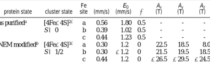
The 4.2 K Mo¨ssbauer spectrum of the as-purified FTR recorded in a weak magnetic field of 50 mT shows a quadrupole doublet with a prominent shoulder on the side of the high-energy line (Figure 1, hatched marks). A spectrum recorded in a strong magnetic field of 8 T (not presented) indicates that the cluster is diamagnetic, consistent with a [4Fe-4S]2+ assignment. Both spectra can be deconvoluted into three components with an intensity ratio of 1:1:2 (Figure 1), corresponding to three distinct Fe sites, a, b, and c, respectively (Table 1). Typically, the Fe atoms of a [4Fe-4S]2+ cluster can be grouped into two valence-delocalized Fe2.5+Fe2.5+ pairs that are antiferromagnetically coupled to form a diamagnetic ground state,7 and accordingly the Mo¨ssbauer spectrum consists of a symmetric quadrupole doublet with parameters (δ ) 0.40-0.45 mm/s, and∆EQ) 1.0-1.2 mm/s) that are indicative of Fe2.5+ ions with tetrahedral sulfur coordination.8,9The absorption intensity determined for site c of FTR indicates that it represents one of the two Fe2.5+Fe2.5+pairs. Therefore, sites a and b must represent the other pair. The parameters determined for both sites b and c are within the ranges observed for [4Fe-4S]2+clusters, indicating Fe sites of regular coordination. The larger δ and ∆EQ of site a, however, indicate a unique Fe site with atypical coordination
environment. This observation is consistent with the X-ray structure of Synechocystis FTR4which shows that the sulfur atom of one of the cysteine residues forming the active-site disulfide is in van der Waals contact (3.1 Å) with both the Fe atom ligated by C52(spinach enzyme sequence number) and the sulfur atom of C52, resulting in the Fe site being distorted from tetrahedral coordination with a (C52)S-Fe-S angle of 129° that is open toward the disulfide (Scheme 1). The presence of a unique Fe site in [4Fe-4S]2+clusters has been detected in both model compounds and proteins. In model complex studies, the increases inδ and ∆EQ are correlated with increases in coordination number at the unique Fe site.10In proteins, unique Fe sites were observed with increasedδ upon binding of substrates to the [4Fe-4S]2+cluster in aconitase,11and upon binding of S-adenosylmethionine to the clusters in pyruvate formate-lyase activating enzyme,12and in biotin synthase.13Thus, it is tempting
§Emory University. †University of Georgia. ‡Universite´ de Neuchaˆtel.
Figure 1. Mo¨ssbauer spectrum of as-purified FTR (0.24 mM) recorded at 4.2 K in a field of 50 mT applied parallel to theγ beam (hatched marks). The spectrum can be deconvoluted into three components with an intensity ratio of 1:1:2 representing three Fe sites a, b, and c (Table 1). The individual components are shown above the spectrum as two solid lines (a and b sites) and a dotted line (c). The solid line overlaid with the experimental spectrum is the sum of the three components.
Table 1. Mo¨ssbauer Parameters of As-Purified and NEM-Modified Spinach FTR
protein state cluster state Fe site (mm/s)δ (mm/s)∆EQ η Ax (T) Ay (T) Az (T) as purifieda [4Fe-4S]2+ a 0.56 1.80 0.5 - - -S ) 0 b 0.39 1.02 0.5 - - -c 0.44 1.23 0.5 - -
-NEM modifiedb [4Fe-4S]3+ a 0.30 1.2 0 22.5 18.5 8.0
S ) 1/2 b 0.30 -1.2 0 21.5 19.5 18.5
c 0.44 1.2 0 -26.5 -29.5 -24.5 aThe spectrum shown in Figure 1 can also be fitted with two quadrupole doublets with an intensity ratio of 1:3. However, the line shape of the high-energy line is better fitted with three components.bTo minimize the number of parameters in our analysis, theδ of the two ferric sites and the magnitude of the∆EQof all three sites are assumed to be the same. The∆EQof site
c can be determined accurately from the spectrum shown in Figure 2.
Spectroscopic
Evidence
for
Site
Specific
Chemistry
at
a
Unique
Iron
Site
of
the
[4Fe
-
4S]
Cluster
in
Ferredoxin:Thioredoxin
Reductase
Guy N. L. Jameson,§Elizabeth M. Walters,†Wanda Manieri,‡Peter Schu¨ rmann,‡Michael K. Johnson,*,†and Boi
HanhHuynh*,§
Department of Physics, Emory UniVersity, Atlanta, Georgia 30322, Department of Chemistry and Center for Metalloenzyme Studies, UniVersity of Georgia, Athens, Georgia 30602, and Laboratoire de Biochimie Ve´ge´tale, UniVersite´ de Neuchaˆtel, CH-2007 Neuchaˆtel, Switzerland
1 Published in Journal of the American Chemical Society (JACS) 125, issue 5, pp. 1146–1147, 2003,
to speculate that the larger δ and ∆EQ of site a reflect a weak interaction between the active-site disulfide and the C52-bound Fe in as-purified FTR.
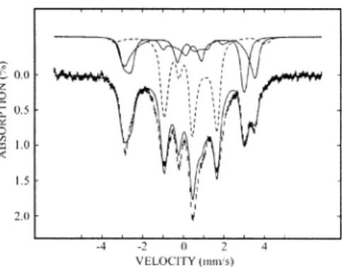
Figure 2 shows the 4.2 K spectrum of the NEM-modified FTR recorded in a magnetic field of 8 T (hatched marks). The spectrum is paramagnetic, consistent with the S )1/
2 state determined by previous EPR investigation.3,5Three distinct components with a 1:1:2 intensity ratio are also observed, indicating that the unique Fe site persists in the NEM-modified FTR. The components corresponding to the two individual Fe sites a and b can be clearly seen to produce a splitting in the absorption in the region between +3 and +4 mm/s (Figure 2). Comparison of the parameters of the NEM-modified form of FTR with those of the as-purified form shows a general reduction inδ for sites a and b with the larger reduction (∼0.26 mm/s) occurring at the unique site a. This observation suggests that upon NEM-modification a reducing equivalent is removed from the cluster, mostly from site a, and supports previous spectroscopic evidence3,5 that the cluster is formally in the [4Fe-4S]3+state. Further, the signs and magnitudes of the magnetic hyperfine coupling tensors compare well to those observed for [4Fe-4S]3+ clusters in high-potential iron-sulfur proteins14and reveal the antiparallel alignment of the spin of the mixed valence pair with those of the two ferric sites.
Taken together, the Mo¨ssbauer data provide key insights into the FTR mechanism that may be understood in terms of a donor-acceptor approach involving the active-site disulfide and the unique
Fe site of the [4Fe-4S] cluster. Partial bonding of the disulfide to the unique iron in the resting state of the cluster promotes charge buildup on that iron, making it an electron donor with increased ferrous character, which, in turn, explains the increased isomer shift. Concomitantly, this interaction between the unique Fe and the C84 sulfur polarizes the S-S bond making the interactive sulfur an electron acceptor (Scheme 1). The system is therefore primed and ready to accept an electron from ferredoxin to break the disulfide bond. When this occurs, C84binds to give a five-coordinate Fe site with two cysteinate ligands, thereby freeing C54 to attack the disulfide of thioredoxin to form the heterodisulfide intermediate. This one-electron reduced state is modeled by the NEM-modified form (Scheme 1). The binding of an additional cysteine to the unique Fe reverses the donor-acceptor properties, and charge is drawn away from the iron. The cluster is then formally in the [4Fe-4S]3+ oxidation state, and the unique Fe becomes more ferric, leading to a dramatic decrease in the isomer shift of that iron. This novel site-specific cluster chemistry provides molecular-level insight into how the [4Fe-4S] cluster mediates disulfide reduction in two one-electron steps in FTR and the related methanogenic hetero-disulfide reductases.15In addition it may provide a paradigm for understanding the mechanism of reductive cleavage of S-adenos-ylmethionine to yield methionine and the 5′-deoxyadenosyl radical in the radical SAM family of Fe-S enzymes.
Acknowledgment. This work was supported by Grants from
the NIH (GM47295 to B.H.H. and GM62542 to M.K.J.) and the Schweizerischer Nationalfonds (31-56761.99 to P.S.)
References
(1) Dai, S.; Schwendtmayer, C.; Johansson, K.; Ramaswamy, S.; Schu¨rmann, P.; Eklund, H. Q. ReV. Biophys. 2000, 33, 67-108.
(2) Chow, L.-P.; Iwadate, H.; Yano, K.; Kamo, M.; Tsugita, A.; Gardet-Salvi, L.; Stritt-Etter, A.-L.; Schu¨rmann, P. Eur. J. Biochem. 1995, 231, 149-156.
(3) Staples, C. R.; Ameyibor, E.; Fu, W.; Gardet-Salvi, L.; Stritt-Etter, A.-L.; Schu¨rmann, P.; Knaff, D. B.; Johnson, M. K. Biochemistry 1996, 35, 11425-11434.
(4) Dai, S.; Schwendtmayer, C.; Schu¨rmann, P.; Ramaswamy, S.; Eklund, H. Science 2000, 287, 655-658.
(5) Staples, C. R.; Gaymard, E.; Stritt-Etter, A.-L.; Telser, J.; Hoffman, B. M.; Schu¨rmann, P.; Knaff, D. B.; Johnson, M. K. Biochemistry 1998, 37, 4612-4620.
(6) Overexpression and purification of spinach FTR was performed according to the published procedure (Gaymard, E.; Franchini, L.Manieri, W.; Stutz, E.; Schu¨rmann, P. Plant Sci. 2000, 158, 107-113).57Fe incorporation
was achieved by addition of57Fe ferric ammonium citrate to
chelex-resin-treated LB media to a final Fe concentration of 5 mg/L. NEM-modified FTR was prepared as previously described,3using reduced methyl viologen
as the reductant.
(7) Noodleman, L.; Peng, C. Y.; Case, D. A.; Mouesca, J.-M. Coord. Chem.
ReV. 1995, 144, 199-244.
(8) Yoo, S. J.; Angove, H. C.; Burgess, B. K.; Hendrich, M. P.; Mu¨nck, E.
J. Am. Chem. Soc. 1999, 121, 2534-2545.
(9) Trautwein, A. X.; Bill, E.; Bominaar, E. L.; Winkler, H. Struct. Bonding 1991, 78, 1-95.
(10) Ciurli, S.; Carrie, M.; Weigel, J. A.; Carney, M. J.; Stack, T. D. P.; Papaefthymiou, G. C.; Holm, R. H. J. Am. Chem. Soc. 1990, 112, 2654-2664.
(11) Beinert, H.; Kennedy, M. C.; Stout, C. D. Chem. ReV. 1996, 96, 2335-2373.
(12) Krebs, C.; Broderick, W. E.; Henshaw, T. F.; Broderick, J. B.; Huynh, B. H. J. Am. Chem. Soc. 2002, 124, 912-913.
(13) Cosper, M. M.; Jameson, G. N. L.; Eidsness, M. K.; Huynh, B. H.; Johnson, M. K. J. Am. Chem. Soc. 2002, 124, 14006-14007. (14) Middleton, P.; Dickson, D. P.; Johnson, C. E.; Rush, J. D. Eur. J. Biochem.
1980, 104, 289-296.
(15) Duin, E. C.; Madadi-Kahkesh, S.; Hedderich, R.; Clay, M. D.; Johnson, M. K. FEBS Lett. 2002, 512, 263-268.
Figure 2. Mo¨ssbauer spectrum of NEM-modified FTR (0.23 mM) measured at 4.2 K in a field of 8 T applied parallel to theγ beam (hatched marks). The spectrum has been deconvoluted into three components (shown above the spectrum) with an intensity ratio of 1:1:2, corresponding to two distinct Fe sites of the ferric pair (sites a and b, solid lines) and a delocalized mixed-valence pair (site c, dashed line). The solid line overlaid with the experimental data is the sum of the three components.
Scheme 1. Illustration of Unique Iron-Site Chemistry of the [4Fe-4S] Cluster in the As-Purified FTR and the
One-Electron-Reduced Heterodisufide Intermediate, as Modeled by NEM-Modified FTR