HAL Id: hal-02411388
https://hal.archives-ouvertes.fr/hal-02411388
Submitted on 24 Nov 2020
HAL is a multi-disciplinary open access
archive for the deposit and dissemination of
sci-entific research documents, whether they are
pub-lished or not. The documents may come from
teaching and research institutions in France or
abroad, or from public or private research centers.
L’archive ouverte pluridisciplinaire HAL, est
destinée au dépôt et à la diffusion de documents
scientifiques de niveau recherche, publiés ou non,
émanant des établissements d’enseignement et de
recherche français ou étrangers, des laboratoires
publics ou privés.
Bedsides
Amine Belaïd, Papa Diogop Ndiaye, Harilaos Filippakis, Jérémie Roux, Eric
Röttinger, Yacine Graba, Patrick Brest, Paul Hofman, Baharia Mograbi
To cite this version:
Amine Belaïd, Papa Diogop Ndiaye, Harilaos Filippakis, Jérémie Roux, Eric Röttinger, et al..
Au-tophagy : Moving Benchside Promises to Patient Bedsides. Current Cancer Drug Targets, Bentham
Science Publishers, 2015, 15 (8), pp.684-702. �10.2174/156800961508151001102452�. �hal-02411388�
1568-0096/15 $58.00+.00 © 2015 Bentham Science Publishers
Autophagy : Moving Benchside Promises to Patient Bedsides
Amine Belaid
1,2,3,4, Papa Diogop Ndiaye
1,2,3,#, Harilaos Filippakis
4,#, Jérémie Roux
1,2,3,
Éric Röttinger
1,2, Yacine Graba
5, Patrick Brest
1,2,3, Paul Hofman
1,2,3,6,7and Baharia Mograbi
1,2,3,*
1Institute of Research on Cancer and Ageing of Nice (IRCAN), INSERM U1081, CNRS UMR7284,
Nice, F-06107, France;
2Université de Nice-Sophia Antipolis, Faculté de Médecine, Nice, F-06107,
France;
3Equipe Labellisée par l’ARC 9 rue Guy Môquet F- 94803 Villejuif, France;
4Brigham and
Women's Hospital, Harvard Medical School, Boston, Massachusetts, USA;
5CNRS, Aix Marseille
Université, IBDML, UMR 7288, Campus de Luminy, Marseille, cedex 09, 13288, France;
6Centre
Hospitalier Universitaire de Nice, Pasteur Hospital, Laboratory of Clinical and Experimental
Pathology, Nice, F-06002, France;
7Centre Hospitalier Universitaire de Nice, Pasteur Hospital,
Human Biobank, Nice, F-06002, France
Abstract: Survival rates of patients with metastatic or recurrent cancers have remained virtually
unchanged during the past 30 years. This fact makes the need for new therapeutic options even more urgent. An attractive
option would be to target autophagy, an essential quality control process that degrades toxic aggregates, damaged
organelles, and signaling proteins, and acts as a tumor suppressor pathway of tumor initiation. Conversely, other
fascinating observations suggest that autophagy supports cancer progression, relapse, metastasis, dormancy and resistance
to therapy. This review provides an overview of the contradictory roles that autophagy plays in cancer initiation and
progression and discusses the promises and challenges of current strategies that target autophagy for cancer therapy.
Keywords: Antineoplastic agents/drug effects/autophagy targeted therapy, autophagy addiction/KRAS/BRAF-driven cancers,
tumor metabolism, tumor resistance, tumor relapse, tumor metastasis, tumor dormancy, translational medicine.
AUTOPHAGY AND CANCER
On December 1, 2014, a PubMed search of “autophagy
and cancer” yielded 5,470 entries, which constitutes 31% of
all 17,588 articles published on the topic of “autophagy.”
This amount of literature illustrates how autophagy has both
positive and negative roles in tumorigenesis: promoting
tumor growth or tumor suppression (Fig. 1). Not surprisingly,
all the signaling pathways that control cancer development
regulate autophagy, which is exploited by both tumor
suppressor and oncogenic mechanisms. However, the results
of fifteen years of research do not clarify whether cancer
therapies can suppress or upregulate autophagy, and whether
upregulation of autophagy can favor tumor cell survival or
death. The exact role that autophagy plays
in cancer is
therefore complex and warrants a further unifying model. In
the following sections, we provide a detailed discussion of
the ways by which autophagy can be both tumorigenic and
tumor suppressive.
AUTOPHAGY AT A GLANCE
The beginning of the research on autophagy can be
tracked back to 1956 when Sam Clark observed irregularly
shaped vacuoles containing amorphous materials and
occasional mitochondria [1]. Soon after that, Christian de
*Address correspondence to this author at Institute of Research on Cancer and Ageing of Nice (IRCAN); Université de Nice-Sophia Antipolis; Centre Antoine Lacassagne, Avenue de Valombrose; 06107 Nice Cedex 02, France; Tel: +33.4.92.03.12.45; Fax: +33.4.92.03.12.41;
E-mail: [email protected]
#These authors contribute equally to this work.
Duve established the nomenclature for different lysosomal
pathways (Nobel Prize in Physiology or Medicine, 1974).
Autophagy, as suggested by its Greek acronym
‘self-eating’, targets intracellular organelles and constituents for
lysosomal degradation. So far, three different types of
autophagy have been described; namely,
chaperone-mediated autophagy (CMA), microautophagy, and macro-
autophagy. These three types essentially differ in the
mechanism by which they deliver substrates (cargo) to the
lysosomal lumen. Chaperone-mediated autophagy ensures
the translocation of a KFERQ-motif bearing protein into the
lysosome by the chaperone heat shock protein cognate 70
(HSC70) [2]. Microautophagy involves the direct
internalization of small cytoplasmic material into the
lysosome through invagination of its membrane [3].
Macroautophagy delivers proteins and organelles to
lysosomes for degradation upon sequestration in a
double-membrane vesicle – termed the autophagosome.
The complementarity, speed, specificity, and most
importantly the reversibility of these pathways allow for the
complete adaptation of a cell to its environment.
Complementarity
All three of these pathways are constitutively active at
basal levels to maintain cell homeostasis. In response to
environmental stress, such as those encountered in cancer,
including nutrient starvation, hypoxia, and other forms of
metabolic stress, both macroautophagy and CMA are
upregulated, but their activation does not occur
simultaneously. In as early as 30 minutes, cells overcome
nutrient depletion by dramatically up-regulating autophagy.
Even under persistent starvation, cells do not cannibalize
themselves. The upregulation of autophagy is transient,
reaches maximal activity around the first 4–6 hours, and then
gradually declines to basal levels [4]. The decrease in
macroautophagy is concomitant with a progressive switch to
CMA that may allow the cell to degrade only unwanted
proteins for cell survival [2]. In this review, we will focus
the discussion on the role of macroautophagy (hereafter
referred to as autophagy) in cancer.
Reversibility
It is known that the autophagosomes are formed and
degraded within only eight minutes [5]. As a result, the
autophagic pathway is highly dynamic, rapidly increased and
suppressed once the stress is removed, providing evidence
that unlike other stress states, namely apoptosis, necrosis or
senescence, autophagy is a reversible phenomenon.
Cargo Specificity
Since its discovery in the 1950s, macroautophagy was
first thought to be a bulk, non-selective “self-eating” process.
Emerging evidence now suggests that autophagy is a quality
control process, which selectively degrades damaged and
unneeded proteins and organelles that would otherwise
unnecessarily accumulate during the life of the cell.
Autophagic substrates include organelles such as
mitochondria (mitophagy), peroxisomes (pexophagy), large
protein aggregates (aggrephagy), and even portions of the
nucleus and micronuclei (nucleophagy, chromatophagy). Far
from simply being a housecleaner, we as well as others
recently provided evidence that autophagy also negatively
regulates signaling pathways by degrading kinases,
cell-cycle regulators, G-protein and transcription factors [6].
The destiny of the autophagic cargo can be modulated
even after it is degraded as part of the adaptive autophagic
process. Under most circumstances, nutrients of digested
cytoplasmic material are recycled into biosynthetic
pathways. However, under metabolic stress, the products of
autophagy can be further catabolized to fuel ATP synthesis
required for survival.
With all of these features (adaptation, speed, reversibility),
autophagy is not only a housekeeping process that suppresses
tumor initiation by removing harmful components and the
unneeded signaling proteins from the cells, but also the
supplier of all intracellular nutrients absolutely required for
the survival, proliferation, metastasis, and dormancy of
cancer cells.
INSIDE AUTOPHAGY: A TRIO OF VESICLES WITH
36 PLAYERS
The autophagy pathway begins with the formation of a
double-membrane compartment, termed the “phagophore”
that sequesters a portion of the cytosol. The phagophore
expands into a matured vesicle, the “autophagosome”.
During the maturation step, the autophagosome acquires an
acidic pH and hydrolases by fusing with a lysosome to
generate an “autolysosome” where the content is then
degraded. The products generated by degradation are then
transferred back to the cytosol by permeases in the
autolysosomal membrane and recycled into different
metabolic pathways (Fig. 2).
At the molecular level, autophagy is orchestrated by a
family of 36 genes that were originally identified in yeast
and are called autophagy-related genes (Atg), many of which
have mammalian orthologues [7].
The nucleation of the phagophore is critically dependent
on the class III PI3K (phosphatidylinositol 3-kinase)
complex containing an enzyme Vps34, together with
Vps15/p150, Becn1 (Beclin-1/Atg6), and Atg14. This PI3K
complex catalyzes the production of phosphatidylinositol
3-phosphate [PI3P], thereby generating a signal that recruits
several WIPI (WD-repeat domain
phosphoinositide-interacting) proteins. WIPI together with ATG2, regulates
the trafficking of ATG9 vesicles, the only core ATG protein
with a transmembrane domain.
The elongation of the isolation membrane and subsequent
closure of the autophagosome require two ubiquitin-like
conjugates. Atg12 is conjugated to Atg5 by the sequential
activity of Atg7 and Atg10. The resulting Atg5-Atg12
conjugate then associates with Atg16L1, to form a ~800-kDa
multimeric complex (referred to as the Atg16L complex).
A fraction of the Atg16L complex localizes to the
phagophore and mediates the binding of the
Lc3/Atg8-phosphatidylethanolamine conjugate (microtubule-associated
protein 1 light chain 3-II, Lc3-II) via the activity of Atg4,
Atg7 and Atg3. Upon recruitment, the incorporation of
Lc3-II to the isolation membrane governs its elongation,
curvature, and closure as well as the substrate recruitment
into the autophagosome. While the unprocessed form of Lc3
(Lc3-I) is diffusely distributed throughout the cytoplasm, the
lipidated form of Lc3 (Lc3-II) specifically accumulates on
nascent autophagosome until its degradation and thus
represents a marker to monitor autophagy.
The autophagosome eventually seals off and fuses with
lysosomes through mechanisms that remain poorly
characterized. Upon completion of the autophagosome, the
Atg16L complex and most components of the autophagic
machinery are released from the membrane. Some regulators
of the autophagosome-lysosome fusion include Lc3, the
lysosomal proteins Lamp-1 and Lamp-2
(lysosomal-associated membrane protein), the small GTP-binding
protein RAB7, the SNARE protein Syntaxin 17 and the
AAA-type ATPase SKD1. Autophagosome-lysosome fusion
then results in the activation of hydrolases which completely
degrade the autophagosomal cargo.
REGULATION OF AUTOPHAGY BY ONCOGENES
AND TUMOR SUPPRESSORS
To maintain cellular homeostasis while adapting to
environmental conditions, the autophagic flux (from the
formation to the degradation of autophagosomes) must be
highly dynamic, rapidly increased and suppressed by
signaling pathways. Within minutes, reactions of
Fig. (2). Autophagy (macroautophagy) pathway. Shown are the vesicular and molecular steps of autophagy, enabling the cell to digest its
own cytosol. Inset: At ultrastructural level, the double membrane–enclosed autophagosomes sequestrating morphologically intact cytoplasm
and entire organelles (mitochondria) is a hallmark of autophagy.
phosphorylation, acetylation, ubiquitination, lipidation, and
proteolytic cleavage increase the activity of the autophagic
machinery [8, 9]. This is followed by a general increase in
the expression of autophagy and lysosomal proteins
(ATP6V1B2, ATG5, BECN1, LC3…) by the transcription
factors Tp53, NFκB, FOXO3a, ATF4 and TFEB [10].
Not surprisingly, the mammalian target of rapamycin
(mTOR) kinase, a sensor of the nutritional status of the cell,
controls the initiation of autophagy. When nutrients and
growth factors are available, mTORC1 inhibits autophagy by
phosphorylating and maintaining ULK1 in an inactive state,
which is required for the formation of the phagophore.
mTOR and its direct regulators are among the most
frequently mutated oncogenes and tumor suppressors in
cancer. Specifically, tumor suppressors that negatively
regulate mTOR, such as TSC1/2, LKB1, PTEN, and AMPK
stimulate autophagy, while oncogenes that activate mTOR,
such as the tyrosine kinase EGFR, RAS, the class I PI3K,
and AKT inhibit autophagy, suggesting that inhibition of
autophagy likely contributes to the onset to tumor
development [11].
AUTOPHAGY AS A TUMOR SUPPRESSOR PATHWAY
The decade-old discovery that the essential autophagy
gene BECN1 suppresses tumor development has been
enthusiastically entertained because of its potential to lead to
new therapeutic strategies in the treatment of cancer. The
first evidence came from the monoallelic deletion of BECN1
in 40% - 75% of breast, ovarian, colon, and prostate cancers
[12, 13]. Consistently, allelic loss of Becn1 was demonstrated
in mice to predispose to lymphomas, hepatocellular
carcinomas, and lung carcinomas [14, 15]. Similarly, several
partners of Becn1 that positively regulate autophagy, such as
Ambra1 [16, 17], Bif1/Sh3glb1 [18], and Uvrag [19, 20],
have been shown to display tumor suppressive effects. Soon
thereafter, this tumor suppressor function was extended to
Atg5, Atg7, and Atg4C [21-23] (Table 1). It is thus widely
accepted that the entire autophagy pathway could suppress
tumorigenesis, while a growing list of underlying
mechanisms has been proposed.
Mechanisms by which Autophagy Suppresses Tumor
Development
Switching off Oncogenic Signaling
The degradation of key signaling proteins is one of the
most powerful tumor-suppressive mechanisms by which a
cell can control its own growth, survival, and motility. We
and others have provided evidence that autophagy limits
several key signaling pathways (mTOR, Wnt, NFκB,
RHOA) by degrading kinases, G proteins, downstream
components, and transcription factors. We therefore propose
the term "SIGNALphagy" to indicate a dedicated type of
macroautophagy that degrades and thereby maintains the
appropriate level of active signaling proteins to achieve
tumor suppression [6, 24, 25].
Degrading the SQSTM1 Oncoprotein
Since 1996 [26], SQSTM1 (sequestosome-1; also known
as p62) has emerged as a critical oncoprotein involved in a
Table 1. Phenotypes of transgenic mice defective for autophagy (see also table 2).
Gene Mice Phenotypes Refs.
Atg4c tumor suppressor
Atg4c –/– é carcinogen-induced fibrosarcomas [23]
Atg5 tumor suppressor
mosaic deletion of Atg5 é benign liver adenomas [21]
Atg7 tumor suppressor
liver-specific Atg7−/− é benign liver adenomas [21]
Becn1 Haploinsufficient tumor suppressor
Becn1 +/– é lung cancer, lymphoma, hepatocellular carcinoma [14, 15]
Uvrag tumor suppressor
Ectopic expression Xenograft model
ê tumorigenicity of HCT116 tumor cells [19, 20]
Sh3glb1/Bif-1 tumor suppressor
Sh3glb1 –/– é lymphoma, HCC, sarcomas, duodenal adenocarcinomas, small cell lung carcinomas, and esophageal squamous cell carcinomas
[18]
Ambra1 tumor suppressor
Ambra1 +/− é lung carcinomas, liver carcinomas [16, 17]
Rb1cc1/Fip200 Protumoral role
Conditional deletion of FIP200 ê oncogene-driven mammary tumorigenesis Tumor cell glycolysis and proliferation
host anti-tumor immune surveillance
myriad of cellular functions. This multifunctional role of
SQSTM1 is explained by its ability to recruit and activate
key signaling proteins that control cell survival (NFκB [27]),
nutrient sensing (mTOR [28]), oxidative detoxifying stress
(NRF2 [29]), and migration (Twist1 [30]); all crucial events
that have a direct impact on cancer development. Not
surprisingly, SQSTM1 is essential for KRASG12D-induced
tumorigenesis of lung and pancreas tumors in mice [27, 31].
In humans, SQSTM1 overexpression was associated with
worsening
of
cancer-specific
survival
in
lung,
gastrointestinal, prostate, liver, kidney and breast cancers,
suggesting a broader role for SQSTM1 overexpression in
cancer progression [32].
Besides these signaling functions, SQSTM1 was the first
identified autophagic substrate and the first characterized
autophagic receptor [33]. Indeed, the presences of the LIR
(LC3-interacting region) and UBA (ubiquitin-associated)
domains enable SQSTM1 to serve as an adaptor for selective
autophagy of ubiquitinated substrates (misfolded proteins,
signaling proteins and damaged organelles). Autophagy
therefore mediates the clearance of SQSTM1 and
impairment of autophagy results in SQSTM1 accumulation
and oncogenesis in multiple cellular settings [21, 34, 35].
Overall, the emerging concept is that autophagy is required
to degrade SQSTM1 and thereby suppress the inappropriate
activation of oncogenic signaling pathway, which can
promote survival and tumorigenesis [35].
Limiting Oxidative Stress and DNA Damage
Given their highly active metabolism, cancer cells have
higher levels of Reactive Oxygen Species (ROS) than
normal cells. A high ROS level can damage the cell’s DNA,
and activates signaling pathways, thus stimulating
carcinogenesis, cancer initiation, and cancer progression.
Mitochondria are the main source of ROS and their ROS
production increases as these organelles age or become
damaged. As a quality control mechanism, mitophagy is a
form of selective autophagy, which degrades compromised
mitochondria that would otherwise induce genotoxic stress
and DNA mutations [36, 37]. Autophagy is also an integral
part of the DNA damage response that favors DNA repair
not only by recycling key proteins involved in this process
[38], but also by maintaining the pools of ATP and dNTPs
[39, 40]. This process is achieved by degrading the damaged
DNA (chromatophagy) [41], and ultimately contributes to
cell death when hyperactivated [42].
Maintaining Genomic Stability
Genomic instability is present in
∼90% of solid human
tumors, and associated with tumor progression, aggressiveness,
drug resistance and poor patient outcome. Evidence to date
suggests that autophagy supports genomic stability by
impeding retrotransposon insertions [43] and by controlling
cytokinesis [25, 36, 37]. Each time a cell divides, it must
duplicate its entire genome, distribute one copy of each
chromosome to each mitotic pole, and then split the
cytoplasm into two identical daughter cells, during
cytokinesis. Recent elegant studies reveal that the autophagy
pathway also functions as a “guardian” of cellular genome.
Defects of the entire autophagy pathway [irrespective of the
defect: i.e., either at the step of autophagosome formation
(Atg5−/−, Becn1−/+, ATG7 shRNA), sequestration (SQSTM1
shRNA) or degradation (V-ATPase a3−/−)] drive cytokinesis
failure, multi-nucleation, and losses/gain of entire
chromosomes [25, 36, 37]. Furthermore, activation of
autophagy was shown to maintain genome stability in yeast
[44] and reduce genomic instability in hepatocellular
carcinoma [45]. For faithful inheritance of a diploid genome,
we have shown that autophagy is essential to degrade and
thereby maintain the appropriate level of the active RHOA
GTPase at midbody, a key regulator of the contractile ring
that separates the two daughter cells during cytokinesis [25].
Promoting Autophagic Cell Death and Senescence
Although autophagy first serves as a survival mechanism,
a sustained autophagy can act as an accomplice that
accelerates the mitotic catastrophe or behaves as an actual
killer that commits the cell to an autophagic suicide in
several eukaryotic models [46, 47]. This non-apoptotic cell
death, the type II (autophagic cell death) is defined by
enlarged vesicles, and demonstrated dependence on the
autophagy machinery [48]. It is noteworthy that the
overexpression of the HRASV12 oncogene induces the
demise of several cell types through a type II cell death
pathway. Type II cell death pathway depends on the
autophagy genes BECN1 and ATG5 as well as the BH3-only
protein NOXA, which ultimately limits the oncogenic
potential of RAS [49-50]. This is in line with the observation
that autophagy can be induced during the oncogene-induced
senescence (OIS) [51] and depending on the level of
induction, the RAS-induced autophagy can dictate the
cellular response to RAS: to senesce or proliferate [52] (see
below). Additionally, in response to chemotherapy,
autophagy has also been connected to senescence, but its role
remains controversial. Depending on the cancer cell type and
the drug, autophagy activation seems to suppress cellular
senescence (imatinib, leukemia cells [53]), favor it
(microtubule poison, colon cancer cells and melanoma cells
[54]; temozolomide in glioma cells [55]; lymphoma cells
[56]) or even occur in parallel but in an independent manner
[57].
Limiting Inflammation
Chronic inflammation has long been recognized as an
important
factor
for
tumorigenesis.
The
tumor
microenvironment, rich in inflammatory cells and
pro-inflammatory cytokines supports all stages of tumor
development, from the initial growth, the stimulation of
angiogenesis and metastasis to the reduced response to
therapy [58]. In several models, autophagy impairment has
been shown to lead to exacerbated inflammation, and tumor
growth [59]. It turns out that autophagy may contribute to
tumor suppression by controlling the intensity and duration
of the inflammatory responses at multiple levels:
By Promoting the Rapid Removal of Apoptotic Remains
The activation of autophagy in a dying cell transmits
two major signals: A signal that reads “eat-me”, translated
by the presence of phosphatidylserine on the cell surface,
and the secretion of the “come-get-me” signal, by
lysophosphatidylcholine [60].
By Blocking Cell Necrosis
By blocking cell necrosis and thereby the extracellular
release of danger signals, such as HMGB1 and ATP. During
cellular stress, HMGB1 moves from the nucleus to the
cytosol where it interacts with BECN1 and ATG5 to prevent
their cleavage by calpain, allowing autophagy to proceed
[61].
By Limiting the Production of the Inflammatory Cytokines
(IL-1β, IL-18, TNF-α and IL-6) [62-63]. At the
inflammatory site the massive release of pro-inflammatory
cytokines, reactive oxygen species, and HMGA1 activates
autophagy [64]. In turn autophagy limits the activation of the
inflammasome (a platform that triggers the maturation of
pro-inflammatory cytokines) by inhibiting the release of
mitochondrial DNA from damaged mitochondria [65-66]
and by ultimately targeting ubiquitinated inflammasome
components, as well as pro-IL-1
β, for degradation [67-68].
In parallel, autophagy also dampens the transcription of the
pro-inflammatory cytokines through the degradation of
SQSTM1 and the key signaling proteins of the NFκB
pathway [6]. Through this arsenal of destruction, autophagy
may safeguard against excessive inflammation.
Polemic on the Tumor-suppressive Function of
Autophagy
It is important to note that skeptics have correctly pointed
out that all the above-discussed mechanisms explaining the
tumor suppressive function of autophagy have been mainly
reported in vitro, and there is little evidence that they
actually occur in vivo. Perhaps the most puzzling observation
is that the BECN1 gene is located on chromosome 17q21
next to BRCA1, a known tumor-suppressor gene. Using the
Cancer Genome Atlas (https://tcga-data.nci.nih.gov/tcga/),
Laddha et al. have evidenced that large deletions
encompassing both BRCA1 and BECN1, and deletions of
only BRCA1 but not BECN1, are found in breast and ovarian
cancers, consistent with BRCA1 loss being a primary driver
mutation in these cancers [69].
In response to these concerns, it might be answered that
deletions of the essential autophagy genes, Atg5, or Atg7 in
mice, similarly to Becn1, do produce multiple benign tumors
(adenomas, PanIn), but only in the liver [21], in the lung
[70-72] and in the pancreas [22] and not in any other tissues
(Table 1). In light of these finding, the role of autophagy in
cancer needs to be revisited: rather than a general
mechanism, these studies support the notion that autophagy
may play an important role in tumor suppression but
primarily at early stages and specifically to certain cancer
types. As only benign lesions did emerge and fail to
progress, these findings also reveal the Janus-faced nature of
autophagy in tumor progression (see below).
AUTOPHAGY AS A TUMOR PROMOTER: FUELING
TUMOR PROGRESSION
Autophagy “Addiction” of RAS-driven Tumors
The RAS GTPases (HRAS, KRAS and NRAS) are the most
commonly mutated oncogenes in human cancers, associated
with worse prognosis, early metastasis, and resistance to
therapy. Activating RAS mutations are highly prevalent in
colorectal, pancreatic, and lung cancers (∼ 35% of all human
cancers). There is currently no effective therapy to treat the
RAS-driven cancers. Thus, beyond RAS, there is clearly an
urgent need to identify downstream vulnerabilities directly
involved in tumor progression.
Classically, the rapidly proliferating tumor cells are
viewed as being critically dependent on a single oncogene
that reprograms their metabolism towards aerobic glycolysis
and glutaminolysis to generate the “building blocks” that are
needed to produce a new cell. However, it is apparent that
autophagy degradation also allows the cells to acquire these
essential metabolites, particularly under stressful conditions,
as found in cancer progression. Supporting this idea, the
White [70, 73-74], Debnath [75], and Kimmelman [76]
laboratories elegantly demonstrated that i) autophagy is
constitutively activated by the RAS and BRAF oncogenes and
ii) the genetic inhibition of autophagy is sufficient to block
their tumorigenicity in lung and pancreas, giving rise to
benign oncocytomas [70, 72-73, 76] (Table 2). This has led
to the notion that cancer cells are “addicted” to autophagy
for their progression: the tumor cells that activate autophagy
and thereby successfully adapt to their hostile
micro-environment, survive and proliferate whereas the losers that
do not activate autophagy, die. This autophagy addiction is
not limited to the RAS pathway, as inactivation of Fip200
(also known as Rb1cc1) in the polyoma middle T (PyMT)
mammary cancer model impairs breast tumor growth [77],
and deletion of Atg5 or Atg7 in the liver causes hepatoma
formation without progression to hepatocellular carcinoma
[21].
Fueling Mitochondrial Metabolism
To support rapid cell growth, cancer cells require an
increased levels of ATP and metabolic intermediates to
maintain energy status and a higher biosynthesis of
macromolecules. Both these metabolic requirements are
satisfied by increasing autophagy.
Maintaining a Pool of Functional Mitochondria
Contrary to the conventional wisdom, mitochondria are
not defective in tumor cells, and still have an important role
in tumorigenesis: Cancer cells are highly dependent on
mitochondria to produce the ATP and the tricarboxylic cycle
(TCA) cycle intermediates required for biosynthesis of
lipids, proteins, and nucleic acids. Remarkably, only
autophagy is capable of degrading damaged mitochondria, in
a selective process termed mitophagy [78].
Supplying Metabolic Substrates
Mitochondria can generate cellular energy from different
fuel sources, including glucose, fatty acids, and amino acids.
Through the degradation of proteins and lipids, the
upregulation of autophagy supplies the nutrient required to
maintain aerobic glycolysis [75, 79], fatty acid oxidation [70,
73] and glutaminolysis [72]. Of note, neoplastic tissues
produce high levels of ammonia as a result of an intense flux
through glutaminolysis [80]. Ammonia then diffuses to the
extracellular space and acts as a signaling molecule that
activates autophagy in both the tumor cells and the adjacent
stromal cells to optimize via autophagy the supply of
nutrients [81, 82]. This intricate relationship between
metabolism and autophagy is critical for cancer cell growth
and survival (as discussed below).
Deletion of Atg5 or Atg7 results in the accumulation of
damaged mitochondria, which leads to multiple defects in
mitochondrial metabolism. Decreased production of TCA
cycle intermediates, reduced mitochondrial respiration, and
diminished ATP production are among the hallmarks of
mitochondrial damage. Impaired production of these
metabolic substrates ultimately impedes the growth and the
tumorigenesis of cancer cells expressing RAS/BRAF, and the
PyMT oncogenes [70-77].
Increasing Resistance to Apoptosis
Apoptotic cell death is a crucial defense mechanism
against malignancy initiated by a diverse range of signals,
many of which are autophagy activators. Of note, nutrient
deprivation, matrix detachment, and DNA damage play a
crucial role in apoptosis and autophagy initiation.
Enabling Survival During Starvation
During tumorigenesis, cancer cells frequently endure
limited supply of growth factors, nutrients and oxygen,
especially in the inner area of the tumor. Autophagy is then
activated and inhibition of autophagy by monoallelic loss
Table 2. Summary of the studies showing the two facets of autophagy in RAS/BRAF-driven cancers.
Oncogene Autophagy Function Models Ref
HRAS V12 KRAS V12 KRAS G12D
PRO-TUMORAL
ÓProliferation, tumor progression
ÓMitochondrial respiration (fatty acid oxidation) ÓGlutamine-dependent Lung Cancer NSCLC Mouse model Atg7– , Tp53 – [70, 73-74] KRAS G12D PRO-TUMORAL Lung Cancer NSCLC Mouse model Atg5– [71]
HRAS V12 TUMOR SUPPRESSOR
ÓAutophagic cell death
Cells
fibroblast [49-50]
BRAF V600E
TUMOR SUPPRESSOR – Early stages âOxidative stress (NRF2)
PRO-TUMORAL – Advanced stages ÓProliferation, tumor progression ÓMitochondrial respiration ÓGlutamine-dependent Lung Cancer NSCLC Mouse model Atg7– [72] HRAS V12 KRAS PRO-TUMORAL ÓGlycolysis
ÓTransformation (soft agar)
Cells [75]
KRAS V12
PRO-TUMORAL ÓATG5 ATG7
ÓTransformation (soft agar, xenograft) ÓMitophagy
Liver cancer (HCC)
Cells rat2 [79]
KRAS G12D PRO-TUMORAL
independent of p53 status
Pancreas cancer (PDAC) Mouse model patient-derived xenografts, CQ
[22, 76]
KRAS G12D
DUAL ROLES dependent of p53 status TUMOR SUPPRESSOR – TP53– PRO-TUMORAL – TP53+
Lung ADK Atg5– Pancreas cancer (PDAC)
Atg5–, Atg7–, HCQ
[71] [147]
of Becn1 promotes cell death (necrosis and apoptosis)
specifically in the hypoxic regions of tumors [59]. These
observations suggest a role for autophagy in promoting
tumor cell survival under conditions of metabolic stress.
Enabling Survival During Metastasis
Metastasis is responsible for more than 90% of
cancer-related deaths. Disseminated tumor cells circulating
throughout the body and metastasizing to a distant organ site,
need to overcome anoikis, a form of apoptosis that takes
place when cells are detached from the extracellular matrix
(ECM). Following ECM detachment, autophagy was found
to be robustly activated and essential to protect against
anoikis a large array of cancer cell-lines with RAS-oncogenic
mutations [83, 84]. Besides survival, we have shown that
autophagy promotes the migration of lung cancer cells with
KRAS mutation by degrading RHOA at the lamellipodia
[24]. Consistently, there are clinical associations between
hyperactive autophagy (BECN1 overexpression, punctate
LC3B expression) and early metastases in melanoma and
colorectal cancers [85-87].
Enabling Chemoresistance
Radiation and chemotherapeutic drugs, such as imatinib,
paclitaxel, cisplatin, 5-fluorouracil (5-FU), arsenic trioxide
(As2O3), and TRAIL, induce autophagy along with
apoptosis in in vitro and in vivo models (Table 3, [39, 42,
88-130]). A key contributor to drug and/or radiation resistance
in several cancer types (melanoma, brain cancer, gastric
cancer, prostate cancer or non-small-cell lung cancer) is
undoubtedly autophagy: autophagy precedes apoptosis and
helps cancer cells to escape apoptosis by degrading the drug
molecules, the mitochondria, and the activated caspase-8
[131, 132]. Inversely, once apoptosis is initiated the caspases
3 and 8 cleave BECN1, thus inhibiting autophagy initiation
[133, 134]. As a result, all chemo-sensitive cell lines turn out
to exhibit apoptosis, whereas cell populations that respond to
drugs by inducing autophagy are more drug-resistant and
will recover after the withdrawal of the chemotherapeutic
agents [135].
Fueling Cancer Stem Cell Dormancy
Tumor dormancy is the leading factor in treatment
failure, metastasis, and tumor recurrence even decades after
resection of the primary tumor. The tumor microenvironment
following metastasis or therapy is not sufficiently protective,
allowing a small population of surviving tumor cells, likely
the cancer stem cells, to survive by entering a dormant state.
Given their dormancy, these tumor cells are resistant to the
chemotherapies that exploit the rapid cell division of cancer
cells. Since dormancy is a reversible process, cancer cells
can resume proliferation when the stressful environment has
improved. These surviving cells can therefore become the
source of tumor relapse at a later time.
The regulation of cancer dormancy is poorly understood.
Recently, transcriptomic analyses reveal that the dormancy
of cancer stem cells is characterized by major metabolic
changes with an increased autophagy flux [136, 137]. During
the dormancy period, ARHI/DIRAS3 is the molecular switch
that promotes initiation of autophagosome formation in
cancer cells [138, 139]. Importantly, inhibition of autophagy
using chloroquine or silencing (Atg7 and Atg12 shRNA)
suppresses the increase in ATP levels, impedes cancer cell
survival, and reduces tumor regrowth [138, 140], all of which
could be partially rescued by the addition of glutamine as an
energy source [141]. Once again this supports the notion
that autophagy provides nutrients necessary to meet the
metabolic needs absolutely required for cancer stem cell
dormancy; thus providing the rationale for targeting this
pathway to eradicate these aggressive cancer cells.
AUTOPHAGY AS A THERAPEUTIC TARGET
From a therapeutic perspective, the ever-expanding roles
of autophagy in tumorigenesis provide the rationale to
prioritize this pathway as a new target for drug development.
More than 500 patent applications for autophagy regulators
have been filed so far. This massive investment has been
fueled by the realization that all classes of anticancer
treatments including DNA damaging agents,
microtubule-targeted drugs, glycolytic inhibitors, death receptor agonists,
hormonal agents, anti-angiogenic agents, proteasome
inhibitors, histone deacetylase inhibitors, and targeted kinase
inhibitors affect autophagy [142] (Table 3). It was originally
proposed that autophagic cell death may be part of their
cytotoxicity, particularly in apoptotic-defective cancer cells.
However, similar to its seemingly contradictory
tumor-suppressive and tumor-promoting effects, autophagy is also
exploited by dying cancer cells to deal with the cytotoxicity
of anticancer agents rather than a cause of cell death [143].
Activators of Autophagy
Clinically available drugs that upregulate autophagy
include the inhibitors of receptor tyrosine kinases (for
example, EGFR, ERBB2, PDGFR and VEGFR2: imatinib,
gefitinib and erlotinib), the inhibitors of PI3K pathway
(PIK3CA, Akt) and the inhibitors of mTOR (rapamycin, and
its analogues temsirolimus and everolimus). These three
groups of oncogenic targets are mechanistically linked
because receptor tyrosine kinases activate the PI3K/Akt
pathway and then mTORC1, which is a negative regulator of
autophagosome formation. Moreover, treatment of cancer
cells with the proteasome inhibitor bortezomib can induce
the expression of endogenous mTOR inhibitor sestrin-2,
while perifosine can induce the degradation of mTORC1
complex components. Likewise, the activation of the
PI3K/AKT/mTOR pathway can be suppressed indirectly
by the sirtuin activator resveratrol, the glucocorticoid
dexamethasone and the anti-diabetic drug metformin.
Whatever the underlying mechanism is, these kinase
inhibitors and drugs which are currently used in oncology
exert their antitumor effect by inducing autophagy (Table 3).
Other autophagy activators directly target BECN1 itself.
A good example comes from BH3 mimetics (ABT-737),
which disrupt the interaction between BCL2 proteins and
BECN1 to induce autophagy [118-120]. Direct regulation of
BECN1 is also responsible for autophagy induction by
tyrosine kinase inhibitors, such as erlotinib, that inhibits the
phosphorylation of BECN1 by EGFR [144]. Tamoxifen, a
well-recognized antitumor drug for breast cancer treatment,
can increase the level of BECN1 to stimulate autophagy
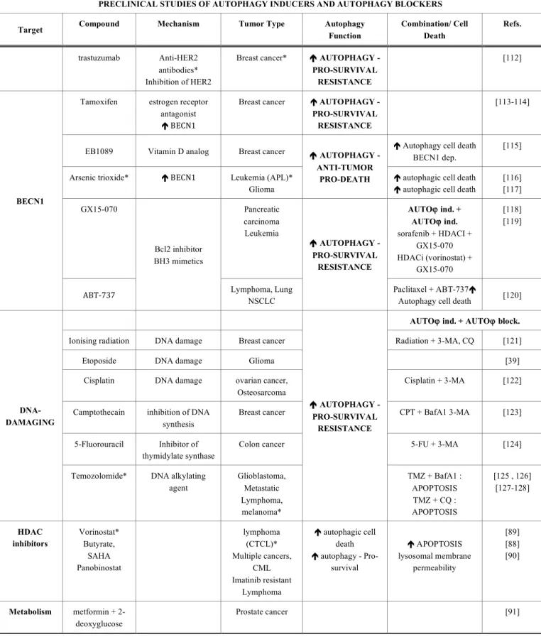
Table 3. Summary of preclinical studies showing enhanced cytotoxic response of chemotherapy in combination with
pharmacological modulators of autophagy.
PRECLINICAL STUDIES OF AUTOPHAGY INDUCERS AND AUTOPHAGY BLOCKERS
Target Compound Mechanism Tumor Type Autophagy
Function
Combination/ Cell Death
Refs.
AUTOϕ ind. + AUTOϕ ind.
Rapamycin* mTORC1 inhibitor Glioma
Leukemia (CML)*
RAPA + PI3K inh, LY294002, RAPA +
Akt inh, UCN-01 Apoptosis Indep.
[92] [93]
Temsirolimus* mTORC1 inhibitor Mantle cell lymphoma glioma
breast cancer
Temsirolimus + HDACi Apoptosis
[94]
Everolimus* mTORC1 inhibitor Leukemia (ALL) Everolimus +
vincristine Apoptosis
[95]
AUTOϕ ind. + AUTOϕ block. mTORC1
Perifosine Degradation of mTOR, raptor, and Akt
Lung cancers Perifosine + CQ
é apoptosis [96] PROTEASOME inhibitors Bortezomib NPI-0052 ä mTOR pathway émTOR inhibitor sestrin-2 Multiple myeloma* Prostate cancer é AUTOPHAGY - PRO-SURVIVAL RESISTANCE Bortezomib + CQ [97 , 98 , 99]
NVP-BEZ235 Dual PI3KCI/Akt inh. Glioma é Autophagy - ? [105]
PI-103 Dual PI3KCI/Akt inh. Glioblastoma é Autophagy - ? [106]
Quercetin ä Akt-mTOR pathway Gastric cancer Colon cells é AUTOPHAGY - PRO-SURVIVAL RESISTANCE Quercertin + CQ é apoptosis [101]
Resveratrol ä Akt-mTOR pathway Breast cancer Ovarian cancer
é Autophagy cell death [102]
Curcumin ä Akt-mTOR pathway Leukemia (CML), glioma
é Autophagy cell death [103] PI3KCI
Dexamethasone ä Akt-mTOR pathway Leukemia (ALL)
é AUTOPHAGY - ANTI-TUMOR PRO-DEATH é Autophagy - Pro-apoptotic [104] Imatinib (Gleevec)* KIT, BCR-ABL, PDGFR inh Gastrointestinal stromal tumor*, leukemia (CML)* é AUTOPHAGY - ANTI-TUMOR PRO-DEATH
é Autophagy cell death [100]
Linifanib VEGFR, PDGFR inhibitor ä Akt-mTOR pathway hepatocellular carcinoma (HCC) AUTOϕ ind. + AUTOϕ block. Linifanib + HCQ [107] Erlotinib* Gefitinib
EGFR inh Glioma*
NSCLC* Glioma AUTOϕ ind. + AUTOϕ ind. Erlotinib + rapamycin é Autophagic cell death Erlotinib + imatinib [108] [109] [110] TYROSINE KINASES Inhibitors
Cetuximab head and neck
squamous cell carcinomas (HNSCC)* é AUTOPHAGY - PRO-SURVIVAL RESISTANCE Cetuximab +PKI-587 (PI3K/mTOR inhibitor) [111]
Table 3. contd….
PRECLINICAL STUDIES OF AUTOPHAGY INDUCERS AND AUTOPHAGY BLOCKERS
Target Compound Mechanism Tumor Type Autophagy
Function Combination/ Cell Death Refs. trastuzumab Anti-HER2 antibodies* Inhibition of HER2
Breast cancer* é AUTOPHAGY - PRO-SURVIVAL
RESISTANCE
[112]
Tamoxifen estrogen receptor antagonist é BECN1
Breast cancer é AUTOPHAGY - PRO-SURVIVAL RESISTANCE
[113-114]
EB1089 Vitamin D analog Breast cancer é Autophagy cell death
BECN1 dep.
[115]
Arsenic trioxide* é BECN1 Leukemia (APL)* Glioma
é AUTOPHAGY - ANTI-TUMOR
PRO-DEATH é autophagic cell death é autophagic cell death
[116] [117] GX15-070 Pancreatic carcinoma Leukemia AUTOϕ ind. + AUTOϕ ind. sorafenib + HDACI + GX15-070 HDACi (vorinostat) + GX15-070 [118] [119] BECN1 ABT-‐737 Bcl2 inhibitor BH3 mimetics Lymphoma, Lung NSCLC é AUTOPHAGY - PRO-SURVIVAL RESISTANCE Paclitaxel + ABT-737é
Autophagy cell death [120] AUTOϕ ind. + AUTOϕ block.
Ionising radiation DNA damage Breast cancer Radiation + 3-MA, CQ [121]
Etoposide DNA damage Glioma [39]
Cisplatin DNA damage ovarian cancer,
Osteosarcoma
Cisplatin + 3-MA [122]
Camptothecain inhibition of DNA synthesis
Breast cancer CPT + BafA1 3-MA [123]
5-Fluorouracil Inhibitor of thymidylate synthase
Colon cancer 5-FU + 3-MA [124]
DNA-DAMAGING
Temozolomide* DNA alkylating agent Glioblastoma, Metastatic Lymphoma, melanoma* é AUTOPHAGY - PRO-SURVIVAL RESISTANCE TMZ + BafA1 : APOPTOSIS TMZ + CQ : APOPTOSIS [125 , 126] [127-128] HDAC inhibitors Vorinostat* Butyrate, SAHA Panobinostat lymphoma (CTCL)* Multiple cancers, CML Imatinib resistant Lymphoma é autophagic cell death é autophagy - Pro-survival é APOPTOSIS lysosomal membrane permeability [89] [88] [90] Metabolism metformin + 2-deoxyglucose Prostate cancer [91]
Abbreviations: AUTOϕ ind, autophagy inducer; AUTOϕ block, autophagy blocker; BafA1, Bafilomycin A1; CQ, Chloroquine; HCQ, Hydroxychloroquine; 3-MA, 3-methyladenine;
5-FU, 5-fluorouracil; Indep, independent; inh, inhibitor; CPT, Camptothecin; RAPA, rapamycin; *, FDA (FDA, US Food and Drug Administration) approved drugs; HDACi, Histone deacetylase inhibitors; TMZ; Temozolomide;
Acute promyelocytic leukemia (APL); acute lymphoblastic leukemia (ALL), cutaneous T cell lymphoma (CTCL); chronic myeloid leukemia (CML), head and neck squamous cell carcinomas (HNSCC), non-small cell lung cancer (NSCLC).
[113, 114]. As a chemotherapeutic vitamin D analog, EB1089
may trigger and induce BECN1-dependent autophagy in
MCF-7 cells [115].
Because BECN1 and other ATGs are haploinsufficient
tumor suppressor genes that undergo heterozygous loss in a
substantial proportion of human tumors, it might be expected
that drugs that may upregulate the remaining copy and
thereby restore autophagy back to its physiological levels of
activation may suppress initial tumor growth. Despite the
excitement surrounding these promises, clinical progress has
been uneven. Autophagy activators have been least effective
in treating the cancer types that have the highest mortality
rates, such as lung, colorectal, pancreatic, skin, and brain
cancer.
Why has the clinical application of autophagy activators
been so challenging? One reason is that many of these drugs
(for instance DNA-damaging drugs) confer resistance by a
cytoprotective autophagy [109, 112, 121, 123, 124]. Intuitively,
overcoming this resistance will require targeting tumor cells
through a cocktail of autophagy activators that efficiently
upregulate the autophagic flux to a cytotoxic level when
applied as concurrent treatment.
Inhibitors of Autophagy
A general hallmark of KRAS/BRAF-driven cancer is the
upregulation of basal autophagy, when compared to their
normal counterparts. A plethora of evidence suggests that
upregulation of autophagy intimately accompanies and
allows for all different facets of malignant progression
(growth, survival, invasion, and dormancy; section 5).
Therefore, autophagy is a central aspect of tumor biology
that might be turned into cancer's Achilles heel.
Various autophagy inhibitors have been developed such
as the inhibitors of PI3KCIII (3-methyladenine, wortmannin,
and LY294002 [125]), of microtubule (vinblastine,
colchicine [142]), of Vacuolar-ATPase (bafilomycin A1
[145]), of lysosomal proteases (pepstatin A, [146]) and weak
bases (chloroquine, hydroxychloroquine, and Monessen) to
block the formation of autophagosome, its maturation/fusion
with lysosome and then its degradation, respectively.
Currently, there are 40 clinical trials targeting autophagy
addiction
using
primarily
chloroquine
(CQ)
and
hydroxychloroquine (HCQ) to kill cancer cells (http://www.
clinicaltrial.gov Fig. 3). Indeed these antimalarial drugs are
already approved for human use, relatively safe, and
inexpensive. Both are weak bases that accumulate in the
lysosomes causing a rise in lysosomal pH and thus
preventing the activity of autolysosomes or the fusion of
autophagosomes with lysosomes.
The rationale of combining a chemotherapy and a
lysosomal autophagy inhibitor is that the former induces
massive autophagic flux and the latter prevents autophagic
contents from being degraded, leading to an accumulation of
ineffective autophagic vesicles and a burst of ROS,
presumably from the damaged mitochondria within
autolysosomes. This ROS in turn produces permeabilization
of lysosomal membrane, release of cathepsin and activation
of apoptotic cell death. A large body of preclinical results
show that the success of CQ as a therapeutic agent lies in its
ability to trigger senescence, apoptosis, and autophagic cell
death of cancer cells and cancer stem cells.
FUTURE
DIRECTIONS:
TRANSLATIONAL
CHALLENGES
TOWARDS
INDIVIDUALIZED
HEALTH CARE
Despite the great promises of autophagy-based therapies,
there are, undoubtedly, many critical issues to address. In
particular, it will be important to identify the cancer type that
might respond best to these therapies and evaluate the
efficacy, safety and long-term outcomes of modulating
autophagy.
What are the Best Patient Populations for
Autophagy-based Therapies?
The concept of “autophagy addiction” was first suggested
for KRAS-, and BRAF-driven tumors [72, 73]. In transgenic
mice, the inhibition of autophagy induces the regression of
adenocarcinomas to a more benign oncocytoma, providing a
rationale for targeting autophagy to treat these refractory
KRAS-mutated patients. Importantly, it was demonstrated
that autophagy ablation demonstrates efficacy to advanced
tumors that have grown to an considerable size in mice [74],
with possible extensions in patients receiving therapy.
However, at present it is unclear whether all cancer-bearing
patients with a mutated RAS or activation of the RAS
pathway will respond similarly to autophagy inhibition. Two
recent papers evidenced that inhibition of autophagy (by CQ,
Atg5 or Atg7 knockout) inhibits the growth of Kras–driven
tumors in pancreas and lung when the status of Tp53 is wild
type but stimulates tumor growth in Ras mutant, Tp53 null
cells [71, 147] (Table 2). In the near future, these puzzling
findings may revolutionize the stratification of patients that
will receive autophagy inhibition treatment. However, in the
ongoing clinical trials no strategy for patient selection is
being pursued.
What are the Main Safety Concerns?
The ultimate goal of any cancer therapy is to robustly
target malignant cells while sparing normal cells. Currently,
the major concern of using autophagy modulators is their
lack of selectivity, targeting both the cancer and the healthy
tissue. A recent study by Karsli-Uzunbas and colleagues
indicates that the systemic inhibition of autophagy for 5
weeks in mice is not only efficacious in regressing
established lung adenocarcinomas to benign oncocytoma but
also safe. However, if the inhibition is sustained for 6 to 12
weeks, this strategy is extremely toxic, causing severe liver,
muscle and brain degenerations [74]. This suggests that, with
proper control of the extent and/or timing of autophagy
inhibition, there is a therapeutic window to suppress
tumorigenesis while mostly sparing normal tissue.
What are the Best Autophagy-based Therapies?
The diverse outcome of cancer treatments designed to
either upregulate or silence autophagy makes autophagy an
attractive therapeutic target, but its unpredictable potential
might also make such targeted therapy challenging and risky.
For instance, CQ displays dramatic effects for some
drugs/tumor models and modest or no effects in others, even
in a panel of KRAS-mutated cancer cell lines [148, 149]. Of
particular concern, CQ might also exacerbate the progression
of established cancers, as suggested by certain studies [24,
147, 150, 151]. Furthermore, the possibility that CQ fulfills
an autophagic-independent role cannot be excluded yet
[152]. All of the above allows us to stipulate that
autophagy-based therapies have to be improved by a specific
modulation. Among the druggable targets are the autophagic
kinases ULK1/2, and the cysteine protease ATG4. In
addition to pharmacological inhibitors, specific gene
interference using siRNA or CRISPR/Cas technology against
various ATG genes may improve the effectiveness of
autophagy inhibition.
What are the Best Clinical Markers for Monitoring
Autophagy?
Actually, autophagy markers in tumor samples are the
detection of autophagic vesicles, the expression of
autophagy-related genes, and the degradation of the
autophagy substrate SQSTM1. However, it is challenging to
predict from these markers whether the drug activates or
blocks autophagy and whether autophagy upregulation
would be beneficial or detrimental for patients. We have
developed methods and guidelines to measure this dynamic
process in vitro [153], but these approaches are not suitable
for application in patient biopsies. For instance, a major
caveat is the difficulty to distinguish by immuno-
histochemistry the LC3-I positive aggregates from LC3-II
positive “real” autophagosomes. Moreover, autophagosomes
represent a mid-point in the dynamic autophagic flux, i.e.
rapidly formed and degraded within 8 minutes [5].
Accumulation of autophagosomes can occur throughout
induction of autophagy, but can also arise through inhibition
of autophagic degradation. As a result, only a few
autophagosomes and autolysosomes are observed in normal
tissues even under nutrient starvation. Thus, the robust
accumulation of LC3 and SQSTM1 spots might be
interpreted as an impaired autophagy flux in cancer [25, 154],
while recognizing that any delay in the processing/fixation of
biospecimens would artificially upregulate autophagy.
CONCLUSION
In conclusion, we are embarking on an exciting journey.
All the remarkable studies that have been performed over the
past 14 years on the role of autophagy in cancer are
culminating in clinical trials. Still in its infancy, the
challenge for the cancer research community will now be to
identify those patients who are more likely to respond and
the combination of autophagy modulators that would be
most effective.
CONFLICT OF INTEREST
The author(s) confirm that this article content has no
conflict of interest.
Fig. (3). Summary of 40 clinical trials involving chloroquine and hydroxychloroquine for cancer treatment (data obtained from
http://www.cancer.gov/clinicaltrials). Abbreviations: CQ, Chloroquine; HCQ, Hydroxychloroquine; 5-FU, 5-fluorouracil; inh, inhibitor;
non-small cell lung cancer (NSCLC).
ACKNOWLEDGEMENTS
This work was supported by the “Institut National de la
Santé et de la Recherche Médicale”, “Region Provence
Alpes Côte d'Azur”, “Agence régionale santé Provence Alpes
Côte d'Azur”, “Direction régionale de l’Environnement, de
l’aménagement et du logement Provence Alpes Côte d'Azur”
(A B: plan régional santé environnement PRSE PACA
n°6.3.3.3 and 6.3.3.4), “Association pour la Recherche
contre le Cancer” (ARC Grants n° SL220110603478), Marie
Curie International Incoming Fellowship within the 7th
European Community Framework Programme (J R:
“SysBioDRez”, N° 626190, call reference: FP7-
PEOPLE-2013-IIF), and French national research agency (“Investments
for the Future” LABEX SIGNALIFE : program reference
#ANR-11-LABX-0028-01 and “STEATOX”
#ANR-13-CESA-0009-01)
ABBREVIATIONS
ATG
=
autophagy-related
BECN1
=
beclin 1
CQ
=
chloroquine
HCQ
=
Hydroxychloroquine
CMA
=
chaperon-mediated autophagy
LAMP
=
lysosomal-associated membrane protein
LC3
=
microtubule-associated protein 1 light
(MAP1LC3)
chain 3
mTOR
=
mammalian target of rapamycin
shRNA
=
short hairpin RNA
SQSTM1
=
sequestosome 1/p62
ROS
=
reactive oxygen species
TCA
=
tricarboxylic acid
ATPase
=
lysosomal V0 subunit A3
H+ transporting
v-ATPase
=
vacuolar-ATPase
REFERENCES
[1] Clark, S. L., Jr. Cellular differentiation in the kidneys of newborn mice studies with the electron microscope. J Biophys Biochem Cytol 1957, 3 (3), 349-362.
[2] Arias, E.; Cuervo, A. M. Chaperone-mediated autophagy in protein quality control. Curr Opin Cell Biol 2011, 23 (2), 184-189. [3] Li, W. W.; Li, J.; Bao, J. K. Microautophagy: lesser-known
self-eating. Cell Mol Life Sci 2012, 69 (7), 1125-1136.
[4] Cuervo, A. M.; Knecht, E.; Terlecky, S. R.; Dice, J. F. Activation of a selective pathway of lysosomal proteolysis in rat liver by prolonged starvation. Am J Physiol 1995, 269 (5 Pt 1), C1200-1208.
[5] Pfeifer, U. Inhibition by insulin of the formation of autophagic vacuoles in rat liver. A morphometric approach to the kinetics of intracellular degradation by autophagy. J Cell Biol 1978, 78 (1), 152-167.
[6] Belaid, A.; Ndiaye, P. D.; Klionsky, D. J.; Hofman, P.; Mograbi, B. Signalphagy Scheduled signal termination by macroautophagy. Autophagy 2013, 9 (10), 1629-1630.
[7] Parzych, K. R.; Klionsky, D. J. An Overview of Autophagy: Morphology, Mechanism, and Regulation. Antioxid Redox Signal 2014, 20 (3), 460-473.
[8] McEwan, D. G.; Dikic, I. The Three Musketeers of Autophagy: phosphorylation, ubiquitylation and acetylation. Trends Cell Biol 2011, 21 (4), 195-201.
[9] Morselli, E.; Marino, G.; Bennetzen, M. V.; Eisenberg, T.; Megalou, E.; Schroeder, S.; Cabrera, S.; Benit, P.; Rustin, P.; Criollo, A.; Kepp, O.; Galluzzi, L.; Shen, S.; Malik, S. A.; Maiuri, M. C.; Horio, Y.; Lopez-Otin, C.; Andersen, J. S.; Tavernarakis, N.; Madeo, F.; Kroemer, G. Spermidine and resveratrol induce autophagy by distinct pathways converging on the acetylproteome. J Cell Biol 2011, 192 (4), 615-629.
[10] Pietrocola, F.; Izzo, V.; Niso-Santano, M.; Vacchelli, E.; Galluzzi, L.; Maiuri, M. C.; Kroemer, G. Regulation of autophagy by stress-responsive transcription factors. Semin Cancer Biol 2013, 23 (5), 310-322.
[11] Choi, A. M.; Ryter, S. W.; Levine, B. Autophagy in human health and disease. N Engl J Med 2013, 368 (19), 1845-1846.
[12] Aita, V. M.; Liang, X. H.; Murty, V.; Pincus, D. L.; Yu, W. P.; Cayanis, E.; Kalachikov, S.; Gilliam, T. C.; Levine, B. Cloning and genomic organization of beclin 1, a candidate tumor suppressor gene on chromosome 17q21. Genomics 1999, 59 (1), 59-65. [13] Liang, X. H.; Jackson, S.; Seaman, M.; Brown, K.; Kempkes, B.;
Hibshoosh, H.; Levine, B. Induction of autophagy and inhibition of tumorigenesis by beclin 1. Nature 1999, 402 (6762), 672-676. [14] Qu, X.; Yu, J.; Bhagat, G.; Furuya, N.; Hibshoosh, H.; Troxel, A.;
Rosen, J.; Eskelinen, E. L.; Mizushima, N.; Ohsumi, Y.; Cattoretti, G.; Levine, B. Promotion of tumorigenesis by heterozygous disruption of the beclin 1 autophagy gene. J. Clin. Invest. 2003, 112 (12), 1809-1820.
[15] Yue, Z.; Jin, S.; Yang, C.; Levine, A. J.; Heintz, N. Beclin 1, an autophagy gene essential for early embryonic development, is a haploinsufficient tumor suppressor. Proc Natl Acad Sci USA 2003, 100 (25), 15077-15082.
[16] Fimia, G. M.; Stoykova, A.; Romagnoli, A.; Giunta, L.; Di Bartolomeo, S.; Nardacci, R.; Corazzari, M.; Fuoco, C.; Ucar, A.; Schwartz, P.; Gruss, P.; Piacentini, M.; Chowdhury, K.; Cecconi, F. Ambra1 regulates autophagy and development of the nervous system. Nature 2007, 447 (7148), 1121-1125.
[17] Cianfanelli, V.; Fuoco, C.; Lorente, M.; Salazar, M.; Quondamatteo, F.; Gherardini, P. F.; De Zio, D.; Nazio, F.; Antonioli, M.; D'Orazio, M.; Skobo, T.; Bordi, M.; Rohde, M.; Dalla Valle, L.; Helmer-Citterich, M.; Gretzmeier, C.; Dengjel, J.; Fimia, G. M.; Piacentini, M.; Di Bartolomeo, S.; Velasco, G.; Cecconi, F. AMBRA1 links autophagy to cell proliferation and tumorigenesis by promoting c-Myc dephosphorylation and degradation. Nat Cell Biol 2015, 17 (1), 20-30.
[18] Takahashi, Y.; Coppola, D.; Matsushita, N.; Cualing, H. D.; Sun, M.; Sato, Y.; Liang, C.; Jung, J. U.; Cheng, J. Q.; Mulé, J. J.; Pledger, W. J.; Wang, H. G. Bif-1 interacts with Beclin 1 through UVRAG and regulates autophagy and tumorigenesis. Nat Cell Biol. 2007, 9, 1142-1151.
[19] Liang, C.; Feng, P.; Ku, B.; Dotan, I.; Canaani, D.; Oh, B. H.; Jung, J. U. Autophagic and tumour suppressor activity of a novel Beclin1-binding protein UVRAG. Nat Cell Biol 2006, 8 (7), 688-699.
[20] Liang, C.; Lee, J. S.; Inn, K. S.; Gack, M. U.; Li, Q.; Roberts, E. A.; Vergne, I.; Deretic, V.; Feng, P.; Akazawa, C.; Jung, J. U. Beclin1-binding UVRAG targets the class C Vps complex to coordinate autophagosome maturation and endocytic trafficking. Nat Cell Biol 2008, 10 (7), 776-787.
[21] Takamura, A.; Komatsu, M.; Hara, T.; Sakamoto, A.; Kishi, C.; Waguri, S.; Eishi, Y.; Hino, O.; Tanaka, K.; Mizushima, N. Autophagy-deficient mice develop multiple liver tumors. Genes Dev 2011, 25 (8), 795-800.
[22] Yang, A.; Rajeshkumar, N. V.; Wang, X. X.; Yabuuchi, S.; Alexander, B. M.; Chu, G. C.; Von Hoff, D. D.; Maitra, A.; Kimmelman, A. C. Autophagy Is Critical for Pancreatic Tumor Growth and Progression in Tumors with p53 Alterations. Cancer Discov 2014, 4 (8), 905-913.
[23] Mariño, G.; Salvador-Montoliu, N.; Fueyo, A.; Knecht, E.; Mizushima, N.; Lopez-Otin, C. Tissue-specific autophagy alterations and increased tumorigenesis in mice deficient in Atg4C/autophagin-3. J. Biol. Chem. 2007, 282, 18573-18583. [24] Belaid, A.; Ndiaye, P. D.; Cerezo, M.; Cailleteau, L.; Brest, P.;
Klionsky, D. J.; Carle, G. F.; Hofman, P.; Mograbi, B. Autophagy and SQSTM1 on the RHOA(d) again Emerging roles of autophagy