УДК 599.323.4(5-012)
NEW SPECIES OF RED-BACKED VOLE (MAMMALIA: RODENTIA: CRICETIDAE) IN FAUNA OF RUSSIA: MOLECULAR AND MORPHOLOGICAL EVIDENCES
N.I. Abramson, A.V. Abramov and G.I. Baranova
Zoological Institute of the Russian Academy of Sciences, Universitetskaya Emb., 1, St. Petersburg, 199034, Russia, e-mail: natalia_abr@mail.ru
ABSTRACT
The new species for the fauna of Russia, Hokkaido red-backed vole (Myodes rex), has been identified at the south of Sakhalin Island (Dolinsk District). Its identification was reliably confirmed by molecular and morphological methods. Undoubtedly, this species is much more widespread in islands of the Far East. Some records of M. sikotanensis from Sakhalin including the so-called “microtinus” form, actually, should be reidentified as M. rex. The voles with complex molars from Shikotan Island and probably those from Zelenyi (= Sibotsu) Island also belong to M. rex.
Key words: Myodes rex, mitochondrial DNA, Sakhalin, teeth pattern РЕЗЮМЕ На южной оконечности о. Сахалин (Долинский р-н) обнаружен новый для фауны России вид рыжих полевок – Myodes rex. Достоверность определения этого вида подтверждена как молекулярным, так и морфологическим методами. Несомненно, этот вид имеет более широкое распространение на островах Дальнего Востока. Часть находок M. sikotanensis с о. Сахалин, включая и т.н. форму “microtinus”, долж-ны быть переопределедолж-ны как M. rex. Полевки со слождолж-ным строением зубов с о. Шикотан и, вероятно, с о. Зеленый (= Шиботцу) также должны быть отнесены к M. rex. INTRODUCTION
The genus Myodes, or red-backed voles, is widely distributed in Northern Hemisphere and includes 12 species (Musser and Carleton 2005). The territory of Russia is inhabited by 3 species of Myodes: the bank vole, M. glareolus (Schreber, 1780), northern red-backed vole, M. rutilus (Pallas, 1779), and gray red-backed vole, M. rufocanus (Sundevall, 1846). Many Russian scientists believe Shikotan red-backed vole M. sikotanensis (Tokuda, 1935) distributed in Sikotan, Sakhalin and Zelenyi (or Sibotsu) islands is a separate species (Gromov and Erbaeva 1995; Pavlinov et al. 1995; Frisman et al. 2002; Kostenko et al. 2004). The taxonomic status of this form for a long time was controversial, but recently Motokawa (2008) analyzed the type specimens of M. sikotanen-sis and demonstrated this name is a junior synonym of M. rufocanus.
During the mammalogical survey in Sakhalin Island in 2008, we collected M. rufocanus in few lo-calities in southern part of the island. Based on the molecular and morphological analyses, we found a new species of vole, previously not recorded for the fauna of Russia.
MATERIAL AND METHODS
Fieldwork was conducted between 19 and 29 Au-gust 2008 near Novoaleksandrovsk (vicinity of Yu-zhno-Sakhalinsk), in the lower reach of Yablochnaya River (Kholmsk District), and near Sokol Research Station (Sokol) in Dolinsk District of southern Sakhalin. Totally 11 specimens of M. rufocanus were collected. A variety of small mammal traps, includ-ing pit-fall traps and snap-traps, were used to collect voles, mice and shrews. Specimens were fixed in 70% ethanol. Tissue samples were preserved in 96%
etha-nol. Skulls were extracted and cleaned from ethanol-preserved vole specimens.
Vole skulls were compared with material from Sakhalin (n = 21), Kunashir (n = 50), Shikotan (n = 23) and Hokkaido (n = 1) islands kept in the collections of the Zoological Institute of the Russian Academy of
Sciences, St. Petersburg, Russia (ZIN) and Zoologi-cal Museum of Moscow State University, Moscow, Russia (ZMMU). We also studied the original de-scriptions and figures for skulls of Far Eastern Myo-des kept in the collections of the Kyoto University Museum, Kyoto, Japan (KUZ) and National Science Museum, Tokyo, Japan (NSM) (Tokuda 1935; Imai-zumi 1971; Motokawa 2008).
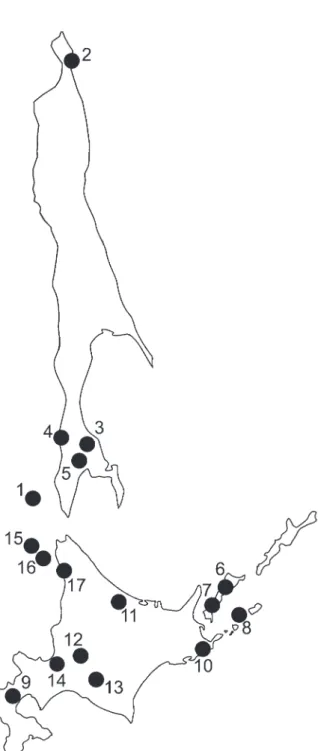
Total genomic DNA was extracted from ethanol-preserved muscles or liver using proteinase K di-gestion, NaCl precipitation of proteins, and DNA precipitation with ethanol, following a modification of Miller et al. (1988). We analyzed the samples col lected during our field work as well as ethanol-preserved tissues housed in the tissue collection of ZIN and sequences available in the Genbank (see Table 1). The collecting localities are shown in Fig. 1. The arvicoline sequences new to this study have been deposited in the EMBL data bank.
A portion of cytochrome b (910 bp) was amplified using primers UCBU/LM (Abramson and Rodchen-kova 2007). Double-stranded polymerase chain reac-tion (PCR) usually entailed 95°C 5 min, followed by 95°C 30 sec, 55°C 30 sec, 72°C 1 min (30 times), with a final extension at 72°C 5 min. All PCR experiments included negative controls. PCR products were visu-alized on 1.5% agarose gel and then purified using the Qiagen QIAquick kit and sequenced on both strands using automatic sequencing (Big Dye Terminator cycle kit) on an ABI 3130 (PE Applied Biosystems). Sequences were aligned and compared manually us-ing the Bioedit v.7.0.3 (Hall 1999).
Estimates of divergence between haplotypes were calculated by using Kimura’s two parameter method, and a neighbor-joining (NJ) phylogenetic tree (Saitou and Nei 1987). Phylogenetic and molecular evolutionary analyses were conducted using MEGA version 4 (Tamura et al. 2007).
RESULTS
DNA analysis. A total of 23 haplotypes were
identified among 41 animals. Of the 817 base pairs (after alignment and removing ambiguous sites), 102 sites were variable and 80 parsimony informative. The NJ reconstruction of phylogenetic relationships between haplotypes is shown in Fig. 2. All studied M. rufocanus specimens from island populations clearly split into two groups with mean divergence between them 2%. The first group includes animals from Fig. 1. Localities from which specimens of Myodes spp. were
Table 1. Collecting localities, specimens numbers, samples codes and Genbank accession numbers of Myodes spp. genetically examined in this study.
Collecting locality Specimen No. Sample code Locality No.
in Fig.1 Accession No. M. rufocanus
Russia, Moneron 91* Mon-1 1 FJ792779**
Russia, Moneron 93* Mon-2 1 FJ792780**
Russia, Moneron 241* Mon-3 1 FJ792781**
Russia, Sakhalin, Okha Sakh-1 2 AB031573
Russia, Sakhalin, Okha Sakh-2 2 AB031574
Russia, Sakhalin, Okha Sakh-3 2 AB031575
Russia, Sakhalin, Okha Sakh-4 2 AB031576
Russia, Sakhalin, Sokol 235* Sakh-5 3 FJ792775**
Russia, Sakhalin, Sokol 921* Sakh-6 3 FJ792776**
Russia, Sakhalin, Sokol 922* Sakh-7 3 FJ792777**
Russia, Sakhalin, Sokol 920* Sakh-8 3 FJ792778**
Russia, Sakhalin, Sokol 1322*, ZIN C.98137 Sakh-13 3 FJ792792**
Russia, Sakhalin, Yablochnaya River 1318*, ZIN C.98187 Sakh-9 4 FJ792788** Russia, Sakhalin, Yablochnaya River 1321*, ZIN C.98125 Sakh-10 4 FJ792789** Russia, Sakhalin, Novoaleksandrovsk 1314*, ZIN C.98123 Sakh-11 5 FJ792790** Russia, Sakhalin, Novoaleksandrovsk 1317*, ZIN C.98188 Sakh-12 5 FJ792791**
Russia, Kunashir, Filatovka 1325*, ZIN C.98127 Kun-1 6 FJ792782**
Russia, Kunashir, Filatovka 1326*, ZIN C.98128 Kun-2 6 FJ792783**
Russia, Kunashir, Filatovka 1328*, ZIN C.98130 Kun-6 6 FJ792787**
Russia, Kunashir, Alekhino 1329*, ZIN C.98131 Kun-3 7 FJ792784**
Russia, Kunashir, Alekhino 1331*, ZIN C.98133 Kun-4 7 FJ792785**
Russia, Kunashir, Alekhino 1333*, ZIN C.98135 Kun-5 7 FJ792786**
Russia, Polonskogo Is. (=Taraku Is.) Tarak 8 AB031564
Japan, Hokkaido, Kuromatsunai Kuro-1 9 AB031557
Japan, Hokkaido, Kuromatsunai Kuro-2 9 AB031558
Japan, Hokkaido, Nemuro Nem-1 10 AB031555
Japan, Hokkaido, Nemuro Nem-2 10 AB031556
Japan, Hokkaido, Takinoue Tak-1 11 AB031561
Japan, Hokkaido, Takinoue Tak-2 11 AB031562
Japan, Hokkaido, Takinoue Tak-3 11 AB031563
Japan, Hokkaido, Tobetsu Tobet 12 AB031560
Japan, Hokkaido, Naganuma Nagan 13 AB031559
Japan, Hokkaido, Ishikari-shi Hok-1 14 AY309416
Japan, Hokkaido, Ishikari-shi Hok-2 14 AY309417
Japan, Hokkaido, Ishikari-shi Hok-3 14 AY309418
Japan, Rebun Rebun 15 AB031554
Japan, Rishiri Rishi-1 16 AB031553
M. rex
Russia, Sakhalin, Sokol 1323*, ZIN C.98136 Sakh-14 3 FJ792793**
Japan, Hokkaido, Takinoue Tak-4 11 AB031582
Japan, Rishiri Rishi-2 16 AB017239
Japan, Hokkaido, Teshio Tesh 17 AB017240
* number in tissue collection of ZIN ** sequences obtained in this study
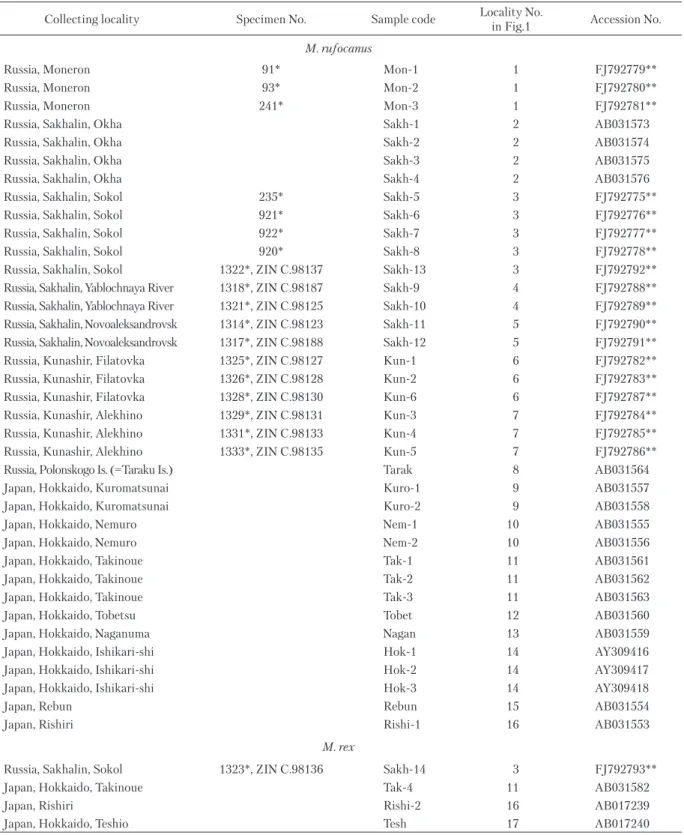
Fig. 2. Cladogram resulted from the phylogenetic analysis of the mitochondrial cytochrome b gene (817 bp) in M. rufo-canus and M. rex from the Far East. A neighbor-joining tree was constructed with pairwise sequence divergences that were calculated by the Kimura’s two parameter method considering all substitutions at all codon positions. Bootstrap values (over 50) related to the nodes are indicated (1000 replicates). Sources of the haplotypes are listed in Table 1.
Japanese islands and Kunashir, the second one – from Moneron and Sakhalin.
Up to now cytochrome b have been sequenced only in three individuals of M. rex, with only partial sequences (402 bp) available in Genbank for two of them. However it is remarkable that one individual collected in Sakhalin, near Sokol (ZIN C.98136, see Table 1), has nearly identical haplotype with the known M . rex. Two species (M. rufocanus and M. rex) differ by 50 informative substitutions and mean diver-gence between M. rex and M. rufocanus comprises 4%.
Morphology. Most of the studied skulls of
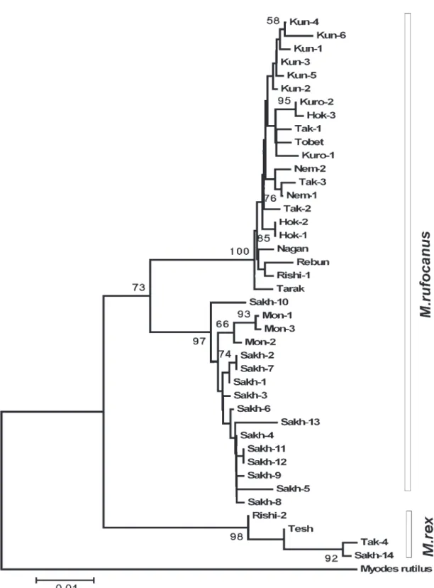
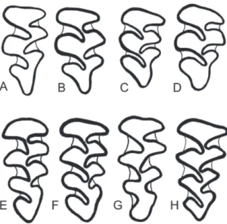
red-backed voles from Sakhalin, Kunashir, Shikotan and Hokkaido islands have simple occlusal pattern of the upper third molar, i.e. third upper molars with two deep reentrant and three prominent salient angles both on inner and outer sides (Fig. 3 A–D). This molar pattern is typical of M. rufocanus. The type specimens of M. sikotanensis also have the same pattern (Tokuda 1935; Motokawa 2008; see Fig. 3A). The voles from Shikotan Island display different patterns on the up-per third molar. Some specimens have “simple” molars as M. shikotanensis and typical M. rufocanus (Fig. 3), whereas others have “complex” molar.
Hokkaido red-backed vole, M. rex (Imaizumi, 1971), has complex shape of the upper third molar, i.e. third upper molars with three deep reentrant and four prominent salient angles both on inner and outer sides (Fig. 3 E-H). The specimen ZIN C.98136 from Sokol has the complex pattern as typical M. rex (Fig. 3F). It is interesting to note that another specimen from Sokol (ZIN C.98137), captured in the same trap-line, has simple molar pattern and typical rufocanus-haplo-type of cytochrome b (see Table 1, Fig. 2).
We have studied 8 specimens from Sakhalin labeled as “Clethrionomys sikotanensis” from the col-lection of ZMMU. Four of them have a simple upper molar pattern, whereas other specimens from Krilion Peninsula in southern Sakhalin have the complex pattern of the upper third molar similar to that in typical M. rex (Fig. 3G). Two of the latter (ZMMU S-67985 and ZMMU S-77229) are labeled by Kuzya-kin as type specimens of Clethrionomys microtinus (Kuzyakin 1963, nomen nudum).
DISCUSSION
Species taxonomy of red-backed voles in the Far East had been very confusing. Tokuda (1935) described the new species Neoashizomys sikotanensis
from Shikotan Island, and pointed out the simple mo-lar pattern for this form (see Fig. 3A). However later, the Japanese authors attributed this name to voles with complex molar pattern, distributed in Daikoku and Rishiri islands of eastern and northern coasts of Hokkaido and, probably, also in mainland Hok-kaido (Imaizumi 1949, 1960; Abe 1973a, b). Later, Imaizumi (1971) described a new form from Rishiri Island, named Clethrionomys rex and distinguished by coloration, larger size and complex molar pattern. Some authors regarded M. sikotanensis most likely conspecific with M. rex (Iwasa et al. 2000, 2001), but according to the current opinion, this name is a junior synonym of M. rufocanus (Kaneko et al. 1998; Musser and Carleton 2005; Motokawa 2008).
Indeed, two forms of voles co-occur in Shikotan Island. In the common taxonomic practice of Russian scientists, M. sikotanensis was regarded as a valid spe-cies with complex third upper molars, whereas voles with simple molar pattern were referred to M. rufo-canus (Kostenko and Allenova 1978; Kostenko et al. 2004). According to this point of view, the voles with Fig. 3. Pattern of the upper third molar in red-backed voles: Myodes rufocanus (A–D), Myodes rex (E–H). A – holotype of Neoaschizomys sikotanensis Tokuda, 1935, Shikotan Island, KUZ M.9144 (after Tokuda [1935, fig. 4] and Motokawa [2008, fig. 2]); B – Sakhalin Island, Novoaleksandrovsk, ZIN C.98123; C – Ku-nashir Island, Alekhino, ZIN C.98135; D – Shikotan Island, ZIN C.83150; E – holotype of Clethrionomys rex Imaizumi, 1971, Rishiri Island, NSM M.10823 (after Imaizumi 1971, fig. 3); F – Sakhalin Island, Sokol, ZIN C.98136; G – Sakhalin Island, Krilion Peninsula, Shebunino, ZMMU S-67985; H – Shikotan Island, ZIN C.87096. Not to scale.
complex molar pattern from Sakhalin were also clas-sified as M. sikotanensis (Gromov and Erbaeva 1995; Pavlinov et al. 1995; Kostenko et al. 2004).
Based on our results, the vole with complex molars from Dolinsk District in Sakhalin should be identified as M. rex. This is the first reliably confirmed record of M. rex in Russia. Undoubtedly, this species is much more widespread in islands of the Far East. Some records of M. sikotanensis from Sakhalin Island including the so-called “microtinus” form are related to M. rex. Voles with complex molars from Shikotan Island and probably from Zelenyi (or Sibotsu) Island belong to M. rex too (see Iwasa et al. 2001).
Proceeding from the data available now the evolutionary scenario suggested earlier by Iwasa et al. (2000) seems very probable. M. rex appears to be a derivative of the common ancestor of M. rufo-canus that colonized the Far-Eastern islands in the Early Pleistocene and evolved in isolation during the Middle Pleistocene interglacial. The populations of M. rufocanus inhabiting the islands nowadays colo-nized them much later during the Late Pleistocene and ecologically superior than the former species (Nakata 1995). Few lines of evidence support this scenario. Based on molecular data, M. rex derivates earlier than M. rufocanus (Lebedev et al. 2007), and despite of insufficient genetic data for this species all studied specimens of M. rex from different localities are dramatically monomorphic in genetic structure. The latter fact suggests not only a small number of founders but also a long time of isolation.
ACKNOWLEDGEMENTS
This study was supported in part by the Russian Foundation for Basic Research (grants 07–04–91202, 06–04–49294 and 08–04–10079). AVA thanks Vladimir Platonov, Mikhail Nazarkin (both from ZIN) and Igor Tumanov (Arctic and Antarctic Research Institute, St. Petersburg, Russia) for their great help during the field work. We thank Tatyana Petrova (ZIN) for technical as-sistance in the laboratory, Nikolai Dokuchaev (Institute of Biological Problems of North FEB RAS, Magadan, Russia) for the additional material from Sakhalin and Moneron islands. We are indebted to Irina Kartavtseva (Institute of Biology and Soil Sciences, FEB RAS, Vladivostok, Russia) and Satoshi Ohdachi (Hokkaido University, Sap-poro, Japan), who supplied photocopies of several papers. We thank Sergei Kruskop (ZMMU) for his help during work with ZMMU specimens. We are thankful to Alexey Tesakov (Geological Institute RAS, Moscow, Russia) and Antonina Smorkatcheva (St. Petersburg State University,
St. Petersburg, Russia) for reviewing the manuscript of this paper and useful comments.
REFERENCES
Abe H. 1973a. Growth and development in two forms of Clethrionomys: tooth characters, with special reference to phylogenetic relationships. Journal of the Faculty of Agriculture, Hokkaido University, 57: 229–253.
Abe H. 1973b. Growth and development in two forms of Clethrionomys: cranial characters, with special reference to phylogenetic relationships. Journal of the Faculty of Agriculture, Hokkaido University, 57: 255–274.
Abramson N.I. and Rodchenkova E.N. 2007. Geneti-cheskaya izmenchivost’ i istoriya populyatsii ryzhei polevki (Clethrionomys glareolus) na territorii Evro-peiskoi chasti Rossii po dannym analiza chastichnykh posledovatel’nostei mitokhondrial’nogo gena tsito-khrom b [Genetic variation and history of populations of European bank vole (Clethrionomys glareolus) on the territory of European Russia as inferred from partial sequences of mitochondrial cytochrome b]. Pp. 7–12 in: V.V. Rozhnov et al. (Eds.). Geneticheskie osnovy sokhraneniya bioraznoobraziya mlekopitayushchikh Golarktiki [Genetic foundations for the conservation of Holarctic mammal biodiversity]. KMK Scientific Press, Moscow. [In Russian]
Frisman L.V., Kartavtseva I.V., Pavlenko M.V., Kosten-ko V.A., Suzuki H., Iwasa M., Nakata K. and Cher-nyavskii F.B. 2002. Gene-geographical variation and genetic differentiation in red-backed voles of the genus Clethrionomys (Rodentia, Cricetidae). Genetika, 38: 655–664. [In Russian with English abstract]
Gromov I.M. and Erbaeva M.A. 1995. Mlekopitayushchie fauny Rossii i sopredel’nykh territoriy. Zaitseobraznye i gryzuny [The mammals of Russia and adjacent territories (lagomorphs and rodents)]. Zoological Institute of the Russian Academy of Sciences, St. Petersburg, 521 pp. [In Russian]
Hall T.A. 1999. BioEdit: a user-friendly biological sequence alignment editor and analysis program for Windows 95/98/NT. Nucleic Acids Symposium Series, 41: 95–98. Imaizumi Y. 1949. The natural history of Japanese mammals.
Yogo-Shobo, Tokyo, 348 pp. [In Japanese]
Imaizumi Y. 1960. Coloured illustrations of the mammals of Japan. Hoikusha, Osaka, 196 pp. [In Japanese]
Iwasa M.A., Serizawa K. and Sato M. 2001. Taxonomic problems of the dark red-backed vole, Clethrionomys rex. Rishiri Studies, 20: 43–53. [In Japanese with Eng-lish abstract]
Iwasa M.A., Utsumi Y., Nakata K., Kartavtseva V., Nevedomskaya I.A., Kondoh N. and Suzuki H. 2000. Geographic patterns of cytochrome b and Sry gene lineages in the gray red-backed vole Clethrionomys rufocanus from Far East Asia including Sakhalin and Hokkaido. Zoological Science, 17: 477–484.
Kaneko Y., Nakata K., Saitoh T., Stenseth N.C. and Bjørn stad O.N. 1998. The biology of the vole Clethri-onomys rufocanus: a review. Researches on Population Ecology, 40: 21–37.
Kostenko V.A. and Allenova T.V. 1978. Osobennosti morfologii i biologii ryzhikh polevok (Clethrionomys) ostrova Shikotan [Characteristics of morphology and biology in the red-backed vole (Clethrionomys) on Shikotan Island]. Pp. 119–125 in: G.F. Bromlei and V.G. Yudin (Eds.). Ekologiya i zoogeografiya nekotorykh pozvonochnykh sushi Dal’nego Vostoka [Ecology and zoogeography of terrestrial vertebrates in Far East]. DVNTs AN SSSR, Vladivostok. [In Russian]
Kostenko V.A., Nesterenko V.A. and Trukhin A.M. 2004. Mlekopitayushchie Kuril’skogo arkhipelaga [Mammals of the Kurile Archipelago]. Dal’nauka, Vladivostok. 186 pp. [In Russian]
Kuzyakin A.P. 1963. K systeme gryzunov fauny SSSR [System of rodents of Russian fauna]. Trudy Mosk-ovskogo Obshchestva Ispytateley Prirody, 10: 105–115. [In Russian]
Lebedev V.S., Bannikova A.A., Tesakov A.S. and Ab-ram son N.I. 2007. Molecular phylogeny of the genus Alticola (Cricetidae, Rodentia) as inferred from the sequence of the cytochrome b gene. Zoologica Scripta, 36: 547–563.
Miller S.A., Dykes D.D. and Polesky H.F. 1988. A simple salting out procedure for extraction DNA from human nucleated cells. Nucleic Acids Research, 16: 1215.
Motokawa M. 2008. Taxonomic status of Neoaschizomys sikotanensis Tokuda, 1935 (Rodentia, Muridae) after re-examination of type specimens. Mammal Study, 33: 71–75.
Musser G.G. and Carleton M.D. 2005. Superfam-ily Muroidea. Pp. 894–1531 in: D.E. Wilson and D.M. Reeder (Eds.). Mammal species of the world. A taxo-nomic and geographic reference. Third edition. Vol. 2. Johns Hopkins University Press, Baltimore.
Nakata K. 1995. Microhabitat selection in two sympatric species of voles Clethrionomys rex and Clethrionomys rufocanus bedfordiae. Journal of the Mammological Society of Japan, 20: 135–142.
Pavlinov I.Ya., Yakhontov E.L. and Agadzhanyan A.K. 1995. Mlekopitayushchie Evrazii. I. Rodentia [Mammals of Eurasia. I. Rodentia]. Izdatelstvo Moskovskogo Uni-versiteta, Moscow. 239 pp. [In Russian]
Saitou N. and Nei M. 1987. The neighbor-joining method: A new method for reconstructing phylogenetic trees. Molecular Biology and Evolution, 4: 406–425.
Tamura K., Dudley J., Nei M. and Kumar S. 2007. MEGA4: Molecular Evolutionary Genetics Analysis (MEGA) software version 4.0. Molecular Biology and Evolution, 24: 1596–1599.
Tokuda M. 1935. Neoaschizomys, a new genus of Microti-nae from Sikotan, a South Kurile Island. Memoirs of the College of Science, Kyoto Imperial University, Series B, 10: 241–250.