HAL Id: tel-01948883
https://tel.archives-ouvertes.fr/tel-01948883
Submitted on 9 Dec 2018
HAL is a multi-disciplinary open access
archive for the deposit and dissemination of sci-entific research documents, whether they are pub-lished or not. The documents may come from teaching and research institutions in France or abroad, or from public or private research centers.
L’archive ouverte pluridisciplinaire HAL, est destinée au dépôt et à la diffusion de documents scientifiques de niveau recherche, publiés ou non, émanant des établissements d’enseignement et de recherche français ou étrangers, des laboratoires publics ou privés.
Morphogenesis at the shoot Apical Meristem
Ursula Citlalli Abad Vivero
To cite this version:
Ursula Citlalli Abad Vivero. Morphogenesis at the shoot Apical Meristem. Morphogenesis. Université de Lyon, 2017. English. �NNT : 2017LYSEN088�. �tel-01948883�
Numéro National de Thèse : 2017LYSEN088
THESE de DOCTORAT DE L’UNIVERSITE DE LYON
opérée par
l’Ecole Normale Supérieure de Lyon
Ecole DoctoraleN° 340
Biologie Moléculaire, Intégrative et Cellulaire (BMIC)
Spécialité de doctorat : Biologie du Développement, Biologie des Plantes Discipline : Sciences de la Vie
Soutenue publiquement le 08/12/2017, par :
Ursula Citlalli ABAD VIVERO
Morphogenesis at the Shoot Apical Meristem
La morphogenèse au sein du méristème apical caulinaire
Devant le jury composé de :
Mme. Angela HAY, Group Leader, MPI for Plant Breeding Research, Köln Rapporteure Mme. Naomi NAKAYAMA, Group Leader, University of Edinburgh, Edinburgh Rapporteure M. Olivier HAMANT, Directeur de recherche, Ecole Normale Supérieure de Lyon Examinateur M. François PARCY, Directeur de recherche, Université de Grenoble Alpes-CNRS Examinateur M. Jan TRAAS, Directeur de recherche, Ecole Normale Supérieure de Lyon Directeur de thèse
Summary
The process of morphogenesis is driven by cell division and expansion, which are controlled in a differential manner among cell types and tissues. In plants, the above ground organs are continuously produced by the shoot apical meristem (SAM), where the initiation of new primordia is triggered by the local accumulation of the plant hormone auxin. We study the process of morphogenesis in the inflorescence of Arabidopsis thaliana, where flowers are formed in a regular pattern from the SAM.
The DNA-binding auxin response factor ARF5/MP plays a central role in the initiation of flowers. After its activation, it induces the expression of LEAFY, AINTEGUMENTA and AINTEGUMENTA-LIKE6 transcription factors necessary for the specification of floral identity and proliferative growth. However, at the cellular level, the initiation of lateral outgrowths depends on regional differences in growth. In plant cells, these processes are regulated via modifications of the cell wall. Auxin and its downstream targets are also involved in these processes, by activating changes in the dynamics of the cortical microtubules, which result in changes in growth direction. Auxin also slightly reduces wall rigidity prior to organ outgrowth in the SAM, which results in changes in growth rate. This is correlated with the transcriptional activation of a number of cell wall modifying genes.
Thus, auxin signaling regulates primordium initiation by integrating the activation of a transcriptional regulatory network and both the stiffness and anisotropy of the cell wall, which directly influence the rate and direction of growth.
The findings of this thesis provide evidence indicating that the mechanisms of organ initiation at the SAM involve feedbacks where changes in the local properties of the cell wall influence the molecular regulation of the transcriptional regulatory network. Our results suggest that this might require the influence from other hormones, different from auxin, that funnel the initiation of lateral outgrowths.
Résumé
Le phénomène de morphogenèse est le fruit de la division des cellules et de leur expansion, qui sont controllées de façon différentielle selon les types cellulaires et les tissus. Dans le cas des plantes, le méristème apical caulinaire (MAC) produit de façon continue les organes aériens à partir de primordia qui sont initiés suite à l’accumulation locale d’une hormone végétale, l’auxine. Pour étudier le processus de formation des organes aériens, nous utilisons l’inflorescence d’Arabidopsis thaliana, dont les fleurs sont mises en place selon un patron régulier à partir de cellules dérivées de cellules souches. Au cours de ce processus, ARF5/MP – un facteur de réponse à l’auxine se liant à l’ADN – joue un rôle central. Une fois activé, il induit l’expression des facteurs de transcription LEAFY, AINTEGUMENTA et AINTEGUMENTA-LIKE6, qui sont nécessaires pour la spécification de l’identité florale et pour la croissance proliférative.
A l’échelle cellulaire, des excroissances latérales sont initiées suite à des hétérogénéités locales de croissance. Dans les cellules végétales, ces différences sont dues à des modifications de la paroi cellulaire impliquant l’auxine et ses cibles, qui induisent des variations dans la dynamique des microtubules corticaux résultant en des changements de direction de croissance. Dans une moindre mesure, l’auxine diminue la rigidité des parois cellulaires préalablement à la formation d’un nouvel organe, conduisant à des changements de taux de croissance. Ceci est corrélé à l’activation transcriptionnelle de nombreux gènes qui sont impliqués dans les modifications de la paroi. Ainsi, la voie de signalisation de l’auxine régule l’initiation des primordia en intégrant d’une part l’activation d’un réseau de régulation transcriptionnelle et, d’autre part, la rigidité et l’anisotropie de la paroi cellulaire, impactant directement le taux et la direction de croissance.
Cette thèse soutient l’idée selon laquelle l’initiation des organes dans le MAC repose sur des boucles de rétroaction là où des changements locaux de propriétés de la paroi cellulaire influent sur le réseau moléculaire. Il est probable que d’autres hormones soient nécessaires afin de canaliser l’initiation des organes.
Acknowledgements
I would like to thank Naomi Nakayama, Angela Hay, François Parcy and Olivier Hamant for reviewing this thesis. Also thanks to my thesis committee, who guided me through my PhD, Patrick Laufs and Olivier Hamant. I am especially grateful to Jan Traas my thesis director, who gave me academic support as well as to Massimiliano Sassi, who assisted in supervising me and answering my questions.
To all the members of the Meristem and Mechanodevo teams, I extend my gratitude to those with whom I worked in the past and that are no longer in the lab and those who are still in the RDP. A special thanks to Amélie who gave me a lot of support in this last part of the thesis. Thanks to Léa who helped me a lot with the summary. To all my lab mates Laia, Mar, Roberta, Massi, Carlos, Jazmin, Ana, Chie, Kateryna, Antoine et tous les copains du foyer, who offered me their friendship and their support both academic and personal. I thank to all the members of the RDP, who through their work and motivation provide the best conditions to work in this laboratory.
I want to express my gratitude to my family, Pilli, Papo, Bin, Naye y familia (Emmanuel, Andres y choquinho(a)), quienes siempre me han apoyado en todos los aspectos de mi vida. Gracias por estar conmigo y ofrecerme su amor incondicional. Gracias tambien a mi familia extendida, Gise, Juan y Jimena, quienes tambien me ofrecen su carino y consejo. A mis amigos, que aun a la distancia siguen cercanos y me reciben siempre con una sonrisa.
Gracias a mi Uke, que es el que mas me aguanta y del que mas aprendo en muchos aspectos. Gracias port tu compania, tu apoyo y tu carino.
Finally, I want to thank anyone who I could have forgotten to mention, to all those who have somehow shared this project with me.
Table of Contents
List of Abbreviations and Glossary of Terms
9Introduction
151. Generalities of pattern formation and morphogenesis in multicellular organisms 17
1.1. On growth and form 17
1.2. Reaction-diffusion model 18
1.3. Positional-information model 20
1.4. Epigenetic landscape 23
1.5. General concepts: some concluding remarks 24
2. Generalities of pattern formation and morphogenesis in plants 26 2.1. Molecular regulation: the role of hormones in plant development 26
2.1.1. Auxin 27
2.1.2.Cytokinins 34
2.1.3. Brassinosteroids 36
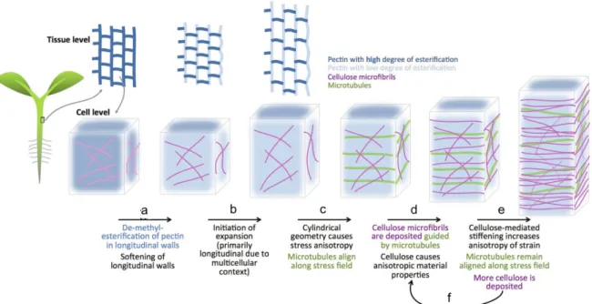
2.1.4. Hormonal crosstalk. The case of auxin and brassinosteroids 38 2.2. The cell wall in plant development and morphogenesis 41 2.2.1. Cellulose microfibrils as cell wall load-bearing elements 42 2.2.2. Hemicelluloses and their role in cell wall architecture 45 2.2.3. Pectins and their role in cell wall expansion 48 2.2.4. Cellulose/xyloglucan and pectin act together during morphogenesis 50
3. Pattern formation and morphogenesis at the shoot apical meristem 52 3.1. Shoot Apical Meristem set up during the embryonic stage 52 3.2. Shoot Apical Meristem functional domains 52 3.2.1. The Central Zone and the maintenance of a group of 53
undifferentiated stem cells
3.2.2. The Peripheral Zone and the generation of organ primordia 57
4. Flower initiation as model system to understand plant morphogenesis 61 4.1. The molecular regulation of flower initiation: a central role for auxin 61 4.2. The physical regulation of flower initiation: a central role for the cell wall 66 4.2.1. Control of growth anisotropy the dialog between mechanical forces 66
and the cytoskeleton
4.2.2. Control of growth rate: cell wall remodeling proteins 68
Chapter I
73Summary 75
Introduction 76
Results 77
Discussion 87
Conclusion and Perspectives 90
Experimental procedures 90
Chapter II
95 Summary 97 Introduction 99 Results 102 Discussion 116Conclusion and Perspectives 118
Experimental procedures 118 Supplementary material 122
Chapter III
135 Summary 137 Introduction 138 Results 139 Discussion 150Conclusion and Perspectives 152
Experimental procedures 153
Supplementary material 155
Discussion
163References
171List of Abbreviations
ABP1 AUXIN BINDING PROTEIN 1
AG AGAMOUS
AGL AGAMOUS-LIKE AGO10 ARGONAUTE 10
AHK ARABIDOPSIS HISTIDINE KINASE
AHP ARABIDOPSIS HISTIDINE PHOSPHOTRANSFERASE AIL6/PLT3 AINTEGUMENTA-LIKE6/PLETHORA3
ANT AINTEGUMENTA AP1 APETALA1
AP2/ERF APETALA2/ETHYLENE RESPONSE FACTOR ARF AUXIN RESPONSE FACTOR
ARR ARABIDOPSIS RESPONSE REGULATOR AS1 ASYMMETRIC LEAVES 1
AT ACETYLTRANSFERASE
AUX/IAA AUXIN/INDOLE ACETIC ACID
BAS1 PHYB ACTIVATION TAGGED SUPRESSOR 1 BAK1 BRI1-ASSOCIATED RECEPTOR KINASE 1 BES1 BRI1-EMS-SUPRESSOR1
BIM1 BES1 INTERACTING MYC-LIKE1 BIN2 BRASSINOSTEROID INSENSITIVE 2 BKI1 BRI1 KINASE INHIBITOR
BOP1 BLADE ON PETIOLE
BRI1 BRASSINOSTEROID INSENSITIVE1 BRM BRAHMA
BSK1 BRASSINOSTEROID-SIGNALING KINASE 1 BSU1 BRI1-SUPPRESSOR 1
BZR1 BRASSINAZOLE RESISTANT1 CAL CAULIFLOWER
CESA CELLULOSE SYNTHASE A
CDG1 CONSTITUTIVE DIFFERENTIAL GROWTH1 CKX CYTOKININ DEHYDROGENASE/OXIDASE CLV3 CLAVATA3
CMT CORTICAL MICROTUBULES
CPD CONSTITUTIVE PHOTOMORPHOGENIC AND DWARF CRF2 CYTOKININ RESPONSE FACTOR 2
CSI1/POM2 CELLULOSE SYNTHASE INTERACTING CSC CESA COMPLEX
CSLC CELLULOSE SYNTHASE-LIKE CUC1 CUPSHAPED COTYLEDON 1 DREB2A DRE-BINDING PROTEIN 2A DWF4 DWARF4
ER ERECTA
EXPA A-TYPE EXPANSIN FD FLOWERING LOCUS D FER FERONIA
FIL FILAMENTOUS FLOWER FT FLOWERING LOCUS T GH GLYCOSIDE HYDROLASES GT GLYCOSYLTRANSFERASES
GRF9 GROWTH-REGULATING FACTOR 9
HD-ZIP III HOMEODOMAIN LEUCINE ZIPPER CLASS III HEC1 HECATE 1
HG HOMOGALACTURONANS HTA13 HISTONE H2A 13
IAA INDOLEACETIC ACID
IPT ISOPENTENYLTRANSFERASE IWS1 INTERACT WITH SPT6 1 JLO JAGGED LATERAL ORGANS KAN KANADI
KNAT1 KN1-LIKE IN ARABIDOPSIS THALIANA 1 KTN1 KATANIN
LBD LATERAL ORGAN BOUNDARIES DOMAIN LFY LEAFY
LOB LATERAL ORGAN BOUNDARIES LOG LONELY GUY
LRR LEUCINE-RICH REPEAT RECEPTOR KINASES LSH4 LIGHT-DEPENDENT SHORT HYPOCOTYL 4 MBD MICROTUBULE-BINDING DOMAIN
MAP4 MICROTUBULE-ASSOCIATED PROTEIN 4 MP MONOPTEROS
MT METHYLTRANSFERASES NAC NAM-ATAF1/2-CUC2 NPA N-1-naphthylphthalamic acid NUC NUCLEAR CAGE
PDF1 PROTODERMAL FACTOR 1 PI PISTILLATA PID PINOID PIN1 PIN-FORMED1 PHB PHABULOSA PHV PHAVOLUTA
PME PECTIN METHYL ESTERASE
PMEI PECTIN METHYL ESTERASE INHIBITOR PUP PURINE PERMEASE
REV REVOLUTA
RIC1 ROP-INTERACTIVE CRIB MOTIF-CONTAINING PROTEIN 1 RLP44 RECEPTOR-LIKE PROTEIN 44
RLK RECEPTOR-LIKE KINASE
ROP6 RHO-LIKE GTPASE FROM PLANTS 6 SEP SEPALLATA
SOC1 SUPPRESSOR OF OVEREXPRESSION OF CO 1 SPK1 SPIKE
SPL10 SQUAMOSA PROMOTER BINDING PROTEIN-LIKE 10 STM SHOOT MERISTEM LESS
SVP SHORT VEGETATIVE PHASE
SWI/SNF SWITCH/SUCROSE NONFERMENTING SYD SPLAYED
THE THESEUS
TIR1/AFB TRANSPORT INHIBITOR RESISTANT1/AUXIN SIGNALING F-BOX TMK TRANSMEMBRANE KINASE
TMO3 TARGET OF MONOPTEROS TOC1 TIMING OF CAB EXPRESSION1 TPL TOPLESS
VIP1 VIRE2-INTERACTING PROTEIN1 WAK WALL-ASSOCIATED KINASES WUS WUSCHEL
XET XYLOGLUCAN ENDOTRANSGLUCOSYLASES
XTH XYLOGLUCAN ENDO-TRANSGLUCOSYLASE/HYDROLASE XXT XYLOGLUCAN XYLOSYL-TRANSFERASES
Glossary of Terms
activator a short-ranging substance that promotes its own production and the
synthesis of its antagonist
anisotropy the existence of directions with distinctive properties
anisotropic growth growth with a maximal and minimal direction
canalization a valley in Waddington’s epigenetic landscape that represents a
cluster of similar trajectories
complex system a system made of many elements that exhibits emerging global
properties not directly predictable from the properties of the individual components
crosstalk specific interactions between components of more than one pathway
emergent property a feature that is characteristic of system-level dynamics that cannot
be attributed to any of its components
epigenetic landscape visual depiction of a set of developmental choices that is faced by a
cell in the embryo
elastic deformation reversible extension of the cell wall
feedback regulation control mechanism that uses the consequence of a process to
regulate the rate at which the process occurs
feed-forward loop a biochemical pattern in a transcription network , a three-gene
pattern, composed of two input transcription factors, one of which regulates the other, both jointly regulating a target gene
hydrogel network of polymer chains that are hydrophilic, they are highly
flexible due to their significant water content
inhibitor rapidly difussing antagonist of an activator, it slows down the
production of the activator or catalyzes its decay
lateral inhibition strategy for emphasizing differences between inputs, a chemical
inhibitor diffusing faster through neighboring cells prevents the accumulation of the activator creating a zone of lateral inhibition
microtubule anisotropy
indicates a dominant microtubule orientation over a population of microtubules
morphogen a diffusible signal that acts at a distance to regulate pattern
phyllotaxis the pattern at which new leaf and flower primordia emerges
plastic deformation irreversible extension of the cell wall
plastochron interval between the initiation of two consecutive primordia
self-organization evolution of a system into an organized form in the absence of
external pressures
strain deformation of an object induced by stress, corresponds to growth
rate in living organisms
stiffness the extent to which an object resists deformation
stress force applied on a surface normalized by the surface area upon
which it is exerted
stress anisotropy stress with maximal and minimal directions
tensile strength the resistance of a material to breaking under tension
yield threshold level of stress that needs to be applied to a structure to induce an
irreversible deformation
wall creep cell wall extension that involves the breaking of hydrogen bonds
1. Generalities of pattern formation and morphogenesis in
multicellular organisms
One of the most fundamental questions in biology is that of biological pattern formation: how do the individual cells of a multicellular organism differentiate and how is this related to the overall structures and shapes that arise during development? In this first part of the introduction I will present the main theories that have addressed the problem of biological pattern formation and that have largely influenced modern developmental biology.
1.1. On Growth and Form
In 1917 D’Arcy Thompson published an extensive study on growth and form during development. He hereby underlined the fact that biological form is the consequence of physical processes and mechanical forces (Thompson, 1917). Thompson eloquently described form as a direct product of growth, and emphasized that growth and form are inseparably associated. According to his view the form of an organism is determined by its rate of growth in various directions. “Every growing organism; and every part of such a growing organism, has its own specific rate of growth, referred to this or that particular direction; and it is by the ratio between these rates in different directions that we must account for the external forms of all save certain very minute organisms”.
On Growth and Form is Thompson’s most famous work containing its most influential ideas. In the book, he offers a descriptive explanation of the shapes of various parts of multicellular organisms. These descriptions were more than anything else mathematical descriptions. Due to his preference for mathematical and biophysical concepts, but most likely also because ideas about molecular regulation were not known at that time, Thompson did not invoke biochemical explanations for his thesis. In this way, he excluded any explanation for the function of such shapes. Since then, the elaboration of biological knowledge has made it possible to address the question of pattern formation not only as a consequence of biophysical forces, but also in terms that consider biochemical signaling and downstream molecular regulation.
Regardless of the limitations (McClung, 1942), Thompson’s ideas provided an important basis for shaping modern developmental biology, and have gained renewed interest. I will now present a number of concepts that have focused more on biochemical regulation.
1.2. Reaction-diffusion model
The assembly of basic physical laws served Alan Turing to propose a key explanation for the formation of biological patterns (Turing, 1952). Turing’s hypothesis was that patterns in biological tissues can arise from simple chemical processes that can be described precisely and mathematically. He suggested “a system of chemical substances, which he termed morphogens, reacting together and diffusing through a tissue, is adequate to account for the main phenomena of morphogenesis”.
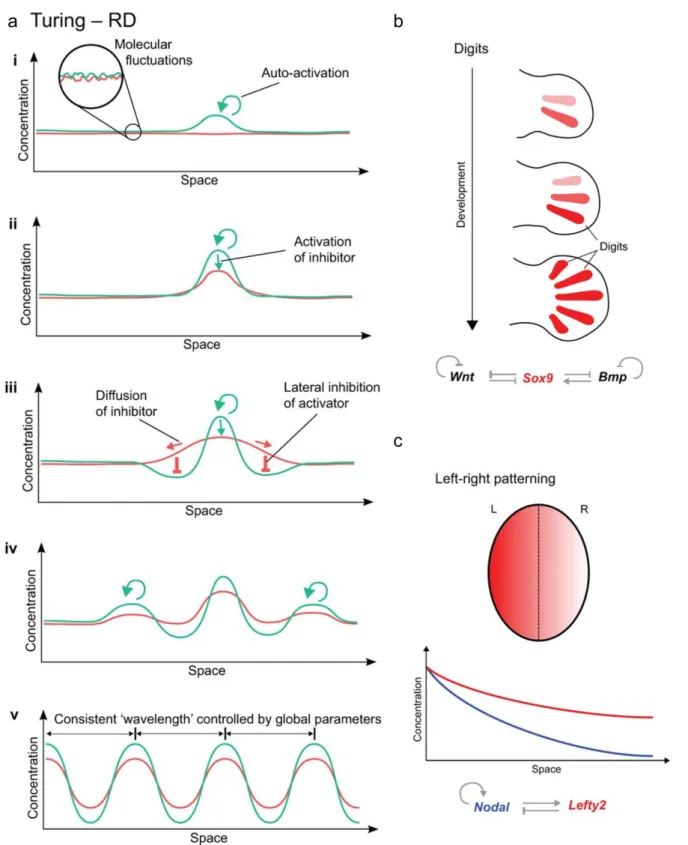
The essential feature of the reaction-diffusion model proposed by Turing is that a small perturbation in the concentration of two substances, initially distributed homogeneously can become spatially distributed heterogeneously given the differences in their diffusion properties and cross-regulation. Over time, the theory of Turing of biological pattern formation was further developed by Gierer and Meinhardt, who introduced to the reaction-diffusion model, the role of autocatalysis in conjunction with lateral inhibition (Gierer and Meinhardt, 1972; Meinhardt and Gierer, 2000). They proposed that one of the two substances is a short-range “activator”, a chemical that can make more of itself; the other one a long-range “inhibitor”, slows the production of the “activator”. Each of these substances acts on itself as well as the other. This dynamical interaction between the morphogens allows the mechanism to become self regulated and endows the ability to produce spontaneously a pattern when starting from a uniform field of cells. The system could be illustrated as follows (Figure 1a). (i) Molecular fluctuations of the morphogens, will cause some cells to accumulate slightly higher levels of activator. (ii) The activator self-regulation will increase its concentration enhancing also the production of the inhibitor. (iii) The activator positive feedback stabilises its own levels. (iv) However, since the inhibitor diffuses faster, it will increase its level in neighbouring cells, preventing the accumulation of the activator, creating a zone of “lateral inhibition” where no new peaks of activator can form (v) The whole system dynamically changes until a regular array of peaks and valleys is formed across the whole field of cells (Green and Sharpe, 2015).
In principle such a reaction-diffusion system can account for many patterning events. This could be the case, for example, for the patterning of digits during limb development in mouse embryos. This mechanism depends on the feedback loop between Wnt and Bmp signaling and the transcription factor Sox9 (Figure 1b)(Newman and Frisch, 1979; Raspopovic et al., 2014). Another example of a potential reaction-diffusion based mechanism is the left-right patterning of the early vertebrate embryo (Figure 1c). The distinction between the left and right side of the body is driven by the interaction between the protein Nodal, the activator, and Lefty 2, the repressor. Their interaction creates a broad gradient that allows cells to distinguish in which side of the embryo they are. Nodal-Lefty network forms spontaneously from an initial maternal bias through local auto-activation and long range inhibition (Green and Sharpe, 2015). In plant systems, probably the best example of a mechanism potentially based on reaction-difussion is phyllotaxis, which describes the pattern at which new leaf and flower primordia emerge (Meinhardt, 1994). Various patterns can be created depending on the range of activation and inhibition, either an alternating (distichous), 90° rotated (decussate) and even spiral (Meinhardt, 1996). Later on Kuhlmeier and collegues discovered that actively transported auxin is the instructive signal determining the induction and positioning of lateral organs. Although in this example, patterning is not through an inhibitor but through a redistribution of an activator by transport (see also below). Good evidence for an activator – inhibitor system also exists for the initiation of leaf hairs (Hulskamp, 2004).
Figure 1. The principles of reaction-diffusion and examples of real patterning systems (adapted from Green and Sharpe, 2015).
(a) Morphogen fluctuation generates higher levels of activator, lower and more diffuse inhibitor levels. Although the inhibitor fails to repress the activator, it prevents the activator region from growing and imposes a zone of “lateral inhibition”. The system dynamically changes until it reaches an equilibrium.
(b) Mouse limb buds are created as a Turing pattern guided by a feedback loop between the signalling of Wnt and Bmp and Sox9.
(c) Mouse embryo body sides, left (L) and right (R) are dictated by a RD system, which include Nodal and Lefty.
1.3. Positional-information model
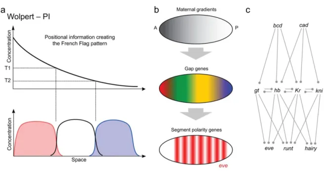
Another notable contribution to the concept of pattern formation was the notion of morphogen concentration and gradients proposed by Wolpert. In his aim to understand how more complex patterns were constructed from earlier tissue heterogenities, he proposed that differences in morphogen concentration across space could be enough to define different positions. In the gradient model there is a fixed source of morphogens. The morphogens leave this site by diffusing within the tissue. Since they are also degraded, they will form a concentration gradient (Wolpert, 1969, 1971). Cells that are responsive to the morphogen interpret the local concentration, whereby different threshold concentrations would hereby give different reponses (Ashe and Briscoe, 2006; Kondo et al., 2009; Kondo and Miura, 2010).
The positional information concept explains how a prior asymmetry results in a graded distribution of a morphogen, and how cells use this distribution to acquire different identities. This concept is commonly illustrated with a French Flag pattern (Figure 2a), in which the field of cells are divided into three different regions of cell fates (red, white and blue). After the interpretation of the morphogen threshold levels (T1, T2), cells react differently to these concentrations and adopt diverse fates. It has been proposed that the development of the
Drosophila embryonic segments is based on a positional information system (Figure 2b and c). Each stripe is defined independently by its unique anterior-posterior position in a succession of local concentration gradients of the gap genes. Differences in morphogen concentration at each position of the field provide distinct inputs to the gap gene network, which convert the smooth spatial differences into more discrete molecular patterns. This more complex molecular pattern of gap genes then provides the positional information for the next level of gene regulation, the segment polarity genes, which are each expressed as a series of stripes (Figure 2b and c) (Green and Sharpe, 2015).
Both, Turing’s reaction-difussion and Wolpert’s positional information models are able to explain biological patterns. The key feature that differentiates a reaction-difussion system from a positional information system is that the gradient is self-organized through the dynamics of the activator-inhibitor pair, unlike the positional information concept, which explains how a prior asymmetry is converted into a specific pattern. Therefore, the two
processes rather than mutually exclusive may be complementary and could function together as regionalizing mechanisms (Green and Sharpe, 2015).
A number of morphogens have been well described in animals (Wolpert, 2011). The first one discovered was the concentration gradient of Bicoid (Bcd) protein in Drosophila, which patterns embryo segmentation (Driever and Nusslein-Volhard, 1988; Frigerio et al., 1986). On the other hand, in plants the morphogen concept has remained a subject of debate. The signaling molecules closest to this concept are hormones. Particularly, auxin fulfils the characteristics of a morphogen, since it functions in diverse patterning events in a concentration-dependent manner and directly regulates target cells (Benkova et al., 2009; Bhalerao and Bennett, 2003; Sabatini et al., 1999).
Figure 2. The principles of positional information and examples of real patterning systems (adapted from Green and Sharpe, 2015)
(a) The concept of positional information arises from molecular asymmetries that result in the graded distribution of a morphogen, cells make use of this information to acquire different cell fates, represented in the scheme by the three different colours.
(b) Initial asymmetries in the anterior-posterior axis of the early Drosophila embryo result in the graded distribution of morphogens that in turn regulate the expression of the gap genes providing the positional information for the segment polarity genes, expressed as a series of stripes.
(c) Differences in the concentration of the morphogens Bicoid and Caudal across the embryo inputs the gap genes network (giant, hunchback, Krüppel, and knirps). In turn, the complex interactions among gap genes create molecular patterns of expression of the segment polarity genes (even-skipped, runt, hairy).
1.4. Epigenetic landscape
A third theory worth to mention, is the epigenetic landscape concept proposed by Conrad Waddington. At the time he started to develop his ideas, the Mendelian laws of heredity were well accepted. Waddington agreed with the distinction between the individual’s physical appearance, or the phenotype and the hereditary information contained in the germ cells and passed to the next generation, better known as the genotype. He considered the phenotype as the result of the interrelations among genetic processes, their potentialities and constraints, and the external environment.
Waddington studied the development of embryonic cells triggered by given stimuli, through a process he called induction. He proposed a hypothesis in which he emphasized the importance of the reacting tissue, in the sense that change occurs not only because cells receive a particular signal, but also because they have the ‘potency’ to react. In other words, cells react to different stimuli, biochemical or environmental, in a way allowed by their state at that time. With each new reaction, the cell might differentiate further into a state with more constraints or more possibilities and potentialities.
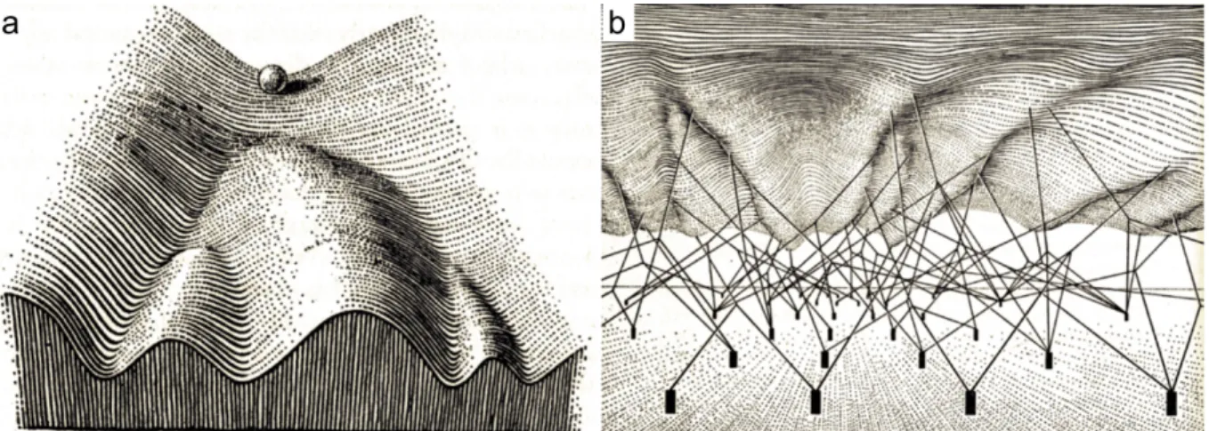
He argued that the various developmental pathways a cell might take follow an epigenetic path. Each step is defined by instructions in the genotype that interact to produce a system that moves along a trajectory. The diverse paths in development are protected or canalized by threshold reactions, providing stability and direction. Waddington illustrated this canalization concept, as a landscape, an epigenetic landscape formed by a series of ridges and valleys a cell can traverse on its way to a final tissue type (Waddington, 1956, 1957) (Figure 3a). The landscape thus represents the tendency of cells to pass from an immature stage to an adult and specified condition. The path of a cell would start from a totipotent state, passing via a pluripotent state to a lineage -committed state that leads it into one of many possible fates.
The steepness of the walls, represents the stability of the path. If the walls are very high, it is hard for the cells to escape from their developmental faith and even big mutational or environmental perturbations will not be able to bring the cell out of its path. The control of the steepness of the walls in turn depends on the underlying genetic landscape (Figure 3b). Importantly, not only genes and their products, but also gene-gene interactions and gene – environment interactions are in control of development. According to Waddington’s ideas,
genes not only regulate, but they are also regulated by non-genetic factors (Van Speybroeck, 2002).
Figure 3. Waddington’s epigenetic landscape (adapted from Waddington 1957).
(a) The classical view of an epigenetic landscape, in which the differentiation of cell in an embryo is illustrated as a pebble that begins at the top of a hill and rolls down the epigenetic landscape though a series of branching points that represent decision events. Cells evolve according to the same laws, but because of the existence of inducing signals, cells in different regions would follow different pathways and end up at different states of differentiation represented as valleys. The effect of these signals is restricted to reacting tissue and ultimately trigger the cells select one of a few possible developmental pathways.
(b) The genetic landscape underlying the architecture of the epigenetic landscape. The valleys are formed by tension on ropes attached to gene complexes represented as cylindrical pins stuck in the ground.
1.5. General concepts: some concluding remarks
According to these theories there are multiple factors directing pattern formation. The physical properties and form diversity in D’Arcy Thompson’s theory, the self-organization and biochemical patterning in Turing patterns and the importance of the interaction of these factors at multiple scales. However, in order to understand pattern formation it is necessary to analyze how are these elements acting in concert. These interactions lead to the emergence of collective properties that cannot be deduced from adding up local behaviour. These are properties common to all multicellular organisms comprised under the concept of complex systems.
Plants as complex systems are hierarchically organized and composed by interactive elements: molecules assemble into cells, cells into tissues and organs. The interactions between the individual components and the multiple feedbacks between the diverse levels of organization are fundamental for pattern formation. Therefore, these systems can only be understood by analysing them at multiple scales, leading to the use of more interdisciplinary approaches. In addition, certain constraints imposed by the developing system might limit the possible final shapes. In plant development some specific emergent properties should be considered when studying pattern formation and morphogenesis. Of them I will speak in the following section.
2. Generalities of pattern formation and morphogenesis in plants
The generation of form in plants is distinguished by a number of specific features (Hernandez-Hernandez et al., 2012; Niklas, 2000; Niklas and Kutschera, 2009). First, plant architecture is characterized by an open and indeterminate ontogeny, with multiple growing points (or meristems) where cell proliferation persist continuously producing new tissues and organs (Esau, 1965). This characteristic relates to the sessile nature of plants. It provides them the opportunity to adjust to their external environment, adapting their shape and architecture in relation to it.
A notable characteristic of plant cells that highly influences development is the presence of a relatively rigid extracellular matrix, the cell wall. The presence of this cell wall provides plants with specific mechanical properties. As a result, morphogenesis must occur in the absence of cell migration. Therefore, cell expansion, cell division and, to a lesser extent, programmed cell death, are of major importance in plant morphogenesis (De Smet and Beeckman, 2011; Van Hautegem et al., 2015). Given that plant cells are immobilized they rely on mobile signals to trigger the local differences that guide tissue morphogenesis. A major category of these mobile signals is plant hormones, which largely control plant growth and development and represent excellent candidates for plant morphogens.
In what follows I will review the basic characteristics of plant development that define pattern formation in plants. I will firstly focus on the role of signaling molecules, hormones and their effect on establishing molecular expression patterns. Secondly, I will describe the role of the extracellular matrix in controlling growth patterns. A good amount of our knowledge in these topics has been obtained from the plant experimental system Arabidopsis thaliana (hereafter called Arabidopsis); therefore I will mainly focus on this species.
2.1. Molecular regulation: the role of hormones in plant development
As mentioned previously, positional information is perceived and transmitted via communication between different parts of the organism, locally and over long distances. This communication is based on the perception and production of mobile signals. In plants, the distribution and perception of hormones as instructive mobile signals of growth and development has been well established, involving in particular cytokinins, auxins,
gibberellins, brassinosteroids and strigolactones (Santner et al., 2009; Santner and Estelle, 2009; Wolters and Jurgens, 2009).
Major aspects of plant hormone synthesis, degradation, transport and signaling have been extensively studied. In what follows I will briefly summarize the role of hormones in controling gene regulation, in particular at the transcriptional level, where they play a central role (Nemhauser et al., 2006). I will hereby focus on auxins, cytokinins and brassinosteroids, which have major roles in development of the meristems and plant architecture and are a central focus of this thesis.
2.1.1. Auxin
Auxin is certainly one of the most important signals, affecting plant developmental processes at cellular, tissue and organ levels. It has been considered as the closest equivalent to morphogens in plants (Bhalerao and Bennett, 2003; Sabatini et al., 1999). During development, auxin differentially accumulates in different parts of the plant. Auxin gradients are fundamental in the regulation of many developmental processes. From the very early stages of development onwards, auxin accumulation and its graded distribution play fundamental roles in defining plant shape. Already after fertilization, the apical cell of the divided zygote is the site of auxin accumulation and activity. This is maintained this way until the 32-cell-embryo stage. Later on auxin distribution changes to establish the root pole and cotyledons (Friml et al., 2003). Another example of the importance of differential auxin distribution is the accumulation of auxin at the location of organ initiation, either at the root (Figure 4b)(Dubrovsky et al., 2008) or at the shoot (Figure 4c)(Heisler et al., 2005; Meinhardt, 2003). There are numerous examples about the role that auxin distributions and gradients play in the regulation of plant growth and development. But how does these specific distributions arise?
One mechanism for the differential distribution of auxin is attributed to its site of biosynthesis. The most common auxin in vascular plants is indole-3-acetic acid (IAA). IAA is synthesized by one tryptophan (Trp)-independent and four Trp-dependent pathways. Two of them, the tryptamine (TAM) pathway, and the indole-3-piruvic acid (IPA) pathway are most relevant for plant development. Rate-limiting enzymes for these pathways include the flavin monooxygenase-like enzymes of the YUCCA family and the Trp aminotransferase of
Arabidopsis (TAA) (Teale et al., 2006; Woodward and Bartel, 2005) (Figure 5a). Mutations of multiple YUCCA and TAA genes impair local auxin accumulation and result in severe developmental defects, in embryogenesis, leaf venation, and floral organ patterning, among others (Cheng et al., 2006, 2007; Stepanova et al., 2008).
Another major process controling auxin distribution is auxin transport. It has been well established that auxin moves directionally through plant tissues. From the sites of its synthesis it is transported to the whole plant (reviewed in (Peer et al., 2011). A long distance source-to-sink transport occurs by the loading of auxin into the phloem, from young biosynthetically active shoot tissues towards sink tissues (Figure 4a). Cell-to cell transport can also achieve auxin movement over both short and long distances. Cell-to-cell transport was predicted by the chemiosmotic model, based in the physicochemical properties of the auxin molecules (Goldsmith, 1977; Raven, 1975; Rubery and Sheldrake, 1974). Auxins are weak acids, and their ability to penetrate through the membrane, depends on the pH. The plant’s apoplastic pH is approximately 5.5. Under this conditions it is predicted that only a small but signficant fraction (17%) of auxin molecules are proton-associated (HA). Although protonated auxin freely diffuses from the apoplast into the cytoplasm, 83% of the auxin pool remains unavailable for diffusion in its dissociated form. Once in the cytoplasm where pH is approximately 7, the equlibrium of the auxin shifts to the anionic, dissociated form. In this circumstances auxins cannot diffuse across the cell membrane, hence the active transport of auxin is required. Indeed, three main families of transmembrane proteins provide means of active auxin transport in and out of the cell, across the plasma membrane: i) the AUX1/LIKE AUX1 (AUX1/LAX) auxin influx permeases, ii) the P-glycoproteins of the ATP-Binding Cassette family B (ABCB/PGP) efflux transporters, and iii) the PIN-FORMED (PIN) auxin efflux carriers reviewed in (Zazimalova et al., 2010) (Figure 5b). Among them, mainly PIN-mediated transport seems to contribute to polar auxin transport (PAT), which is essential for defining differential auxin distribution (Weijers et al., 2005).
PINs are plant specific proteins with a predicted secondary structure of five transmembrane helices at the N and C terminus (Galweiler et al., 1998; Krecek et al., 2009; Paponov et al., 2005) linked by an intracellular hydrophilic loop that influences protein localization and activity (Bennett et al., 2014; Dhonukshe et al., 2010; Huang et al., 2010). Arabidopsis PIN family consists of eight members, PIN1-8. PIN1, 2, 3, 4 and 7 localize preferentially in the
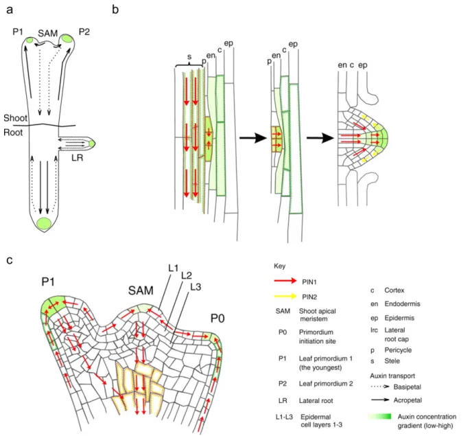
Figure 4. Auxin graded distribution in root and shoot morphogenesis (Petrasek and Friml, 2009).
(a) Directional flow of auxin in the shoot and root of Arabidopsis. The accumulation of auxin at the location of organ initiation (green) is maintained by flow towards root and shoot apices (solid arrows). Reverse flow towards root and shoot basis (dashed arrows).
(b) Auxin transport in the developing lateral root. Auxin maxima specify the founder cells in the pericycle, subsequent coordinated divisions form the lateral root primordium. PIN1 and PIN2 facilitate the transport of auxin that enables the development of the primordia.
(c) Auxin transport in the shoot apical meristem in developing primordia (P1 and P2). Auxin is transported through the epidermis layer L1 by the activity of PIN1, maintaining an auxin maxima at the organ primordium tip. From there, a basipetal transport route is established through the interior of the primordium, marking the future vasculature tissues.
plasma membrane (Adamowski and Friml, 2015; Petrasek et al., 2006; Wisniewska et al., 2006); PIN5 and 8 localization has been reported at the endoplasmic reticulum (Dal Bosco et al., 2012; Ding et al., 2012; Mravec et al., 2009); whereas there are still doubts regarding the localization and function of PIN6 (Nisar et al., 2014). Plasma membrane localized PINs often display polar cellular localization, which notably correlates with the directional flow of auxin and therefore have also been used to deduce such fluxes (Benkova et al., 2003; de Reuille et al., 2006; Friml et al., 2002a; Galweiler et al., 1998; Wisniewska et al., 2006). The auxin transport ability of PINs has been shown in Arabidopsis and heterologous systems (Petrasek et al., 2006; Yang and Murphy, 2009; Zourelidou et al., 2014). The positioning of PINs is highly dynamic. It is crucial to the production of organs during development (Blilou et al., 2005; Friml et al., 2003; Heisler et al., 2005; Reinhardt et al., 2003) and to the modulation of patterns of growth and development in response to the environment, for example in gravitropism (Friml et al., 2002b). After their transcription, PINs are either retained in the endoplasmic reticulum (ER) or translocated through the Golgi Apparatus (GA) to the plasma membrane (Matheson et al., 2006). PINs undergo continuous shuttling between the plasma membrane and the intracellular compartments by rounds of internalization (endocytosis) and polar recycling (exocytosis). These processes together are known as consitutive endocytic cycling (Dhonukshe et al., 2010; Dhonukshe et al., 2008; Geldner et al., 2001; Kleine-Vehn et al., 2011). According to current models, the regulation of PIN trafficking is based on their phosphorylation status, determined by the action of the PINOID (PID) and other AGC3 kinases and the antagonistic action of PP2A/PP6 phosphatases (Figure 5b) (Benjamins et al., 2001; Michniewicz et al., 2007). Unphosphorylated PINs are recycled to the plasma membrane by the ADP-ribosylation factor-guanine nucleotide exchange factor (ARF-GEF) GNOM. Phosphorylated PINs result in GNOM-independent recycling to the opposite plasma membrane (Dhonukshe et al., 2007; Geldner et al., 2001; Kleine-Vehn et al., 2008). Monoubiquitination of PINs induces their endocytosis, which occurs via clathrin-coated vesicles and requires the actin cytoskeleton. Subsequently, polyubiquitination labels PIN proteins for degradation (Leitner et al., 2012). The post-translational modifications of PINs, ubiquitination and phosphorylation, provide an entry point for various external signals, for example gravity (Abas et al., 2006; Friml et al., 2002b) or light (Ding et al., 2011; Michniewicz et al., 2007; Willige et al., 2013). Thereby the abundance of PINs in the plasma membrane or their polarity can be modified in reponse to external signals. Several hormones (auxin included) may influence directly or indirectly the transcription of PINs (Bishopp et al., 2011a; Dello Ioio et al., 2008; Hacham et al., 2012; Liu et al., 2013; Zhang et al., 2011) or
influence their abundance (Crawford et al., 2010; Hacham et al., 2012; Willige et al., 2011). Experimental evidence partially combined with modelling approaches suggests that auxin itself provides feedback regulation on its own distribution influencing transcription, turnover, and plasma membrane localization of PIN proteins (Heisler et al., 2005; Stoma et al., 2008).
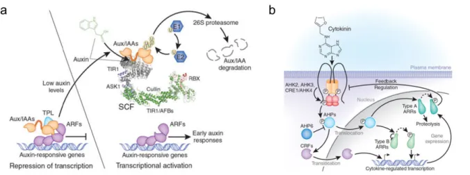
Besides production and transport, auxin perception and downstream signaling have also been extensively studied in a range of developmental processes. Auxin is first perceived via one or more receptors that initiate a signaling cascade that translates the auxin concentration into diverse cellular behaviors. Mainly two auxin receptor systems have been described, involving respectively the TRANSPORT INHIBITOR RESISTANT1/AUXIN SIGNALING F-BOX (TIR1/AFB) complexes (Dharmasiri et al., 2005a; Dharmasiri et al., 2005b; Kepinski and Leyser, 2005) and AUXIN BINDING PROTEIN 1 (ABP1) (Woo et al., 2002) (Figure 5c). From these, the best characterized is the TIR1/AFB pathway that regulates auxin responses within the nucleus (Calderon Villalobos et al., 2012; Chapman and Estelle, 2009; Dharmasiri et al., 2005a; Kepinski and Leyser, 2005; Parry et al., 2009) (Figure 6a). At low auxin concentrations the transcriptional repressors AUXIN/INDOLE ACETIC ACID (Aux/IAAs) negatively regulate auxin signalling. Aux/IAAs carry out their repressor activity by binding to the DNA-binding AUXIN RESPONSE FACTOR (ARF) proteins (Guilfoyle and Hagen, 2007; Kim et al., 1997), and recruiting the transcriptional co-repressors of the TOPLESS (TPL) family to ARF-bound promoters (Ke et al., 2015; Szemenyei et al., 2008). This prevents the ARF mediated transcription of auxin responsive-genes (Figure 6a). At high auxin concentrations, auxin interacts with TIR1 or other AFBs (Dharmasiri et al., 2005a). The binding of auxin occurs within an internal pocket formed from the binding between the F-box protein and the Aux/IAA, forming a complex TIR1/AFB-auxin-Aux/IAA that targets Aux/IAA for ubiquitination and degradation via the 26S proteasome (Figure 6a) (Calderon Villalobos et al., 2012). Once Aux/IAA are degraded, ARF proteins can either activate or repress auxin responsive genes (Chandler, 2016). More than 50 genes encoding ARF and
Aux/IAA have been identified in the Arabidopis genome (Vernoux et al., 2011). Differential expression of each of these players provides combinatorial possibilities for auxin-dependent gene regulation (Chapman and Estelle, 2009). Large-scale analyses of the AUX/IAA-ARF network have been performed in order to try to understand the distribution and perception of auxin signaling; for instance, in the shoot apex of Arabidopsis (Vernoux et al., 2011). Through a combination of expression data, a set of molecular interactions, mathematical
modelling and auxin signaling sensors, Vernoux et al., (2011) have described a key role for local auxin signaling in the regulation of the shoot apex patterning.
Notably auxin functional specificity can be generated at different levels. For instance, at the level of protein-DNA interactions, by the presence of cis regulatory elements in auxin-responsive genes. At the level of chromatin-level it has been shown that ARF5/MP transcriptional regulation requires chromatin state changes target loci. Aux/IAA repressors of auxin signalling together with co-repressors of the TPLs family and the repressive chromatin regulator histone deacetylase HDA19, prevent the expression of ARF5/MP regulated genes (Long et al., 2006; Szemenyei et al., 2008). Local accumulation of auxin, drives Aux/IAA degradation, as well as the dissociation of TPL and HDA19. This in turns leaves ARF5/MP free to recruit the SWITCH/SUCROSE NONFERMENTING (SWI/SNF) chromatin-remodeling complexes, SPLAYED (SYD) or BRAHMA (BRM). SWI/SNF complex unlock the repressed chromatin state at ARF5/MP target loci, which also increases chromatin accessibility for additional transcription factors. In contrast, in the absence of auxin, Aux/IAAs promote chromatin closure by recruiting TPL transcriptional co-repressors to ARF-bound promoters (Wu et al., 2015).
ABP1 was the first auxin-binding protein described in the literature. Its binding capacity was demonstrated by physiological and structural studies (Hesse et al., 1989; Woo et al., 2002). ABP1 localizes mainly to the ER, but a small portion is likely secreted to the cell wall where it is assumed to be active (Sauer and Kleine-Vehn, 2011). Described physiological responses linked to ABP1 are initiated on the outside of the plasma membrane, this requires that the signal is passed into the cell. This role is supposedly accomplished by plasma membrane-localized proteins TRANSMEMBRANE KINASE (TMK) or SPIKE (SPK1) (Lin et al., 2012; Xu et al., 2014), which were reported to transfer auxin signal inside the cell via the RHO OF PLANTS (ROP)-GTPases and the ROP INTERACTIVE CRIB motif-containing (RIC) proteins. ROP-RIC systems regulate endocytosis/exocytosis of PIN proteins on the plasma membrane, thus controlling auxin fluxes. In leaf pavement cells, for example, two ROP-RIC downstream pathways have been described (Fu et al., 2005; Xu et al., 2010). The ROP2-RIC4 pathway acts in the lobe outgrowth through the stabilization of cortical actin microfilaments and further inhibition of PIN1 endocytosis (Nagawa et al., 2012; Xu et al., 2010). The ROP6-RIC1 pathway inhibits the indentation outgrowth by the activation of the microtubule severing protein KATANIN (KTN1), which promotes the bundling of cortical microtubules in
the necks, and further inhibits PIN1 and PIN2 endocytosis (Chen et al., 2012b; Fu et al., 2009; Lin et al., 2013). In the root, ABP1 was reported to control cell cycle entry by regulating the D-type CYCLIN/RETINOBLASTOMA pathway and the PLETHORA (PLT) gradients (Tromas et al., 2009). Although a number of non-transcriptional responses mediated by ABP1 have been reported (Chen et al., 2012a; Chen et al., 2015; Chen et al., 2012b; Lin et al., 2012; Nagawa et al., 2012; Wu et al., 2011; Xu et al., 2014; Xu et al., 2010) the function of ABP1 as an auxin receptor has remained unclear. The recent isolation of two Arabidopsis abp1 mutants with no obvious phenotypes raised strong questions (Gao et al., 2015). Although ABP1 inactivation by inducible antibody- and antisense-based lines present strong phenotypes not caused by ABP1 down-regulation, which might suggest redundancy (Michalko et al., 2016).
Figure 5. Underlying processes of auxin differential distribution (Finet and Jaillais, 2012).
(a) Auxin biosynthesis and storage as inactive conjugates, which involves enzymes of the GH3 family. Intracellular homeostasis of auxin provided by ER localized PINs and PIN-LIKE proteins (PILS) (Barbez et al., 2012).
(b) Polar auxin transport depends on influx (AUX/LAX) and efflux carriers (PIN and ABCB/PGP) that promote the uptake and release of auxin to the apoplast. The endocytic trafficking and polar recycling of PINs is illustrated. Hormonal regulation of these pathways include auxin feedback regulation, cytokinin control over PIN endocytosis (Marhavy et al., 2011), and gibberellin regulation of PIN trafficking to lytic vacuoles (Willige et al., 2011)
Figure 6. Auxin and cytokinin transduction pathways (adapted from Santner 2009).
(a) At low auxin (left) transcription of auxin-responsive genes is prevented by Aux/IAAs. At high auxin (right) the F-box receptor TIR1/AFBs bind auxin and enhances its affinity for Aux/IAAs, promoting their ubiquitination and degradation, ARFs are released to initiate transcription of auxin-responsive genes.
(b) Cytokinin is perceived by the plasma membrane localized receptors AHK. A series of phosphorelay steps follow the AHK activation, which lead the activation and nuclear translocation of the AHP proteins. Once inside the nucleus AHP transfer the phosphoryl group to ARR proteins. CRF proteins are also activated by cytokinin and act as activators of cytokinin-regulated transcription
2.1.2. Cytokinins
Cytokinins are known for their ability to promote cytokinesis, hence their name. Cytokinins are adenine derivatives carrying either an isoprene-derived or an aromatic side chain (Mok and Mok, 2001). Great diversity exists among the predominant CKs between plant species (Sakakibara et al., 2006). Major derivatives present in Arabidopsis are trans-zeatin (tZ), and isopentenyladenine (iP) types. The initial step of CK synthesis is catalyzed by isopentenyltransferases (IPTs), which use ADP, ATP and tRNA as isoprenoid acceptors (Kakimoto, 2001; Takei et al., 2001). In higher plants two pathways for the synthesis of tZ coexist. The iP nucleotide-dependent pathway catalyzed by the cytochrome P450 monooxygenases CYP735A and the nucleotide-independent pathway, catalysed by the CK riboside 5’-monophosphate phosphoribohydrolase, LONELY GUY (LOG) (Kurakawa et al., 2007; Kuroha et al., 2009; Tokunaga et al., 2012; Zurcher and Muller, 2016) Additionally steady-state levels of active CKs are determined by the rate of conjugation and degradation. Active CKs, the free bases, are modified into ribosides and ribotides by O-glycosylation. Modified cytokinins can be activated when needed and seem to be the major long-range transport forms in plants (Zurcher and Muller, 2016). CYTOKININ DEHYDROGENASE/OXIDASE (CKX) proteins catalyse the degradation of CKs; they act by cleaving the CKs side chains. Genes encoding cytokinin degradation and synthesis proteins are widely expressed and active both in the shoot and the root (Nordstrom et al.,
2004). Regulation over the synthesis of CKs depends on the differential expression of the basic elements of its metabolism IPTs, CKX and CYP735A. CKs homeostasis is fine-tuned by other hormones and external factors, such as nitrogen availability (Sakakibara et al., 2006).
The signaling of CKs initiates by its perception by the membrane-bound Arabidopsis HISTIDINE KINASE (AHK) proteins, which serve as CKs receptors (Heyl et al., 2012). Binding of cytokinin to AHK proteins triggers a phosphorelay, in which a phosphoryl group is transferred from a His residue into an Asp residue within the kinase domain of the receptor. Afterwards, the phosphoryl is transmitted to a His residue of an Arabidopsis HISTIDINE PHOSPHOTRANSFERASE (AHP) protein. The previous is true for AHP1-5. AHP6 differs, since it cannot accept an activated phosphoryl group, which makes it unable to perform the phosphorelay (Mahonen et al., 2006). The role of AHP6 is, however, important, since it performs an inhibiting role over cytokinin signaling by competing with the “true” AHPs and contributes to confine the CKs signaling domains (Besnard et al., 2014; Bishopp et al., 2011b). AHP1-5 proteins continuously translocate to the nucleus enabling the phosphorylation of Arabidopsis RESPONSE REGULATOR (ARR) proteins. According to C-terminal differences, the family of ARRs has been classified into type –A –B and C (D'Agostino et al., 2000). Type-B ARRs have a transcription factor domain for DNA binding, once phosphorylated type-B ARRs as DNA-binding transcription factors activate transcription of cytokinin-regulated genes (Kiba et al., 1999; Mason et al., 2004). Type-A ARRs lack the transcription factor domain and instead act as negative regulators of cytokinin by attenuating the signal (Brandstatter and Kieber, 1998; D'Agostino et al., 2000; Rashotte et al., 2003). Transcription of type-A ARRs is under the direct regulation of type-B ARRs (Figure 6b). Type-C ARRs are less characterized, although they might have roles as modulators, since their ectopic expression affects cytokinin signaling (Kiba et al., 2004).
Sites of cytokinin synthesis do not necessarily coincide with the sites of perception, suggesting the transport of cytokinins (Zurcher et al., 2013). Long distance transport of tZ-type cytokinins occurs from the root to the shoot via the xylem, whereas iP-tZ-type cytokinins move through the phloem from the shoot to the root (Bishopp et al., 2011b). Cell-to-cell transports of cytokinins seem to be mediated by the PURINE PERMEASE (PUP) proteins (Burkle et al., 2003).
The components involved in cytokinin biosynthesis, degradation and phosporelay signaling, are encoded by multigene families (Muller and Sheen, 2007). The diverse but specific expression patterns of these components, suggest a broad range of cytokinin functions. Physiological functions of cytokinins include male and female gametophyte development, root and shoot apical meristem maintenance and development, as well as vasculature development and nodule organogenesis (Zurcher and Muller, 2016).
2.1.3. Brassinosteroids
Brassinosteroids (BRs) are steroidal hormones that play a major role in promoting cell expansion and proliferation (Hardtke et al., 2007; Nakaya et al., 2002). Brassinolide (BL) is the most biologically active BR among more than 50 natural BRs (Fujioka and Yokota, 2003). The biosynthesis of BRs involves parallel and highly branched pathways (Fujioka and Yokota, 2003). The initial step is the formation of campestanol (CN) from campesterol (CR). For this step, two branches have been proposed, the early C-22 oxidation, and the late C-22 oxidation. In Arabidopsis the early C-22 oxidation appears to be the major BR biosynthetic pathway (Fujita et al., 2006). Two main pathways have been identified for the biosynthesis of BL from campestanol, the early and late C-6 oxidation pathways (Ohnishi et al., 2009). In the early C-6 oxidation pathway, C-6 oxidation occurs ahead of C-22 hydroxylation. While in the late C-6 oxidation pathway, C-22 hydroxylation takes places before C-6 oxidation (Chung and Choe, 2013). A number of the genes relevant for brassinosteroids biosynthesis or signaling have been cloned taking advantage of BR-deficient mutants. Features such as dwarfism, dark-green and curled leaves, reduced fertility and delayed senescence, are characteristic of these mutants. When grown in the dark, such mutants are de-etiolated with short hypocotyls and open cotyledons (Clouse et al., 1996; Li et al., 1996; Szekeres et al., 1996).
Two different enzymes can perfom the initial modification of CR. The cytochrome P450 monooxygenase (CYP90B1) DWARF4 (DWF4) (Choe et al., 1998); and the cytochrome P450 monooxygenase (CYP90A1) CONSTITUTIVE PHOTOMORPHOGENIC AND DWARF (CPD)(Szekeres et al., 1996). DWF4 acts as a C-22-hydroxylase, whereas CPD functions as a C-3 dehydrogenase (Ohnishi et al., 2012). Depending on the availability of substrates and enzymes, the biosynthesis of BL progresses via either DWF4- or CPD-mediated pathways. Interestingly, both DWF4 and CPD can act on multiple substrates,
constituting multiple biosynthetic parallel pathways. Nevertheless, the overall flux of the BR biosynthetic pathway seems to be determined by the activity of DWF4 (Chung and Choe, 2013). The expression of BR- biosynthetic genes is primarily regulated at the transcriptional level. Interestingly the expression of several biosynthetic genes is subject to feedback regulation from the BRs signaling pathway (Bancos et al., 2002; He et al., 2005; Mathur et al., 1998).
BRs are widely distributed throughout plant tissues, although the active forms seem to accumulate mostly in young growing regions undergoing active cell division and elongation. BRs do not seem to undergo long-distance transport; in contrast they appear to be synthesized and function in the same tissue or even the same cell (Bishop et al., 1996; Shimada et al., 2003; Symons and Reid, 2008). BRs synthesis seems to take place in the endoplasmic reticulum, while its perception is located at the exterior cell surface. Thus movement of BRs is more likely to occur within and between neighbouring cells (Symons and Reid, 2008).
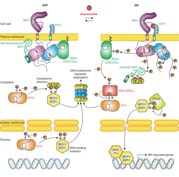
BRs receptors have been described in Arabidopsis as plasma membrane localized leucine-rich repeat receptor kinases (LRR), BRASSINOSTEROID INSENSITIVE1 (BRI1) and its two homologues BRL1 and BRL3 (Li and Chory, 1997). The kinase activity of BRI1 is activated following the binding of BR (Kinoshita et al., 2005; Wang et al., 2001). Upon perception of BRs by BRI1, the inhibitory protein BRI1 KINASE INHIBITOR (BKI1) is phosphorylated and dissociated from BRI1(Jaillais et al., 2011; Wang and Chory, 2006). BRI1 is then free to interact with BRI1-ASSOCIATED RECEPTOR KINASE 1 (BAK1) (Nam and Li, 2002), leading to the autophosphorylation and transphosphorylation between the kinase domains of BRI1 and BAK1 (Wang et al., 2008). Activated BRI1 is able to phosphorylate BRASSINOSTEROID-SIGNALING KINASE 1(BSK1) and CONSTITUTIVE DIFFERENTIAL GROWTH1 (CDG1) kinases (Kim et al., 2011; Tang et al., 2008). Subsequently, phosphorylated BSK1 and CDG1 bind and phosphorylate BRI1-SUPPRESSORS1 (BSU1) phosphatase (Kim et al., 2011; Kim et al., 2009). Phosphorylated BSU1 inactivates by dephosphorylation the GSK3-like kinase BRASSINOSTEROID INSENSITIVE 2 (BIN2) (Kim and Wang, 2010). Activated BIN2 phosphorylates transcription factors BRASSINAZOLE RESISTANT1 (BZR1) and BZR2 (hereafter called BRI1-EMS-SUPRESSOR1 (BES1)) (Wang et al., 2002; Yin et al., 2002). Phosphorylated BZR1 and BES1 are retained in the cytoplasm via the activity of 14-3-3 proteins (Gampala et al., 2007; Vert and Chory, 2006). Thus, under high BR levels, inactivation of BIN2 by BSU1
leads to the dephosphorylation and activation of BZR1 and BES1 (Tang et al., 2011). Unphosphorylated BZR1 and BES1 are free to move into the nucleus and bind the promoter of their target genes (He et al., 2005; Sun et al., 2010; Yin et al., 2005; Yu et al., 2011). The activation of BR signaling requires histone modifications and additional interacting transcription factors, among them, BES1 INTERACTING MYC-LIKE1 (BIM1) and INTERACT WITH SPT6 1 (IWS1) (Figure 7)(Li et al., 2010; Yin et al., 2005; Yu et al., 2008).
A number of target genes of BZR1 and BES1 have been identified, revealing diverse molecular links. Noteworthy, the activity of BZR1 is responsible for the transcriptional regulation of BR biosynthetic genes, such as DWF4 and CPD (Kim et al., 2006; Wang et al., 2002). When activated, BZR1 binds to the BR-responsive elements of the promoter sequence of DWF4 and CPD and repress their transcription (He et al., 2005). BES1 might also repress
DWF4 and CPD transcription to attenuate BR responses in a feedback loop, but this mechanism is primarily dependent on the repression and de-repression of transcription by BZR1 (Yu et al., 2011). Additional targets of BRs signaling are related with cell wall modification (Xie et al., 2011) and cellular transport, in agreement with BRs effects on cell expansion and growth. Not surprisingly, BR signaling converges substantially with other hormonal and environmental signals, such as light and GA signaling (Guo et al., 2013; Zhu et al., 2013). Of special relevance for this thesis are the interactions with auxin signaling, which I will examine next.
2.1.4 Hormonal crosstalk. The case of auxin and brassinosteroids
Multiple hormones are at play during plant growth and development. Their specific functions, however, are sometimes difficult to define, in particular because extensive crosstalk and signaling integration among growth regulating hormones has been demonstrated (Nemhauser et al., 2006). For instance, cell proliferation is regulated by cytokinins and auxin, while, cell expansion is under the control of auxin, BRs and gibberellins. More recently, a role of BRs in cell proliferation has been identified (Hardtke et al., 2007; Nakaya et al., 2002). Antagonistic relationships between cytokinin and auxin have also been described in much detail. This relationship seems to keep a balance between cell proliferation and differentiation, especially during embryogenesis and during shoot and root meristem development (Barkoulas et al., 2007; Dinneny and Benfey, 2008; Muller and Sheen, 2008).
Figure 7. Brassinosteroids signal transduction pathway (adapted from Belkhadir and Jaillais, 2014)
OFF stands for inactive pathway, whilst ON portrays the active pathway. In the presence of Brassinolide, BRI1 kinase phosphorylates BKI1 and in turn interacts with BAK1. A series of phosphorelay steps follows; firstly the phosphorylation and activation of BSK1 and CDG1 kinases, which then leads to the phosphorylation of BSU1 phosphatase. BSU1 dephosphorylates and inactivates BIN2, allowing the nuclear translocation of BZR1 and BES1 transcription factors. Once inside the nucleus BZR1 and BES1 are able to bind the promoter of their target genes aided in certain cases by other transcription factors.
Here I will focus on auxin and BRs, which modulate cell expansion and proliferation and because of their relevance for this thesis. In the following paragraphs I will describe some of the evidence that suggests molecular interactions between auxin and BRs signaling pathways.
Physiological assays of cell elongation have provided the first evidence of the interaction between auxin and BRs. In these assays segments of hypocotyls were promoted to elongate by the application of auxin or BRs. The asymmetrical application of auxin or BRs both triggered bending of the root or hypocotyl, mimicking the tropic response normally achieved by the local accumulation of auxin. This similarity in the responses triggered by both hormones, i.e. elongation by directional expansion as well as certain tropic responses, suggested interaction between the two hormone pathways. (Clouse et al., 1993; Clouse and Sasse, 1998; Zurek et al., 1994).
The close relationship between auxin and BRs very likely reflects several levels of cross-regulation. One possibility is that auxin and BRs control cell elongation through different cellular mechanisms. In this case the signaling pathways and mechanisms used by each hormone can be independent from each other and the interaction is at the level of the physical properties of the system. This can happen without any interaction between the hormone signaling pathways. In addition, interference between the two hormone signaling pathways (Mundy et al., 2006) can be at the biosynthesis level, the components of the signaling pathways might interact, or the signaling pathways might share components (Hardtke, 2007).
In the first case, it has been found that among the targets of BZR1 and BES1 there are genes involved in auxin biosynthesis, transport and signaling (Bao et al., 2004; Kim et al., 2007; Nakamura et al., 2004; Sun et al., 2010; Yu et al., 2011). In turn, there is evidence indicating that auxin regulates BR biosynthesis (Chung et al., 2011; Scacchi et al., 2009) and signaling (Sakamoto et al., 2013).
Nevertheless, the hypothesis which has more support is that auxin and BR pathways converge at the level of common target genes (Hardtke et al., 2007; Nemhauser et al., 2004). Transcriptomic studies identified significant overlap of genes that respond to external application of auxin with genes that respond to external application of BR (Goda et al., 2004; Goda et al., 2002; Mussig et al., 2002; Nemhauser et al., 2004; Yin et al., 2002). Notably, genes repressed by auxin are usually repressed by BRs, while auxin-induced genes are also