HAL Id: hal-02989268
https://hal.archives-ouvertes.fr/hal-02989268
Submitted on 5 Nov 2020
HAL is a multi-disciplinary open access
archive for the deposit and dissemination of
sci-entific research documents, whether they are
pub-lished or not. The documents may come from
teaching and research institutions in France or
abroad, or from public or private research centers.
L’archive ouverte pluridisciplinaire HAL, est
destinée au dépôt et à la diffusion de documents
scientifiques de niveau recherche, publiés ou non,
émanant des établissements d’enseignement et de
recherche français ou étrangers, des laboratoires
publics ou privés.
When Separation Strengthens Ties
Helena Canever, François Sipieter, Nicolas Borghi
To cite this version:
Helena Canever, François Sipieter, Nicolas Borghi. When Separation Strengthens Ties. Trends in Cell
Biology, Elsevier, 2020. �hal-02989268�
Spotlight
When Separation
Strengthens Ties
Helena Canever,
1,2François Sipieter,
1,2and
Nicolas Borghi
1,*
Phase separation underlies func-tional compartmentalization in living systems. Two recent studies (Beutel
et al.andSchwayeret al.) show that
zonula occludens (ZO) proteins of tight junctions (TJs) condense into compartments within the cytoplasm that display liquid properties. This ability to condense predicts normal TJ assembly and epithelial barrier function which are essential for vertebrate embryogenesis.
All forms of life as we know it are likely composed of intermingled liquid phases whose separation results in compartmen-talization of biological functions. At the cellular scale, lipid membranes are liquid crystalfilms that form selectively perme-able boundaries between aqueousfluids, thereby organizing the intracellular ecosys-tem of organelles in eukaryotes. By contrast, proteins typically exist as solutes that may assemble into solids whose shapes, ordered structures, and mechanical properties, such as those of cytoskeletal filaments, are essential to cell physiology. Such protein assemblies may nonetheless be dynamic because of catabolism, and may act as compartments by accumulating specific interacting partners.
Observations on marine animal eggs more than a century ago led to the hy-pothesis that the cytoplasm also consists of a liquid–liquid emulsion [1]. Moreover, liquid–liquid phase separation (LLPS) was long ago proposed as a primordial step in the origin of life [2]. The past de-cade has seen aflurry of studies investi-gating how protein condensates can
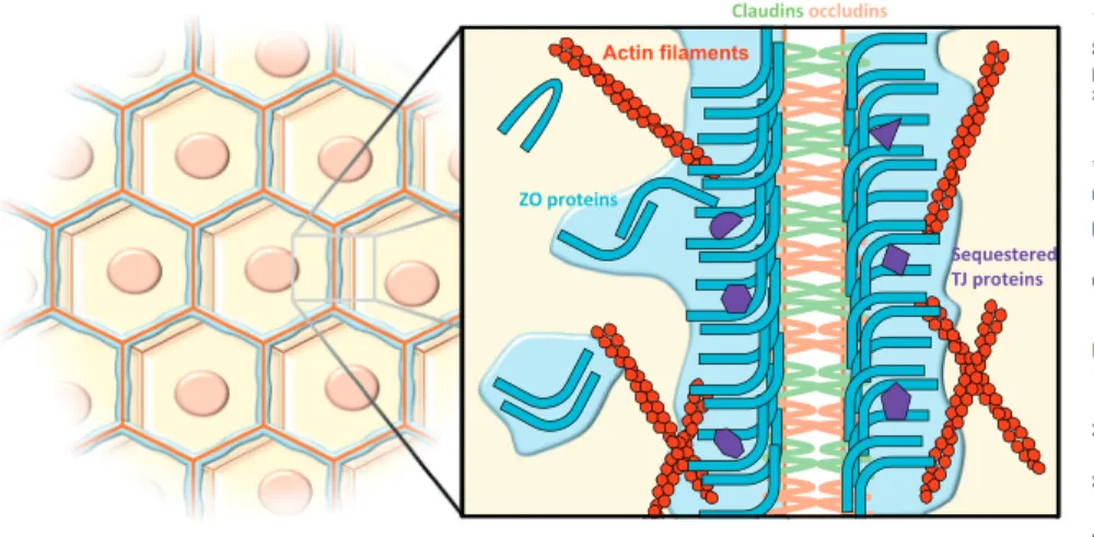
form such emulsions, and how liquid be-havior could make them special [3]. Today, hundreds of proteins have been identified as undergoing LLPS [4]. Two recent studies published in Cell now add the ZO proteins of TJs to the growing list of proteins that are capable of LLPS [5,6]. TJs are intercellular adhesion com-plexes that form a selective diffusion bar-rier between epithelial or endothelial cells, thereby regulating tissue permeability, a crucial function for embryogenesis and compartmentalization at the organ scale. In addition to transmembrane proteins that interact between adjacent cells, TJs require the assembly of scaffolding pro-teins on their cytoplasmic sides, among which are the ZO proteins (Figure 1).
By means of experiments in vitro and in cul-tured epithelial cells, Beutel et al. [5] have provided evidence that ZO proteins can form condensates that display liquid-like properties: their roundish shapesfluctuate, they coalesce, and their internal con-tents are highly mobile. In epithelial cells these condensates are not distinguishable from TJs at physiological concentrations, but can be revealed by cytoskeleton depolymerization or intercellular adhesion disruption. Both treatments disrupt the continuous TJ belt, and this breaks up into disconnected condensates reminis-cent of a fluid thread as a result of Rayleigh–Plateau instability. In gastrulating zebrafish embryos, Schwayer et al. [6] showed that, in the yolk syncytium, ZO proteins form non-junctional condensates that subsequently fuse with TJs at the boundary between the yolk syncytial layer and the enveloping cell layer. The shapes and sizes of these non-junctional conden-sates are externally constrained by the sur-rounding actin cytoskeleton meshwork, reminiscent of how the size and shape of a liquid are limited by its container. Beutel et al. used a collection of partial deletion mutants to demonstrate that
multivalent interactions in ZO1 underlie its phase separation (as in many other pro-teins [4]), and that these essentially involve its PSG (PDZ3-SH3-GuK) supradomain. These interactions are antagonized by the nearby U6 (unique-6) domain, and this autoinhibition is itself opposed by the long and mostly disordered C terminus. De-phosphorylation also promotes phase sep-aration, which thereby could be involved in density-dependent enhancement of epi-thelial cell barrier function [7]. In addition, Beutel et al. show that the C terminus pro-videsfluidity to the condensates. The C terminus also contains an actin-binding re-gion (ABR) that does not strongly influence phase separation but substan-tially contributes to C terminus-dependent fluidity. Within TJs, however, ZO proteins are less mobile than in ectopic or non-junctional condensates.
Functionally, ZO proteins are able to selec-tively accumulate diffusible interactants in in vitro condensates, ectopic condensates in TJ-less cells [5], and non-junctional condensates in zebrafish [6]. Therefore, ZO condensates act as compartments. At the tissue scale, Beutel et al. reveal that the ability of ZO1 protein to phase-separate scales with its abilities to accu-mulate at TJs and build an impermeable epithelial barrier in cysts, in a manner that is mostly independent of the ABR when grown in Matrigel [8]. In zebrafish, how-ever, a function for phase separation is un-clear, but Schwayer et al. show that the ABR is required for efficient incorporation of non-junctional clusters into TJs – which scales with actomyosin tension and retrogradeflow – as well as for normal gastrulation. Because the ABR provides both actin binding andfluidity to ZO con-densates, the respective contributions of each feature remain an open question in both contexts.
Addressing whether these liquid conden-sates result in functions that cannot be achieved by solid counterparts, and
Trends in Cell Biology, Month 2019, Vol. xx, No. xx 1
Trends in Cell Biology
identifying physiological or pathological cues that may cause this transition, are some of the next exciting steps. Indeed, a distinguishing feature of liquids is that their size and shape are instructed from outside; therefore, any functional benefit must outcompete the energy cost. This question is all the more valid for TJs where mechanosensitive stretching organizes ZO proteins into a 2Dfilm that is very different from the shapeless droplets typical of liquid condensates [9]. ZO proteins also function in the nucleus [10], and the findings of
Beutel, Schwayer, and colleagues are likely to have implications beyond cell–cell adhe-sion. In any case, they advance our under-standing of TJ biogenesis and assembly, and provide many exciting questions to be addressed in the future.
Acknowledgments
Our work is supported in part by the Centre National de la Recherche Scientifique (CNRS) and grants from the French National Research Agency (ANR). H.C. re-ceived support from La Ligue contre le Cancer (alloca-tion de recherche doctorale). We thank Mathieu Coppey for critical reading of the manuscript.
1
Université de Paris, Centre National de la Recherche Scientifique (CNRS), Institut Jacques Monod, 15 rue Hélène Brion, 75013 Paris, France
2
Equal contributions. *Correspondence:
[email protected](N. Borghi).
https://doi.org/10.1016/j.tcb.2019.12.002
© 2019 Elsevier Ltd. All rights reserved.
References
1. Wilson, E.B. (1899) The structure of protoplasm. Science 10, 33
2. Oparin, A.I. and Morgulis, S. (1938) The Origin of Life, Macmillan
3. Hyman, A.A. et al. (2014) Liquid–liquid phase separation in biology. Annu. Rev. Cell Dev. Biol. 30, 39–58
4. Li, Q. et al. (2019) LLPSDB: a database of proteins under-going liquid–liquid phase separation in vitro. Nucleic Acids Res. Published online September 6, 2019https://doi.org/ 10.1093/nar/gkz778
5. Beutel, O. et al. (2019) Phase separation of zonula occludens proteins drives formation of tight junctions. Cell 179, 923–936
6. Schwayer, C. et al. (2019) Mechanosensation of tight junc-tions depends on ZO-1 phase separation andflow. Cell 179, 937–952
7. Sallee, J.L. and Burridge, K. (2009) Density-enhanced phosphatase 1 regulates phosphorylation of tight junction proteins and enhances barrier function of epithelial cells. J. Biol. Chem. 284, 14997–15006
8. Odenwald, M.A. et al. (2017) ZO-1 interactions with F-actin and occludin direct epithelial polarization and single lumen specification in 3D culture. J. Cell Sci. 130, 243–259
9. Spadaro, D. et al. (2017) Tension-dependent stretching activates ZO-1 to control the junctional localization of its interactors. Curr. Biol. 27, 3783–3795
10. Bauer, H. et al. (2010) The dual role of zonula occludens (ZO) proteins. J. Biomed. Biotechnol. 2010, 402593
Trends
Trends inin Cell BiologyCell Biology
Figure 1. At Epithelial Cell Intercellular Contacts, Zonula Occludens (ZO) Proteins Condense into a Liquid-Like Phase To Assemble the Tight Junction (TJ) Belt.