HAL Id: hal-01310863
https://hal.sorbonne-universite.fr/hal-01310863
Submitted on 3 May 2016HAL is a multi-disciplinary open access
archive for the deposit and dissemination of sci-entific research documents, whether they are pub-lished or not. The documents may come from teaching and research institutions in France or abroad, or from public or private research centers.
L’archive ouverte pluridisciplinaire HAL, est destinée au dépôt et à la diffusion de documents scientifiques de niveau recherche, publiés ou non, émanant des établissements d’enseignement et de recherche français ou étrangers, des laboratoires publics ou privés.
Optimization of the pediatric head computed
tomography scan image quality: reducing dose with an
automatic tube potential selection in infants
Baptiste Morel, Aurélien Bouëtté, Pierre Lévy, Guillemette Antoni, François
Chalard, Eleonore Blondiaux, Hubert Ducou Le Pointe
To cite this version:
Baptiste Morel, Aurélien Bouëtté, Pierre Lévy, Guillemette Antoni, François Chalard, et al.. Op-timization of the pediatric head computed tomography scan image quality: reducing dose with an automatic tube potential selection in infants. Journal de Neuroradiologie / Journal of Neuroradiol-ogy, Elsevier Masson, 2016, �10.1016/j.neurad.2016.03.005�. �hal-01310863�
Optimization of the pediatric head CT scan image quality: reducing dose with an
automatic tube potential selection in infants
Baptiste Morel, MD1, Aurélien Bouëtté MP1, Pierre Lévy MD2, Guillemette Antoni MD3, François Chalard MD1, Eleonore Blondiaux1, Hubert Ducou Le Pointe MD, PhD1
1
Service de Radiologie, Hôpital Trousseau – Hôpitaux Universitaires de l’Est Parisien (Assistance Publique - Hôpitaux de Paris), Université Pierre et Marie Curie, Paris
2
Service de Santé Publique, Hôpital Tenon (Assistance Publique - Hôpitaux de Paris), Université Pierre et Marie Curie, Paris & Inserm UMR S 1136
3
INSERM CESP Centre de Recherche en Epidémiologie et Santé des Populations, U1018, F-94807, Villejuif, France
Corresponding author:
Baptiste Morel, baptistemorelaphp@gmail.com Service de Radiologie
Hôpital Armand-Trousseau 26 Avenue du Dr Arnold Netter 75012 Paris, France
Phone number: 0033 1 71 73 87 56 Fax number: 0033 1 44 73 65 11
Acknowledgments:
The authors thank John C. Scatarige and Kris Munroe for English language assistance and their friendly support.
Abstract
Purpose
The objective of our study was to evaluate the impact of an automatic tube potential selection (ATPS) on the delivered dose and image quality in unenhanced head CT scans of infants.
Material and Methods
Unenhanced head CT scans were acquired before and after the introduction of an ATPS in full automatic mode in 2 groups of 20 patients under 1 year of age. The delivered dose (CDTIvol) as the quantitative (Contrast-to-Noise Ratio) and qualitative (based on the European CT criteria) image quality were compared on the supra and infratentorial regions by three senior pediatric radiologists. Mann-Whitney and Fisher Exact tests were performed. An interobserver Fleiss’s Kappa agreement was calculated for each criterion.
Results
The use of an ATPS allowed a significant reduction in the delivered dose (-21%, p=0.0005) with no significant difference of the Contrast to Noise Ratio in supra (-5%, p=0.21) and infratentorial region (+16%, p=0.96). In all cases, reduction of dose was obtained with the same value of 100 kV. It maintained a good qualitative image quality (e.g., differentiation between grey and white matter in supra-tentorial region: p=0.470). The interobserver Fleiss's Kappa agreements were good to excellent.
An automatic tube potential selection is a tool that can significantly reduce the delivered dose by choosing the most appropriate tube voltage while maintaining image quality in unenhanced head CT scans of infants.
Keywords:
Pediatric head CT scan; automatic tube potential selection; radiation exposure, image quality
Research Paper
Introduction
Computed tomography (CT) is an extremely valuable diagnostic tool, and its use has rapidly increased over the past decade [1]. We must be vigilant, particularly to children: the increasing risks of leukemia and brain cancer are linked to an increase in radiation exposure, even probably at low dose [2,3]. At high doses, age at exposure greatly affects lifetime risk [4]. But it is still desirable to base decisions on clinical utility for the patient without resorting to individual cancer risk estimation [5]. Head and cervical CT scans represent 60% of the CT examinations done in children under 5 years of age [6]. Efforts are made to conduct only strictly indicated examinations. With our new 64 sections CT scanner and standard acquisition protocol, image quality has been good in daily examinations, with delivered dose lower compared to the national diagnostic reference levels [7]. However, difficulty in interpretation was sometimes encountered, particularly on the children under 1 year of age, due to a subjectively lower image quality. In infants, brain structures are small and the differentiation between grey and white matters is poor. Appropriateness of the scan (proper indication, no other technique as ultrasound or MRI available) associated with technique adjusted to the age
and size of the child (optimization) are important considerations [8–10]. In concert with the Image Gently CT program improvement [8] and practice of ALARA [11], we focused our attention on optimizing the image quality of the unenhanced head CT scan in this particular population.
Iterative reconstruction and automatic exposure control (CareDose4DTM) can significantly improve the reference image quality in a pediatric anthropomorphic whole body phantom [12]. An automatic tube potential selection (ATPS) (CARE kVTM, Siemens Healthcare, Forchheim, Germany) is designed to automatically choose the most appropriated tube voltage and to accordingly adjust intensity to deliver the lowest dose achievable while preserving a constant Contrast-to-noise ratio (CNR) [13]. The estimated dose is calculated based on a specific tube current time product curves for all of the voltage levels to determine the optimal dose efficiency. Using CARE kVTM requires first that the users pick a reference voltage, quality reference mAs, and a number from 1 through 12 reflecting the type of study that they are going to perform (i.e. unenhanced, CTA, etc.).
The purpose of this study was to evaluate the impact of an automatic tube potential selection during an optimization process of the image quality of unenhanced head CT scans in two groups of 20 infants.
Materials and Methods
Study Description
The local institutional review board approved this single center prospective study. Three senior pediatric radiologists blindly and successively examined 40 patients (2 groups of 20 patients) under 1 year of age who underwent unenhanced head CT scans with a 64-section multidetector row CT scanner (Somatom AS+, Siemens AG, Forchheim, Germany) from December 2014 to March 2015. The indications of the CT scan were traumatism, seizures,
and acute headache in emergency. Patients were not sedated. We evaluated the impact of an ATPS on both delivered doses, as well as the qualitative and quantitative measures of brain CT image quality.
Scanning protocols
A lateral scout image with 100kVp and 35 mA was obtained to define the scan area prior to helical CT imaging. The acquisition and reconstruction parameters are given in Table 1. All CT scans were operated with 1-s rotation time, 230-mm field of view, pitch of 0.6 and collimation of 128 × 0.6 mm. Automatic intensity modulation was always activated with quality reference mAs at 400mAs and strong level of modulation. Images were reconstructed with 2 mm section with a medium-smooth J30s kernel for soft-tissue brain images reconstruction. The iterative reconstruction strength (SAFIRE) was set at 3 on a scale of 5. The only modification between the first group and the second group of patients was the activation of an automatic tube potential selection (CARE kVTM, Siemens Healthcare, Forchheim, Germany) with full automatic mode. The cursor was positioned at 3, as recommended for unenhanced CT scan acquisition by the manufacturer, with the minimum possible tube voltage fixed at 80kVp. For the first group, the tube voltage was fixed at 120kVp, while for the second group the selected the tube voltage could vary between 120kVp and 80kVp
Dose assessment
All the following CT scan parameters were reported automatically via the Dose achieving and communication system (DACS) with a patient-dose monitoring software (DosewatchTM, GE): CTDIvol, patient age, tube voltage (kVp), and mean tube intensity (mA) for acquisitions. The accuracy of the displayed CTDIvol and DLP of the manufacturer was regularly tested by a quality control program from our institution with a 16 cm phantom.
Image quality assessment
Patients with images degraded by severe streak artifacts due to foreign bodies or movement of the head were excluded from the study.
Quantitative analysis
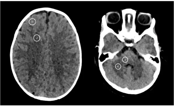
Signal intensity was defined as CT attenuation in Hounsfield units; image noise was determined to be the Standard Deviation (SD) of CT attenuation within a region of interest. Three repeated averaged CT attenuation measurements were acquired by using circular ROIs of 40 mm2 placed in normal cortical frontal anterior grey matter and adjacent normal white matter periventricular structure for the supratentorial parenchyma, and right cerebellar peduncle, cerebellar white matter for infratentorial structure, by using soft-tissue brain images reconstruction. ROIs were adjusted to fit the measured anatomic structure and to avoid volume-averaging artifacts (Figure 1).
Supratentorial contrast (C) was defined as the difference between signal intensity on the supratentorial frontal grey matter and signal intensity in the normal periventricular white matter.
𝐶𝑠𝑢𝑝𝑟𝑎𝑡𝑒𝑛𝑡𝑜𝑟𝑖𝑎𝑙 = 𝑆𝑠𝑢𝑝𝑟𝑎𝑡𝑒𝑛𝑡𝑜𝑟𝑖𝑎𝑙 𝑓𝑟𝑜𝑛𝑡𝑎𝑙 𝑔𝑟𝑒𝑦 𝑚𝑎𝑡𝑡𝑒𝑟 − 𝑆𝑝𝑒𝑟𝑖𝑣𝑒𝑛𝑡𝑟𝑖𝑐𝑢𝑙𝑎𝑟 𝑤ℎ𝑖𝑡𝑒 𝑚𝑎𝑡𝑡𝑒𝑟
Infratentorial contrast was defined as the difference between signal intensity on the right cerebellar peduncle and the right cerebellar white matter.
𝐶𝑖𝑛𝑓𝑟𝑎𝑡𝑒𝑛𝑡𝑜𝑟𝑖𝑎𝑙 = 𝑆𝑟𝑖𝑔ℎ𝑡 𝑐𝑒𝑟𝑒𝑏𝑒𝑙𝑙𝑎𝑟 𝑝𝑒𝑑𝑢𝑛𝑐𝑙𝑒− 𝑆𝑟𝑖𝑔ℎ𝑡 𝑐𝑒𝑟𝑒𝑏𝑒𝑙𝑙𝑎𝑟 𝑤ℎ𝑖𝑡𝑒 𝑚𝑎𝑡𝑡𝑒𝑟
Noises in the supratentorial region and in the cerebellum were calculated using the following formulas:
𝑁𝑠𝑢𝑝𝑟𝑎𝑡𝑒𝑛𝑡𝑜𝑟𝑖𝑎𝑙 = √𝑁𝑠𝑢𝑝𝑟𝑎𝑡𝑒𝑛𝑡𝑜𝑟𝑖𝑎𝑙 𝑓𝑟𝑜𝑛𝑡𝑎𝑙 𝑔𝑟𝑒𝑦 𝑚𝑎𝑡𝑡𝑒𝑟 2+ 𝑁𝑝𝑒𝑟𝑖𝑣𝑒𝑛𝑡𝑟𝑖𝑐𝑢𝑙𝑎𝑟 𝑤ℎ𝑖𝑡𝑒 𝑚𝑎𝑡𝑡𝑒𝑟2
𝑁𝑖𝑛𝑓𝑟𝑎𝑡𝑒𝑛𝑡𝑜𝑟𝑖𝑎𝑙 = √𝑁𝑟𝑖𝑔ℎ𝑡 𝑐𝑒𝑟𝑒𝑏𝑒𝑙𝑙𝑎𝑟 𝑝𝑒𝑑𝑢𝑛𝑐𝑙𝑒2+ 𝑁𝑟𝑖𝑔ℎ𝑡 𝑐𝑒𝑟𝑒𝑏𝑒𝑙𝑙𝑎𝑟 𝑤ℎ𝑖𝑡𝑒 𝑚𝑎𝑡𝑡𝑒𝑟2
Supratentorial CNR was calculated as the ratio between contrast and noise as previously defined [14,15] and was the primary quantitative analysis criterion.
𝐶𝑁𝑅𝑠𝑢𝑝𝑟𝑎𝑡𝑒𝑛𝑡𝑜𝑟𝑖𝑎𝑙 =
𝑆𝑠𝑢𝑝𝑟𝑎𝑡𝑒𝑛𝑡𝑜𝑟𝑖𝑎𝑙 𝑓𝑟𝑜𝑛𝑡𝑎𝑙 𝑔𝑟𝑒𝑦 𝑚𝑎𝑡𝑡𝑒𝑟 − 𝑆𝑝𝑒𝑟𝑖𝑣𝑒𝑛𝑡𝑟𝑖𝑐𝑢𝑙𝑎𝑟 𝑤ℎ𝑖𝑡𝑒 𝑚𝑎𝑡𝑡𝑒𝑟 √𝑁𝑠𝑢𝑝𝑟𝑎𝑡𝑒𝑛𝑡𝑜𝑟𝑖𝑎𝑙 𝑓𝑟𝑜𝑛𝑡𝑎𝑙 𝑔𝑟𝑒𝑦 𝑚𝑎𝑡𝑡𝑒𝑟 2+ 𝑁𝑝𝑒𝑟𝑖𝑣𝑒𝑛𝑡𝑟𝑖𝑐𝑢𝑙𝑎𝑟 𝑤ℎ𝑖𝑡𝑒 𝑚𝑎𝑡𝑡𝑒𝑟2
Infratentorial CNR was calculated as the ratio between contrast and noise.
𝐶𝑁𝑅𝑖𝑛𝑓𝑟𝑎𝑡𝑒𝑛𝑡𝑜𝑟𝑖𝑎𝑙 = 𝑆𝑟𝑖𝑔ℎ𝑡 𝑐𝑒𝑟𝑒𝑏𝑒𝑙𝑙𝑎𝑟 𝑝𝑒𝑑𝑢𝑛𝑐𝑙𝑒− 𝑆𝑟𝑖𝑔ℎ𝑡 𝑐𝑒𝑟𝑒𝑏𝑒𝑙𝑙𝑎𝑟 𝑤ℎ𝑖𝑡𝑒 𝑚𝑎𝑡𝑡𝑒𝑟
√𝑁𝑠𝑢𝑝𝑟𝑎𝑡𝑒𝑛𝑡𝑜𝑟𝑖𝑎𝑙 𝑓𝑟𝑜𝑛𝑡𝑎𝑙 𝑔𝑟𝑒𝑦 𝑚𝑎𝑡𝑡𝑒𝑟 2+ 𝑁𝑝𝑒𝑟𝑖𝑣𝑒𝑛𝑡𝑟𝑖𝑐𝑢𝑙𝑎𝑟 𝑤ℎ𝑖𝑡𝑒 𝑚𝑎𝑡𝑡𝑒𝑟2
Qualitative Analysis
All head CT scans were reviewed blindly in a native axial plan and displayed randomly with the free software ViewDEX at the same diagnostic workstation by 3 pediatric radiologists. ViewDEX is a Java software developed to randomly present anonymized images, without dose assessment, with the possibility of directly answering related questions on screen [16]. Before providing scores, the radiologists were trained for consensus regarding the image quality scoring system on 10 routine head CT examinations. Window-level settings were standardized for initial review (width 74; center 30). The viewing environment was constant.
The choice of structures was based on the structures defined in the European Guidelines on Quality Criteria for Computed Tomography [17] and on the scale previously used by Kilic et al. [18]. The choice concentrated on typical parameters of image quality, including differentiation between grey and white matter in the supra and infratentorial spaces,
delimitation of the peri-mesencephalic cerebrospinal fluid space, delimitation of the shape of the ventricular system, and visualization of the basal ganglia. Each criterion was on a 4-point scale: 0, unacceptable, 1, only acceptable under limited conditions, 2, probably acceptable and 3, fully acceptable. Results of the scoring were summarized on a text file that was automatically generated. We retained the score agreed by at least 2 radiologists if there was a discordance.
Statistical Analysis
The two groups of 20 pediatric CT scans were compared by using quantitative measurements and qualitative modal scores. Mann-Whitney tests were performed to compare age, cranial perimeter, mA, CTDIvol, Contrast, Noise and CNR on supra and infratentorial spaces between the 2 groups. For qualitative variables, a Kruskall Wallis test was performed. An interobserver Fleiss’s Kappa agreement was calculated for each criterion. A statistically significant difference was defined by p<0.05. We used R software to implement the statistical analyses [19].
Results
The two groups were homogenous, with no statistical difference in age and cranial perimeter. Main results are summarized in Table 2. All the CT scans were normal.
Acquisition Parameters and Dose Analysis
All acquisitions in group 1 were performed with tube voltage set at 120 kVp, while the tube voltage selected by CARE kVTM was 100 kVp for the second group in all cases. The activation of an automatic tube potential selection induced a significant decrease of the delivered dose CTDIvol (-21%, p=0.0005). The acquisition parameters that influenced the delivered dose and the mean quantitative ROI measurements are summarized in Table 3.
Quantitative Analysis
Supratentorial contrast and noise were not significantly increased (+6%, p=0.93 and +13%, p=0.097 respectively). Supratentorial CNR was not significantly modified when CARE kVTM was activated (-5%, p=0.21).
Infratentorial contrast was not significantly increased (+23%, p=0.43). Infratentorial noise was significantly increased (+12%, p=0.043). Infratentorial CNR was not significantly modified when CARE kVTM was activated (+16%, p=0.96).
Qualitative Analysis
Activation of the automatic tube potential selection did not induce significant modification in the qualitative analysis of image quality for all criterions (Table 4). The interobserver Fleiss's Kappa agreements were good to excellent (Table 5).
Discussion
Radioprotection for children is one of the major goals of radiologists. Pediatric head CT scans are requested in case of emergency, particularly in the context of head trauma and the search for an ischemic or hemorrhagic lesion. Minimizing the delivered dose while preserving image quality remains a priority. Our study suggests that an automatic tube potential selection is effective in maintaining quantitative and qualitative image quality while significantly reducing the delivered dose on unenhanced cranial CT scans in infants. This a new complementary information, not observed in the Wallace et al. study [20]. Our patients selection is higher than in previous published works: there were 9 children under 3 years of age reported in the McKnight study [21], Santos et al. studied 2 groups of 10 newborns [22]. Our results highlight that, with the use of an ATPS, before the age of one year, the tube
voltage could be lowered to 100 kV for unenhanced head CT. After one year of age, the tube current is often of 120 kV in unenhanced head CT scan [22]. Its use maintains image quality with a significant decrease in the delivered dose, by choosing the most appropriate tube voltage (100 kV in our population). Lowering the tube voltage settings increases contrast resolution (particularly in the infratentorial region) owing to the higher attenuation of lower-energy x-rays produced with this parameter. The ATPS has partially compensated the increase of noise due to the diminution of the voltage by increasing the current to maintain the CNR constant. We did not observe the increase of the noise induced by the shielding of the brain by the skull.
The use of an automatic tube potential selection can also allow having only one pediatric head CT scan protocol, which is easier to use in daily practice, whereas having different CT scan protocols depending on the age of the patients.
These results are consistent with those of previous studies on enhanced thoracic and abdominal [13] and adult [23] or pediatric [22] head CT scans. We focused our analysis on unenhanced pediatric head CT scans to avoid any variation related to the CT image acquisition and contrast injection protocols, which might have resulted in differences in radiation doses delivered. The good to excellent interobserver agreements allowed us to be confident in our qualitative image quality evaluation. In the majority of previous studies, there were generally 2 readers [3,18,21,24,25].
The selection size of our study was limited (40 patients), but allowed us to observe the impact of an ATPS without any age or cranial perimeter bias.
In conclusion, after defining an image quality reference using iterative reconstruction and automatic exposure control, an ATPS can significantly reduce the delivered dose in unenhanced cranial CT scans in infants by choosing the most appropriate tube voltage, while
objectively maintaining image quality, with the interest of a single automatic CT scan acquisition protocol.
Conflict of Interest:
The authors have no conflict of interest to declare.
Acknowledgments:
The authors thank John C. Scatarige and Kris Munroe for English language assistance and their friendly support.
References
[1] Brenner DJ, Hall EJ. Computed tomography--an increasing source of radiation exposure. N Engl J Med 2007;357:2277–84.
[2] Pearce MS, Salotti JA, Little MP, McHugh K, Lee C, Kim KP, et al. Radiation exposure from CT scans in childhood and subsequent risk of leukaemia and brain tumours: a retrospective cohort study. Lancet 2012;380:499–505.
[3] Mathews JD, Forsythe AV, Brady Z, Butler MW, Goergen SK, Byrnes GB, et al. Cancer risk in 680,000 people exposed to computed tomography scans in childhood or
adolescence: data linkage study of 11 million Australians. BMJ 2013;346:f2360. [4] Ozasa K, Shimizu Y, Suyama A, Kasagi F, Soda M, Grant EJ, et al. Studies of the
mortality of atomic bomb survivors, Report 14, 1950-2003: an overview of cancer and noncancer diseases. Radiat Res 2012;177:229–43.
[5] Rehani MM. I Am Confused About the Cancer Risks Associated With CT: How Can We Summarize What Is Currently Known? AJR Am J Roentgenol 2015;205:W2–3.
[6] IRSN. Exposition de la population française aux rayonnements ionisants liée aux actes de diagnostic médical en 2012. 2014.
[7] Vassileva J, Rehani M. Diagnostic reference levels. AJR Am J Roentgenol 2015;204:W1–3.
[8] Goske MJ, Applegate KE, Boylan J, Butler PF, Callahan MJ, Coley BD, et al. The Image Gently campaign: working together to change practice. AJR Am J Roentgenol 2008;190:273–4.
[9] Adamsbaum C, Rolland Y, Husson B. [Pediatric neuroimaging emergencies]. J Neuroradiol J Neuroradiol 2004;31:272–80.
[10] Nievelstein RAJ, van Dam IM, van der Molen AJ. Multidetector CT in children: current concepts and dose reduction strategies. Pediatr Radiol 2010;40:1324–44.
[11] Sodhi KS, Krishna S, Saxena AK, Sinha A, Khandelwal N, Lee EY. Clinical application of “Justification” and “Optimization” principle of ALARA in pediatric CT imaging: “How many children can be protected from unnecessary radiation?” Eur J Radiol 2015. [12] Söderberg M, Gunnarsson M. The effect of different adaptation strengths on image
quality and radiation dose using Siemens Care Dose 4D. Radiat Prot Dosimetry 2010;139:173–9.
[13] Siegel MJ, Hildebolt C, Bradley D. Effects of automated kilovoltage selection technology on contrast-enhanced pediatric CT and CT angiography. Radiology 2013;268:538–47.
[14] Mullins ME, Lev MH, Bove P, O’Reilly CE, Saini S, Rhea JT, et al. Comparison of Image Quality Between Conventional and Low-Dose Nonenhanced Head CT. Am J Neuroradiol 2004;25:533–8.
[15] Udayasankar UK, Braithwaite K, Arvaniti M, Tudorascu D, Small WC, Little S, et al. Low-dose nonenhanced head CT protocol for follow-up evaluation of children with ventriculoperitoneal shunt: reduction of radiation and effect on image quality. AJNR Am J Neuroradiol 2008;29:802–6.
[16] Håkansson M, Svensson S, Zachrisson S, Svalkvist A, Båth M, Månsson LG. VIEWDEX: an efficient and easy-to-use software for observer performance studies. Radiat Prot Dosimetry 2010;139:42–51.
[17] Menzel H, Schibilla H, Teunen D. European guidelines on quality criteria for computed tomography. Luxemb Eur Comm 2000;16262.
[18] Kilic K, Erbas G, Guryildirim M, Arac M, Ilgit E, Coskun B. Lowering the dose in head CT using adaptive statistical iterative reconstruction. AJNR Am J Neuroradiol
2011;32:1578–82.
[19] Team RC. R: A language and environment for statistical computing. R Foundation for Statistical Computing, Vienna, Austria, 2012. ISBN 3-900051-07-0; 2012.
[20] Wallace AN, Vyhmeister R, Bagade S, Chatterjee A, Hicks B, Ramirez-Giraldo JC, et al. Evaluation of the use of automatic exposure control and automatic tube potential selection in low-dose cerebrospinal fluid shunt head CT. Neuroradiology 2015;57:639– 44.
[21] McKnight CD, Watcharotone K, Ibrahim M, Christodoulou E, Baer AH, Parmar HA. Adaptive statistical iterative reconstruction: reducing dose while preserving image quality in the pediatric head CT examination. Pediatr Radiol 2014:1–7.
[22] Santos J, Foley S, Paulo G, McEntee M, Rainford L. The impact of pediatric-specific dose modulation curves on radiation dose and image quality in head computed tomography. Pediatr Radiol 2015:1–9.
[23] Othman AE, Afat S, Brockmann MA, Nikoubashman O, Brockmann C, Nikolaou K, et al. Radiation dose reduction in perfusion CT imaging of the brain: A review of the literature. J Neuroradiol 2015.
[24] Vorona GA, Zuccoli G, Sutcavage T, Clayton BL, Ceschin RC, Panigrahy A. The Use of Adaptive Statistical Iterative Reconstruction in Pediatric Head CT: A Feasibility Study. Am J Neuroradiol 2013;34:205–11.
[25] Rapalino O, Kamalian S, Kamalian S, Payabvash S, Souza LCS, Zhang D, et al. Cranial CT with Adaptive Statistical Iterative Reconstruction: Improved Image Quality with Concomitant Radiation Dose Reduction. Am J Neuroradiol 2012;33:609–15.
Figures captions
Figure 1: Regions of interest for the measurement of contrast and noise. On the left: supratentorial frontal grey matter (ROI 1) and periventricular white matter (ROI 2). On the right: right cerebellar peduncle (ROI 3) and right cerebellar white matter (ROI 4).
Figure 2: Examples of axial CT scan slices of the supratentorial space of the group 1 (image A, CTDIvol = 32.2 mGy) and the group 2 after activation of an automatic tube potential selection (image B, CTDIvol = 18.7 mGy).