British Journal of Nutrition (1998), 80, Suppl. 1 , S77-S112 577
Functional food science and defence against reactive oxidative species
A. T. Diplock’”, J.-L. Charleux2, G. Crozier-Willi3, F. J.
K0k4, C. Rice-Evans1, M. Roberfroid’,
W. Stah? and J. Viiia-Ribes7
’
International Antioxidant Research Centre, UMDS, Guy’s Hospital, St Thomas Street, London S E l 9RT, U K F. HofSmann-La Roche Ltd, Business Unit Carotenoids, Headofice Kaiseruugst V M l Building, CH04002 Basel, SwitzerlandNestec Ltd, Nestle‘ Research Center, Vers-Chez-La-Blanc, P O Box 44, CH-1000 Lausanne 26, Switzerland Division of Human Nutrition and Epidemiology, Wageningen Agricultural University, P O Box 8129, N L 6700 EV,
Wugeningen, The Netherlands
UCL, Ecole de Pharmacie, Tour Van Helmont, Avenue E. Mounier, B-1200 Brussels, Belgium
6Heinrich-Heine-University
Dusseldorf, Medizinische Einrichtungen, Institut f u r Physiologische Chemie I, Postfach 1 0 I0 07, 0 - 4 0 0 0 1 Dusseldorf, GermanyUniversidad de Valencia, Fucultad de Medicina, Departamento de Fisiologia, Avenida Blasco Ibajez 17, E-46010 Valencia, Spain
Contents
Introduction
Oxidative damage, antioxidant defence and the role of prooxidants in disease
2.1. Antioxidant defence system of the human organism 2.1.1 Origins and nature of free radicals and
other oxidants
2.1.2. Enzymic and non-enzymic defence systems in vivo
2.1.3. Dietary antioxidants: nutrient and non- nutrient
Oxidative damage to bodily functions and its implications in disease
2.2.1. Coronary heart disease 2.2.2. Carcinogenesis
2.2.3. Cataract and age-related macular degeneration
2.2.4. Neuronal diseases 2.2.
2.3. Conclusions
Available methodologies for evaluating and quantifying ex vivo damage to DNA, lipids and proteins by prooxidants in vivo
3.1. Oxidative damage to DNA
3. I . 1. Measurement of guanine damage products
in DNA by HPLC and gas chromatography-
mass spectrometry (GC-MS) 3.2. Oxidative damage to lipids
3.2. I Lipid peroxidation
3.2.2. Can some measure of ‘total’ peroxidation be obtained?
3.2.3. LDL oxidation
Measurement of antioxidant nutrients, carotenoids and javonoids extracted from human plasma 3.3. Oxidative damage to proteins
3.4. s79 S80 S80 S80 S80 S80 S82 S82 S83 S83 S83 S84 S84 S84 S84 S85 S85 S86 S86 S87 S87 3.5. Conclusions
antioxidant defence systems
4. Nutritional options modulating oxidative damage and
4.1. 4.2. 4.3. 4.4. 4.5. Introduction Dietary antioxidants
4.2.1. Sources of dietary antioxidants 4.2.2. Antioxidant intake and status 4.2.3. Bioavailability of antioxidants 4.2.4. Fat intake and antioxidant status Epidemiological studies on protective effects of
antioxidants
4.3.1. Cardiovascular disease 4.3.2. Cancer
4.3.3. Other age-related diseases
Human intervention studies of antioxidants 4.4.1. Cardiovascular disease
4.4.2. Cancer Conclusions
5. Potential safety implications related to antioxidant nutritional enhancement
5. I . Introduction 5.2. Vitamin C 5.3. Vitamin E 5.4. Carotenoids
5.5. Non-nutrient antioxidants (Jlavonoids and other related compounds)
5.5.1. Absorption
5.5.2. Possible adverse effects
6. Role of food technology in nutritional and safety aspects of antioxidants 6. I . Introduction 6.2. Physical processes 588 588 588 589 589 589 590 590 590 590 S9 1 S9 1 592 592 592 593 593 593 593 594 596 596 596 597 597 597 598
Abbreviations: AMD, age-related macular degeneration; ATBC study, a-tocopherol p-carotene study; FOX method, ferrous oxidation in xylenol orange
method; GC-MS, gas chromatography-mass spectrometry; 8-OHdG, 8-hydroxy-deoxyguanosine; 8-OHG, 8-hydroxyguanosine; 8-oxodG, 8-0x0-7,8-
dihydro-2’-deoxyguanine; PG, prostaglandin; PUFA, polyunsaturated fatty acids; RDA, recommended daily allowance; RNS, reactive nitrogen species;
ROS, reactive oxygen species; TBA, thiobarbituric acid; TBARS, TBA-reactive substances; a-TE, a-tocopherol equivalents. *Corresponding author: Professor A. T. Diplock, fax +44 (0) 171 403 7195, tel +44 (0) 171 955 4521.
S78 A. T. Diplock et al.
6.2.1. Structural integrity S98
6.2.2. Moisture content S98
6.2.3. Temperature S98
6.2.4. Minimizing oxygen s99
6.2.5. Protection from light s99
6.3. Chemical processes s99
6.3.1. Enzymes s99
6.3.2. Supplementation s99
6.4. Conclusions SlOO
7. Critical assessment of the science base and
conclusions SlOO
7.1. Identi$cation of criteria SlOO
7.2. Critical evaluation of the present knowledge base SlOl SlOl SlOl s102
6.2.6. Irradiation s99
7.2.1. Conclusions from section 2 7.2.2. Conclusions from section 3 7.2.3. Conclusions from section 4
7.2.4. Conclusions from section 5 7.2.5. Conclusions from section 6 7.3. Evaluation of criteria 7.4. Final conclusions s102 s102 s102 S103
8. Recommendations for future research S104
8.1. Introduction S104
8.2. Specific recommendations S104
8.2.1. Oxidative damage and antioxidant defence S104 8.2.2. Ex vivo methodologies for quantitating and
systems of the human organism validating damage in vivo to biological
macromolecules s104
damage S104
enhancement of antioxidants S105 8.3. Priorities for the recommendations made S105
8.2.3. Nutritional options modulating oxidative 8.2.4. Safety implications of nutritional
Abstract
This paper assesses critically the science base that underpins the argument that oxidative damage is a significant causative factor in the development of human diseases and that antioxidants are capable of preventing or ameliorating these disease processes. The assessment has been carried out under a number of headings, and some recommendations for future research are made based on the present day knowledge base.
The knowledge database
(1) Consideration of the basic science that underlies understanding of the role of free radicals in causing cellular pathologies, and the role of antioxidants in preventing this, shows that an imbalance of reactive oxygen species and antioxidant defence systems may lead to chemical modifications of biologically relevant macromolecules. This imbalance provides a logical pathobiochemical mechanism for the initiation and development of several disease states. Experimental data obtained in vivo provide evidence that antioxidants function in systems that scavenge reactive oxygen species and that these are relevant to what occurs in vivo. The relevance in vivo of these observations depends inter alia on knowledge of the uptake and distribution of the antioxidant within the human body, and on what tissue levels of the antioxidant may be expected in relation to dietary levels.
(2) There is some way to go until validated precise methods are available for measuring
biomarkers of oxidative damage in human subjects in vivo under minimally invasive conditions. With respect to oxidative damage to DNA, HPLC and GC-mass spectrophotometry methods
have both merits and limitations. Lipid oxidation products in plasma are best measured as isoprostanes or as lipid hydroperoxides using specific HPLC techniques. Development of isoprostane measurement will advance specificity and precision. The measurement of oxidative damage to proteins has some potential but such methods have not been effectively exploited. (3) Epidemiological studies support the hypothesis that the major antioxidant nutrients vitamin E and vitamin C, and 6-carotene (which may or may not be acting as an antioxidant in vivo), may play a beneficial role in prevention of several chronic disorders. More research is needed on the impact of other non-nutrient compounds, such as other carotenoids and flavonoids, on human health. In general, human intervention studies using hard end-points are the gold standard. Trials are restricted mainly to the major antioxidants and do not allow firm conclusions because of inconsistent findings, an insufficient number of studies and the use of varying doses. There is evidence that large doses of 6-carotene may be deleterious to the health of certain subgroups of the population such as heavy habitual smokers.
(4) With respect to the safety of administration of supplementary vitamins, vitamin C is safe at levels of supplementation up to 600 mg/d, and higher levels, up to 2000 mg/d, are without risk. Vitamin E has a very low human toxicity and an intake of lOOOmg/d is without risk; 3200 mg/d
has been shown to be without any consistent risk. Large intakes of &carotene must be viewed with caution because they have been shown to confer detriment to a population at high risk of lung cancer when administered after many years of high risk (smoking) behaviour. Until further work clarifies the situation in heavy smokers with respect to taking supplements, larger doses should be avoided by such individuals. There is little reliable information about the human toxicology of flavonoids and related non-nutrient antioxidant constituents of the diet.
Defence against reactive oxidative species s79 (5) The food industry has long experience in the control of oxidative damage in foods and this
experience can be used to advantage for the protection of food antioxidants which are beneficial. Some of these, such as vitamins C and E and &carotene, are well known, and strategies for their protection in foods are already exploited by food technologies. Food technology strategies for the preservation of those antioxidants which have been shown to be beneficial to health can be applied in a cost-effective manner.
Research needs
(1) The review of the available scientific database enables the identification of areas where further research is required. Improvement in dietary antioxidant intake in human populations is expected to result in lowering of the risk of a number of degenerative diseases. While desirable as an ultimate objective per se, the impact on public health and the resultant decrease in health-care costs make it imperative that substantial sums of money should be spent on research in this important area.
(2) Direct measurement of prooxidants in vivo is difficult or impossible. It is imperative to establish which are the critical free radical ‘hits’ that are the relevant ones in the aetiology of diseases. Are the processes examined really relevant to the disease causation? There is a need to identify which are the important antioxidants in terms of the maintenance of health, and what is their relationship to one another. Clarification is needed as to whether it is the antioxidant role of the substance that is important, or whether it is some other function, and the possible non- antioxidant effects of antioxidants, in particular with respect to modulation of gene expression, also need further research. The primary aim is to identify the active components in the overall system that promote health.
(3) Ex vivo methodologies for quantitating and validating damage in vivo to biological macromolecules urgently need further attention before meaningful work can proceed on providing evidence of the level of antioxidants needed to maintain health and well-being. It is necessary to refine and validate methods that are already available for measurement of oxidative damage in human subjects in a non-invasive manner. Validation in many centres by measurement of oxidative damage in the same biological material needs to be undertaken using the same methodology. There is a somewhat longer term need for development of techniques to be used ex vivo as measures of protein oxidation in vivo.
(4) Nutritional options modulating oxidative damage. For proper epidemiological research, as well as for human intervention studies, it would be desirable to put special emphasis on the following. (a) Chemical analysis of the antioxidant content of foods. (b) Studies of bioavailability of antioxidants from the diet, and the factors that influence the absorption, distribution and tissue uptake of the compounds and the likely impact of the antioxidants on metabolic processes. (c) Development and validation of biomarkers of intermediate end-points, both biological response markers and early disease markers, and emphasis on the relevance of the biomarker to the disease end-point as well as the disease process. (d) Application of the validated biomarkers of intermediate end-points in randomized controlled trials testing the efficacy of antioxidants in functional foods for the maintenance of health and well-being.
(5) Safety implications of nutritional enhancement of antioxidants. The detailed evidence that is already available which demonstrates that vitamin C and vitamin E are safe at quite high levels of inclusion in the human diet, means that is unnecessary to recommend further work in this area. The safety of p-carotene was not questioned before the results of the Finnish and American intervention studies, which showed an apparent exacerbation in the incidence of lung cancer in heavy smokers who were given supplements of /3-carotene. This observation needs urgent clarification. With respect to the flavonoids and other polyphenols, it is likely that their bioactivity will be explored, and the key question of their bioavailability clarified, in the near future. It will be necessary to examine the safety of such bioflavonoid compounds.
(6) With regard to the role of food technology, there is no particular direction that research needs to take at the present time. Developments in food technology will be based on, and adapt to, nutritional recommendations resulting from biologically driven work.
Prooxidants: Antioxidants: Oxidative damage: Diet and health
‘Good health is more than the mere absence of disease’. Section 2 reviews the basic science concerning free-radical
Mark Twain damage and the ameliorating role at the cellular level of
antioxidants, and introduces the concept of maintenance of health, and the prevention of some major human degenera- tive diseases. In order to assess the extent of free-radical 1. Introduction
This paper presents a comprehensive assessment of the damage in human subjects in vivo, and the modifying effects literature to the end of December 1997 relevant to the role of antioxidants, it is necessary to have valid, precise and importance of dietary antioxidants in human health. biomarkers. Present literature on this topic is reviewed in
S80 A. T. Diplock et al. section 3. An account is given in section 4 of the human
epidemiological and interventional evidence that links a high intake of dietary antioxidants with a low risk of degenerative disease. If it should be proven with reasonable certainty that antioxidants do indeed lower the risk of human degenerative disease, it is essential to be sure that intervention with antioxidants in the diet, by fortification or supplementation of foods, is entirely free from harmful side- effects; this important topic is addressed in section 5, and the possible contribution of the food industry and food technologists to this enhancement is discussed in section 6. The 7th and 8th sections assess the ‘state of the art’ with respect to the foregoing sections and make recommenda- tions for research in the immediate future.
2. Oxidative damage, antioxidant defence and the role of prooxidants in disease
2.1. Antioxidant defence system of the human organism 2.1.1. Origins and nature of free-radicals and other oxidants. The development and existence of an organism in the presence of O2 is associated with the generation of reactive oxygen species (ROS), even under physiological conditions. ROS are responsible for the oxidative damage of biological macromolecules such as DNA, carbohydrates and proteins (Halliwell & Gutteridge, 1989; Sies, 1991; Halliwell, 1996). These processes are discussed as pathobiochemical mechanisms involved in the initiation or progression phase of various diseases (Diplock, 1994; Wiseman & Halliwell, 1996). Some of the most relevant ROS are: peroxyl radicals (ROO’), the nitric oxide radical PO’), the superoxide anion radical (Oi-), singlet oxygen ( 02), peroxynitrite (ONOO-), and hydrogen peroxide (H202). ROS are either radicals (molecules that contain at least one unpaired electron) or reactive non- radical compounds, capable of oxidizing biomolecules. Therefore, these intermediates are also called oxidants or prooxidants (Halliwell & Gutteridge, 1989; Sies, 1991).
There are various sources for specific ROS in the human organism. However, the superoxide radical anion appears to play a central role, since other reactive intermediates are formed in reaction sequences starting with 0,. It is generated by enzymic one-electron reduction of 0 from xanthine oxidase (EC 1.2.3.2), NADPH oxidase, or by leakage of the respiratory chain. It has been estimated that about 1-3 % of the O2 we utilize is converted to 0;- (Fridovich, 1986; Halliwell, 1996).
H 2 0 2 is a non-radical reactive species and can easily diffuse between living cells. It is efficiently converted to water by the enzyme catalase (EC 1.1 1.1.6), a process which determines its half-life. Recent evidence suggests that H 2 0 2 is involved in signal transduction regulating the expression of genes through the nuclear factor KB and apoprotein-1 pathways (Schreck & Baeuerle, 1994; Sen & Packer, 1996). The most reactive species is the hydroxyl radical with an estimated half-life of about lop9 s. It might be formed in vivo on high-energy irradiation (e.g. X-rays) by homo- lytic cleavage of body water or from endogenous H202 in metal-catalysed processes (Fenton reaction: Fe-catalysed Haber-Weiss reaction). u.v.-Light is insufficiently energetic to split water but it can cleave H 2 0 2 to yield
two molecules of the hydroxyl radical. The high reactivity of this radical implies immediate reaction at the place where it is generated.
The peroxyl radical (ROO’) is relatively long lived (seconds) with a considerable diffusion pathlength in bio- logical systems. It can be generated in the process of lipid peroxidation which is initiated by the abstraction of an H atom from polyunsaturated fatty acids (PUFA); the hydro- xyl radical is capable of starting this reaction sequence (Esterbauer et al. 1992; Reaven & Witzum, 1996).
Further products generated in lipid peroxidation are alkoxyl radicals (RO’) and organic hydroperoxides (ROOH). The latter might rearrange to endoperoxide inter- mediates which are cleaved to yield aldehydes. The reaction of aldehydes with amine groups of proteins has been discussed as a mechanism involved in the modification of the protein part of lipoproteins.
Singlet molecular oxygen (lo2) is another non-radical ROS which is suggested to be formed in vivo in light- exposed tissue. Its half-life has been estimated to be s depending on the nature of the surrounding matrix. O2 can interact with other molecules either by transferring its excitation energy or by combining chemically. Preferential targets for chemical reactions are double bonds; e.g. in PUFA or guanine in DNA bases (Kanofsky, 1989; Stahl & Sies, 1993; Cadet et al. 1994).
An interesting ROS which has attracted attention within the past few years is the nitric oxide radical (NO’). It is a signalling compound formed enzymically from arginine and relaxes smooth muscles in blood-vessel walls resulting in lowered blood pressure. It is also produced by activated macrophages contributing to the primary immune defence. An excess of NO’ is cytotoxic. It might react directly with biomolecules or combine with 0;- to form peroxynitrite (ONOO-). Peroxynitnte is capable of inducing lipid per- oxidation in lipoproteins but might also interfere with cellular signalling by nitrating tyrosine residues in proteins (Beckman, 1996; Packer, 1996).
The ROS described here, and also the biological path- ways for their endogenous formation, are examples of a whole class of reactive intermediates and their ways of generation. It should further be noted that the organism is also exposed to ROS from external sources. With the diet many compounds of prooxidant nature, such as quinones capable of redox cycling, are delivered to the organism. Also an array of radicals are inhaled with cigarette smoke; ozone, of which increasing levels are reported due to
air
pollution, isan
ROS which can oxidize lipids (Pryor et al. 1995).
ROS are also produced in the organism as a part of the primary immune defence. Phagocytic cells such as neutro- phils, monocytes, or macrophages defend against foreign organisms by synthesizing large amounts of 0;- or NO as a part of their killing mechanism. Several diseases are accom- panied by excessive phagocyte activation resulting in tissue damage which is at least in part due to the activity of ROS. 2.1.2. Enzymic and non-enzymic defence systems in vivo. To counteract the prooxidant load a diversity of antioxidant defence systems are operative in biological systems including enzymic and non-enzymic antioxidants. An antioxidant has been defined as ‘any substance that, when present in low concentrations compared to that of an
Defence against reactive oxidative species S81
oxidizable substrate, significantly delays or inhibits the oxidation of that substrate’ (Halliwell & Gutteridge, 1989; Sies, 1993; Halliwell, 1995).
The major enzymes directly involved in the detoxification of ROS are superoxide dismutase (EC 1.15.1. l), scavenging Oi-, as well as catalase and glutathione peroxidases (EC 1.1 1.1.9) which reduce H202 and organic hydroperoxides respectively. Several subtypes of glutathione peroxidase are Se-dependent. In animal studies an elevated intake of Se was associated with protective effects against cancer. Its preventive effects in man are still under investigation (Levander & Burk, 1996). Indirect antioxidant functions are mediated by enzymes that restore endogenous anti- oxidant levels; e.g. GSH levels are replenished on reduction of GSSG by glutathione reductase (EC 1.6.4.1). Further, reactive intermediates produced in reactions of prooxidants and biological molecules (e.g. epoxides) are conjugated by phase I1 detoxification enzymes such as g1utathione-S- transferases (EC 2.5.1.18) to favour their excretion. Another strategy to prevent the formation of ROS is the control of the levels of free Fe or Cu ions. Metal-binding proteins respon- sible for the transport of these ions bind them tightly, thus preventing the initiation of lipid peroxidation or DNA damage. Some of the most relevant metal-binding proteins are ferritin, transferrin, and caeruloplasmin.
Various endogenous low-molecular-mass compounds are also involved in antioxidant defence. GSH, the major cytosolic thiol, serves as a cofactor for several detoxifying enzymes (glutathione peroxidases, glutathione-S-trans- ferases), is involved in the reduction of protein disulfides and additionally scavenges ROS, being oxidized to GSSG. Other endogenous compounds such as ubiquinol- 10, urate, or bilirubin also exhibit antioxidant activities (Jacob & Burri, 1996).
2.1.3. Dietary antioxidants; nutrient and non-
nutrient. The human diet contains an array of different
compounds that possess antioxidant activities or have been suggested to scavenge ROS based on their structural properties. The most prominent representatives of dietary antioxidants are ascorbate (vitamin C), tocopherols (vitamin E), carotenoids, and flavonoids. Apart from vitamin C, each group of these antioxidants consists of a number of structurally different compounds; e.g. more than 600 different carotenoids have been identified to date and about fifty of them might occur in the human diet (Sies & Stahl, 1995; Rice-Evans & Miller, 1996; Rock et al. 1996). In the diet, there may be synergistic effects of these various dietary compounds which are difficult to assess at present. Indeed, the diet may be considered as an orchestra where interactions between constituents may bring about effects which are not the necessary properties of the individual constituents.
Vitamin C is considered to be one of the most powerful, least toxic natural antioxidants (Bendich et al. 1986; Weber
et al. 1996). It is water-soluble and is found in high
concentrations in many tissues; human plasma contains about 60pmol ascorbateA. On interaction with ROS it is oxidized to dehydro-ascorbate via the intermediate ascorbyl free radical. Dehydro-ascorbate is recycled back to ascorbic acid by the enzyme dehydro-ascorbate reductase. Thus, dehydro-ascorbate is found in only very low levels
compared with ascorbate. As a scavenger of ROS ascorbate has been shown to be effective against the superoxide radical anion, H202, the hydroxyl radical, and singlet oxygen. In aqueous solutions vitamin C also scavenges reactive nitrogen oxide species efficiently, preventing the nitrosation of target molecules. The major sources of ascorbate in the diet are fruits, especially citrus fruits, kiwi fruit, cherries and melons, and vegetables such as tomatoes, leafy greens, broccoli, cauliflower, Brussels sprouts, and cabbage; its content might exceed 100mg ascorbate/100 g fresh weight. At low dose levels (100 mg) the bioavailability values for vitamin C from synthetic and food sources are very similar (Mangels et al. 1993a); the efficacy of absorption decreases with increasing dose levels (Levine et al. 1996). There is evidence from studies in vitro that vitamin C is capable of regenerating tocopherol from the tocopheroxyl radical which is formed on inhibition of lipid peroxidation by vitamin E (Niki et al. 1982, 1985). This process would allow for the transport of a radical load from a lipophilic compartment to an aqueous compartment where it is taken care of by efficient enzymic defence systems. It should be noted, however, that ascorbate might also act as a prooxidant in vivo. In the presence of free transition metal ions (Fe and Cu) and ascorbate the hydroxyl radical can be generated and initiation of lipid peroxidation may occur. However, the amounts of free transition metals
in vivo are very small because they are efficiently bound to
proteins. Vitamin C has additional well-established biolo- gical functions including cofactor-activity for several important enzymes (Levine et al. 1996).
The term vitamin E is a generic description for all tocols and tocotnenol derivatives which exhibit the biological activity of a-tocopherol (Parker, 1989; Eldin & Appelqvist, 1996; Sokol, 1996; Traber & Sies, 1996). This group of compounds is highly lipophilic, and operative in membranes or lipoproteins. Their most important antioxidant function appears to be the inhibition of lipid peroxidation, scaven- ging lipid peroxyl radicals to yield lipid hydroperoxides and a tocopheroxyl radical. The latter is less reactive than peroxyl radicals towards neighbouring PUFA and acts as a chain-breaking antioxidant. The tocopheroxyl radical might be either reduced by ascorbate and GSH or further oxidized to the respective quinone. Since only small amounts of tocopheryl quinone are detectable in human blood and tissues, the regenerative pathway in vivo appears to be favoured. In comparison with other lipophilic antioxidants, a-tocopherol is probably the most efficient in the lipid phase (Niki, 1987). It contains shielding methyl groups adjacent to the phenolic hydroxyl group and it is optimally positioned in membranes by its phytyl side-chain, which is located in the hydrophobic region of the membrane structure. In addition to its peroxyl-radical scavenging properties, further interac- tions with ROS have been described, including quenching of singlet oxygen and interaction with peroxynitrite. The richest sources of vitamin E in the diet are vegetable oils (soyabean, maize, cottonseed, and safflowerseed), and pro- ducts made from these oils such as margarine and mayon- naise. Further, wheat germ, nuts, and some green leafy vegetables contribute considerable amounts to the vitamin E supply (Parker, 1989). Vitamin E plasma levels in man are about 22 pmolA; the compound is also found in tissues such
S82 A. T. Diplock et al. as liver, kidney, fat and adrenals. In the liver the RRR-
isomer of a-tocopherol is preferentially incorporated into VLDL which are further catabolized in the circulation. Thus, RRR-a-tocopherol is the major form of vitamin E in LDL (Traber & Sies, 1996).
Carotenoids are natural colourants with pronounced anti- oxidant activity (Stahl & Sies, 1993; Olson & Krinsky, 1995). Their chemical properties are closely related to the presence of an extended system of conjugated double bonds which is substituted with various endgroups. ROS which are efficiently scavenged by carotenoids are
lo2
and peroxyl radicals (Palozza & Krinsky, 1992). Two different pathways are operative with respect to the deactivation oflo2:
physical and chemical quenching (Truscott, 1990). Physical quenching implies the deactivation oflo2
by energy trans- fer from the excited oxygen species to the carotenoid, yielding a triplet excited carotenoid. The energy of the excited carotenoid is dissipated through vibrational interac- tions with the solvent to recover ground state carotenoid. The carotenoid remains intact in this process and might undergo further cycles of deactivation. Chemical quenching contributes less than 0.05% to total '02-quenching by carotenoids but is responsible for the eventual destruction of the molecule. Carotenoids are the most efficient naturally occumng quenchers for lo2 with quenching rate constants of about 5-12 x 109/m01 per s. Carotenoids were reported to scavenge peroxyl radicals by chemical interaction (Kennedy & Liebler, 1992). It is suggested that carotene radical intermediates are formed in this process which finally leads to the destruction of the molecule. Like vitamin E, carotenoids belong to the group of lipophilic antioxidants present in lipoproteins such as LDL and HDL. It has been shown that they are consumed when isolated LDL is exposed to the process of lipid peroxidation. Their contribu- tion to the antioxidant defence system of LDL is not clear, since no regeneration pathways for oxidized carotenoids are known at present. A variety of structurally different caroten- oids are present in fruits and vegetables. Some of the major sources are carrots (a-carotene, @-carotene), tomatoes (lycopene), citrus fruits (0-cryptoxanthin), spinach (lutein), and maize (zeaxanthin) (Mangels et al. 1993b). The absorption and transport processes of carotenoids are quite complex. Several factors influencing carotenoid bio- availability from food, such as co-ingestion of fat or fibre, cooking or food processing, have been identified (Erdman et al. 1993).Flavonoids are a large group of polyphenolic antioxidants that occur in several fruits, vegetables, and beverages such as tea, wine and beer mainly as 0-glycosides. They are efficient antioxidants capable of scavenging radical species (peroxyl radicals, hydroxyl radical, Oi-) forming a phenoxy radical (Rice-Evans et al. 1995; Rice-Evans & Miller, 1996). The term flavonoids summarizes a number of structurally different subgroups including flavanols (cate- chin, epicatechin), flavonols (quercetin, myricetin, kaem- pherol), flavanones (naringenin, taxifolin), flavones (apigenin, hesperetin), isoflavones (genestein), or antho- cyanidins (cyanidin, malvidin). Several criteria for optimal radical scavenging properties of flavonoids have been postulated based on pulse radiolysis studies. These include the presence of the 3',4'-dihydroxy structure in ring B, the
presence of the 2,3-double bond in conjugation with the 4- 0x0-group in ring C, and the presence of a 5-hydroxyl group in ring A with a 3-hydroxyl group and a 4-0x0 function in the C-ring. The antioxidant properties of flavonoids have been investigated in various studies in vivo and in vitro. It should be mentioned, however, that the bioavailability of these compounds is rather poor. They are rapidly conjugated in phase I1 detoxification reactions and levels of free flavonoids in human plasma are very low. Further phenolic compounds with antioxidant activity are derivatives of cinnamic acid; e.g. caffeic acid, chlorogenic acid, and ferulic acid (Rice-Evans & Miller, 1996).
In addition to the flavonoids, a number of other phenolic compounds of potential interest occur in foods. Thus, olive oil contains a number of phenolic substances, notably the o-
diphenol tyrosol, which may contribute to the antioxidant content of diets rich in olive oil (Kiritsakis, 1990). Similarly, plants of the Lamiaceae family, notably rosemary, oregano, sage, mint and thyme, contain a range of potential antiox- idants such as carnosol, rosemanol and carvacrol, which can contribute to the antioxidant potential of the diet (Lagouri & Boskou, 1996). As with the flavonoids, however, little is known of the human absorption and tissue distribution of these compounds.
Several other dietary constituents might also be involved in the antioxidant defence system either by direct action as antioxidants or by effects related to the induction of detox- ifying enzymes. Enzymes such as glutathione peroxidase and superoxide dismutase, which require a dietary supply of Se, and of Cu and Zn respectively, contribute to the overall oxidative defence mechanism. Some endogenous sub- stances such as urate also add to the antioxidant potential of living cells, although their significance is only speculat- ive. Enhancement of dietary intake of the minerals identified may be beneficial when their content in the diet is low.
2.2. Oxidative damage to bodily functions and its implications in disease
ROS are suggested, or known, to be involved in pathogenic processes of numerous diseases (Esterbauer et al. 1992; Luis & Navab, 1993; Diplock, 1994; Sies, 1997) such as cardiovascular disease, some forms of cancer, cataract, age-related macular degeneration, rheumatoid arthritis and a number of neurodegenerative diseases. Oxidative damage to important biomolecules is a deleterious pathway, but also influences of ROS on gene regulation or the immune system might impair bodily functions. There is increasing evidence from clinical and intervention studies, as well as from basic research that antioxidants might prevent or delay the development of disease states. There may also be particular population groups that will benefit from enhanced antioxidant intake, such as pregnant women, neonates and children, senior citizens and, perhaps, sportspeople.
The primary cause for most cardiovascular diseases is thought to be arterio- sclerosis, a multifactorial disease of the artery wall. It is suggested that in the early stages of arteriosclerosis lipid deposits, so-called fatty streaks, are formed in the subendothelial space. There is increasing evidence that
Defence against reactive oxidative species S83 oxidative stress, particularly oxidation of LDL, is a risk
factor and plays a role in the pathogenic pathway (Berliner & Heinecke, 1996). LDL oxidation is due to a lipid peroxidation reaction initiated by free radicals. Separate investigations of the lipid and protein parts of oxidized LDL demonstrated that oxidative modifications of both contrib- ute to the proatherogenic properties of oxidized LDL. Several biochemical mechanisms underlying this effect have been discussed. These include the formation of foam cells on the uptake of oxidized LDL via the scavenger receptor by macrophages resident in the subendothelial area, release of cytotoxic lipid peroxidation products from oxidized LDL, or chemoattractant properties of the oxidized lipoprotein. LDL oxidation is efficiently inhibited by lipophilic antioxidants of which a-tocopherol appears to be the most important. Epidemiological studies suggest preventive effects towards atherogenic lesions to be associated with an increased uptake of lipophilic antiox- idants such as vitamin E or carotenoids (Rimm et al. 1993). Additional effects of RRR-a-tocopherol, independent of its antioxidant activity, have been related to the protective properties of this compound. An early event in the onset of arteriosclerosis is the migration of smooth-muscle cells from the media to the intima of the arterial wall followed by proliferation of these cells. There is increasing evidence that RRR-a-tocopherol acts as a negative regulator of smooth-muscle cell proliferation via modulation of protein kinase C activity. Protein kinase C is an important element in the signal transduction cascade mediated by growth factors such as platelet-derived growth factor which are involved in the control of cell proliferation. It should be noted that these effects are limited to RRR-a-tocopherol; RRR-0-tocopherol does not inhibit protein kinase C (Azzi et al. 1995; Ozer et al. 1995).
2.2.2. Curcinogenesis. Carcinogenesis is a complex multistep process including initiation, promotion and progression. The generation of ROS is thought to be linked to tumourigenesis at different levels. Oxidative damage to DNA has been demonstrated in vitro and in
vivo leading to DNA single or double strand breaks and
DNA cross linking, as well as to chromosomal aberrations such as breakage or rearrangement. Modified DNA bases (e.g. hydroxythymidine, or hydroxyguanine) have been determined after exposure of cells to situations of oxidative stress. The modification of DNA bases might result in point mutations, deletions, or gene amplification as a first step of carcinogenesis. Further, ROS are capable of deactivating detoxifying enzymes responsible for the scavenging of potent carcinogens. Data from epidemiological studies support the idea that antioxidants are preventive in carcinogenesis by scavenging ROS (Flagg et al. 1995).
Carotenoids exhibit further biological functions which are not related to their antioxidant activities but might be of importance with respect to their cancer preventive effects (Gerster, 1995). It has been shown that provitamin A and non-provitamin A carotenoids are capable of inhibiting the growth of transformed fibroblasts (Bertram & Bortkiewicz, 1995). There is increasing evidence that growth arrest is due to the stimulation of gap-junctional communication between transformed and surrounding normal cells. These findings suggest that carotenoids or carotenoid-derived
retinoids play a role in intercellular signalling involved in growth control. Inhibitory effects of &carotene and lyco- pene on cell proliferation have also been described for several human cancer cell lines (Sharoni & Levy, 1996).
2.2.3. Cataract and age-related macular degeneration.
Oxidative damage and impaired vision have been discussed in the context of two ophthalmological diseases of the elderly, cataract and age-related macular degeneration (AMD) (Schalch, 1992; Taylor, 1993). Senile cataract indicates the opacity of ocular lenses. Lens proteins are extremely long- lived and often show oxidative damage. This is not surprising, since they are subjected to chronic exposure to light and 02, which is likely to be responsible for the formation of ROS which might react with lens proteins. As a consequence, the damaged proteins may aggregate and precipitate, thus losing their regular function. Supplementation studies support the hypothesis that a higher intake of vitamins including vitamin C and vitamin E prevents or delays the development of cataracts (Seddon et al. 1994b).
AMD is the major cause of visual impairment in Western countries and affects the anatomical region of the retina with the highest degree of visual activity. The macular pigment (yellow spot) represents a colour filter through which light must pass before detection. The carotenoids lutein and zeaxanthin are the predominant pigments in this area (Land- rum, 1997). Carotenes (hydrocarbon carotenoids) are not present in the yellow spot. The function of the macular pigment has not been unequivocally identified but it might protect against photo-oxidation by blue light, mediated by excited triplet state molecules, lo2, or superoxide. There are hints from food-frequency questionnaires that an increased consumption of food rich in lutein and zeaxanthin is associated with a diminished risk of AMD (Seddon et al. 1 9 9 4 ~ ) . Carotenoids are the most efficient natural com- pounds scavenging O2 and excited triplet state molecules. Growing data from experi- mental models and human brain studies add evidence that oxidative stress might play a role in the development of neuronal degeneration related to diseases such as Parkin- son’s disease, amyotrophic lateral sclerosis, and Alzhei- mer’s disease (Kondo, 1996; Simonian & Coyle, 1996). ROS are capable of inducing both necrosis and apoptosis. As a consequence of lipid peroxidation membrane rupture might occur or ion gradients, operative over compartments which are separated by membranes, might be disturbed. Neurons might undergo necrotic cell death as has been demonstrated in cell culture following depletion of intracellular GSH, the major endogenous antioxidant thiol. NO has been hypothesized to be an important mediator of neuronal death under pathological conditions. The ultimate species responsible for NO toxicity may be peroxynitrite which is formed by the reaction of the NO-radical with the superoxide radical.
Beyond the classical aspects of oxidative damage to biologically relevant molecules as pathological mechanisms underlying several diseases, and the protective effects of antioxidants, new fields of research in this area are rapidly developing. This includes effects of prooxidants and anti- oxidants on immune functions (Bendich, 1990) and antiox- idant and redox regulatory properties on gene expression. Both mechanisms may be involved in the development of
S84 A. T. Diplock et al. disease states while protection might be provided by
antioxidants via these pathways. 2.3. Conclusions
An imbalance between ROS and antioxidant defence sys- tems may lead to chemical modifications of biologically relevant macromolecules like DNA, proteins or lipids which are possible pathobiochemical mechanisms in the initiation or development of several disease states. Experimental data provide evidence that dietary antioxidants scavenge ROS and are useful in the prevention of these diseases. Epi- demiological studies clearly show a correlation between the increased consumption of food rich in antioxidants and a decreased risk of several diseases. Thus, an increased intake of fruits and vegetables can be recommended. Data on antioxidant supplementation are contradictory. Further research is necessary to establish whether supplementation beyond dietary intake levels is of benefit.
3. Available methodologies for evaluating and quantifying ex vivo damage to DNA, lipids and proteins
by prooxidants in vivo 3.1. Oxidative damage to DNA
The most abundant base alteration induced in DNA by ROS is the formation of 8-0~0-7,8-dihydro-2’-deoxyguanine (8- oxodG). In vivo this DNA base alteration is repaired by excision and the resulting product 8-oxodG is excreted unchanged, and independently of diet, into the urine. Thus, the rate of excretion of 8-oxodG (as given by the appearance of the metabolite in urine with time) serves as a biological marker of the integrated rate of oxidative DNA damage in the whole body.
DNA damage is usually measured in lymphocytes iso- lated from blood or in urine. Baseline levels of DNA damage are considered to be important because repair may be incomplete (one damaged base per lo6 bases); the actual measurement may therefore provide an estimate of the balance between damage and repair, so the time window is a crucial consideration here. From studies in vitro it is known that when DNA is exposed to the activated oxygen species the product is specific to the oxygen species involved, thus:
lo2, ROO’
-
guanine oxidized ‘OH-
multiplicity of changes to all four basesO,, H202
-
no base changesONOO-
-
xanthine, hypoxanthine, 8-nitroguanine. There are two types of measurement of oxidative DNA damage. First, steady-state damage can be measured when DNA is isolated from human cells and tissues and analysed for base damage products: it presumably reflects the balance between damage and DNA repair. Hence a rise in steady- state oxidative DNA damage (e.g. as has been reported in some human cancerous tissues; Malins & Haimonot, 1991; Olinski et al. 1992) could be due to increased damage and/or decreased repair. Second, several DNA base damage pro- ducts are excreted in human urine, including the nucleoside8-hydroxy-deoxyguanosine (8-OHdG), 8-hydroxy-adenine and 7-methyl-8-hydroxyguanine (Ames, 1989; Stillwell et
al. 1989) but the one most exploited is 8-OHdG, usually measured by a method involving HPLC with electrochem- ical detection (Ames, 1989; Shigenaga et al. 1994).
The validity of these urinary measurements of oxidative DNA damage must be considered. The level of 8-OHdG in urine is presumably unaffected by the diet since nucleosides are not absorbed from the gut. The question of whether any 8-OHdG is metabolized to other products in man has not been rigorously addressed. Additionally, it is possible that some or all of the 8-OHdG excreted in urine may arise not from DNA, but from deoxyGTP in the DNA precursor pool of nucleotides. An enzyme has been described which hydrolyses deoxyGTP containing oxidized guanine to pre- vent its incorporation into DNA (Mo et al. 1992; Sakumi et
al. 1993). These uncertainties require clarification. 3.1. I . Measurement of guanine damage products in DNA
by HPLC and gas chromatography-mass spectrometry
(GC-MS). As mentioned earlier, 8-hydroxyguanine (8-
OHG) and 8-OHdG are the products most frequently used as indicators of oxidative DNA damage. Analysis of 8-OHdG using HPLC coupled to electrochemical detection (Floyd et
al. 1986), is a highly sensitive technique that is frequently used after release of 8-OHdG from DNA, usually by enzymic hydrolysis. GC-MS with selective ion monitoring has also been used to characterize oxidative DNA base damage by the identification of a spectrum of products (Dizdaroglu, 1993a), including 8-OHG, after formic acid hydrolysis of DNA and derivatization (often by trimethyl- silylation) to generate volatile products. When GC-MS is used to measure modified DNA bases, a quantitative analysis of these bases in a DNA sample can be achieved by adding a suitable internal standard to that sample at an early stage of the analysis, such as before the hydrolysis of the DNA (Dizdaroglu, 1993b). Stable-isotope-labelled analogues of the modified bases can also be used as internal standards (Dizdaroglu, 19936).
One advantage of the GC-MS approach is that measure- ment of a wide range of base damage products allows more accurate quantification of DNA damage and can help to identify the ROS and reactive nitrogen species (RNS) that caused the damage (Malins & Haimonot, 1991); O2 selectively attacks guanine whereas OH’ attacks all four DNA bases. However, the levels of 8-OHdG measured in DNA by HPLC with electrochemical detection are often (Halliwell & Dizdaroglu, 1992) (but not always; Lunec
et al. 1994; Herbert et al. 1996) less than the levels of
8-OHG measured by GC-MS with selective ion monitor- ing. HPLC could underestimate the real amount of 8- OHdG in DNA if the enzymic hydrolysis was incomplete; the action of the exonucleases and endonucleases used to hydrolyse the DNA may be affected by the modification of the bases (Halliwell & Dizdaroglu, 1992; Turk & Weitzman, 1995) and the acid pH often used for nuclease digestions might induce hydrolysis of 8-OHdG to 8-OHG, resulting in the loss of HPLC-detectable material. In contrast, GC-MS might overestimate 8-OHG (and perhaps other base damage products) as a result of their artifactual formation during the heating step involved in classical silylation-based derivatization procedures (Halliwell &
Defence against reactive oxidative species S85
Dizdaroglu, 1992; Ravanat et al. 1995). A ‘cold’ derivatization procedure has been developed that should avoid this problem (Hamberg & Zhang, 1995). The important factor is that any necessary heating stages should be done anoxically: heating DNA bases in the presence of O2 inevitably results in oxidation. Hence some of the claimed artifacts (Hamberg & Zhang, 1995; Ravanat et al. 1995), are possibly due to failure to remove 02. However, it is difficult to remove O2 completely. Indeed, a major problem to be considered in all these techniques is the possibility that DNA is oxidatively damaged during its isolation from cells and tissues, particularly if phenol-based methods are used, since oxidizing phenols generate ROS (Claycamp, 1992; Finnegan et al. 1996). However, rigorous control of isolation procedures and avoidance of phenol in many labora- tories (e.g. by studying isolated chromatin or by using different DNA isolation methods) does not abolish oxidative damage detected in isolated DNA (Halliwell & Dizdaroglu, 1992; Dizdaroglu, 1993a; Harris et al. 1994; Shigenaga et al. 1994; Finnegan et al. 1996), strongly supporting the view that there is a low steady-state DNA damage in vivo. Indeed the presence of a DNA repair enzyme system and the excretion of base damage products support the view that oxidative damage really does occur in vivo.
As an alternative means of avoiding possible problems with derivatization an HPLC method with electrochemical detection has been developed that allows measurement of 8- OHG and three of the other oxidized base products in acid- hydrolysed DNA, thus avoiding the need for derivatization. Liquid chromatography-mass spectrometry techniques are under development in several laboratories: this is another approach to avoiding derivatization problems if sufficient sensitivity can be achieved.
3.2. Oxidative damage to lipids
3.2.1. Lipid peroxidation. There is a range of methods available for measurement of markers of lipid peroxidation and products of peroxidation in vivo that can be measured in blood and urine as indicators of oxidative stress. There are, however, a number of problems that need to be resolved before it is possible to be confident that measurements made are valid and reproducible, and inter-laboratory studies using the same reference material are urgently needed to resolve the remaining difficulties. These include variability of the standards used, and small differences in the technique employed which can have a marked effect on the result achieved; furthermore, no single method can, by itself, provide an unambiguous indicator of levels of lipid peroxidation, whether by measuring lipid hydroperoxides, or degradation products therefrom.
Evidence for damage to lipids in vivo is derived from measurement of peroxides or isoprostanes in blood and urine. Such indicators of peroxides in vivo are important
vis-2-vis, for example, the relationship of plasma peroxides
to vessel wall oxidation of LDL in the context of athero- sclerosis. The major problem which has yet to be addressed with some consolidated approach is the differentiation in identification of peroxides formed as a consequence of in
vivo oxidative stress and those ingested from dietary
sources.
Peroxide levels in cells and tissues present a balance between peroxide formation and peroxide metabolism or decomposition, i.e. they are essentially a ‘steady-state’ measurement.
With respect to measurement of levels of lipid hydro- peroxides, a number of methods are available. Ex vivo
measurement of the lipid hydroperoxide products directly is best achieved by HPLC determination following parti- tioning of the hydroperoxide into a polar solvent, which achieves a primary separation between less polar triacylgly- cerol and cholesterol hydroperoxides and the more polar free fatty acids and phospholipid hydroperoxides (Rice- Evans et al. 1991). Chemiluminescence-based detection has proved very satisfactory in providing a reliable assay procedure (Yamamoto, 1994); an alternative is luminol- chemiluminescence.
The steady-state levels of peroxides in human body fluids, such as blood plasma, appear very low, usually
<
100 nmol/l. These data come from assays that measure ‘real’ lipid peroxides (Holley & Slater, 1991; Akasaka et al. 1995) viz by HPLC with chemiluminescence detection rather than notoriously-unspecific methods such as diene conjugation or the simple thiobarbituric acid (TBA) test (Halliwell & Chirico, 1993). HPLC-based TBA tests can, however, record comparably-low values, provided that butylated hydroxytoluene is added with the TBA reagents (Halliwell & Chirico, 1993).Several other different approaches have been used for measurements of the lipid hydroperoxide products of peroxidation of PUFA. The most simple method concep- tually involves direct iodometric determination of lipid hydroperoxide. A further alternative is provided by the ferrous oxidation in xylenol orange (FOX) method in its two variants which provide methods for measuring low levels of soluble hydroperoxides in the aqueous phase, or lipid hydroperoxides derived from membranes of lipopro- teins in the lipid phase (Wolff, 1994). Assays of human tissues and body fluids by simple ‘peroxide-determinations’ such as those involving xylenol orange (Jiang et al. 1992) or iodometric methods (Thomas et al. 1989) could measure protein peroxides; this could conceivably explain why levels of alleged ‘lipid peroxides’ measured by such tech- niques in human body fluids tend to be higher (often in the PM range) than those revealed by the more-specific tech- niques for measuring lipid peroxides that were discussed earlier (see section 3.2.3).
A more recently introduced type of assay concerns measurement of isoprostanes which are derived from PUFA by a non-cyclooxygenase-mediated free-radical- catalysed mechanism. Formation of the arachidonic acid- derived compounds involves formation of four positional peroxyl radical isomers of the fatty acid which undergo endocyclization to prostaglandin (PG)G2-like compounds that are then reduced to PGF2-like compounds. Four F2- isoprostane isomers are formed, each of which can, in theory, comprise eight diastereoisomers. Quantification of F2-isoprostanes represents a reliable and useful approach to assessment of lipid peroxidation and oxidant stress in vivo (Morrow & Roberts, 1994).
Human body fluids also contain low levels of F2-isopros- tanes, compounds isomeric to prostaglandins that appear to
S86 A. T. Diplock et al. arise by free-radical oxidation of phospholipids containing
arachidonic acid (Morrow & Roberts, 1994; Morrow et al. 1995). Isoprostanes appear to exist in human plasma largely esterified to phospholipids rather than ‘free’, and sensitive assays to measure them have been described (Morrow & Roberts, 1994; Wang et al. 1995). Isoprostanes and their metabolites can be measured in human urine (Morrow & Roberts, 1994; Morrow et ul. 1995) by GC-MS and this may prove a valuable assay of whole-body lipid peroxida- tion if a confounding effect of diet can be ruled out. These compounds are useful ‘markers’ of lipid peroxidation and can be measured in plasma (35 (SD 6) pg/ml) and urine (1600 (SD 600) pg/mg creatinine) of healthy volunteers, indicative of ongoing lipid peroxidation even in healthy human subjects (Halliwell, 1996).
3.2.2. Can some measure of ‘total’ peroxidation be
obtained? Approaches to measurements of ‘total-body’
lipid peroxidation have been by measurements of urinary TBA-reactive substances (TBARS), using the HPLC TBA method (Chirico & Halliwell, 1994), measurements of hydrocarbon gas excretion and by measurements of Fz- isoprostanes in urine. Urinary TBARS measurements have been found to be confounded by a multiplicity of urinary constituents that react with TBA, and this problem is further complicated by contributions from dietary constituents, particularly cooked meats. Most of the lipid-related TBARS appearing in urine seem to arise from lipid peroxides or aldehydes in ingested food, which are presumably largely generated during cooking (Dhanakoti & Draper, 1987; Brown et al. 1995). Hence urinary TBARS is not a suitable assay to assess whole-body lipid peroxidation in response to changes in dietary composition, although it could theoret- ically be used to look at effects of supplementary antioxidants in individuals on a standardized ‘fixed diet’ (Dhanakoti & Draper, 1987). In any case, HPLC must be used to separate the real (TBA), malondialdehyde adduct since the majority of the TBARS in urine are not even lipid- derived (Gutteridge & Tickner, 1978) or derive from a wide variety of aldehydes other than malondialdehyde.
Measurement of hydrocarbon gases (alkanes and alkenes), degradation products of lipid peroxidation in
vivo (Burk & Ludden, 1989; Springfield & Levitt, 1994),
can be confounded by interference from air pollutants and from the products of gastrointestinal bacterial metabolism. They are unreliable also because of the low level of the metabolites to be measured, which challenges the sensitivity of the assay, and the possibility of metabolism of the metabolite before excretion, so that the measured amount represents only a portion of the true level; co-elution of other metabolites which are indistinguishable from the products of interest is a further problem; the method is therefore not considered further here.
There appears to be a general consensus that the most reliable assay procedures available are the chemilumines- cence-linked HPLC determination of lipid hydroperoxides (Yamamoto, 1994) and the HPLC-linked TBA measure- ment (Yamamoto, 1994). The isoprostane assay is gaining momentum and has been excellently reviewed recently (Morrow & Roberts, 1996).
3.2.3. LDL oxidation. Particular techniques have been developed to determine the oxidation of LDL and a wide
range of methods is now available (Esterbauer et al. 1992). A critical review of practical approaches to LDL oxidation has also appeared recently (Rice-Evans et al. 1996).
The xylenol orange assay or FOX assay describes a sensitive spectrophotometric system for detecting authentic peroxides in LDL and has been successfully applied to the measurement of lipid hydroperoxides in LDL. Hydroper- oxides oxidize ferrous to ferric ions in dilute acid and the resultant ferric ions are determined using ferric sensitive dyes as an indirect measure of hydroperoxide concentration. Xylenol orange [o-cresolsulfonephthalein 3’3”-bis (methyl- imino) diacetic acid sodium salt) binds ferric ions with high selectivity to produce a coloured (blue-purple) complex with an extinction coefficient of 1.5 x 10⁴/mol per cm and absorbance maximum of 560 nm. The method compares favourably with the iodometric assay, TBA assay and conjugated diene measurement. No extraction step is required for analysis of lipoprotein in the 900mV1 methanol-25 m - H 2 S 0 4 environment in which the assay is performed.
Triphenylphosphine is used as a specific reductant of hydroperoxides, converting them to the corresponding alco- hol. This allows the measurement of authentic hydroper- oxides reacting in the assay and removes any background signal generated. Each sample is therefore measured with and without triphenylphosphine, the difference between the two being the lipid hydroperoxides in the sample. Back- ground values for plasma have been reported to be high. The mean value obtained for native LDL is reported to be 13.3
(SD 8.8) nmoVmg LDL (Rice-Evans et al. 1996). Lipid hydroperoxides can also be measured easily in LDL using the tri-iodide assay (El-Sadaani, 1989). The lipids of LDL are dispersed by the detergent used in an enzymic choles- terol assay kit and the hydroperoxides oxidize I- to 1, which is detected spectrophotometrically. The values given for native LDL for the iodometric method correspond to 25 nmol/mg LDL protein (El-Sadaani, 1989), 18.6 (SD
9.4)nmoVmg LDL protein (Esterbauer et al. 1992) and in the range of 10-20nmoYmg LDL protein (O’Leary et ul. 1992). This sort of range was deemed to be at the borderline of the detection limit for the iodometric assay (Esterbauer et ul. 1992).
The HPLC procedure for detecting lipid hydroperoxides has the advantage that the identity and mass of specific lipid peroxides may be determined down to very low concentra- tions, well below that available by the colorimetric methods. This sensitivity is dependent on the availability of chemi- luminescence detection (Kritharides et al. 1994; Stocker et
al. 1991). It has recently been reported (Kritharides et al. 1994) that LDL freshly isolated from healthy subjects was free from detectable amounts of cholesterol-ester hydroper- oxides and phospholipid hydroperoxides as measured by HPLC with post-column chemiluminescence detection, suggesting that if lipid hydroperoxides are present at all, the levels must be below 1 nmol/mg LDL protein. This method of detection does require considerable dedication of technical resources and is unlikely ever to be suitable for any routine assays in large-scale clinical research. In some cases, U.V. detection can be used but with lower sensitivity. A comment should be made on the wide-ranging differences between the detected endogenous peroxide levels in LDL
Defence against reactive oxidative species ,387
applying the HPLC-chemiluminescence method compared with the spectrophotometric assays. (A direct comparison of the methods has yet to be carried out in a single laboratory on the same LDL samples.) No one is sure whether the FOX and El-Sadaani (1989) methods are determining additional unidentified components (which might include protein hydroperoxides) or whether the HPLC assay is missing some contributing features. On the other hand, it may relate to the methods applied for isolating the LDL; for example, it has been reported that applying the FOX assay after rapid isolation procedures gives 3 nmoVmg LDL protein, compared with 13.3nmoVmg LDL using the sequential isolation methods (Kritharides et al. 1995).
3.3. Oxidative damage to proteins
Oxidative damage to proteins is of particular importance in
vivo both in its own right (affecting the function of recep-
tors, enzymes, transport proteins etc. and perhaps generating new antigens that provoke immune responses), and because it can contribute to secondary damage to other bio- molecules, e.g. inactivation of DNA repair enzymes and loss of fidelity of DNA polymerases in replicating DNA. The chemical reactions resulting from attack of ROS or RNS on proteins are complex. Free-radical attack can generate protein peroxides, which can decompose in com- plex ways (Ambe & Tappel, 1961; Fu et al. 1995).
Most use has been made of the carbonyl assay, a general assay of oxidative protein damage (Levine et al. 1995) to assess steady-state protein damage in human tissues and body fluids. The carbonyl assay is based on the fact that amino acid residues in proteins (particularly histidine, arginine, lysine and proline) are particularly susceptible to attack by ROS producing carbonyl functions. Such increased carbonyl content can be measured after reaction with 2,4-dinitrophenylhydrazine. The carbonyl assay has become widely used and many laboratories have developed individual protocols for it (Levine et al. 1994, 1995). Sometimes the assay procedures used in a particular labora- tory are not precisely specified in published papers and even when they are, they often differ from those used originally by the group of Stadtman (Oliver et al. 1987; Levine et al. 1994, 1995). This point is important because there is a considerable variation in the ‘baseline’ levels of protein carbonyls in certain tissues, depending on how the assay is performed (Cao & Cutler, 1995; Lyras et al. 1996). Contrariwise, broadly comparable values for protein carbo- nyls in human plasma, of < 1 nmoVmg protein have been reported by most groups, so plasma protein carbonyls should be a useful marker of oxidative protein damage for nutritional studies. More work needs to be done to identify the molecular nature of the carbonyls, namely, which amino acid residues have been damaged and in which proteins they reside. Westem-blotting assays based on the use of anti- dinitrophenylhydrazine antibodies have also been developed in
an
attempt to identify oxidatively-damaged proteins in tissues and body fluids (Keller et al. 1993; Levine et al. 1994). A cautionary note is the covalent binding of certain aldehyde end-products of lipid peroxidation to proteins, gen- erating ‘carbonyls’.
Indeed, many oxidized molecules contain carbonyls which will interfere in the protein carbonyl assay.Several in vitro assays for damage to specific amino acid residues in proteins have been developed including assays of 3-hydroxy-~-tyrosine (L-DOPA) (produced by tyrosine hydroxylation) (Giseg et al. 1993), valine hydroxides derived from valine hydroperoxides (Giseg et al. 1993), ring-opening products of tryptophan oxidation (Griffiths et
al. 1992; Maskos et al. 1992) 8-oxohistidine (Uchida & Kawakishi, 1993, 1994), dityrosine (Giulivi & Davies, 1993) and ortho and meta-tyrosines, products of attachment of O H to phenylalanine (Karam et al. 1991; Wells-Knecht
et al. 1993). The levels of any one (or, preferably, of more
than one) of these products in proteins could, in principle, be used to assess the balance between oxidative protein damage and the repair or (more likely) hydrolytic removal of damaged proteins. The only products exploited to date have been the hydroxylated phenylalanines (Wells-Knecht
et al. 1993).
Attack of various RNS (ONOO-, NO; and possibly some other species) on tyrosine (both free and in proteins) leads to production of 3-nitrotyrosine, which can be measured immunologically or by HPLC or GC-MS techniques (reviewed by Wells-Knecht et al. 1993). Reduction of nitrotyrosine to aminotyrosine increases the sensitivity of measurement, since the latter compound can be measured using highly-sensitive electrochemical detection. Nitro- tyrosine is also excreted in human urine (Oshima et al. 1990), although the possible confounding effect of dietary nitrotyrosine (if any) and of dietary nitrate and/or nitrite is yet to be evaluated.
For measures of total ongoing protein damage, urinary nitrotyrosine (Oshima et al. 1990) might be useful as a generalized index of attack by RNS. Very little research has been carried out on the presence of oxidized amino acids and their metabolites in urine, except that bityrosine has been detected and can be measured by HPLC with fluores- cence detection. More work needs to be done in this area, and the possible confounding effects of oxidized proteins and amino acids in the diet (e.g. in irradiated foods; Halliwell & Chirico, 1993) must be considered.
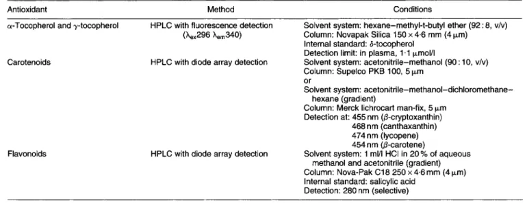
3.4. Measurement of antioxidant nutrients, carotenoids and jlavonoids extracted from humun plasma
Several antioxidants are routinely measured in plasma: a- tocopherol (and y-tocopherol) whose antioxidant roles are well clarified in vivo; @-carotene, lycopene and other dietary carotenoids for which there is, as yet, little evidence of antioxidant activity in vivo; ascorbic acid, the most efficient reducing agent in vivo, its redox potential defining its central role as an aqueous phase antioxidant. There is, as yet, little information as to the importance of dietary flavonoids as antioxidants in vivo, nor evidence for such activity in vivo, although these polyphenols are highly efficacious free- radical scavengers in vitro. Furthermore, it is only recently that it has become possible to detect and identify flavonoids (and other glycosides) in human plasma, in non-supplemented subjects.
Table 1 indicates the most favourable systems for detec- tion, identification and quantification of the antioxidants by HPLC. Plasma samples can be stored at -70”.