Hypertension and microvascular remodelling
Franc
¸ois Feihl
1*, Lucas Liaudet
2, Bernard I. Levy
3, and Bernard Waeber
11
Division de Physiopathologie Clinique, De´partement de Me´decine, Centre Hospitalier Universitaire Vaudois and Universite´ de Lausanne, Rue du Bugnon 46, BH10-701, CH-1011 Lausanne, Switzerland;2Service de Me´decine Intensive Adulte, Centre Hospitalier Universitaire Vaudois and Universite´ de Lausanne, Rue du Bugnon 46, CH-1011 Lausanne, Switzerland; and3
Centre de Recherche Cardiovasculaire Inserm Lariboisie`re, U689, Hoˆpital Lariboisie`re, 41 Bd de la Chapelle, 75475 Paris, France
Received 18 September 2007; revised 22 January 2008; accepted 31 January 2008; online publish-ahead-of-print 4 February 2008 Time for primary review: 32 days
In the present review, microvascular remodelling refers to alterations in the structure of resistance vessels contributing to elevated systemic vascular resistance in hypertension. We start with some his-torical aspects, underscoring the importance of Folkow’s contribution made half a century ago. We then move to some basic concepts on the biomechanics of blood vessels, and explicit the definitions pro-posed by Mulvany for specific forms of remodelling, especially inward eutrophic and inward hyper-trophic. The available evidence for the existence of remodelled resistance vessels in hypertension comes next, with relatively more weight given to human, in comparison with animal data. Mechanisms are discussed. The impact of antihypertensive drug treatment on remodelling is described, again with emphasis on human data. Some details are given on the three studies to date which point to remodelling of subcutaneous resistance arteries as an independent predictor of cardiovascular risk in hypertensive patients. We terminate by considering the potential role of remodelling in the pathogenesis of end-organ damage and in the perpetuation of hypertension.
KEYWORDS
Hypertension; Microcirculation; Resistance artery; Structure
Hypertension elicits two different kinds of diffuse structural changes in the systemic microcirculation. One, termed rare-faction, consists in an abnormally low spatial density of arterioles, capillaries, and possibly venules. The other con-cerns structural modifications of resistance small arteries and arterioles, which lead to a reduction in lumen diameter and are grouped under the generic name of remodelling. We have recently reviewed rarefaction in detail.1The focus of
the present paper is on remodelling which probably accounts for the major part of long-term elevation of systemic vascu-lar resistance (SVR) in hypertensive patients.
1. Historical note
The first intellectual association of hypertensive disease with diffuse abnormalities of the microcirculation may be traced back to Richard Bright, a father of nephrology, who in the third decade of the 19th century brilliantly described the natural course of what became to be known as Bright’s disease, in fact a heterogeneous class of chronic nephropa-thies. Bright noted the presence of a hard pulse2and the fre-quent autopsy finding of left ventricular hypertrophy unexplained by gross cardiac or aortic defect.3 In the excerpt shown in Figure 1, one may follow the line of thought which led Bright to hypothesize a diffuse
microcirculatory derangement. In 1869, George Johnson provided the first histological evidence of wall thickening in small arteries of various organs obtained at the autopsy of patients who suffered from Bright’s disease (Figure 2).4 Johnson was well aware that blood pressure in Bright’s disease was abnormally high. A decade later, the concept that heightened blood pressure may precede and be a cause rather than a consequence of altered microvascular architecture was introduced by Ewald,5,6who was also the
first to propose the ratio of wall thickness to lumen diameter for the assessment of abnormal microvessel structure.7Of
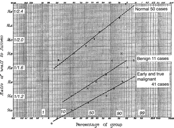
note, it is only later, i.e. in the first two decades of the 20th century, that essential hypertension became estab-lished as a nosographic entity.6The first quantitative study of arteriolar structure in hypertensive disease, using modern histological methods as well as a relatively crude form of morphometry, was published in 1929 (Figure 3).8
In the ensuing decades, the discovery of the Goldblatt’s models and the renin–angiotensin system were important factors explaining that the focus of hypertension research shifted towards functional and away from structural aspects of resistance vessel abnormalities.6,9The pioneering work of Bjo¨rn Folkow started the pendulum moving in a different direction, demonstrating that hypertension was associated with abnormally high resistance to blood flow, even in maximally dilated vascular beds, a strong evidence for the haemodynamic importance of altered microvascular structure (as opposed to altered vascular tone) in the
*Corresponding author. Tel: +41 21 314 14 23; fax: +41 21 314 14 32. E-mail address: [email protected] or [email protected]
Published on behalf of the European Society of Cardiology. All rights reserved.&The Author 2008. For permissions please email: [email protected].
hypertensive circulation.10 It took however a good additional 20 years before systematic interest progressively developed in other groups of investigators for the vascular structural factor in hypertension.11–14
2. Definitions and basic concepts
2.1 Resistance vessels
Resistance vessels are those which concentrate the major part of the pressure drop that must occur between large conduit arteries and capillaries. It has been underscored that their exact anatomical location is hard to define pre-cisely,15 but they are commonly believed to encompass small arteries and arterioles, with diameters ranging from 300 to 15 mm.16 Resistance vessels are characterized by the presence of myogenic tone, i.e. their intrinsic ability to contract in response to a sudden increase of transmural pressure.1 Important to note, myogenic tone becomes
more and more vigorous as vessel size decreases.17
2.2 Structural design and adaptability of blood
vessels
A fundamental tool to understand the varying structure of different blood vessels in different conditions is the Laplace law, which for any tubular element with cylindrical geometry relates intramural stress (s), wall thickness (W ),
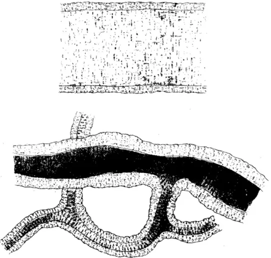
Figure 2 First description of wall thickening of small arteries in hypertension. These drawings are taken from the 1868 paper by George Johnson.4They
rep-resent microscopic views of small skin arteries, one normal (upper drawing) and one from a patient who at autopsy prep-resented with an abnormal kidney and left ventricular hypertrophy (lower drawing). Reproduced with permission by RSM Publishing.
Figure 1 Title and excerpt from Richard Bright’s original report The black bar in the margin draws attention to the mention made of the microcircula-tion. The excerpt straddles pages 396 and 397 in the original publicamicrocircula-tion.3 Reproduced with permission by ISS Enquiry.
lumen radius (r), and transmural pressure (P, the difference between luminal and extraluminal pressures) according to the equation:
s ¼Pr W
which may be rewritten in terms of the W to lumen diameter (D) ratio as
s ¼ 0:5 P ðW =DÞ
Laplace law dictates vascular structure so as to maintain s within a relatively tight domain across vessel type, species, and conditions. Within an individual, there is high plasticity of vascular structure, which continuously adapts to accom-modate changing conditions of mechanical load, as in fact do other organs such as skeletal muscle and bone. This is well exemplified by the thickness difference between sys-temic and pulmonary conduit arteries, which is non-existent in the foetus, and develops only after closure of the ductus arteriosus.18Evidence of plasticity beyond the developmen-tal stage is given by the wall thickening of venous grafts exposed to systemic pressure.19
2.3 The Folkow hypothesis
In his pioneering work already mentioned, Folkow used plethysmography to measure forearm blood flow under
conditions of maximal vasodilation, and from that he derived the minimal resistance (Rmin) of this vascular bed.9,10Forearm Rmin was found abnormally high in hyper-tensive subjects, an observation which, along with other data gathered in these experiments, was taken as evidence that a structural factor contributed to elevate systemic vas-cular resistance in these patients. In a bold intellectual move, Folkow proposed that structural alterations could lead to increased resistance even at normal levels of vascu-lar tone. In simplified form, reasoning was as follows. First, even minor reductions in lumen diameter (D) may have major effects on vessel resistance, because the latter is inversely proportional on the fourth power of the radius (Poiseuille law). Second, hypertensive structural alterations of resistance arteries consist in wall thickening and increased W/D ratio, with wall elements encroaching on the lumen, hence structurally reducing D, and increasing Rmin. Third, in conditions of increased W/D the same level of vascular tone, causing the same shortening of SMCs, leads for simple geometrical reasons to a greater reduction of D. In other words, the structurally modified resistance arteries in hypertension function as amplifiers of vascular tone, which thus needs not be augmented for vascular resistance and blood pressure to be higher than in the normotensive state.
Folkow’s view permeates all contemporary thinking on the pathogenesis of elevated blood pressure in chronic hypertension. It is now widely accepted that, important as its is, vascular tone is only a short-term modulator, while structural adaptation of resistance vessels is an obligatory
Figure 3 First quantitative evidence of abnormal resistance vessel structure in hypertension.8Data from biopsies of the pectoral muscle carried out in 50
norm-tensive subjects (upper curve), 11 patients with “benign” hypertension (middle curve), and 41 patients with malignant hypertension (lower curve). The mean ratio of wall thickness to lumen diameter was measured in arterioles and small arteries (25–100 mm). To help legibility, the original figure has been overlaid with repeats of some labels. Reproduced with permission from the American Medical Association&1929. All Rights reserved.
requirement for elevated blood pressure to be maintained for any prolonged period.20–23
2.4 Further concepts
Folkow’s hypothesis has both the advantages and disadvan-tages of simplicity. Notably, it focuses on s as the single determinant of vascular structural reshaping, while the shear stress (t) generated on endothelial lining by flowing blood is now of recognized importance. A durable increase in s is counteracted by an augmented W/D ratio,9 but a durable increase in t results in structural augmentation of D (thus tending to reduce blood velocity).24Adding to com-plexity, s and t are highly interdependent. For example, the higher D resulting from an increase in t tends to raise s by virtue of the Laplace law. Pries et al.25obtained extensive geometrical and mechanical data in the relaxed mesenteric vasculature of anesthetized rats. This enabled them to con-struct a sophisticated mathematical model of this vascular bed, which notably included the following: (i) several hun-dreds of vessels, arteriolar, capillary, and venular, (ii) the non-Newtonian nature of blood flow in small vessels (D , 300 mm);26(iii) quantitative rules which applied uniformly across all vessels in the network, everywhere relating s, t, geometry, blood flow, transmural pressure, and additional factors such as metabolic demand.27 This model faithfully
reproduced the structural changes observed experimentally in the mesenteric circulation of hypertensive rats.
2.5 Vascular remodelling
2.5.1 Nomenclature
In the historical work cited above, the increased W of hyper-tensive resistance vessels was uniformly ascribed to a higher volume of wall material per unit length of vessel [increased wall cross-sectional area (CSA)], or ‘hypertrophy’. It was assumed that smooth muscle cells in resistance vessels behaved as did left ventricular myocytes in the face of the increased pressure load, and that growth took place mainly on the luminal side, leading to a structural reduction of internal diameter (D). In 1966, Short28 carried out a careful quantitative geometrical analysis of intestinal small arteries and arterioles in patients with essential hypertension. He observed that, although W of these vessels was higher, and D smaller than in normotensive con-trols, media CSA was the same in the two groups of subjects. This finding was not compatible with a structural alteration of hypertensive vessels due to growth alone. The term remo-delling was first applied to resistance vessels by Baumbach and Heistad,29based on observations made in pial arterioles
from stroke-prone spontaneously hypertensive rats (SPSHRs), to indicate a structural rearrangement of existing wall material around a smaller lumen.
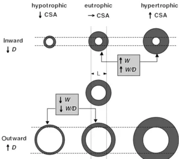
Noting that the narrow definition adopted by Baumbach and Heistad conflicted with other usages of the same term (i.e. myocardial remodelling), Mulvany30proposed that
vas-cular remodelling should encompass any change in D noted in a fully relaxed vessel, not explained by a change in transmural pressure or compliance, and therefore due to structural factors. In addition, a two-way classification scheme was put forward (Figure 4). Remodelling could be inward or outward, depending on whether D decreased or increased, and hypertrophic, eutrophic, or hypotrophic if respectively contributed to by net growth (increased CSA),
no net change in amount of tissue (constant CSA), or net loss of tissue (decreased CSA). With that terminology, the structural abnormalities noted by Short in intestinal arterioles28 are of course described as inward eutrophic
remodelling. For elementary geometrical reasons, inward eutrophic and hypertrophic remodelling are always associ-ated with increased W and W/D ratio, the opposite being true of outward eutrophic and hypotrophic remodelling30
(Figure 4).
The terminology just described has spurred some contro-versy.31,32 Importantly, eutrophic remodelling should not be construed as necessarily and exclusively implying rearrangement of existing components.33Mulvany’s32 termi-nology may indeed pose problems if automatically equated with distinct pathophysiological processes, but remains useful as long as no implicit assumptions are being made on underlying mechanisms.
2.5.2 Methodology
To be operational, the classification shown in Figure 4 necessitates appropriate methods for the measurement of resistance vessels dimensions. This problem is much harder than would seem at first sight.31
To meet the definition of remodelling given above, the respective sizes of hypertensive and normotensive small arteries and arterioles must be compared with the influence of the following factors either removed or controlled for: (i) vascular tone, (ii) transmural pressure, and (iii) vessel com-pliance. Obviously, none of these requirements would be met by geometrical measurements made on standard histo-logical sections prepared without perfusion of the tissue sample (e.g. shrinking artefacts).
One widely used approach, possible with small arteries (100 and 300 mm), is to carry out geometrical measurements on dissected segments put in standardized conditions in vitro. The segments are mounted in an organ chamber
Figure 4 Subtypes of vascular remodelling. Each pair of concentric circles represents a vessel in cross-sectional view. Depicted in the centre of the figure is the reference state, with respect to which structural changes occur. CSA wall (or media) cross-sectional area. D, lumen diameter. W, Wall thickness. Modified from ref.30, with author’s permission.
(myograph), fully relaxed by applying a vasodilator in maximal concentration, and then distended in a standar-dized fashion. There are two variants of this approach, one using the wire myograph (the most ancient one),34 and the other the pressure myograph. In addition to standar-dized geometry, both methods allow detailed measurements of passive mechanical properties, thus making it possible to distinguish alterations in geometry due to changes in wall elasticity from those due to other causes (i.e. remodelling stricto sensu). The disadvantages of myography are 2-fold. The first one has been called the sampling problem.33 Whether the arterial segments assessed with this approach are representative of resistance vessels in the studied organ (not to speak of the whole organism) is open to ques-tion. More subtly, for meaningful comparison of hyperten-sive and normotenhyperten-sive state, it is necessary that the vascular tree be sampled at identical branching level in both conditions, but verification of this requirement is not easy. The second disadvantage of myography resides in the partly arbitrary choice of transmural pressure under which geometry is measured. In particular, one may question the appropriateness of setting this variable at the same level for hypertensive and normotensive vessels, as most studies reviewed below have done.
3. Morphologic evidence for remodelling of
small vessels in untreated hypertension
3.1 Animal data
A considerable amount of studies have investigated struc-tural alterations of resistance arteries and arterioles in a large variety of hypertensive models, mostly in the rat, with relatively little work done in other species. The majority of studies have used a popular model of genetically determined hypertension, the SHR. A fundamental problem with this model is the lack of rigorous normotensive con-trols, since the Wistar-Kyoto rat (WKY) used to that effect differs from the SHR by many genetic markers, only a min-ority of which are linked to hypertension.35 To mitigate
this concern, alterations of microcirculatory structure broadly similar to those demonstrated in SHRs have been found in several other models for which fully adequate con-trols exist, such as transgenic rats expressing the mouse Ren-2 renin gene (mRen-2),36–39or rats made hypertensive by surgical (Goldblatt)40–42or pharmacological means.43–45
In a variety of hypertensive animal models (both genetic and secondary), media hypertrophy seems progressively less important as vessel size decreases, whereas eutrophic remodelling is most readily observed in the more distal part of arterial microcirculation.36,42,46–48 For example, Miller et al.46 have examined perfusion-fixed submucosal
intestinal 1A (largest) to 4A (smallest) arterioles from SHRs. While the W/D ratio was abnormally high in hyperten-sive vessels of all sizes, only at the 1A level was media CSA increased in comparison with WKY controls. In mRen-2 trans-genic rats,36impressive media hypertrophy was found in the large main mesenteric arteries,37 while inward eutrophic
remodelling predominated further downstream in this vas-cular bed.36Similar observations of large vs. small
mesen-teric47and kidney48arteries are available in SHRs. Below, we shall provide a simple mechanical explanation to this
pattern of proximal hypertrophic, progressively blending into distal eutrophic (at times hypotrophic) remodelling.
The definition of remodelling includes the requirement that changes in D and W/D at comparable distending pressure should not be due to altered wall elasticity. In the paper that originated the concept of eutrophic remodel-ling, as already mentioned, pial arterioles of SPSHRs had reduced D increased W/L and increased distensibility in comparison with those of normotensive controls, thus ruling out stiffening of vessel wall as a cause of the observed morphological alterations.29 Similar conclusions were reached in several,49,50but not all studies.38,51
Whether an abnormally high media CSA reflects cellular hypertrophy or hyperplasia has received variable answers. In general, there has been evidence of smooth muscle cells hyperplasia (with or without hypertrophy) in SHRs43,47 and SPSHRs,52i.e. genetic models, whereas SMC hypertro-phy without hyperplasia was found in models of secondary hypertension.41,43This difference between models may be related to the particular ability of SHR’s fibroblasts and SMCs to multiply rapidly under the influence of growth factors.33 However, this trait is of doubtful relevance to the pathogeny of hypertension, because it does not cosegre-gate with blood pressure in SHR/WKY F2 hybrids.33
3.2 Clinical data
The most feasible possibility for quantitative structural studies of resistance vessels in humans relies on the examin-ation of small muscular (presumably resistance) arteries from biopsies of subcutaneous gluteal fat carried out under local anaesthesia. Small arteries can also be obtained from omental fat excised at the time of abdominal surgery.14,53,54The dissected vessels are mounted in a wire or pressure myograph and characterized with the aforemen-tioned methodology. Due to the invasive character of these procedures, most relevant studies are of modest size, typi-cally involving between 10 and 20 subjects per group (with a few notable exceptions55–57). Furthermore untreated
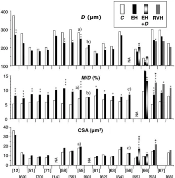
hypertensives are often patients in whom medication was withdrawn for a few weeks, rather than being newly diagnosed. With these caveat in mind, one may read in Figure 5 summary of the available information on the com-parative structure of small subcutaneous12,14,51,53,55,56,58–71 or omental14,53 arteries from untreated hypertensive and
normotensive human subjects, as obtained with the wire12,14,53,55,56,58–68or pressure51,69–71myograph
method-ology. In non-diabetic essential hypertensives, all data con-verge on a pattern of reduced D, increased M/D ratio (M denoting media width, measured in preference to the total wall thickness W in all these studies), and constant media CSA. Not all of these studies have checked vascular mechanics, but when this was done, the wall stiffness of hypertensive vessels was either unchanged54,71 or slightly decreased.51These aggregated data indicate that small
sub-cutaneous arteries of non-diabetic hypertensives undergo inward eutrophic remodelling. In contrast, it appears that diabetes on top of essential hypertension is associated with media hypertrophy, without a reduction of lumen diam-eter as measured in passive conditions.65,66The same hyper-trophy was also shown by one of these studies66 in
normotensive diabetics, supporting a pressure-independent effect of diabetes on resistance vessel morphology.23
Finally, the limited data available suggest that, contrary to the essential form, hypertension secondary to renovascular disease could promote media growth in human small subcu-taneous arteries.53,67,68
There are at least two caveats regarding the interpret-ation of these clinical data. First, the extent to which they might be contaminated by the aforementioned sampling problem is impossible to assess. Second, the sub-cutaneous vasculature is not necessarily representative of other vascular beds. There are a few observations to miti-gate the latter concern. We may recall here the evidence of eutrophic remodelling in the intestinal microcirculation of hypertensive patients presented by Short.28In addition,
a positive correlation has been found in hypertensive patients between coronary flow reserve and the M/D ratio of subcutaneous arteries, indeed supporting that hypertensive changes of microvascular structure were not limited to the subcutis.72 Finally, Harazny et al.73 have very recently been able to evaluate the W/D ratio of retinal arterioles in patients with treated hypertension and without advanced retinopathy (stages III or IV). To that effect, they used laser Doppler imaging whereby outer and inner diameters were respectively determined from reflection and perfusion images. Results indicated a higher ratio when blood pressure control was poor than when it was satisfactory.
4. Mechanisms of altered resistance vessel
structure in hypertension
It is thought that hypertensive remodelling of resistance vessels results from complex, yet incompletely understood interactions between genetic factors, intrinsic adaptation of vascular wall to altered mechanical conditions, and neu-rohumoral as well as local trophic influences.9
4.1 Genetic factors
Considering the polygenic nature of essential hypertension, it would be consistent that particular traits would exist that would favour alterations of vascular structure. Some evidence along this line is given by comparison of the same vessels in different strains of genetically hypertensive animals. Specifically, medial hypertrophy of large mesen-teric arteries seems much more marked in mRen-2 trans-genic rats than in age-matched SHRs.36 Another way to search for ‘remodelling traits’ is to examine the ontogeny of microvascular structure in genetic hypertension. Results have been heterogeneous. A structural reduction of lumen diameter has been detected in the small mesenteric arteries of young SHRs, when their blood pressure did not yet differ from that of age-matched WKY controls74but normal struc-ture of these same vessels has been reported in mRen-2 transgenic rats at the prehypertensive stage.39 Regarding human data, forearm Rmin was higher in young normoten-sive subjects with than without a family history of essential hypertension.75,76In neither study however was it clear that structural changes antedated hypertension, because blood pressure, although in the normotensive range, was some-what higher in the offsprings of hypertensive parents. In a similar group of subjects, subcutaneous small arteries failed to demonstrate any structural abnormality when examined with wire myography (Figure 5).60The view that essential hypertension could be initiated by a primary, genetically determined remodelling of resistance vessels is now largely abandoned.
4.2 Pressure-dependent factors
Structural narrowing of resistance vessels occurs in all like-lihood posterior to the onset of essential hypertension, although possibly prior to left ventricular hypertrophy or any other end-organ damage.71Posteriority is furthermore
self-evident in case of secondary hypertension. In addition, the evidence for a causal pathway leading from high blood pressure to microvascular remodelling is fairly strong. In the early 70s, Folkow et al.77reported that the abnormal
elevation of hindquarter Rmin seen in adult SHR was abol-ished by prior partial ligation of the abdominal aorta below the renal arteries carried out at the pre-hypertensive stage, an intervention that protected the lower limb vessels from exposure to high blood pressure. Also in pre-hypertensive SHRs, Bund et al.78partially ligated the left
external iliac artery, let the animals develop hypertension, and then observed with wire myography that distal femoral arteries excised from the right leg (unprotected) showed hypertrophic remodelling in comparison with those taken from the left leg (protected). Similar evidence exists with models of secondary hypertension.40,79
We have previously reviewed the general tendency of chronic hypertension to produce media growth in larger as
Figure 5 Morphologic characteristics of small subcutaneous arteries in untreated hypertensive patients vs. normotensive human subjects. Studies examined vessels isolated from biopsy material, using either the pressure myograph or the wire myograph. D internal diameter. M/D ratio of media thickness to internal diameter. CSA, cross-sectional area. C, normotensive controls. EH, untreated essential hypertension. EH+D, essential hypertension and diabetes; RVH, renovascular hypertension. NA, data not available from original publication. Data are the mean values reported in the various studies (references in square brackets). *P , 0.05, **P , 0.01, ***P , 0.001 vs. C. +P , 0.05, ++P , 0.01, +++P , 0.001 vs. EH. Notes: a) Means and P-values recomputed by pooling the two EH groups reported separately in the original publication. b) Group EH consisted of normotensive offspring of hypertensive parents. c ) The largest study, comprising 59 controls and 159 non-diabetic patients with essential hypertension.
opposed to inward eutrophic remodelling in smaller resist-ance vessels. It is believed that this pattern may be explained by the general mechanisms that regulate wall stress (s) in blood vessels.1,42,80 By virtue of the Laplace law, the same acute elevation of transmural pressure pro-duces a greater increase of s in larger arteries, due to their smaller W/D ratio in comparison with more distal nar-rower vessels. Furthermore, the larger vessels have a very limited, or even no myogenic response, so that their only way to regulate s in the face of persistent distension is to increase wall thickness, by way of a growth response. In con-trast, smaller vessels have vigorous myogenic tone, which together with a larger M/D ratio allows them to control s in the short term by mere active variations of lumen size and wall thickness. If the elevation of transmural pressure persists, myogenic constriction progressively gives way to structural rearrangement of wall material around a smaller lumen, i.e. the pattern of inward eutrophic remodel-ling.81,82 The aforementioned greater hypertrophy and lesser eutrophic remodelling of resistance vessels in hyper-tensive diabetics65,66 may be understood in this context, because diabetes impairs myogenic tone.66
A final remark relates to the differential effects on vascu-lar biology of pulsatile vs. steady mechanical forces.83In rabbit aorta for example, pulsatile variations of transmural pressure did not trigger the same intracellular signalling events [interestingly related to activation of kinases associ-ated with focal adhesion (FA) points, see below] as did a steady increase of this variable.84It is now realized that pul-satile pressure and flow penetrate much deeper into micro-vascular networks than previously thought,16and we may thus speculate that hypertensive resistance vessels may not be exposed to the same pulsatile stress/strain than are normotensive ones. Whether this factor contributes to remodelling could become an active area for future research.
4.3 Pressure-independent factors
In hypertensive models, the strategy of protecting a specific vascular bed from high blood pressure in order to sort out pressure-mediated from non-pressure mediated mechanisms of remodelling has in fact yielded some contradictory results.79,85,86The most cited evidence for the importance of pressure-independent factors in hypertensive remodelling of resistance vessel has been obtained in rats receiving a low-rate infusion of angiotensin II. In such conditions, blood pressure remains initially normal, and then slowly increases over several days, after which media hypertrophy of small mesenteric arteries is observed. Co-treatment of these animals with an infusion of hydralazine completely prevented hypertension, but had no effect on the develop-ment of vascular hypertrophy.44
In the following, we shall briefly discuss relevant neurohu-moral as well as local non-mechanical factors. These might act independent of pressure, interact with pressure-dependent mechanisms, or constitute essential mediators thereof, with the current state of knowledge not allowing to fully distinguishing between these possibilities.
4.4 Sympatho-adrenergic system
Sympathetic activation is a hallmark of human essential hypertension, and plays a well-known, in part
pressure-independent role in the development of left ventri-cular hypertrophy. A trophic impact of adrenergic stimu-lation on vascular smooth muscle has also been demonstrated in vitro. For example, stimulation of a1 adre-noreceptors enhanced the production of extracellular matrix (ECM) and the expression of transforming growth factor b (TGF-b) by cultures of primary human aortic smooth muscle cells, while b1 stimulation had the opposite effects.87The recent suggestion of a role for dysregulated production of TGF-b in hypertensive structural narrowing of resistance vessels must be noted here.88
On the other hand, there are no conclusive data, whether from animal models or clinical studies, to support an inde-pendent role of sympathetic activation in the pathogenesis of hypertensive remodelling of resistance vessels (for review, see22). In adult SHRs who underwent neonatal sym-pathectomy plus ablation of the adrenal medulla at 4 weeks, vascular structure was normal, but so was blood pressure.89In Sprague-Dawley rats made hypertensive with a slow low dose infusion of angiotensin II, neonatal sym-pathectomy+medullectomy influenced neither the time-course of blood pressure, nor the development of medial hypertrophy in small mesenteric arteries.90In the clinical arena, the structure of small subcutaneous arteries did not differ between patients with a hyperactivated sympatho-adrenergic system due to pheochromocytoma and patients with essential hypertension.91 b-blockers have consistently failed to influence the structure of these arteries in essential hypertensive patients (see below).
4.5 Angiotensin II
Beyond contraction of smooth muscle, angiotensin II has the ability to promote many processes in cardiovascular tissue, including cell growth, migration, differentiation, and apop-tosis, as well as modulation of ECM composition and turn-over. For these reasons, angiotensin II is a likely candidate for a pivotal role in remodelling throughout the cardiovascu-lar system.92Interestingly, the growth-promoting properties of angiotensin II have been demonstrated at physiological concentrations (10210M)93
in cultures of primary smooth muscle cells obtained from human small subcutaneous arteries.94In this respect, cells from patients with essential hypertension were more sensitive than cells from normoten-sive controls.94 The aforementioned results with slow low-dose infusion of angiotensin II in the rat have convin-cingly implicated this peptide as a primary, pressure-independent mediator of small mesenteric artery medial hypertrophy in this model.44It has been speculated that angiotensin II may participate in inward eutrophic remodelling by differentially favouring growth on the luminal side and apoptosis on the abluminal side of vascular wall.95
4.6 Reactive oxygen species
It is now well accepted that vascular production of reactive oxygen species (ROS) is abnormally high in hypertension.96
ROS in turn may trigger many cellular events, including growth. For example, the angiotensin II-induced growth of rat aortic smooth muscle cells was suppressed either by treatment with diethyldithiocarbamate, as ROS scavenger, or by overexpression in these cells of superoxide dismutase, an antioxydant enzyme.97 On in vitro administration of
angiotensin II, SMCs isolated from human small subcutaneous arteries also had a ROS-mediated growth response, which very interestingly was more marked in cells from hyperten-sive than from normotenhyperten-sive subjects.98
4.7 Nitric oxide
A low bioavailability of nitric oxide (NO), linked to endo-thelial dysfunction and possibly mediated in part by excess ROS, seems widely implicated at multiple levels in the patho-genesis of hypertension.99 NO has myriads of biological effects, among which a negative control exerted on cellular growth. Mice with deletion of the gene coding for the endo-thelial form of nitric oxide synthase (eNOS) are mildly hyper-tensive. In these mice, the inward hypotrophic remodelling of conduit arteries, which normally follows a chronic reduction in blood flow, is replaced by a hyperplastic response.100 Hypertrophic remodelling of small coronary arteries has been observed in rats made hypertensive by the chronic administration of NG-methyl-l-arginine, an inhibitor of NOS, and this structural abnormality was not reversed when blood pressure was normalized by concomitant treatment with hydralazine,45 suggesting that suppression of NO could enhance vascular growth in a pressure-independent manner. Interestingly, a simulation carried out with the aforemen-tioned computational model of rat mesenteric microcircula-tion by Pries et al.27 suggested that even minor degrees of endothelial dysfunction can powerfully potentiate the impact of hypertension on microvascular structure. In hyper-tensive humans, endothelial dysfunction of the microvascula-ture has been demonstrated in the skin.101
4.8 The extracellular matrix–integrin–
cytoskeleton axis
Integrins are ubiquitous dimeric transmembrane receptors which ligate specific sites of various ECM proteins, for example fibronectin and collagens.80,102,103 Their
cyto-plasmic part binds cytoskeletal proteins such as actin. Within the cell membrane, ligated integrin complexes occur in clusters called FA points. This arrangement allows for a tight mechanical connexion between ECM and cytoske-leton. FAs also comprise a complex signalling apparatus, able to influence all major cell functions, including cell cycle, gene expression, substrate adhesion, motility, and membrane ion channel permeability. In a multicellular environment, any change in cell number, shape, or position requires the active participation and dynamic restructuring of the ECM–integrin–cytoskeleton axis. This axis is the major mean that cells have to sense their mechanical environment and react accordingly. For example, it has been shown that the aVb3 and a5b1 integrins are the
stretch sensors responsible for initiating myogenic contrac-tion in resistance vessels.104
With this background, it seems logical that the ECM– integrin–cytoskeleton axis should be profoundly implicated in hypertensive remodelling of resistance vessels. Rat mesenteric arteries were pressurized in vitro and exposed to endothelin-1 for 3 days; eutrophic remodelling ensued an effect blocked by an antibody directed against the b3
integrin subunit.105 In eutrophically remodelled small mesenteric arteries of hypertensive mRen-2 transgenic rats, expression of the aVintegrin subunit was increased,
in comparison with similar vessels from normotensive
controls.39 In that study furthermore, some mRen-2 rats received at the pre-hypertensive stage a peptide antagonist of aV: this treatment had no effect on the further time
course of blood pressure, but modified the remodelling of mesenteric arteries from mainly eutrophic to mainly hypertrophic.
4.9 Adventitial cells
The vascular adventitia has traditionally been considered as a passive supportive structure. There is mounting evidence that it also actively participates in general remodelling pro-cesses.106In rat carotid artery subjected to balloon injury, adventitial fibroblasts differentiated to myofibroblasts and migrated through the media to participate in the formation of neointima.107 In rats with hypertension secondary to
chronic pharmacologic inhibition of NOS, the basilar artery was eutrophically remodelled, and its adventitial layer revealed a greatly increased cellular density.108 Eutrophi-cally remodelled mesenteric arteries of SPSHRs also had abnormally dense adventitial cellularity, and in addition dis-played a striking number of advential-looking cells in the media.109 Considering the potential role of sympathetic activation evoked above, it is relevant that adventitial fibroblasts highly express a1-adrenergic receptors, and that noradrenaline can stimulate their proliferation and differentiation to myofibroblasts, at least in vitro.110
5. Effects of antihypertensive drug treatment
on the structure of resistance arteries
This topic was systematically reviewed by Christensen and Mulvany111 in 2001. Thirty clinical studies were identified
where resistance vessel structure was evaluated before and after blood pressure lowering with antihypertensive medication. All major classes of drugs were tested, over periods ranging from 1 to 84 months, in all cases with sub-stantial improvement of blood pressure control. Twenty-one studies inferred structure from the measurement of Rmin with forearm plethysmography and the remaining 9 used myography of biopsied small subcutaneous arteries. It came out quite clearly that at least partial reversal of hypertensive structural changes could be obtained with all drug classes, except with b-blockers. As shown in Table 1, more recent studies of biopsied subcutaneous arteries have mostly borne out this conclusion. Very recently, Mathiassen et al.112 presented data in 28 patients with
essential hypertension. In these subjects, forearm Rmin had remained unchanged and abnormally high despite excel-lent blood pressure control obtained with a b-blocker for one full year. For the next year, medication was switched to an AT1 antagonist (eprosartan), a period during which blood pressure remained stable while Rmin decreased by 16% (P , 0.01).
The blatant inability of b-blockers to affect the structure of resistance vessels has been attributed to their mode of action through reduction of cardiac output rather than vaso-dilation.111,112 Considering our previous discussion on mechanisms, it is also possible that vasodilator antihyper-tensive drugs, especially those interfering with the renin-angiotensin system, influence vascular structure by mechanisms independent of haemodynamic factors, but this issue is completely unsolved at present.
6. Prognostic implication of hypertensive
resistance vessel remodelling
We now have three relevant and concordant clinical studies. The first one was published in 2003 by Rizzoni et al. Within an 8-year period, 128 patients with either essential or secondary hypertension had had a gluteal or omental fat biopsy after being weaned from antihyperten-sive medication for at least 3 weeks. Follow-up ensued for a mean duration of 5 years (range 2.6–9.9), with a compo-site endpoint of various major and minor cardiovascular events, which occurred in 33 cases. On multivariable analy-sis, a high M/D ratio of the small artery and a high pulse pressure were the only independent predictors for event occurrence. A few years later, the authors extended their survey as follow-up included more subjects. The expanded cohort comprised 303 patients observed for a mean dur-ation of 6.9 years (range 0.6–13.9). It was thus possible to distinguish major (sudden death, stroke, myocardial infarction, n = 25) from other cardiovascular events (n = 23). Here, a high M/D ratio was an independent predictor for the occurrence, not only of any event as before, but also of any major one.57Both studies contained a mix of diabetic and non-diabetic hypertensive patients, which may be seen as a weak point considering the possible inde-pendent impact of diabetes on small artery morphology (see above). However, Mulvany’s group reported essentially the same results in a more homogeneous cohort of 159 non-diabetic subjects with essential hypertension (mean follow up time 10 years, composite outcome of cardiac, cerebro-vascular, renal, and peripheral arterial events, 30 events in total).56
In view of these data, should evaluation of small artery structure become part of clinical evaluation in hypertensive disease? In the present state of knowledge, probably not, if only because biopsies of subcutaneous arteries are not prac-tical on a large scale.113The aforementioned non-invasive structural assessment of retinal arterioles73might seem in that respect a promising approach, which deserves further evaluation.113
7. In form of conclusion: Should we aim to
correct resistance vessel structure when
treating hypertension?
At the time of writing the present review, this issue has not been resolved. The answer could come from two different types of considerations. First, does remodelling of resistance vessels participate in hypertensive organ damage? Second, does this remodelling hamper the proper control of blood pressure?
The possible involvement of resistance vessel remodelling in hypertensive end-organ damage has received the greatest attention in the case of the myocardium.72,114,115Notably, in patients with essential hypertension, coronary flow reserve improved concomitantly with normalization of small subcu-taneous artery structure following 1 year of treatment with perindopril, an ACE inhibitor. In contrast, coronary flow reserve worsened, and small subcutaneous artery struc-ture remained unchanged in a parallel group in whom the same level of blood pressure reduction was achieved over the same period, by the b-blocker atenonol.115 In that study, interestingly, myocardial blood flow was measured with position emission tomography, allowing expression of flow per unit mass of tissue, thus removing at least in part the influence of left ventricular hypertrophy (which regressed with the ACE inhibitor only). Assuming a corre-lation between the remodelling of subcutaneous and intra-myocardial resistance arteries, as suggested by another clinical study,72correction of this anomaly would contribute to improve the functional status of the coronary circulation. That altered vascular structure could hamper the thera-peutic control of blood pressure would be suggested by an oversimplified version of the Folkowian view—i.e. high blood pressure breeding remodelling which in turn breeds high blood pressure. However, Folkow9himself has stressed that this positive feed-back loop would be unsustainable without intervention of powerful counter-regulatory mech-anisms, the nature of which remains incompletely under-stood. As also underscored by Mulvany,21 the control of blood pressure involves: (i) one or several setpoints for Table 1 Effect of antihypertensive treatment on blood pressure and on resistance artery structure
Primary drug Drug type %change MBP %change M/D Treatment duration (months) Reference
Amlodipine CaB 28%** 39%* 12 116a Atenolol b+ 211% 7% 12 117 Atenolol b+ 218%*** 0% 12 115 Perindopril ACE 217%*** 215%* 12 115 Losartan ARB 210%** 215%* 12 116a Candesartan ARB 27%* 216%** 12 118 Valsartan ARB 213%* 219%* 12 117 Enalapril ACE 213%* 221%** 12 118 Irbesartan ARB 0% 223%** 12 119b
Recent clinical studies which documented changes in mean blood pressure (MBP) and in media/lumen ratio (M/D) of small subcutaneous arteries following antihypertensive treatment. Calcium antagonists (CaB), b+ antagonist of beta-adrenergic receptors, ACE inhibitors of the angiotensin-converting enzyme, ARB antagonist of the AT1 angiotensin receptor.
aOne study reporting the M/D ratio of vessels ,100 mm, measured with histology of skin biopsies.116In all others, 200–300 mm arteries were mounted in a
wire115,118or pressure117,119myograph. b
One study in which patients whose blood pressure was well controlled with a b-blocker were crossed-over to an ARB.119 *P , 0.05.
**P , 0.01. ***P , 0.001.
this variable, (ii) fast processes (i.e modulation of vessel tone through local and neurohumoral mechanisms) to keep blood pressure closest to this (these) setpoint(s), and (iii) slow processes (remodelling) which allow adaptation to a sustained setpoint change, in energetically economical terms (i.e. reduced lumen diameter at normal activation level of smooth muscle in resistance vessels). What deter-mines the setpoint(s) is presently unknown, although the need to maintain appropriate blood and oxygen supply to various organs, notably the kidney, is probably involved.21 In the hypertensive circulation, correcting the structure without also changing the setpoint would only activate neu-rohumoral and other mechanisms to keep blood pressure high. Consistent with these considerations, we have noted above a marked dissociation between the impacts of antihy-pertensive therapy on blood pressure and on resistance vessel structure (Table 1).111 If and how the various drug classes affect the setpoint for blood pressure might become a focus of future research.
Funding
This work has been funded in part by the Swiss National Science Foundation (grant 3200B0-116511).
Acknowledgements
The authors wish to thank Franc¸oise Bilat for excellent secretarial assistance.
Conflict of interest: none declared.
References
1. Feihl F, Liaudet L, Waeber B, Levy BI. Hypertension, a disease of the microcirculation? Hypertension 2006;48:1012–1017.
2. Bright R. Cases and observations illustrative of renal disease accompanied with the secretion of albuminous urine. Guy’s Hosp Rep 1836;1:338–379.
3. Bright R. Tabular view of the morbid appearances in 100 cases con-nected with albuminous urine, with observations. Guy’s Hosp Rep 1836;1:380–400.
4. Johnson GI. On certain points in the pathology of Bright’s disease of the kidney. II. On the influence of the minute blood vessels upon the circula-tion. Tr Medico-Chir Soc Lond 1868;51:57–76.
5. Ewald CA. Ueber die Vera¨nderungen kleiner Gefa¨sse bei Morbus Brightii und die darauf bezu¨glichen Theorien. Virchows Arch 1877;71:453–499. 6. Folkow B. Physiological aspects of primary hypertension. Physiol Rev
1982;62:347–504.
7. Newton NM, Fine LG. Inference of the existence of high blood pressure as a cause of renal disease in the mid-19th century: observations on vas-cular structures in the kidney. Am J Nephrol 1999;19:323–332. 8. Kernohan JW, Anderson EW, Keith NM. The arterioles in cases of
hyper-tension. Arch Intern Med 1929;44:395–423.
9. Folkow B. The ‘structural factor’ in hypertension, with special emphasis on the hypertrophic adaptation of the systemic resistnace vessels. In: Laragh JH, Brenner BM (ed.), Hypertension: Pathophysiology, Diagnosis and Treatment. New York, Raven Press, Ltd; 1990. p565–581. 10. Folkow B, Grimby G, Thuslesius O. Adaptive structural changes of the
vascular walls in hypertension and their relation to the control of the peripheral resistance. Acta Physiol Scand 1958;44:255–272.
11. Mulvany MJ, Hansen OK, Aalkjaer C. Direct evidence that the greater contractility of resistance vessels in spontaneously hypertensive rats is associated with a narrowed lumen, a thickened media, and an increased number of smooth muscle cell layers. Circ Res 1978;43:854–864. 12. Schiffrin EL, Deng LY, Larochelle P. Morphology of resistance arteries and
comparison of effects of vasoconstrictors in mild essential hypertensive patients. Clin Invest Med 1993;16:177–186.
13. Rizzoni D, Castellano M, Porteri E, Bettoni G, Muiesan ML, Agabiti-Rosei E. Vascular structural and functional alterations before and after the development of hypertension in SHR. Am J Hypertens 1994;7:193–200.
14. Rosei EA, Rizzoni D, Castellano M, Porteri E, Zulli R, Muiesan ML et al. Media: lumen ratio in human small resistance arteries is related to forearm minimal vascular resistance. J Hypertens 1995;13:341–347. 15. Christensen KL, Mulvany MJ. Location of resistance arteries. J Vasc Res
2001;38:1–12.
16. Levy BI, Ambrosio G, Pries AR, Struijker-Boudier HAJ. Microcirculation in hypertension—a new target for treatment? Circulation 2001;104: 735–740.
17. Davis MJ. Myogenic response gradient in an arteriolar network. Am J Physiol 1993;264:H2168–H2179.
18. Glagov S, Vito R, Giddens DP, Zarins CK. Micro-architecture and compo-sition of artery walls: relationship to location, diameter and the distri-bution of mechanical stress. J Hypertens Suppl 1992;10:S101–S104. 19. Dilley RJ, McGeachie JK, Prendergast FJ. A review of the histologic
changes in vein-to-artery grafts, with particular reference to intimal hyperplasia. Arch Surg 1988;123:691–696.
20. Korner PI, Angus JA. Vascular remodeling. Hypertension 1997;29: 1065–1066.
21. Mulvany MJ. Small artery remodeling in hypertension. Curr Hypertens Rep 2002;4:49–55.
22. Simon G. Pathogenesis of structural vascular changes in hypertension. J Hypertens 2004;22:3–10.
23. Mulvany MJ. Small artery structure: time to take note? Am J Hyperten-sion 2007;20:853–854.
24. Lehoux S, Tedgui A. Cellular mechanics and gene expression in blood vessels. J Biomech 2003;36:631–643.
25. Pries AR, Reglin B, Secomb TW. Structural adaptation of vascular net-works: role of the pressure response. Hypertension 2001;38:1476–1479. 26. Nichols WW, O’Rourke MF. Chap. 2: The nature of flow of a fluid. McDo-nald’s blood flow in arteries. London, Edward Arnold; 1990. p12–53. 27. Pries AR, Reglin B, Secomb TW. Remodeling of blood vessels: responses
of diameter and wall thickness to hemodynamic and metabolic stimuli. Hypertension 2005;46:725–731.
28. Short D. Morphology of the intestinal arterioles in chronic human hyper-tension. Br Heart J 1966;28:184–192.
29. Baumbach GL, Heistad DD. Remodeling of cerebral arterioles in chronic hypertension. Hypertension 1989;13:968–972.
30. Mulvany MJ. Vascular remodelling of resistance vessels: can we define this? Cardiovasc Res 1999;41:9–13.
31. Bund SJ, Lee RM. Arterial structural changes in hypertension: a con-sideration of methodology, terminology and functional consequence. J Vasc Res 2003;40:547–557.
32. Mulvany MJ. Structural abnormalities of the resistance vasculature in hypertension. J Vasc Res 2003;40:558–560.
33. Heagerty AM, Aalkjaer C, Bund SJ, Korsgaard N, Mulvany MJ. Small artery structure in hypertension. Dual processes of remodeling and growth. Hypertension 1993;21:391–397.
34. Mulvany MJ, Aalkjaer C. Structure and function of small arteries. Physiol Rev 1990;70:921–961.
35. St Lezin E, Simonet L, Pravenec M, Kurtz TW. Hypertensive strains and normotensive ‘control’ strains. How closely are they related? Hyperten-sion 1992;19:419–424.
36. Thybo NK, Korsgaard N, Mulvany MJ. Morphology and function of mesen-teric resistance arteries in transgenic rats with low-renin hypertension. J Hypertens 1992;10:1191–1196.
37. Struijker-Boudier HA, van Essen H, Fazzi G, De Mey JG, Qiu HY, Levy BI. Disproportional arterial hypertrophy in hypertensive mRen-2 transgenic rats. Hypertension 1996;28:779–784.
38. Dunn WR, Gardiner SM. Differential alteration in vascular structure of resistance arteries isolated from the cerebral and mesenteric vascular beds of transgenic [(mRen-2)27], hypertensive rats. Hypertension 1997;29:1140–1147.
39. Heerkens EH, Shaw L, Ryding A, Brooker G, Mullins JJ, Austin C et al. AlphaV integrins are necessary for eutrophic inward remodeling of small arteries in hypertension. Hypertension 2006;47:281–287. 40. Hashimoto H, Prewitt RL, Efaw CW. Alterations in the microvasculature
of one-kidney, one-clip hypertensive rats. Am J Physiol 1987;253: H933–H940.
41. Korsgaard N, Mulvany MJ. Cellular hypertrophy in mesenteric resistance vessels from renal hypertensive rats. Hypertension 1988;12:162–167. 42. Stacy DL, Prewitt RL. Effects of chronic hypertension and its reversal on
43. Lee RM. Structural alterations of blood vessels in hypertensive rats. Canad J Physiol Pharmacol 1987;65:1528–1535.
44. Griffin SA, Brown WC, MacPherson F, McGrath JC, Wilson VG, Korsgaard N et al. Angiotensin II causes vascular hypertrophy in part by a non-pressor mechanism. Hypertension 1991;17:626–635. 45. Numaguchi K, Egashira K, Takemoto M, Kadokami T, Shimokawa H,
Sueishi K et al. Chronic inhibition of nitric oxide synthesis causes coron-ary microvascular remodeling in rats. Hypertension 1995;26:957–962. 46. Miller BG, Connors BA, Bohlen HG, Evan AP. Cell and wall morphology of
intestinal arterioles from 4- to 6- and 17- to 19-week-old Wistar-Kyoto and spontaneously hypertensive rats. Hypertension 1987;9:59–68. 47. Owens GK, Schwartz SM, McCanna M. Evaluation of medial hypertrophy
in resistance vessels of spontaneously hypertensive rats. Hypertension 1988;11:198–207.
48. Smeda JS, Lee RM, Forrest JB. Structural and reactivity alterations of the renal vasculature of spontaneously hypertensive rats prior to and during established hypertension. Circ Res 1988;63:518–533.
49. Dunn WR, Wallis SJ, Gardiner SM. Remodelling and enhanced myogenic tone in cerebral resistance arteries isolated from genetically hyperten-sive Brattleboro rats. J Vasc Res 1998;35:18–26.
50. Bund SJ. Spontaneously hypertensive rat resistance artery structure related to myogenic and mechanical properties. Clin Sci 2001;101: 385–393.
51. Intengan HD, Deng LY, Li JS, Schiffrin EL. Mechanics and composition of human subcutaneous resistance arteries in essential hypertension. Hypertension 1999;33:569–574.
52. Amann K, Gharehbaghi H, Stephen S, Mall G. Hypertrophy and hyperpla-sia of smooth muscle cells of small intramyocardial arteries in spon-taneously hypertensive rats. Hypertension 1995;25:124–131. 53. Rizzoni D, Porteri E, Castellano M, Bettoni G, Muiesan ML, Muiesan P
et al. Vascular hypertrophy and remodeling in secondary hypertension. Hypertension 1996;28:785–790.
54. Rizzoni D, Porteri E, Guefi D, Piccoli A, Castellano M, Pasini G et al. Cel-lular hypertrophy in subcutaneous small arteries of patients with reno-vascular hypertension. Hypertension 2000;35:931–935.
55. Rizzoni D, Porteri E, Boari GE, De Ciuceis C, Sleiman I, Muiesan ML et al. Prognostic significance of small-artery structure in hypertension. Circu-lation 2003;108:2230–2235.
56. Mathiassen ON, Buus NH, Sihm I, Thybo NK, Morn B, Schroeder AP et al. Small artery structure is an independent predictor of cardiovascular events in essential hypertension. J Hypertens 2007;25:1021–1026. 57. De Ciuceis C, Porteri E, Rizzoni D, Rizzardi N, Paiardi S, Boari GE et al.
Structural alterations of subcutaneous small-resistance arteries may predict major cardiovascular events in patients with hypertension. Am J Hypertension 2007;20:846–852.
58. Rizzoni D, Muiesan ML, Porteri E, Castellano M, Zulli R, Bettoni G et al. Effects of long-term antihypertensive treatment with lisinopril on resistance arteries in hypertensive patients with left ventricular hyper-trophy. J Hypertens 1997;15:197–204.
59. Rizzoni D, Porteri E, Guelfi D, Muiesan ML, Valentini U, Cimino A et al. Structural alterations in subcutaneous small arteries of normotensive and hypertensive patients with non-insulin-dependent diabetes melli-tus. Circulation 2001;103:1238–1244.
60. Aalkjaer C, Heagerty AM, Bailey I, Mulvany MJ, Swales JD. Studies of iso-lated resistance vessels from offspring of essential hypertensive patients. Hypertension 1987;9:III155–III158.
61. Aalkjaer C, Heagerty AM, Petersen KK, Swales JD, Mulvany MJ. Evidence for increased media thickness, increased neuronal amine uptake, and depressed excitation–contraction coupling in isolated resistance vessels from essential hypertensives. Circ Res 1987;61:181–186. 62. Korsgaard N, Aalkjaer C, Heagerty AM, Izzard AS, Mulvany MJ. Histology
of subcutaneous small arteries from patients with essential hyperten-sion. Hypertension 1993;22:523–526.
63. Izzard AS, Cragoe EJ Jr, Heagerty AM. Intracellular pH in human resist-ance arteries in essential hypertension. Hypertension 1991;17:780–786. 64. Thurmann PA, Stephens N, Heagerty AM, Kenedi P, Weidinger G, Rietbrock N. Influence of isradipine and spirapril on left ventricular hypertrophy and resistance arteries. Hypertension 1996;28:450–456. 65. Endemann DH, Pu Q, De Ciuceis C, Savoia C, Virdis A, Neves MF et al.
Persistent remodeling of resistance arteries in type 2 diabetic patients on antihypertensive treatment. Hypertension 2004;43:399–404. 66. Schofield I, Malik R, Izzard A, Austin C, Heagerty A. Vascular structural
and functional changes in type 2 diabetes mellitus: evidence for the roles of abnormal myogenic responsiveness and dyslipidemia. Circula-tion 2002;106:3037–3043.
67. Muiesan ML, Rizzoni D, Salvetti M, Porteri E, Monteduro C, Guelfi D et al. Structural changes in small resistance arteries and left ventricular geo-metry in patients with primary and secondary hypertension. J Hypertens 2002;20:1439–1444.
68. Kvist S, Mulvany MJ. Reduced medication and normalization of vascular structure, but continued hypertension in renovascular patients after revascularization. Cardiovasc Res 2001;52:136–142.
69. Schiffrin EL, Deng LY. Structure and function of resistance arteries of hypertensive patients treated with a beta-blocker or a calcium channel antagonist. J Hypertens 1996;14:1247–1255.
70. Schiffrin EL, Park JB, Intengan HD, Touyz RM. Correction of arterial structure and endothelial dysfunction in human essential hypertension by the angiotensin receptor antagonist losartan. Circulation 2000;101: 1653–1659.
71. Park JB, Schiffrin EL. Small artery remodeling is the most prevalent (earliest?) form of target organ damage in mild essential hypertension. J Hypertens 2001;19:921–930.
72. Rizzoni D, Palombo C, Porteri E, Muiesan ML, Kozakova M, La Canna G et al. Relationships between coronary flow vasodilator capacity and small artery remodelling in hypertensive patients. J Hypertens 2003; 21:625–631.
73. Harazny JM, Ritt M, Baleanu D, Ott C, Heckmann J, Schlaich MP et al. Increased wall:lumen ratio of retinal arterioles in male patients with a history of a cerebrovascular event. Hypertension 2007;50:623–629. 74. Lee RM. Vascular changes at the prehypertensive phase in the
mesen-teric arteries from spontaneously hypertensive rats. Blood Vessels 1985;22:105–126.
75. Takeshita A, Imaizumi T, Ashihara T, Yamamoto K, Hoka S, Nakamura M. Limited maximal vasodilator capacity of forearm resistance vessels in normotensive young men with a familial predisposition to hypertension. Circ Res 1982;50:671–677.
76. Giannattasio C, Cattaneo BM, Mangoni AA, Carugo S, Stella ML, Failla M et al. Cardiac and vascular structural changes in normotensive subjects with parental hypertension. J Hypertens 1995;13:259–264.
77. Folkow B, Gurevich M, Hallback M, Lundgren Y, Weiss L. The hemody-namic consequences of regional hypotension in spontaneously hyperten-sive and normotenhyperten-sive rats. Acta Physiol Scand 1971;83:532–541. 78. Bund SJ, West KP, Heagerty AM. Effects of protection from pressure on
resistance artery morphology and reactivity in spontaneously hyperten-sive and Wistar-Kyoto rats. Circ Res 1991;68:1230–1240.
79. Stacy DL, Prewitt RL. Attenuated microvascular alterations in coarcta-tion hypertension. Am J Physiol 1989;256:H213–H221.
80. Heerkens EH, Izzard AS, Heagerty AM. Integrins vascular remodeling hypertension. Hypertension 2007;49:1–4.
81. Martinez-Lemus LA, Hill MA, Bolz SS, Pohl U, Meininger GA. Acute mechanoadaptation of vascular smooth muscle cells in response to con-tinuous arteriolar vasoconstriction: implications for functional remodel-ing. FASEB J 2004;18:708–710.
82. Bakker EN, van der Meulen ET, van den Berg BM, Everts V, Spaan JA, VanBavel E. Inward remodeling follows chronic vasoconstriction in iso-lated resistance arteries. J Vasc Res 2002;39:12–20.
83. Safar ME, Lacolley P. Disturbance of macro- and microcirculation: relations with pulse pressure and cardiac organ damage. Am J Physiol 2007;293:H1–H7.
84. Lehoux S, Esposito B, Merval R, Tedgui A. Differential regulation of vas-cular focal adhesion kinase by steady stretch and pulsatility. Circulation 2005;111:643–649.
85. Plunkett WC, Overbeck HW. Increased arteriolar wall-to-lumen ratio in a normotensive vascular bed in coarctation hypertension. Am J Physiol 1985;249:H859–H866.
86. Liu JL, Bishop SP, Overbeck HW. Morphometric evidence for non-pressure-related arterial wall thickening in hypertension. Circ Res 1988;62:1001–1010.
87. O’Callaghan CJ, Williams B. The regulation of human vascular smooth muscle extracellular matrix protein production by alpha- and beta-adrenoceptor stimulation. J Hypertens 2002;20:287–294. 88. August P, Suthanthiran M. Transforming growth factor beta signaling,
vascular remodeling, and hypertension. N Engl J Med 2006;354: 2721–2723.
89. Lee RM, Borkowski KR, Leenen FH, Tsoporis J, Coughlin M. Combined effect of neonatal sympathectomy and adrenal demedullation on blood pressure and vascular changes in spontaneously hypertensive rats. Circ Res 1991;69:714–721.
90. Simon G, Csiky B. Effect of neonatal sympathectomy on the develop-ment of structural vascular changes in angiotensin II-treated rats. J Hypertens 1998;16:77–84.
91. Porteri E, Rizzoni D, Mulvany MJ, De Ciuceis C, Sleiman I, Boari GE et al. Adrenergic mechanisms and remodeling of subcutaneous small resist-ance arteries in humans. J Human Hypertens 2003;21:2345–2352. 92. Touyz RM. Intracellular mechanisms involved in vascular remodelling of
resistance arteries in hypertension: role of angiotensin II. Exp Physiol 2005;90:449–455.
93. Jankowski V, Vanholder R, van der Giet M, Tolle M, Karadogan S, Gobom J et al. Mass-spectrometric identification of a novel angiotensin peptide in human plasma. Arterioscler Thromb Vasc Biol 2007;27: 297–302.
94. Touyz RM, He G, Wu XH, Park JB, Mabrouk ME, Schiffrin EL. Src is an import-ant mediator of extracellular signal-regulated kinase 1/2-dependent growth signaling by angiotensin II in smooth muscle cells from resistance arteries of hypertensive patients. Hypertension 2001;38:56–64. 95. Intengan HD, Schiffrin EL. Vascular remodeling in hypertension: roles of
apoptosis, inflammation, and fibrosis. Hypertension 2001;38:581–587. 96. Fortuno A, Jose GS, Moreno MU, Diez J, Zalba G. Oxidative stress and
vascular remodelling. Exp Physiol 2005;90:457–462.
97. Zafari AM, Ushio-Fukai M, Akers M, Yin Q, Shah A, Harrison DG et al. Role of NADH/NADPH oxidase-derived H2O2 in angiotensin II-induced vascu-lar hypertrophy. Hypertension 1998;32:488–495.
98. Touyz RM, Schiffrin EL. Increased generation of superoxide by angioten-sin II in smooth muscle cells from resistance arteries of hypertensive patients: role of phospholipase D-dependent NAD(P)H oxidase-sensitive pathways. J Hypertens 2001;19:1245–1254.
99. Marin E, Sessa WC. Role of endothelial-derived nitric oxide in hyperten-sion and renal disease. Curr Opin Nephrol Hypertens 2007;16:105–110. 100. Rudic RD, Shesely EG, Maeda N, Smithies O, Segal SS, Sessa WC. Direct evidence for the importance of endothelium-derived nitric oxide in vas-cular remodeling. J Clin Invest 1998;101:731–736.
101. de Jongh RT, Serne EH, Ijzerman RG, Stehouwer CD. Microvascular func-tion: a potential link between salt sensitivity, insulin resistance and hypertension. J Hypertens 2007;25:1887–1893.
102. Giancotti FG, Ruoslahti E. Integrin signaling. Science 1999;285: 1028–1032.
103. Mitra SK, Hanson DA, Schlaepfer DD. Focal adhesion kinase: in command and control of cell motility. Nat Rev Mol Cell Biol 2005;6:56–68. 104. Martinez-Lemus LA, Crow T, Davis MJ, Meininger GA. Alphavbeta3- and
alpha5beta1-integrin blockade inhibits myogenic constriction of skeletal muscle resistance arterioles. Am J Physiol 2005;289:H322–H329. 105. Bakker EN, Buus CL, VanBavel E, Mulvany MJ. Activation of resistance
arteries with endothelin-1: from vasoconstriction to functional adap-tation and remodeling. J Vasc Res 2004;41:174–182.
106. Siow RC, Churchman AT. Adventitial growth factor signalling and vascu-lar remodelling: Potential of perivascuvascu-lar gene transfer from the outside-in. Cardiovasc Res 2007;75:659–668.
107. Siow RC, Mallawaarachchi CM, Weissberg PL. Migration of adventitial myofibroblasts following vascular balloon injury: insights from in vivo gene transfer to rat carotid arteries. Cardiovasc Res 2003;59:212–221. 108. Arribas SM, Gonzalez C, Graham D, Dominiczak AF, McGrath JC. Cellular changes induced by chronic nitric oxide inhibition in intact rat basilar arteries revealed by confocal microscopy. J Hypertens 1997;15: 1685–1693.
109. Arribas SM, Hillier C, Gonzalez C, McGrory S, Dominiczak AF, McGrath JC. Cellular aspects of vascular remodeling in hypertension revealed by confocal microscopy. Hypertension 1997;30:1455–1464. 110. McGrath JC, Deighan C, Briones AM, Shafaroudi MM, McBride M, Adler J
et al. New aspects of vascular remodelling: the involvement of all vas-cular cell types. Exp Physiol 2005;90:469–475.
111. Christensen KL, Mulvany MJ. Vasodilatation, not hypotension, improves resistance vessel design during treatment of essential hypertension: a literature survey. J Hypertens 2001;19:1001–1006.
112. Mathiassen ON, Buus NH, Larsen ML, Mulvany MJ, Christensen KL. Small artery structure adapts to vasodilatation rather than to blood pressure during antihypertensive treatment. J Hypertens 2007;25:1027–1034. 113. Touyz RM. Vascular remodeling, retinal arteries, and hypertension.
Hypertension 2007;50:603–604.
114. Brilla CG, Janicki JS, Weber KT. Impaired diastolic function and coronary reserve in genetic hypertension. Role of interstitial fibrosis and medial thickening of intramyocardial coronary arteries. Circ Res 1991;69: 107–115.
115. Buus NH, Bottcher M, Jorgensen CG, Christensen KL, Thygesen K, Nielsen TT et al. Myocardial perfusion during long-term angiotensin-converting enzyme inhibition or beta-blockade in patients with essential hypertension. Hypertension 2004;44:465–470.
116. Gomez-Garre D, Martin-Ventura JL, Granados R, Sancho T, Torres R, Ruano M et al. Losartan improves resistance artery lesions and prevents CTGF and TGF-beta production in mild hypertensive patients. Kidney Int 2006;69:1237–1244.
117. Savoia C, Touyz RM, Endemann DH, Pu Q, Ko EA, De Ciuceis C et al. Angiotensin receptor blocker added to previous antihypertensive agents on arteries of diabetic hypertensive patients. Hypertension 2006;48:271–277.
118. Rizzoni D, Porteri E, De Ciuceis C, Sleiman I, Rodella L, Rezzani R et al. Effect of treatment with candesartan or enalapril on subcutaneous small artery structure in hypertensive patients with noninsulin-dependent diabetes mellitus. Hypertension 2005;45:659–665. 119. Schiffrin EL, Park JB, Pu Q. Effect of crossing over hypertensive patients
from a beta-blocker to an angiotensin receptor antagonist on resistance artery structure and on endothelial function. J Hypertens 2002;20: 71–78.