A Quantitative Approach to Membrane Binding of Human
Ubiquitous Mitochondrial Creatine Kinase Using
Surface Plasmon Resonance
Uwe Schlattner
1,2and Theo Wallimann
1Received January 14, 1999; accepted June 1, 1999
We have evaluated surface plasmon resonance with avidin–biotin immobilized liposomes to characterize membrane binding of ubiquitous mitochondrial creatine kinase (uMtCK). While the sarcomeric sMtCK isoform is well known to bind to negatively charged phospholipids, especially cardiolipin, this report provides the first experimental evidence on the membrane interaction of an uMtCK isoform. Qualitative measurements showed that liposomes containing 16% (w/w) cardiolipin bind octameric as well as dimeric human uMtCK and also cytochrome
c, but not bovine serum albumin. Quantitative parameters could be derived only for the
membrane interaction of octameric human uMtCK using an improved analytical approach. Association and dissociation kinetics of octameric uMtCK fit well to a model for heterogeneous interaction suggesting two independent binding sites. Rate constants of the two sites differed by one order of magnitude, while their affinity constants were both about 80–100 nM. The data obtained demonstrate that surface plasmon resonance with immobilized liposomes is a suitable approach to characterize the binding of peripheral proteins to a lipid bilayer and that this method yields consistent quantitative binding parameters.
KEY WORDS: Mitochondrial creatine kinase; protein–lipid interaction; membrane binding; cardiolipin;
surface plasmon resonance.
INTRODUCTION affinity to the outer surface of the inner mitochondrial membrane (reviewed in Stachowiak et al., 1998).
Membrane binding of MtCK is of considerable Mitochondrial isoforms of creatine kinase
physiological relevance, since it creates a microenvi-(MtCK) are localized between the inner and outer
mito-ronment containing octameric MtCK together with the chondrial boundary membranes and in the cristae
inter-integral membrane proteins adenylate translocator membrane space (reviewed in Wyss et al., 1992).
(ANT, inner mitochondrial membrane) and porin While sarcomeric MtCK (sMtCK) is restricted to
mus-(outer mitochondrial membrane; Beutner et al., 1996, cle, ubiquitous MtCK (uMtCK) is found in many
tis-1998). These complexes allow the efficient export of sues, including brain, gut, kidney, and placenta (Payne
energy in form of phosphocreatine (PCr) into the
cyto-et al., 1991). Out of the two stable oligomeric forms
of MtCK, dimers and octamers, the latter predominates sol (reviewed in Wallimann et al., 1992; Schlattner et
in vivo. Octameric sMtCK is well known as a peripher- al., 1998) and, in addition, play a role in the formation
ally binding membrane protein with especially high and regulation of the mitochondrial permeability pore, which is involved in the execution pathway of apoptosis (Beutner et al., 1996, 1998; O’Gorman et
al., 1997). Destabilization of octameric structure and
1Institute of Cell Biology, ETH Zu¨rich, CH-8093 Zu¨rich,
membrane-binding capacity of MtCK lead to an Switzerland.
impairment of these important functions (Khuchua et 2Author to whom all correspondence should be sent. email:
[email protected]. al., 1998, O’Gorman et al., 1997).
123
In contrast to the uMtCK isoform, membrane not allow one to measure interactions with transmem-brane proteins. An alternative approach is the capturing binding of sMtCK isoforms has been studied
exten-sively. It is generally accepted now that cardiolipin is of defined biotinylated liposomes (large unilamellar vesicles) on avidin-coated standard carboxymethyl the main binding partner of sMtCK in vivo and that
binding is predominantly electrostatic (reviewed in sensorchips (Masson et al., 1994; Stachowiak et al., 1996). This method is very suitable for the character-Stachowiak et al., 1998). However, it has been shown
in vitro that the octameric enzyme can also bind to ization of MtCK–membrane interactions, since it allows the adjustment of properties of the sensorchip other negatively charged phospholipids (Rojo et al.,
1991a; Vacheron et al., 1997), and is able to cross- surface by changing vesicle (ligand) density, lipid com-position or incorporation of reconstituted transmem-link membranes (Rojo et al., 1991b). Nevertheless, a
number of problems still remain largely unsolved. The brane proteins. Using such a SPR approach, the interaction of chicken sMtCK with cardiolipin-con-membrane-binding characteristics of the uMtCK
iso-form are unknown and inconsistent results were taining membranes has been studied to some extent (Stachowiak et al., 1996). It has to be stressed that, obtained for dimeric sMtCK (Marcillat et al., 1987;
Rojo et al., 1991a). Various methods failed to detect while the SPR measurement itself can be entirely auto-mated by commercially available instruments like the an interaction between ANT and MtCK, although both
proteins are present in isolated mitochondrial com- BIAcore, experimental design and mathematical treat-ment of the data are still crucial steps and must be plexes (Beutner et al., 1996, 1998). Further, some
con-flicting results were obtained for the binding mode of carefully chosen (Myszka, 1997; Schuck and Min-ton, 1996).
MtCK. In particular, it is not clear if MtCK is entirely
surface-bound (Cheneval et al., 1989) or penetrates The aim of this study was to validate our experi-mental SPR setup and to gather quantitative informa-partially into the lipid bilayer (Rojo et al., 1991a;
Vacheron et al., 1997) and whether hydrophobic inter- tion on the binding of a ubiquitous MtCK isoform (human uMtCK) to liposomes mimicking the inner actions play a significant role in the binding process
(Brooks and Suelter, 1987; Saks et al., 1986; Stachow- mitochondrial membrane. Our approach included improvement of data quality and kinetic analysis as iak et al., 1996). Some candidates for
membrane-bind-ing epitopes of MtCK have been proposed (Fritz-Wolf well as control measurements for peripheral membrane binding. We could show that dimeric and octameric
et al., 1996; Schlattner et al., 1998), but they still await
experimental verification. uMtCK, like cytochrome c, bind to CL-containing ves-icles. Finally, SPR data for octameric human uMtCK A prerequisite to re-examine some of these
perti-nent questions, e.g., by a site-directed mutagenesis analyzed by a heterogeneous interaction model yielded consistent rate and equilibrium constants.
approach, is an appropriate screening method to char-acterize membrane binding. Such a method should be efficient, very sensitive, applicable to membranes
MATERIAL AND METHODS containing integral proteins, and capable of completely
characterizing the interaction in terms of rate and equi- Materials librium constants. Surface plasmon resonance (SPR)
Egg yolk phosphatidylcholine was from Lipid fulfills these conditions. It allows on-line biomolecular
Products (South Nutfield, Great Britain), avidin, interaction analysis (BIA) of analyte binding to
immo-cardiolipin, and cytochrome c from Sigma (Buchs, bilized ligand without the necessity of labeling
(Mys-Switzerland), Biotin-X-DHPE from Molecular Probes zka, 1997; Salamon et al., 1997). All constants
(Leiden, Netherlands), and all other chemicals from characterizing a given interaction, i.e., kinetic and
Fluka (Buchs, Switzerland). Human ubiquitous MtCK equilibrium data, are provided by a single series of
(uMtCK) was expressed in E. coli and purified to experiments. SPR analysis has been already
success-homogeneity (Furter et al., 1992; Schlattner et al., fully applied to protein–membrane interactions, e.g.,
unpublished). by using supported lipid bilayer (Heyse et al., 1997;
Salamon et al., 1997) or lipid monolayer on
hydropho-bic surfaces (e.g., BIAcore HPA sensorchip; Steffner Large Unilamellar Vesicles and Markey 1997). However, both approaches have
important disadvantages. While the first strategy is not A lipid stock solution containing 0.1% (w/w) Bio-tin-X-DHPE together with 16% (w/w) cardiolipin (CL) yet easily implemented for BIAcore, the second does
and 83.9% (w/w) phosphatidylcholine (PC) or 99.9% pler just prior to injection. Octamer content in stock solutions and dilution series was routinely checked (w/w) PC was produced as follows. Lipids dissolved
in chloroform:methanol (2:1) were mixed in the appro- by gel filtration chromatography on a BioCad HPLC (Perseptive Biosystems, Perkin-Elmer, Rotkeuz, Swit-priate ratios, dried in a rotary evaporator, and
main-tained under reduced pressure for at least 5 h. The zerland) using a Superose 12 column (Pharmacia, Du¨bendorf, Switzerland). For qualitative control experi-lipid residue was resuspended in a running buffer (10
mM TES, 50 mM NaCl, pH 7.0) to a final concentra- ments, cytochrome c and BSA were used in higher molarities (1.2mM), since their much smaller Mrresults
tion of 5 mg-ml21 using glass beads to remove the
lipid film from the round bottom flask. The suspension in a weaker SPF signal upon binding as compared to octameric uMtCK. Protein was determined according to was finally vortexed for 15 min and could be stored
at2208C. Large unilamellar vesicles (liposomes) with Bradford (1976) using the Bio-Rad assay (Glattbrugg,
Switzerland) and BSA as standard. a diameter of approximately 160 nm were produced
by a combination of freeze/thawing and extrusion tech-niques (Stachowiak et al., 1996). Briefly, 1-ml aliquot
of stock suspension was subjected to 15 freeze thaw General Data Analysis cycles using liquid nitrogen (5 min) and a water bath
at 308C (5 min). The suspension was then passed 21
Data were analyzed using BIAcore Evaluation times through a polycarbonate membrane (Avestin,
Software (Biacore) for fitting and Sigma Plot (Jandel Milsch Equipment, Germany) with a pore diameter of
Scientific) for regression analysis. Kinetics were fitted 200 nM mounted in a mini-extruder (Avestin). The
for a time span of 100 s, which was determined as liposome suspension was stocked at 48C and used
follows. A minimum delay of 10 s after addition or within 2 days.
removal of uMtCK was necessary to avoid mixing effects and bulk refractive index changes. Additional problems with mass transport limitation might occur Surface Plasmon Resonance
at low concentrations of bound analyte, i.e., at the beginning of association and at the end of dissociation. SPR was measured with BIAcore 2000 (Biacore,
Thus, dissociation phase was fitted in the time interval Uppsala, Sweden) and a carboxymethyl sensorchip
from 10 to 110 s, after the end of MtCK injection. To CM5 (Biacore). The chip was coated with 20000 RU
determine the starting point for fitting during contact avidin using 1 mg-ml21 avidin in water and routine
phase, we used the quality of our fit to the SPR data amine coupling (Johnson et al., 1991). A stable
lipo-(see below). Since deviations from the applied model some surface of about 500 RU was generated according
occurred between 0 and 20 s after uMtCK injection, to Masson et al. (1994) by injection of biotinylated
contact phase was fitted in the time interval from 20 liposomes (1:250 dilution in running buffer). BIAcore
to 120 s. Only fits with residuals smaller than 1 RU response units (RU) are proportional to the amount of
were retained for further analysis. Resonance units at material bound at the sensorchip surface. Programmed
equilibrium (Req) were extrapolated from these fittings.
measurement cycles at 258C and a flow rate of 0.3
ml-For each MtCK concentration, at least three different h21(Fig. 1) consisted of vesicle immobilization (1:250
data sets of different experiments were pooled and dilution of vesicles in running buffer), wash (running
mean6 standard deviation (SD) were calculated using
buffer), contact phase (different MtCK concentrations
an Mr of 345 for octameric human uMtCK.
in running buffer), dissociation phase (running buffer), and chip regeneration (1% SDS).
Calculation of Binding Constants MtCK and Control Protein Preparations
SPR data were fitted with double exponential inte-grated rate equations. They correspond to a heterolo-Measurement cycles were run with dilutions of
MtCK stock solution (5 mg-ml21) in running buffer gous interaction model comprising two independent binding sites, which are identified by the indexes 1 covering the range from 0.03 to 0.3 mM. Since such
low concentrations favor spontaneous octamer dissoci- and 2. The dissociation kinetics is described by the rate equation
autosam-y5 (R02 R1)? e2kd1?t1 R1? e2kd2?t (1)
where kd1 and kd2 are the dissociation rate constants (off rates), R0is the response at the beginning of
disso-ciation, and R1 the response associated with kd2. The contact phase, where association and dissociation of MtCK takes place, is described by the rate equation
y5 Req1? (1 2 e2(ka1c1kd1)? t)
1 Req2? (1 2 e2(ka2?c1kd2)? t) (2)
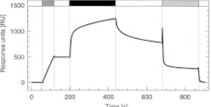
Fig. 1. A typical BIAcore measurement cycle of human uMtCK.
where and ka1and ka2are the association rate constants
The cycle consisted of vesicle immobilization ( ), buffer wash (on rates), kd1 and kd2 the dissociation rate constants
(M), contact (association) phase with 0.15 mM human uMtCK (m), (off rates, already calculated from the dissociation dissociation phase (M), chip regeneration with 1% SDS ( ) and phase), and Req1 and Req2 the equilibrium responses buffer wash (M). For details see Materials and Methods. (Req is reached when bound and free analyte are in
equilibrium or steady state). The dissociation
equilib-1% SDS removes the vesicles and recovers the avidin rium constants or affinity constants were calculated as
surface for repeated immobilization cycles. Avidin also
Kd5 kd/ka (3) minimizes nonspecific binding of basic analyte pro-teins like MtCK to the sensorchip surface. Since avidin To verify the obtained rate and equilibrium constants,
has a basic isoelectric point, it neutralizes remaining nega-both were independently calculated with linear plots
tive charges of the carboxymethyl chip and leads to elec-derived from contact phase kinetics. A modified
equa-trostatic repulsion of any basic analyte protein at the given tion (2), where kaand kdwere replaced by the
concen-pH of 7. However, unspecific binding increased with the tration dependent “apparent” rate constant
number of measurement cycles and was then corrected
kobs5 ka? c 1 kd (4) by blank runs without immobilized vesicles.
Bovine serum albumin (BSA) and cytochrome c was fitted to contact phase kinetics. Slope and y-axis
were used to check the specificity of our experimental intercept of a linear fit to the plot kobsversus c yielded
setup for peripherally binding membrane proteins. estimates for the rate constants ka and kd. The slope
BSA (1.2mM) did not show any specific interaction of the plot Req/c versus c, analogous to a Scatchard
with liposomes after correction for background binding plot, provided estimates for the equilibrium constant
(Fig. 2d). By contrast, injection of 1.2mM cytochrome (or affinity constant) Kd.
c, a basic peripheral membrane protein, resulted in
pronounced association and dissociation kinetics (Fig. 2a). Using 16% CL-vesicles, we could estimate the RESULTS
dissociation equilibrium constant Kdof cytochrome c to be in the lowermM range (1–10 mM).
We used immobilization of biotinylated
lipo-somes on an avidin-coated surface to characterize We further verified if the presence of CL is neces-sary for the strong interaction of human uMtCK octam-membrane interaction of human ubiquitous MtCK
(uMtCK) and other proteins with the surface plasmon ers with liposomes. The results shown in Fig. 2 demonstrate that binding to pure PC vesicles was very resonance technique (BIAcore). The liposomes
con-tained 84% phosphatidylcholine (PC) and 16% cardio- weak (Fig. 2c) as compared to vesicles containing 16% CL (Fig. 2b). The binding properties of dimeric and lipin (CL), thus mimicking the mitochondrial inner
membrane. octameric human uMtCK were compared at low pro-tein concentrations (12.5mg-ml21). Under this condi-A typical measurement cycle with human uMtCK
is shown in Fig. 1. Visual inspection of the kinetic tion, dimeric uMtCK can be obtained by simple dilution and incubation of octamers at 48C for 24 h in data already reveals that the enzyme indeed interacts
with the CL-containing vesicle surface. The quantity running buffer. Dissociation of octameric MtCK with the transition state analog complex (TSAC, Milner-of immobilized liposomes and thus the binding
capac-ity of the surface could be exactly adjusted to 500 RU. White and Watts, 1971) was avoided, since the latter is less efficient in case of human uMtCK (Schlattner The SPR tracing also illustrates how washing with
octameric control uMtCK (Fig. 3a). The response of dissociated uMtCK was clearly higher than could be expected from the remaining 5% octamers. We can, therefore, conclude that dimeric uMtCK also has bound to the 16% CL-vesicles.
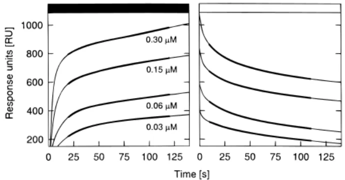
To obtain exact binding parameters, association and dissociation kinetics were recorded in a concentra-tion range of uMtCK from 0.03 to 0.30mM (Fig. 4a, b). The obtained SPR kinetics did not follow a simple 1:1 interaction model, as already observed in other studies with immobilized vesicles (Stachowiak et al., 1996; Lange and Koch, 1997). Therefore, we analyzed the data using a heterogeneous interaction model, which implies two independent binding sites. We have
Fig. 2. Binding and dissociation kinetics of cytochrome c, BSA,
taken some precautions to avoid the possibility that and uMtCK. Contact phase (m) and dissociation phase (M) of 1.2
mM cytochrome c (a), 1.2 mM BSA (d) or 0.15 mM octameric heterogeneity is artificially introduced in our experi-uMtCK (b,c) with liposomes containing 16% CL and 84% PC mental system, e.g., by mass transport limitations or (a,b,d) or 100% PC (c). Data, recorded at 258C and a flow rate of
rebinding effects during dissociation (Myszka, 1997; 0.3 ml-h21, were corrected for unspecific binding. For details see
Schuck and Minton, 1996). We routinely used a low Materials and Methods.
binding capacity (immobilization of only 500 RU vesi-cles) and analyzed only those parts of the kinetics that are least likely to be influenced by mass transport
et al., unpublished) and since TSAC components may, limitations (see solid lines in Figure 4a, b and Materials
in addition, interfere with the binding process. A fresh and Methods for details). Additional control experi-uMtCK dilution contained .95% octamers (control ments were performed at higher flow rate (0.9 ml-h21,
uMtCK); the octamer content was decreased to about not shown). Although these measures enhanced data 5% after 24 h of incubation (dissociated uMtCK). The quality, as judged by improved fitting, they did not SPR tracings, corrected for unspecific interactions, allow description of the SPR kinetics by a model sim-revealed binding of dissociated, dimeric uMtCK (Fig. pler than heterogeneous interaction. The integrated rate 3b) during contact phase, albeit weaker than for the equations of such a model, consisting of double
expo-Fig. 4. Typical binding and dissociation kinetics of octameric
human uMtCK. Contact phase (m) and dissociation phase (M) of octameric human uMtCK, recorded at 258C and a flow rate of 0.3 ml-h21. MtCK was applied in running buffer (10 mM TES, 50 mM NaCl) at four different concentrations (for details see Materials and
Fig. 3. Comparative binding kinetics of dimeric and octameric Methods). The response units (RU) are proportional to the amount of MtCK bound at the vesicle surface (faint lines). Parts of the human uMtCK. Contact phase (m) of dilutions containing 12.5 mg
ml21human uMtCK in (a) the octameric form (.95% octamers) kinetics unlikely to be influenced by refractive index changes, mass transport, or rebinding effects were used for mathematical analysis or (b) the dimeric form (95% dimers), recorded at 258C and a flow
rate of 0.6 ml-h21. Data were corrected for unspecific binding. For of rate and equilibrium constants (bold lines). For details see Materi-als and Methods.
nentials (see Materials and Methods) properly fitted most kinetic data with residuals below 1 RU.
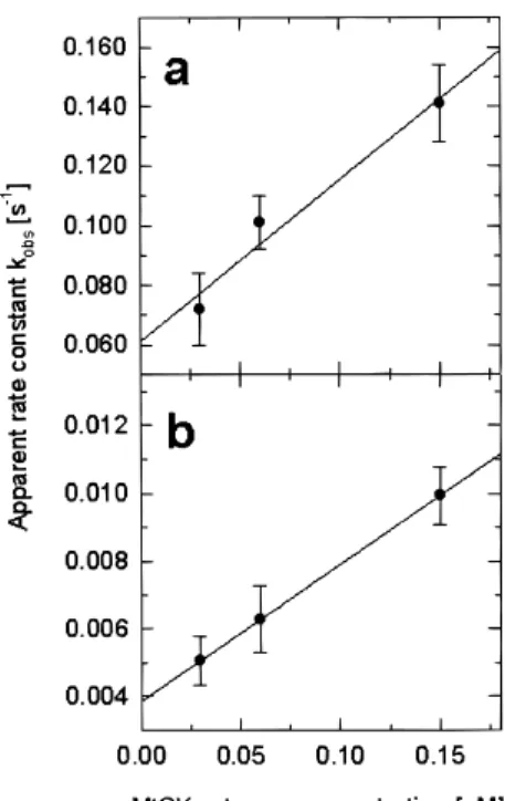
Association and dissociation rate constants (ka and kd) as well as the dissociation equilibrium constants (Kd) were calculated by two independent methods and are summarized in Table I. First, rate constants were directly derived from the fit of the double exponential rate equations to SPR data of contact and dissociation phase [see Eqs. (1) and (2) in Materials and Methods]. Independently, both rate constants were also estimated from contact phase kinetics. The rate equation applied in the latter method uses the global apparent rate con-stants kobs1and kobs2 with kobs5 ka ? c 1 kd [see Eq. (4) in Materials and Methods]. As seen in a plot of
kobsversus c (Fig. 5a,b), kobsindeed showed a linear
dependency on analyte concentration. The rate con-stants of both interaction sites could thus be calculated from slope and y-axis intercept of a linear fit to this plot. Dissociation equilibrium constants (or affinity constants) were directly calculated from the above rate
constants [see Eq. (3) in Materials and Methods]. Inde- Fig. 5. Concentration dependence of the apparent rate kobsduring pendently from rate constants, Kdwas derived from the contact phase. Plots for (a) the first and (b) the second binding site, derived by fitting the contact phase kinetics of octameric human concentration dependency of the equilibrium response
uMtCK (Fig. 2) to a heterogeneous interaction model. Each data
Req. Although equilibrium was not entirely reached
point represents the mean value6 SD of at least three experiments. in our experiments, it could be estimated from the
extrapolation of the contact phase kinetics (Fig. 5a,
b). Kd was then directly calculated from the slope of terize the binding of peripheral membrane proteins to the Scatchard plot Req/c versus Req(Fig. 5c,d). lipid bilayers.
Our experimental setup was validated with cyto-chrome c and BSA as positive and negative binding DISCUSSION controls, respectively (Fig. 2). The interaction of the basic protein cytochrome c with lipids is considered a paradigm for the electrostatic binding of peripheral This study presents first experimental evidence
and provides quantitative data on the membrane-bind- proteins to biological membranes (e.g., Brown and Wu¨thrich, 1977; Heimburg and Marsh, 1995) and has ing properties of the human ubiquitous mitochondrial
CK isoform (uMtCK). It further provides an evaluation been studied intensively (e.g., DeKruijff and Cullis, 1980; Rytomaa, Mustonen, and Kinnunen, 1992; Sala-of surface plasmon resonance with biotin–avidin
immobilized vesicles as a method to generally charac- mon and Tollin, 1996). Similar to MtCK, cytochrome
Table I. Binding Parameters for the Interaction of Octameric Human uMtCK with 16% CL Vesicles
Rate constants Dissociation equilibrium constants ka1[M21-s21] ka2[M21-s21] kd1[s21] kd2[s21] Kd1[nM] Kd2[nM] (a)6.16 2.2 105 3.86 2.2 104 4.86 0.1 1022 2.86 0.9 1023 786 37 746 34 (b)5.46 1.1 105 4.16 0.1 104 6.16 1.0 1022 3.86 0.1 1023 1036 22 846 49
(a)Rate constants derived from the direct fit of SPR data to dissociation and association rate equations and K
dvalues calculated as Kd5
kd/ka(for details see Materials and Methods).
(b)Rate constants derived from linear plots of k
c binding to membranes shows highest affinity for CL in vivo (Soboll et al., 1999). However, the
dimer-specific properties may not only depend on a lack of (Rytomaa, Mustonen, and Kinnunen, 1992), but also
includes some hydrophobic component (Salamon and membrane interaction, since differences in membrane binding of the two oligomeric forms are quantitative Tollin 1996). Our SPR method detected pronounced
binding to CL-containing membranes with cytochrome rather than qualitative.
The binding kinetics of human uMtCK dimers
c and uMtCK and, as expected, no binding with the
soluble protein BSA. Thus, the system is perfectly could neither be fitted with single nor double exponen-tial rate equations, thus hampering the calculation of suited for qualitative analysis of peripheral membrane
proteins binding to defined lipid bilayers. Furthermore, quantitative binding parameters and indicating a more complex binding mode. Such a binding might consist the Kdestimated for the cytochrome c–CL interaction
is similar to data recently obtained with a different of multiple steps, e.g., low-affinity attachment of dimers followed by membrane-mediated octameriza-SPR method (Salmon and Tollin, 1996), indicating
that our experimental setup is also capable of yielding tion, which finally leads to high-affinity binding. An effect of membranes on MtCK octamerization has also meaningful quantitative binding parameters.
Human uMtCK octamers, while strongly binding been observed upon rebinding of sMtCK dimers to mitoplast membranes (Schlegel et al., 1990).
to CL-containing vesicles, showed only a weak
interac-tion with pure PC vesicles. This result confirms that Quantitative analysis of the interaction between human uMtCK octamer and 16% CL-containing lipo-binding of uMtCK depends on the presence of acidic
phospholipids, as do several sMtCK isoforms (Mu¨ller somes suggested the presence of two independent bind-ing sites and yielded rate and equilibrium (affinity)
et al., 1985; Vacheron et al., 1997; Schlattner et al.,
1998). In contrast to octamers, sMtCK dimers were constants for both of them (summarized in Table I). It is important that constants calculated by a direct fit claimed to be unable to interact with mitochondrial
membranes (Marcillat et al., 1987). Other studies to the SPR data were the same as the values derived from the linear plots kobsversus c and Req/c versus Req.
detected a weak binding of sMtCK dimers, although
the ability to cross-link membranes was drastically This is an important prerequisite for internal consis-tency of SPR data analysis (Schuck and Minton 1996). reduced compared to the octamer (Schlegel et al.,
1990, Rojo et al., 1991a,b). In agreement with these Our results for human uMtCK do not, however, entirely coincide with the values obtained with SPR for latter results, we find a weak but specific binding of
uMtCK dimers to CL-containing liposomes. Earlier binding of chicken sMtCK to cardiolipin membranes (Stachowiak et al., 1996). This may be, in part, due reports probably failed to detect dimer binding because
of insufficient sensitivity of the method and the use to differences between the two isoforms. In addition, the present study used a refined analytical approach, of basic pH, which is known to diminish membrane
affinity of MtCK (Marcillat et al., 1987). In fact, the taking into account the concentration dependence of MtCK binding and the influence of dissociation during weaker membrane interaction of sMtCK dimers as
compared to octamers was often ascribed to the more the contact phase.
The dissociation equilibrium constants (Kd1 and acidic pI of dimers (Wyss et al., 1992). However, in
case of the human MtCK isoforms, the difference in Kd2often also called affinity constants) are a measure of the binding strength or affinity of the interaction. pI amounts only to 0.3 pH units (Schlattner et al.,
unpublished), which may be insufficient to explain the All determined Kd values were in the range of 80 to 100 nM and, thus, we can consider that, within difference in membrane interaction observed here.
A different membrane binding behavior of the experimental error, both binding sites have the same affinity. The calculated values are in the same order two oligomeric forms of MtCK would be in line with
other diverging and physiologically relevant proper- of magnitude as the more recent literature data for sMtCK isoforms (Table II), especially the study of ties. sMtCK dimers, in contrast to octamers, are unable
to maintain full creatine-stimulated respiration in iso- Vacheron et al. (1997) using very similar CL-con-taining liposomes.
lated mitochondria (Khuchua et al., 1998) and to delay
the opening of the permeability transition pore in vitro Association and dissociation rate constants (ka and kd) are measures of the velocity of the interaction. (O’Gorman et al., 1997). Recently, we have found an
increase of sMtCK dimers in two different animal In contrast to Kd, the two binding sites differed in their rate constants by one order of magnitude. Fast models of ischemic heart and proposed the
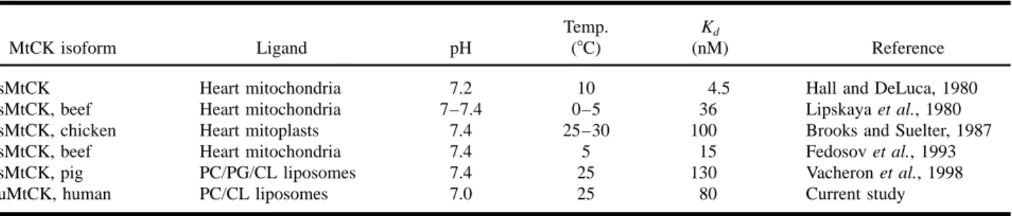
Table II. Dissociation Equilibrium Constants (Kd) for the Binding of Different MtCK Isoforms to Mitochondrial Membranes and
Liposomes
Temp. Kd
MtCK isoform Ligand pH (8C) (nM) Reference
sMtCK Heart mitochondria 7.2 10 4.5 Hall and DeLuca, 1980
sMtCK, beef Heart mitochondria 7–7.4 0–5 36 Lipskaya et al., 1980 sMtCK, chicken Heart mitoplasts 7.4 25–30 100 Brooks and Suelter, 1987
sMtCK, beef Heart mitochondria 7.4 5 15 Fedosov et al., 1993
sMtCK, pig PC/PG/CL liposomes 7.4 25 130 Vacheron et al., 1998
uMtCK, human PC/CL liposomes 7.0 25 80 Current study
and, conversely, slow association (ka2) was linked to static, slow binding could involve specific CL patches, forming nonbilayer structures (hexagonal HIIphases;
slow dissociation (kd2) (Table I). We may therefore
distinguish “fast” and “slow” binding sites. They con- de Kruijff and Cullis, 1980) and MtCK undergoing structural changes to expose a hydrophobic stretch in tribute about 60 and 40%, respectively, to the total
amount of binding sites, as estimated from the equilib- the C-terminus (Schlattner et al., 1998). This process would lead to partial penetration of MtCK into the rium response of both kinetics (Fig. 6a,b). At present,
it is difficult to decide if the phenomenological occur- lipid bilayer as already observed upon sMtCK binding to cardiolipin (Rojo et al., 1991a; Vacheron et al., rence of two binding sites is due to our experimental
design or if it reflects the actual binding mode of 1997). Partial integration of MtCK into the membrane would also explain its resistance against full detach-human uMtCK. If the latter is true, fast and slow
binding may be the key to elucidate the physical nature ment by high ionic strength and the presence of such a bulky enzyme in the narrow mitochondrial intermem-of the two binding sites. For example, they may
repre-sent two different binding sites at the MtCK surface brane space (Schlattner et al., 1998).
We conclude that SPR with immobilized lipo-or, vice versa, two different kinds of CL-patches on
the liposomes. However, it is possible that both may somes, which we have presented here, is an appropriate method to discriminate the binding behavior of periph-be the case. While fast binding might periph-be purely
electro-eral membrane proteins and is able to yield quantitative data, including rate and equilibrium constants. It will be suitable to describe the membrane binding of differ-ent CK isoforms or mutant proteins and to further examine the hydrophobic binding component. The experimental setup will also allow analysis of interac-tions with more complex membranes, including differ-ent lipid mixtures and reconstituted transmembrane proteins like the adenylate translocator. Schlame and Augustin (1985) described binding sites of MtCK on mitoplasts differing in their affinity by two orders of magnitude. The single Kdof the MtCK–CL interaction will be an experimental advantage when trying to resolve additional binding sites in more complex model membranes.
Fig. 6. Affinity of octameric human uMtCK for 16% cardiolipin ACKNOWLEDGMENTS
vesicles: (a,b) concentration dependence of the equilibrium response Reqand (c,d) corresponding Scatchard plots for (a,c) the
This work was supported by the Swiss National first and (b,d) the second binding site. Reqvalues for the two binding
Science Foundation (Grant no. 3100-5082.97 to T.W. sites were derived from the contact phase fit. Each data point
Mu¨ller, M., Moser, R., Cheneval, D., and Carafoli, E. (1985). J. REFERENCES
Biol. Chem. 260, 3839–3843.
Myszka, D. G. (1997). Current Opinions Biotechnol. 8, 50–57. O’Gorman, E., Beutner, G., Dolder, M., Koretsky, A. P., Brdiczka,
D., and Wallimann, T. (1997). FEBS Lett. 414, 253–257. Beutner, G., Ru¨ck, A., Riede, B., Welte, W., and Brdiczka, D.
(1996). FEBS Lett. 396, 189–195. Payne R. M., Haas, R. C., and Strauss A. W. (1991). Biochim. Biophys. Acta 1089, 352–361.
Beutner, G., Ru¨ck, A., Riede, B., and Brdiczka, D. (1998). Biochim.
Biophys. Acta 1368, 7–18. Rojo, M., Hovius, R., Demel, R., Wallimann, T., Eppenberger, H. M., and Nicolay, K. (1991a). FEBS Lett. 281, 123–129. Bradford, M. M. (1976). Anal. Biochem. 72, 248–254.
Brooks, S. P. J., and Suelter, C. H. (1987). Arch. Biochem. Biophys. Rojo, M., Hovius, R., Demel, R. A., Nicolay, K., and Wallimann, T. (1991b) J. Biol. Chem. 266, 20290–20295.
253, 122–132.
Brown, L. R., and Wu¨thrich, K. (1977). Biochim. Biophys. Acta Rytomaa, M., Mustonen, P., and Kinnunen, P. K. (1992). J. Biol. Chem. 267, 22243–22248.
468, 389–410.
Cheneval, D., Carafoli, E., Powell, G. L., and Marsh, D. (1989). Saks, V. A., Khuchua, Z. A., Kuznetsov, V. A., Veksler, V. I., and Sharov, V. G. (1986) Biochem. Biophys. Res. Commun. Eur J. Biochem 186, 415–419.
De Kruijff, B., and Cullis, P. R. (1980). Biochim. Biophys. Acta 139, 1262–1271.
Salamon Z., and Tollin, G. (1996). Biophys. J. 71, 848–857.
602, 477–490.
Fedosov, S. N., Belousova, L. V., and Plesner, I. W. (1993). Biochem. Salamon, Z., Macleod, H. A., and Tollin, G. (1997). Biochim. Biophys. Acta 1331, 131–152.
Biophys. Acta 1153, 322–330.
Fritz-Wolf, K., Schnyder, T., Wallimann, T., and Kabsch, W. (1996). Schlame, M., and Augustin, W. (1985). Biomed. Biochim. Acta
44, 1083–1088.
Nature (London) 381, 341–345.
Furter, R., Kaldis, P., Furter-Graves, E. M., Schnyder, T., Eppenb- Schlattner, U., Forstner, M., Eder, M., Stachowiak, O., Fritz-Wolf, K., and Wallimann, T. (1998). Mol. Cell. Biochem. 184, erger, H. M., and Wallimann, T. (1992). Biochem. J. 288,
771–775. 125–140.
Schlegel, J., Wyss, M., Eppenberger, H. M., and Wallimann, T. Hall, N., and DeLuca, M. (1980). Arch. Biochem. Biophys. 201,
674–677. (1990). J. Biol. Chem. 265, 9221–9227.
Schuck, P., and Minton, A. P. (1996). Trends Biochem. Sci. 252, Heimburg T., and Marsh, D. (1995). Biophys. J. 69, 536–546.
Heyse, S., Ernst, O. P., Dienes, Z., Hofmann, K. P., and Vogel, H. 458–460.
Soboll, S., Brdiczka, D., Jahnke, D., Schmidt, A., Schlattner, U., (1997). Biochemistry 37, 507–522.
Johnson, B., Lo¨fas, S., and Lindquist, G. (1991). Anal. Biochem. Wendt, S., Wyss, M., and Wallimann T. (1999). J. Mol. Cell. Cardiol. 31, 857–866.
198, 268–277.
Khuchua, Z. A., Qin, W., Boero, J., Cheng, J., Payne, R. M., Saks, Stachowiak, O., Dolder, M., and Wallimann, T. (1996). Biochemis-try 35, 15522–15528.
V. A., and Strauss, A. W. (1998). J. Biol. Chem. 273,
22990–22996. Stachowiak, O., Schlattner, U., Dolder, M., and Wallimann, T. (1998). Mol. Cell. Biochem. 184, 141–151.
Lange C., and Koch, K.-W. (1997). Biochemistry 36, 12019–12026.
Lipskaya, T. Y., Templ, V. D., Belousova, L. V., Molokova, E. V., Steffner, P., and Markey, F. (1997). When the chips are down. J. Biomol. Interact. Anal. 4, 11–15.
and Rybina, I. V. (1980). Biochimia USSR 45, 877–886.
Marcillat, O., Goldschmidt, D., Eichenberger, D., and Vial, C. Vacheron, M.-J., Clottes, E., Chautard, C., and Vial, C. (1997). Arch. Biochem. Biophys. 344, 316–324.
(1987). Biochem. Biophys. Acta 890, 233–241.
Masson, L., Mazza, A., and Brousseau, R. (1994). Biochemistry Wallimann, T., Wyss, M., Brdiczka, D., Nicolay, K., and Eppenb-erger, H. M. (1992). Biochem. J. 281, 21–40.
218, 405–412.
Milner-White, E. J., and Watts, D. C. (1971). Biochem. J. 122, Wyss, M., Smeitink, J., Wevers, R. A., and Wallimann, T. (1992). Biochim. Biophys. Acta 1102, 119–166.