HAL Id: lirmm-02105827
https://hal-lirmm.ccsd.cnrs.fr/lirmm-02105827
Submitted on 21 Apr 2019
HAL is a multi-disciplinary open access
archive for the deposit and dissemination of
sci-entific research documents, whether they are
pub-lished or not. The documents may come from
teaching and research institutions in France or
abroad, or from public or private research centers.
L’archive ouverte pluridisciplinaire HAL, est
destinée au dépôt et à la diffusion de documents
scientifiques de niveau recherche, publiés ou non,
émanant des établissements d’enseignement et de
recherche français ou étrangers, des laboratoires
publics ou privés.
Towards unified dataset for Modeling and Monitoring of
Computer Assisted Medical Interventions
Fabien Despinoy, Sandrine Voros, Nabil Zemiti, Nicolas Padoy, Pierre Jannin
To cite this version:
Fabien Despinoy, Sandrine Voros, Nabil Zemiti, Nicolas Padoy, Pierre Jannin. Towards unified dataset
for Modeling and Monitoring of Computer Assisted Medical Interventions. SURGETICA, Nov 2017,
Strasbourg, France. �lirmm-02105827�
F. DESPINOY et al. Surgetica 2017 Strasbourg, France – 20 - 22 Nov.
Towards unified dataset for
Modeling and Monitoring of
Computer Assisted Medical
Interventions
Fabien DESPINOY
1, Sandrine VOROS
2, Nabil ZEMITI
3, Nicolas PADOY
4and Pierre
JANNIN
11 : LTSI, UMR 1099, University of Rennes 1, Inserm, F-35043, France
2 : TIMC-IMAG, UMR 5525, University of Grenoble-Alpes, CNRS, F-38706, France
3 : LIRMM, UMR 5506, University of Montpellier, Inserm, F-34095, France
4 : ICube, UMR 7357, University of Strasbourg, CNRS, F-67000, France
Contact: fabien.despinoy@univ-rennes1.fr
M
2CAMI is a working group dedicated to Modeling and Monitoring of Computer Assisted Medical Interventions within the CAMI Labex. It aims at unifying data ac-quired from different surgical trainers and pro-cedures for collaborative research. In this pa-per, we propose a generic structure for multi-modal dataset that allows faster development and easier processing. With such formalization, our objective is to go beyond the state of the art by sharing various types of data between inter-national institutions and merge methodological approaches for better detection and understand-ing of surgical workflows.1
Introduction
For the past decade, the medical community has shown a growing interest in biomedical international compe-titions, named challenges. Since 2007, more than 120 challenges have been proposed on the grand-challenge platform1 where various research fields are addressed such as image segmentation and registration, anatomi-cal structure loanatomi-calization and tracking or surgianatomi-cal work-flow detection and understanding. Through such plat-form, multiple types of data are shared going from point cloud depth image acquisition to CT or MRI segmented images. These challenges share the same goal: use pre-clinical and clinical data in order to validate and evaluate methodologies that are developed for biomedi-cal analysis purposes.
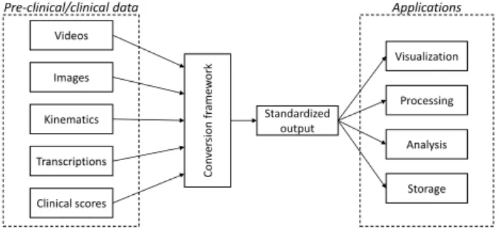
1https://grand-challenge.org/ Images Videos Kinematics Transcriptions Clinical scores Con ver si on fr ame w or k Standardized output Visualization Processing Analysis Storage
Pre-clinical/clinical data Applications
Figure 1: Pipeline for data conversion into a standardized output for M2CAMI applications.
Focusing on surgical workflow detection and under-standing, various datasets have been released over the last years, where multiple types of data are accessible. For each dataset, data representation and storage are unique because of the acquisition setup and the per-sonal requirements. However, this sharing model is not suitable for collaborative research due to the heteroge-neous representation of the same data type. Moreover, such approach does not promote the use of multimodal data for multiscale processing because of the complex integration of various data types and formats.
In this paper, we address the need of a standard for-malism for multimodal data representation and storage. By taking into account the available datasets in the M2CAMI community, we propose a generic structure and a first implementation for data conversion to a generic format that is readable and usable in the dif-ferent applications developed into the M2CAMI group (fig. 1).
F. DESPINOY et al. Surgetica 2017 Strasbourg, France – 20 - 22 Nov.
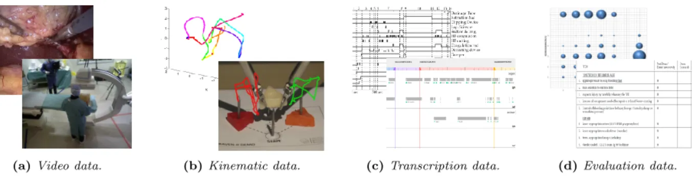
(a) Video data. (b) Kinematic data. (c) Transcription data. (d) Evaluation data.
Figure 2: Multimodal data of surgical workflow available into the M2CAMI collaborative research group.
2
Methods
In the M2CAMI group, data acquisition and analysis are oriented towards surgical workflow detection and understanding. In order to build a unified framework to convert data into a generic representation, we pro-posed a two-step approach. The first step was the inventory of the different available data into the work-ing group. Then, in the second step, we described a common formalism for data representation and storage. From the M2CAMI community, two datasets are pub-licly available: NeuroSurgicalTools [1] and Cholec80 [2] where the latter has been already used for a previous scientific challenge2. To generalize our work, we used also three other available datasets: JIGSAWS [3] as well as acquisitions performed during previous CAMI works [4][5].
2.1
Data inventory
These datasets contain various data types that can be sorted into four distinct categories. Videos are (fig. 2a) automatically captured from the surgical field by record-ing the endoscopic point of view, or from the entire operating room with additional cameras. They can also be expressed as a set of images. Kinematic data (fig. 2b) is automatically captured from robotic systems or additional tracking devices to describe the surgical instrument motions over the time. Transcription data (fig. 2c), or annotations, is most of the time manually generated by human observers and describes the differ-ent actions, instrumdiffer-ents or evdiffer-ents taking place during the procedure. Evaluation data (fig. 2d) quantifies per-formance of the surgeon to achieve a clinical task or procedure. It can be automatically calculated from sur-gical trainers or manually generated by human experts and is necessary for surgical workflow referenced-based evaluations. As an extension to these four categories, unusual clinical data types are emerging and combine annotations with kinematics such as human body pose annotations for medical staff analysis [6]. To cover the full spectrum from training to interventional clinical data available into the M2CAMI group, we developed an inventory form based on a previous formalism [7] to make a list of the different data and structures available.
2http://camma.u-strasbg.fr/m2cai2016/
2.2
Generic data representation
As previously mentioned, a same data type can be rep-resented in different ways. For instance, video is a set of images that is embedded into a streaming container. However, depending on the format, the encoder and the output container it can be difficult to use it for process-ing purpose. Thus, our objective is to define a standard representation that rely on open source protocols (e.g. x265 for video codec, Boost.PropertyTree for configura-tion file) to ease broadcast of our developments. This standard representation can be developed at two differ-ent scales. At the global level, we provide guidelines and tools to uniformly structure dataset in order to ease sharing, processing and hard data storage. Additionally, standardized header files are integrated into datasets to regroup data properties for conversion management. At a second level, we propose a C++ language-based container that stores data synchronously into memory for processing (e.g. as an extended map for data series). The main advantage of the presented structure is to be easily extended to add new types of data. The only as-sumption we rely on is the data synchronization where for each timestamp in the timeline there is at least one sample from each data type available in the dataset.
3
Conclusion and Perspectives
At the current stage, data conversion seems feasible to provide larger datasets for each partner of the M2CAMI group. Relying on open source protocols, the current implementation is shareable between members and im-provements can be easily undertaken. The added value of the current work will be emphasized by the con-nection of various developments coming from different teams, including surgical context detection [2] that will provide entry frame for instrument tracking [8]. The output information from the tool tracking will be used for gesture recognition [9] in order to, at the end, ob-tain a complete comprehension of the surgical context. These are the first steps towards generation of new decision-making systems where multimodal data allows to capture and understand surgical events for training, teaching and surgical assistance purposes.
F. DESPINOY et al. Surgetica 2017 Strasbourg, France – 20 - 22 Nov.
References
[1] D. Bouget, R. Benenson, M. Omran, L. Riffaud, B. Schiele and P. Jannin (2015). Detecting Surgical Tools by Modelling Local Appearance and Global Shape. IEEE Transactions on Medical Imaging, 34(12):2603–2617.
[2] A.P. Twinanda, S. Shehata, D. Mutter, J. Marescaux, M. de Mathelin and N. Padoy (2016). EndoNet: A Deep Architecture for Recognition Tasks on Laparoscopic Video. IEEE Transactions on Medical Imaging, 36(1):86–97.
[3] Y. Gao, S. S. Vedula, C. E. Reiley, N. Ahmidi, B. Varadarajan, H. C. Lin, L. Tao, L. Zappella, B.B´ejar, D. D. Yuh, C. C. G. Chen, R. Vidal, S. Khudanpur and G. D. Hager (2014). The JHU-ISI Gesture and Skill Assessment Working Set (JIG-SAWS): A Surgical Activity Dataset for Human Motion Modeling. Modeling and Monitoring of Computer Assisted Interventions (M2CAI) - MIC-CAI Workshop.
[4] R. Wolf (2014). Quantification de la qualit´e d'un geste chirurgical `a partir de connaissances a priori. Universit´e Grenoble Alpes.
[5] F. Despinoy (2016). Analyse, reconnaissance et r´ealisation des gestes pour l’entraˆınement en chirurgie laparoscopique robotis´ee. Universit´e de Montpellier.
[6] A. Kadkhodamohammadi, A. Gangi, M. De Math-elin and N. Padoy (2017). Articulated clinician detection using 3D pictorial structures on RGB-D data. Medical Image Analysis 35:215–224.
[7] P. Jannin, C. Grova and C. R. Maurer Jr. (2006). Model for defining and reporting reference-based validation protocols in medical image processing. International Journal of Computer Assisted Radi-ology and Surgery, 1(2):63–73.
[8] A. Agustinos and S. Voros (2015). 2D/3D Real-Time Tracking of Surgical Instruments Based on Endoscopic Image Processing. Computer-Assisted and Robotic Endoscopy, 9515:90–100.
[9] F. Despinoy, D. Bouget, G. Forestier, C. Penet, N. Zemiti, P. Poignet and P. Jannin (2016). Unsuper-vised Trajectory Segmentation for Surgical Gesture Recognition in Robotic Training. IEEE Transac-tions on Biomedical Engineering, 63(6):1280–1291.