Journal of Fundamental and Applied Sciences is licensed under aCreative Commons Attribution-NonCommercial 4.0 International License.Libraries Resource Directory. We are listed underResearch Associationscategory.
SYNTHESIS, QUANTUM CHEMICAL COMPUTATIONS AND X-RAY CRYSTALLOGRAPHIC STUDIES OF A NEW COMPLEX BASED
OF MANGANESE (+II)
N. Benyza1,2*, A. Messai1,, A. Hamdaoui1, T. Lanez2and K. Sayin3
1
Laboratoire d’Ingénierie et Sciences des Matériaux Avancés (ISMA) Institut des Sciences et Technologie Université Abbés Laghrour Khenchela ; 40000, Algeria
2
VTRS Laboratory, Department of Chemistry, Faculty of Exact Sciences, University of El-Oued, PO Box 789, El-Oued 39000, Algeria
3
Department of Chemistry, Institute of Science, Cumhuriyet University 58140 Sivas – Turkey
Received: 30 January 2017 / Accepted: 25 April 2017 / Published online: 01 May 2017
ABSTRACT
The ligand oxime, C7H9N5O2, was Synthesis and characterises with different characterization methods such as 1H NMR and FTIR spectroscopy. The complexation of this ligand with manganese (II) perchlorate yielded pink crystals of formula [Mn (C7H9N5O2)2]2+, 2[ClO4]-, which crystallized in the monoclinic space group P21/n with a = 12.824(3), b=13.799(2),
c=15.441(4)Å, β = 100.17(2), and Z = 4. The complex consists of cations (+II) and two perchlorate anions, the cations part existing in a slightly distorted octahedral complex. Computational investigations of manganese (II) complex are done by using the DFTmethod with B3LYP functional in conjunction with the 6-31G(d,p) and lanl2dz basis sets in the gas phase imposing the C1and C2vsymmetries.
Keywords: Manganese complex; Crystal structure; DFT method; B3LYP functional;
6-31G(d,p) and (LANL2DZ) basis.
Author Correspondence, e-mail:nabsat1979@yahoo.fr
doi:http://dx.doi.org/10.4314/jfas.v9i2.11 ISSN 1112-9867
1. INTRODUCTION
The chemistry of oxime-based ligand is diverse [1]. Introduced first in 1905 by Tschugaeff [2], e.g. preparation of nickel (II) dimethylglyoximato and recognition of the chelate five-membered character of this complex by Chugaev [3-4], the oximes as potential ligands have been increasingly expanding its horizon in coordination chemistry. Stability structure and reactivity of molecules, analytical and organometallic chemistry and particularly syntheses of molecules with unusual electronic properties are the aspects that kindled the interest for the oxime-based ligands [5–9]. Oxime ligands are used to obtain polynuclear compounds with molecular magnetism and supramolecular structure Due to there ability to form bridges with metal ions [10, 11]. Also; the presence of additional donor group together with the oxime group in the ligand molecule resulting a significant increase in chelating efficiency and ability to form polynuclear complexes [12].
The literature contains reports of some amide oximes which can co-ordinate as bi-,tri-tetra-and hexa-dentate ligands[13-18]. The majority of these oximes are aliphatic compounds which form five-membered rings utilizing the diimine moiety, -N=C-C=N-, involving oxime and imine nitrogens as the co-ordination sites with transition-metal atoms [19].
Pyridine-2,6-dicarboxamide oxime (pyridine-2,6-diamidoxime), C7H9N5O2 (L), possesses the structural requirements to react as a polydentate ligand with the heterocyclic and two oxime nitrogen atoms forming five-membered rings with metal ions [20].
This research was carried out with the specific purpose of determining the structures of manganese (+II) co-ordination with pyridine-2,6-dicarboxamide oxime. We report here the synthesis, the single crystal X-ray structure of the complex and the Optimization of the structure using DFT method with B3LYP functional at 6-31G (d,p) and (LANL2DZ) levels in vacuo.
2. RESULTS AND DISCUSSION
The crystal structure of [Mn(L)2+2,2(ClO4)-] has been reported and discussed with X-ray single crystal data, the hydrogen-bonding networks has been also studies. Followed with a
quantuim chemical computations of the geometrical parameters of our compound and compared with experimental results.
2.1. Synthesis
The ligand (L) pyridine-2,6-diamidoxime has been prepared as well as [19-20] by the reaction of of 2,6-dicyanopyridine solution with a neutralized aqueous solution of hydroxylamine hydrochloride . The 1H NMR shifts and the infrared spectum bands are listed in the Experimental section.
Reaction of (L) with the manganese perchlorate in alcohool gave the corresponding coordination complex in 83.5% yield.
2.2 Structure
Crystals of complexe suitable for X-ray study were grown by slow evaporation from alcoholic solution, a summary of crystal data and parameters for structure refinement details are given in Table 1. The atomic coordinates and equivalent thermal parameters are also given in Table 2.
Table 1. Crystal data and parameters for structure refinement
Crystal data Complex
Empirical formula Formula weight (g mol-1) Temperature (K)
Crystal system Space group C2/c Hall symbo
Unit cell dimensions (A°) a b c β° Volume (A3) Za Calculated density (g/cm3) Absorption coefficient (mm-1) F(000) Crystal size (mm3) Color Shape
Cell parameters from Wavelength (Mo Ka) (A)
θmax- θmin
Independent reflections reflections with I > 2σ(I) Rint Limiting indices h k l Refinement method Final R indicesb[F2 > 2σ(F2)] R1, wR2 Goodness-of-fit on F2c parameters H atoms
Largest difference peak and hole (e A_3) Δρmax, Δρmin MnC14H8Cl2N10O12(a) Mr = 634.14 293 Monoclinic p21/n -P 2yn 12.824(3) 13.799(2) 15.441(4) 114.691(19) 100.17(2) 4 1.566 0.763 1268 0.05 × 0.0 2× 0.01 mm pink Block 13138 reflections 0.71073 29.27°- 2.95° 6163 3734 0.0237 −12→17 k = −16→17 l = −20→17 Full-matrix Least-squares on F2 0.11, 0.26 1.031 309 a constrained refinement 0.952e , -1.168e
(a) The asymmetric unit contains 0.5 of the chemical formula. (b) R1 =P|Fo -Fc|/Fo. wR2 = {P[w(Fo2 - Fc2)2]/P[w(Fo2)2]}1/2. ( c) S = {P[w(Fo2 - Fc2)2]/(Nobs - Nvar)}1/2.
Table 2. Coordinates and Equivalent Isotropic Displacements Parameters of the
non-Hydrogen atoms (A2)
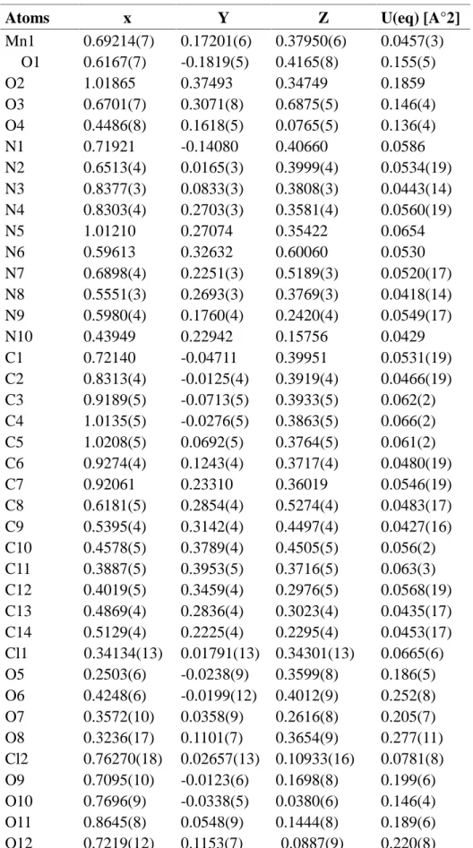
Atoms x Y Z U(eq) [A°2]
Mn1 0.69214(7) 0.17201(6) 0.37950(6) 0.0457(3) O1 0.6167(7) -0.1819(5) 0.4165(8) 0.155(5) O2 1.01865 0.37493 0.34749 0.1859 O3 0.6701(7) 0.3071(8) 0.6875(5) 0.146(4) O4 0.4486(8) 0.1618(5) 0.0765(5) 0.136(4) N1 0.71921 -0.14080 0.40660 0.0586 N2 0.6513(4) 0.0165(3) 0.3999(4) 0.0534(19) N3 0.8377(3) 0.0833(3) 0.3808(3) 0.0443(14) N4 0.8303(4) 0.2703(3) 0.3581(4) 0.0560(19) N5 1.01210 0.27074 0.35422 0.0654 N6 0.59613 0.32632 0.60060 0.0530 N7 0.6898(4) 0.2251(3) 0.5189(3) 0.0520(17) N8 0.5551(3) 0.2693(3) 0.3769(3) 0.0418(14) N9 0.5980(4) 0.1760(4) 0.2420(4) 0.0549(17) N10 0.43949 0.22942 0.15756 0.0429 C1 0.72140 -0.04711 0.39951 0.0531(19) C2 0.8313(4) -0.0125(4) 0.3919(4) 0.0466(19) C3 0.9189(5) -0.0713(5) 0.3933(5) 0.062(2) C4 1.0135(5) -0.0276(5) 0.3863(5) 0.066(2) C5 1.0208(5) 0.0692(5) 0.3764(5) 0.061(2) C6 0.9274(4) 0.1243(4) 0.3717(4) 0.0480(19) C7 0.92061 0.23310 0.36019 0.0546(19) C8 0.6181(5) 0.2854(4) 0.5274(4) 0.0483(17) C9 0.5395(4) 0.3142(4) 0.4497(4) 0.0427(16) C10 0.4578(5) 0.3789(4) 0.4505(5) 0.056(2) C11 0.3887(5) 0.3953(5) 0.3716(5) 0.063(3) C12 0.4019(5) 0.3459(4) 0.2976(5) 0.0568(19) C13 0.4869(4) 0.2836(4) 0.3023(4) 0.0435(17) C14 0.5129(4) 0.2225(4) 0.2295(4) 0.0453(17) Cl1 0.34134(13) 0.01791(13) 0.34301(13) 0.0665(6) O5 0.2503(6) -0.0238(9) 0.3599(8) 0.186(5) O6 0.4248(6) -0.0199(12) 0.4012(9) 0.252(8) O7 0.3572(10) 0.0358(9) 0.2616(8) 0.205(7) O8 0.3236(17) 0.1101(7) 0.3654(9) 0.277(11) Cl2 0.76270(18) 0.02657(13) 0.10933(16) 0.0781(8) O9 0.7095(10) -0.0123(6) 0.1698(8) 0.199(6) O10 0.7696(9) -0.0338(5) 0.0380(6) 0.146(4) O11 0.8645(8) 0.0548(9) 0.1444(8) 0.189(6) O12 0.7219(12) 0.1153(7) 0.0887(9) 0.220(8)
The reaction of manganese (II) perchlorate with pyridine-2,6-dicarboxamide oxime produced a pink 1:2 complex (1), the crystal structure of this complex confirms the 1:2 MnII+ligand co-ordination where the perchlorates anions are not involved in the co-ordination.
The complex crystallizes in the monoclinic space group P21/n with the unit cell parameters a = 12.824(3)A°, b = 13.799(2)A°, c = 15.441(4)A° and β= 114.691(19).with four molecules in the unit cell (one per asymmetric unit) . The asymmetric unit, formed by one Mn2+ metal ion, two crystallographically independent oxime ligands and two perchlorate counter-ion located on general positions. The molecular structure of the compound showing the atom numbering scheme is shown in Figure. 1
Fig.1. The molecular structure of the complex Mn(L)2
Manganese (II) ion is six-co-ordinated through the amine and heterocyclic nitrogens of the two oxime molecules. The resulting complexe is the slightly distored octahedron, caused by non-equivalent Mn-N bond length (Table 3 and 4). The two Mn-N (imine) bond length are almost equivalent, but shorter than the four Ni-N (amine) wich are not mutually equivalent. The two co-ordinated oximes are at 85.28° are almost orthogonal to each other. A comparison of the same bond lengths and angles for the free ligand and the manganese (II) complex shows little change in (L) and (1)[21].
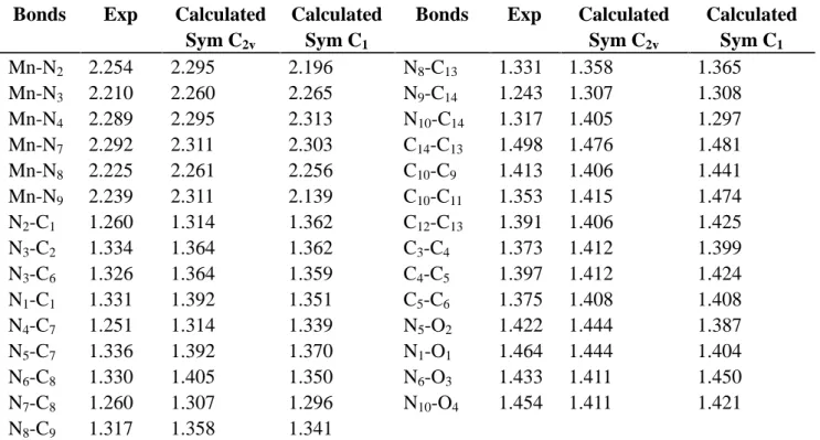
Table 3. Experimental and calculated bond lengths (Å) of Mn(II) complex by using
DFT/UB3LYP/LanL2DZ method
Bonds Exp Calculated
Sym C2v
Calculated Sym C1
Bonds Exp Calculated
Sym C2v Calculated Sym C1 Mn-N2 2.254 2.295 2.196 N8-C13 1.331 1.358 1.365 Mn-N3 2.210 2.260 2.265 N9-C14 1.243 1.307 1.308 Mn-N4 2.289 2.295 2.313 N10-C14 1.317 1.405 1.297 Mn-N7 2.292 2.311 2.303 C14-C13 1.498 1.476 1.481 Mn-N8 2.225 2.261 2.256 C10-C9 1.413 1.406 1.441 Mn-N9 2.239 2.311 2.139 C10-C11 1.353 1.415 1.474 N2-C1 1.260 1.314 1.362 C12-C13 1.391 1.406 1.425 N3-C2 1.334 1.364 1.362 C3-C4 1.373 1.412 1.399 N3-C6 1.326 1.364 1.359 C4-C5 1.397 1.412 1.424 N1-C1 1.331 1.392 1.351 C5-C6 1.375 1.408 1.408 N4-C7 1.251 1.314 1.339 N5-O2 1.422 1.444 1.387 N5-C7 1.336 1.392 1.370 N1-O1 1.464 1.444 1.404 N6-C8 1.330 1.405 1.350 N6-O3 1.433 1.411 1.450 N7-C8 1.260 1.307 1.296 N10-O4 1.454 1.411 1.421 N8-C9 1.317 1.358 1.341
Table 4. Experimental and Calculated angles (deg.) of Mn(II) complex by using
DFT/UB3LYP/LanL2DZ method
Angels Exp Calc
Sym C2v
Calc Sym C1
Angels Exp Calc
Sym C2v Calc Sym C1 N2-Mn-N3 071.39 078.58 075.90 Mn –N3-C2 120.20 119.33 121.30 N2-Mn-N4 142.11 147.17 149.20 Mn –N3-C6 120.40 119.33 120.09 N2-Mn-N9 093.01 092.17 091.50 Mn –N4-C7 116.50 116.11 117.21 N2-Mn-N8 109.00 101.41 108.70 C1–C2-N3 111.80 109.50 110.50 N2-Mn-N7 098.84 092.17 090.40 N3–C6-C7 112.40 109.50 108.59 N3-Mn-N4 070.89 078.58 077.40 Mn –N9-C14 118.80 115.13 114.15 N3-Mn-N7 105.75 101.06 102.60 Mn –N8-C13 118.60 118.83 119.03 N3-Mn-N8 176.11 180.00 170.90 Mn –N8-C9 120.60 118.83 120.73 N3-Mn-N9 112.44 101.06 100.16 Mn –N7-C8 118.10 115.13 117.33 N4-Mn-N7 094.63 092.17 93.30 C14–C13-N8 113.00 110.03 111.09 N4-Mn-N8 108.87 101.41 102.10 N8–C9-C8 113.30 110.03 111.93 N4-Mn-N9 097.90 092.17 098.40 C2–C3-C4 118.70 118.14 117.10 N7-Mn-N8 070.37 078.93 078.10 C3–C4-C5 119.30 120.60 119.99 N7-Mn-N9 141.81 147.87 141.90 C6–N3-C2 119.40 121.32 120.52 N8-Mn-N9 071.44 078.93 074.50 C13–N8-C9 120.70 122.33 123.03 Mn –N2-C1 117.90 116.11 128.01 C12–C11-C10 122.20 121.03 120.93
Both of the ligands molecules are almost planers. Deviation from two plans compresing all non-H atoms: for the N3-ligand molecule, 0.041 A°; for the N8 molecule, 0.092 A°. The Mn-N bond lengths to the two ligands are comparable, averaging 2.30 A° to the slightly longer bonds to the amine atoms and 2.21 A° to the pyridine nitrogen atoms. These are comparable to[Mn(C9H11N3O2)2][2(ClO4)-]. ( avg. Mn-Noxime=2.29A°; avg Mn-Npyr= 2.18,[21].
The perchlorate anion is not disordered at 293 K, and it is stabilized by strong interactions with its environment. The averages Cl-O bond distances; in the two perchlorate ions present in the asymmetric unit are 1.3555 A° and 1.370 A° respectively and the averages O-Cl-O bond angles are 109° and 109.3° (17), respectively, confirming a tetrahedral configuration , similar to other perchlorates studied at low temperature) [20, 22-24].
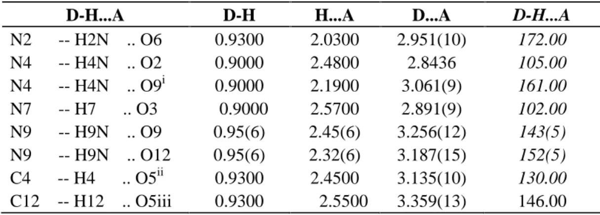
Due of the presence of two perchlorates (ClO4-) ions, the crystal pickings of the complex are mainly consolidate by N-H…O, O-H…O and C-H…O intra and intramolecular hydrogen bonds. Therefore, a total of eight hydrogen bonds are present in the unit (Figure.2) and (Table.5). Significant hydrogen bonds are formed between the oxime NH functions and the perchlorates ions to link molecules into parallel chaines (Figure.3).
Table 5. Hydrogen bond lengths (A) and angles (°)
i : [3/2-x,1/2+y,1/2-z] ; ii: [1+x,y,z] ; iii: [1/2-x,1/2+y,1/2-z]
D-H...A D-H H...A D...A D-H...A
N2 -- H2N .. O6 0.9300 2.0300 2.951(10) 172.00 N4 -- H4N .. O2 0.9000 2.4800 2.8436 105.00 N4 -- H4N .. O9i 0.9000 2.1900 3.061(9) 161.00 N7 -- H7 .. O3 0.9000 2.5700 2.891(9) 102.00 N9 -- H9N .. O9 0.95(6) 2.45(6) 3.256(12) 143(5) N9 -- H9N .. O12 0.95(6) 2.32(6) 3.187(15) 152(5) C4 -- H4 .. O5ii 0.9300 2.4500 3.135(10) 130.00 C12 -- H12 .. O5iii 0.9300 2.5500 3.359(13) 146.00
Fig.2. Crystal packing of the complex showing the H-bonding patterns
Fig.3. A fragment of the structure showing the hydrogen bonds as dashed lines
2.3 Fully optimizations
package [25] by means of the resources provided by GridChem Science Gateway [26]. GaussView 5.o8 [27] was used for visualization of the structure and simulation of the vibrational spectra. Optimized structures of complex optimized using the DFT method with B3LYP functional in conjunction with the 6-31G(d, p) and lanl2dz basis sets in the gas phase imposing the C1 and C2v symmetries. IR spectrum of this complexe is calculated at same level of theory and examined in detail [28-30].
Table 1, Table 2 and figure.4 summarizes the optimized structure of manganese complex [C14H18MnN10O4]2+ with differents symmetries and its bond lengths and angles respectively. For [C14H18MnN10O4]2+, the optimized bond distances and angles are well correlated with the X-ray bond parameters are given in (figure.4).
Fig.4. Ortep diagram of [C14H18MnN10O4]2+ complex 50% probability thermal ellipsoids.showing the atomic numbering scheme and theoretically geometry optimization with C2V symmetry for nominated complex by using DFT/UB3LYP method with LanL2DZ level.
According to Table 1 and Table 2, there are good agreements between experimental and calculated results. It is clear from these results there are very few differences between the optimized structure and the structure determined by the X-ray diffraction and the largest
difference between the experimental and calculated bond lengths and bond angles for the C2V and C1 symmetry with the lanl2dz level are respectively 0,05 Å and 0,1 Å (Mn-N) and 11,38 deg ,12,28 deg ( N-Mn-N ).
Distribution graphs between experimental and calculated results for bond lengths and bond angles are presented in Figure.5.
[C14H18MnN10O4]2+with C2Vsymmetry
[C14H18MnN10O4]2+with C1symmetry
Fig.5. The distribution graphs between experimental and calculated results for [C14H18MnN10O4]2+in symmetries C2Vand C1respectively.
According to the Fig. 5, the correlation constants (R2) are calculated for the C2V and C1 symmetries are respectively o.992 and o.985 for bond lengths and 0.937 and 0.97 for bond angles.
The IR spectrum of Mn(II) complex is calculated at the same level of theory and represented in Figure. 6.
Fig.6. Calculated IR spectrum of Mn(II) complex with C2V symmetry at DFT/UB3LYP/(LANL2DZ) level in vacuo.
Calculated stretching frequencies are obtained as: νNH(3430 and 3433 cm-1), νCH(3244 – 3252 cm-1), νN=O(1534 cm-1), νC=Nand νC=C(1120 cm-1), νC=Nin pyridine (966.62 cm-1), νMn-N (677 and 399.39 cm-1). These frequencies are not scaled and these are anharmonic frequencies and harmonic frequencies are scale by 0.961.
As for the values of the theoretical frequencies of vibration, there are all real and positive, indicating that the optimized structure corresponds to stationary state having a minimum of the hypersurface of the potential energy.
The diagrams of molecular orbitals and Contour plots of selected molecular orbitals of [C14H18MnN10O4]2+are summarized in figure.7.
-11.0 --11.50 -12.0 -12.50 -13.0 -13;50 E(eV) 2.236 eV -14.0 -10.75 1.061 eV
Molecular orbitals (MOs) and their energy diagrams are important to determine the molecular properties. Especially, frontier molecular orbitals (FMOs) are playing important role in determination of chemical reactivity. Multiplicity of investigated compound is two in mentioned Mn complex. The energy diagrams from HOMO-2 to LUMO+2 are given in Figure. 7.
Fig.7. Molecular orbital energy diagrams from HOMO-2 to LUMO+2 and Contour diagrams
of some orbital of mentioned Mn(II) complex at DFT/UB3LYP/LANL2DZ level in gas phase. The highest energy occupied molecular orbital (HOMO) and the lowest unoccupied molecular orbital (LUMO) of Mn complexe are concentrated on the Mn atom and ligand and the
HOMO–LUMO energy gap is 2.236 eV and 1.06 eV for the α-spin and β-spin molecular orbitals respectively for the [C14H18MnN10O4]2+ complex in the DFT/UB3LYP LanL2DZ level.
3. EXPERIMENTAL 3.1 Synthesis
2,6-Dicyanopyridine was synthesized from pyridine-2,6- dicarboxylic acid (Aldrich Chemical Co.) according to the procedure of Banks and Brookes [31].
3.1.1 Preparation of pyridine-2,6-diamidoxime, C7H9N5O2(L)
50 cm3of an aqueous hydroxylamine hydrochloride solution (1.215 g, 17.5 mmol) neutralized with sodium hydroxide (0.7 g, 17.5 mmol) was added to 50 cm3 of 2,6-dicyanopyridine (0.975, 7.5 mmol) dissolved in 50 cm3 of ethanol. The reaction mixture was heated at 70°C with stirring for 1 h, and upon cooling to 10°C yielded the ligang (L) (2.605 g, 89%), m.p. 213 °C . υmax/Cm-1 (KBr disc): 3482 (asym) and 3414 (sym) (NH); and 3352 (sym) (OH); 1653 (sym) (C=N); 958 (sym) (NO). NMR [(CD3)2SO; dH(250 MHz) 9.86 (s, 2 H, NO-H),
7.85 (m, AB2, 3 H, pyridinering), 6.29 (s, 4 H, NH2).
3.1.2 Preparation of bis(pyridine-2,6-diamidoxime)nickel(II) Perchlorate, [Mn(C7H9N5O2)]2+, 2[ClO4]
-50 cm3 of an aqueous solution of manganese (II) Perchlorate (0.655g, 5 mmol) was added to a heated alcoholic solution of the ligand (L) (0.725 g, 5 mmol). The resulting perpel solution was stirred at 40–50 °C for 1 h and allowed to stand 24 hours at room temperature.
The resulting pink crystals of (1) were filtered off and washed with absolute ethanol and diethyl ether.
3.2 Instrumentation
Using Mo Kα radiation (λ = 0.71073 A°) in the range of 3.0<θ<29.3° at 293 K , a pink block selected crystal of the complex, with dimensions of 0.01 × 0.02 × 0.04 mm3was mounted on an Oxford Diffraction Xcalibur, Atlas, Gemini ultra diffractometer, equipped with the required cooling.The unit cell determination and data reduction were performed using the CrysAlis program [32]. A total of 13138 reflections were collected, of which 6163 wre independent and
3734 reflections with I > 2σ (I). The structure was solved by direct methods using the program SIR2004 [33] and was refined by full-matrix least squares technique on F2 including all reflections with SHELXL- 1997 program [34]. Both softwares were included within the WingX crystallographic software package [35].
All non-hydrogen atoms were anisotropically refined. All of the hydrogen atoms were located from the difference Fourier map and were fixed in calculated positions with distances constraints of C-H = 0.93 A and
N-H = 0.86 A, and refined in riding mode with Uiso(H) = 1.2 Ueq(C,N). The refinements converged at conventional R factor of 0.0747 and wR of 13.17%.. Structural representations of the complex were drawn using ORTEP-3 [36] and MERCURY [37]. Analyses were carried out by the program PLATON [38], as incorporated in the WinGX [39] pack.
The FTIR spectrums has been recorded in frequency region between 4000-400 cm-1 with a FTIR NEXUS NICOLET Spectrometer in KBr pellets.
The 1H NMR has been recorded on a Bruker 300 MHZ instrument at 23°C to confirm the molecular structure of our ligand.
Computational calculations were performed by using GaussView 5.0.8 [25] and Gaussian 09 AM64L-G09RevD.01 package program [26]. B3LYP functionnal was selected as computational method and LANL2DZ, 6-31G(d,p) levels where selected for the whole atoms in the molecule. Some stretching frequencies were selected from IR spectrum and these are examined in detail.
4. CONCLUSION
The experimental and theoretical structural investigations of an octahedral Mn(II) complex with oxime ligand were successfully performed by single-crystal XRD, the experimental values in the solid phase were recorded in the presence of intermolecular interactions. The theoretical study of the various properties structural, vibrational and electronic properties by means of the functional theory of the density (DFT) using the functional hybrid B3LYP at the 6-31G (d, p) and LanL2dz levels. To summarize, the following conclusions can be drawn: 1. The complex crystallizes in the monoclinic system, space group P21/n.
2. Crystals of the complex (1) suitable for X-ray study were grown from alcohol solution. Shows the expected 6-coordinate metal complex with two (L) ligands coordinated to the manganese ion, with a degree of distortion from an octahedral geometry.
3. The crystal structure of this complex shows the oxime nitrogens are not available for tridentate co-ordination in conjunction with the pyridine nitrogen.
4. The structure is held together through N-H…O and O-H…O and C-H…O hydrogen bonds occurring between the coordinated oxime molecules and the perchlorate counter-ions
5. Calculations concerning the optimization of the geometry (bond lengths and bond angles) have shown a good agreement between the results obtained by quantum chemical computations and experimental results the calculated lengths are slightly larger than those encountered in experimental data due to the fact the computations were performed for a single molecule in vacuo, where as the experimental values in the solid state
6. The vibration frequencies are all real for this complex, which implies that is a stationary state and the simplest spectrum is that which correspond to the highest symmetry.
7. The diagram of orbital molecular obtained by means of DFT/UB3LYP/6-31G (d, p) method showed that the bond gap energy HOMO-LUMO is important for the alpha and beta spin and the highest occupied molecular orbital has a sigma character.
5. SUPPLEMENTARY MATERIALS
CCDC 1401465 contains the supplementary crystallographic data for this paper. These data can be obtained free of charge via http://www.ccdc.cam.ac.uk/conts/retrieving.html (or from the Cambridge Crystallographic Data Centre, 12, Union Road, Cambridge CB2 1EZ, UK; fax: 00441223 336033.
6. ACKNOWLEDGEMENTS
This research is made possible by TUBITAK ULAKBIM, High Performance and Grid Computing Center (TR-Grid e-Infrastructure).
7. REFERENCES
[1] Stamatatos TC, Escuer A, Abboud KA, Raptopoulou CP, Perlepes SP, Christou G . Inorg Chem. 2008, 47 (24), 11825-11838, DOI: 10.1021/ic801555e
[2]Tschugaeff L. Ueber den Einfluss der Association der Flüssigkeiten auf das optische Drehungsvermögen derselben. Chem. Ber. 1898, 31 (2), 2451-2454
[3] Wang B, Côté AP, Furukawa H, O'Keeffe M, Yaghi OM. Nature. 2008; 8;453(7192):207-11. doi: 10.1038/nature06900
[4] Rocha J, Carlos LD, Almeida Paz FA, Ananias D. Chem. Soc. Rev. 2011, 40, 926–940, DOI: 10.1039/C0CS00130A
[5] Chakravorty A.Structural chemistry of transition metal complexes of oximes. oord. Chem. Rev. 1974, 13(1), 1-46 [6] Keeney M E, Osseo-Asare K, Woode K A. Transition metal hydroxyoxime complexes. Coord. Chem. Rev. 1984, 59, 141-201
[7] Schrauzer GN. New developments in the field of vitamin B12: Reactions of the cobalt atom in corrins and in vitamin B12 model compounds. Angew. Chem. Int. Ed. Engl. 1976, 15:417-426
[8] Kukushkin VY, Pombeiro AJL. Oxime and oximate metal complexes: unconventional synthesis and reactivity.Coord. Chem. Rev. 1999, 181(1):147-175
[9] Biswas B, Salunke-Gawali S, Weyhermu¨ller T, Bachler V, Bill E, Chaudhuri P. Inorg Chem. 2010, 49(2), 626-641, DOI: 10.1021/ic9018426
[10] Jnan P N, Chiranjan B, Liping L, Miaoli Z. J. Chem. Crystallogr. 2011, 41,502–507, DOI 10.1007/s10870-010-9909-1
[11] Costes J. P, Dahan F, Dupuis A. J Chem Soc DaltonTrans. 1998, (8), 1307- 1314 , DOI: 10.1039/A708374B
[12] W.-K. Dong, Sh.-Sh. Gong, Y.-X. Sun, J.-F. Tong, and J. Yao. Structural characterisation of two copper (II) complexes with oxime-type ligands. Journal of structural Chemistry. 2011, 52(5) 1018-1024
[13] Pearse G A, Raithby P R, Lewis J. Synthesis and X-ray crystal structure of pyridine-2-amidoxime, C6H7N3O, and aqua-bis-(pyridine-2-amidoxime) copper (II) chloride, [Cu(C6H7N3O)(H2O)]Cl2. Polyhedron. 1989, 8(3), 301-304
[14] Pearse G A, Raithby P R, Hay C M , Lewis J. Synthesis and X-ray crystal structure of two complexes of nickel(II) nitrate with pyridine-2-amidoxime (C6H7N3O): [Ni(C6H7N3O)2(NO3)2] and [Ni(C6H7N3O)3](NO3)2·H2O. Polyhedron. 1989, 8(3), 305-310 [15] Nasakkala M, Saarinem H, Korvenranta J and Orama M. Acta Crystallogr. 1989, C 45:
1514-1517, doi:10.1107/S0108270189003331
[16] Cullen D L. Liugafelten E C. Inorg. Chem. 1970, 9 (8), 1865-1877, DOI: 10.1021/ic50090a017
[17] Pearse G A, Raithby P R, Maughan M M J. Synthesis and x-ray crystal structure of 2,2′,2″-iminotris(acetamidoxime) copper(II) sulphate monohydrate. [CuN(CH2CNH2: NOH)3(SO4)]·H2O. Polyhedron. 1994, 13(4), 553-558
[18] Pearse G A, Pfluger C E. The synthesis and crystal structure of ethylenedinitrilotetraacetamidoxime nickel(II) sulfate trihydrate. Inorg Chim Acta. 1994, 227(1), 171-174
[19] Brad A B, George A P. J. Chem. Soc . Dalton. Trans. 1997, 2793–2797, DOI: 10.1039/A608599G
[20] Hamdaoui A, Messai A, Benyza N. Lanez T, Sayin K. Synthesis, crystal structures, hydrogen bonding graph-sets and theoretical studies of nickel (+ii) co-ordinations with
pyridine-2,6-dicarboxamide oxime. J. Fundam. Appl. Sci., 2017, 9(1), 183-205.
[21] Christopher W. Glynn and Mark M. Turnbull . Complexes of 2,6-diacetylpyridine dioxime (dapdoH2). Crystal structures of [M(dapdoH2)2](ClO4)2 (M=Cu and Mn) . Transition Metal Chemistry. 2002, 27: 822–831
[22] Messai A, Direm A, Benali-Cherif N, Luneau D, Jeanneau E. Acta Cryst. 2009, E65, o460, doi:10.1107/S1600536809003171
[23] Bendjeddou L, Cherouana A, Dahaoui S, Benali-Cherif N, Lecomte C. Acta Cryst. (2003).E59(5),o649-o651, doi: 10.1107/S1600536803008080
[24] Bendjeddou L, Cherouana A, Berrah F,Benali-Cherif N. Acta.Acta.Cryst.(2003),E59(4),o574-o576, doi: 10.1107/S1600536803006457
[25] M.J. Frisch, G.W. Trucks, H.B. Schlegel, G.E. Scuseria, M.A. Robb, J.R. Cheeseman,G. Scalmani, V. Barone, B. Mennucci, G.A. Petersson, H. Nakatsuji M. Caricato, X. Li, H.P.
Hratchian, A.F. Izmaylov, J. Bloino, G. Zheng, J.L. Sonnenberg, M Hada, M. Ehara, K. Toyota, R. Fukuda, J. Hasegawa, M. Ishida, T. Nakajima, Y. Honda, O. Kitao, H. Nakai, T. Vreven, J.A. Montgomery Jr., J.E. Peralta, F. Ogliaro, M. Bearpark, J.J. Heyd, E. Brothers, K.N. Kudin, V.N. Staroverov, R. Kobayashi, J. Normand, K. Raghavachari, A. Rendell, J.C. Burant, S.S. Iyengar, J. Tomasi, M. Cossi, N. Rega, J.M. Millam, M. Klene, J.E. Knox, J.B. Cross, V. Bakken, C. Adamo, J. Jaramillo, R. Gomperts, R.E. Stratmann, O. Yazyev, A.J. Austin, R. Cammi, C. Pomelli, J.W. Ochterski, R.L. Martin, K. Morokuma, V.G. Zakrzewski, G.A. Voth, P. Salvador, J.J. Dannenberg, S. Dapprich, A.D. Daniels, Farkas, J.B. Foresman, J.V. Ortiz, J. Cioslowski, D.J. Fox, Gaussian 09, Revision A.01, Gaussian, Inc., Wallingford CT, 2009. [26] R. Dooley, K. Milfeld, C. Guiang, S. Pamidighantam, G. Allen. From Proposal to Production: Lessons Learned Developing the Computational Chemistry Grid Cyberinfrastructure. J. Grid. Comput. 2006, 4(2), 195–208.
[27] R.D. Dennington, T.A. Keith, J.M. Millam, GaussView 5.0.8, Gaussian Inc, 2008.
[28] K. Sayin, D. Karakaş. Structural, spectral, NLO and MEP analysis of the [MgO2Ti2(OPri)6], [MgO2Ti2(OPri)2(acac)4] and [MgO2Ti2(OPri)2(bzac)4] by DFT method.
Spectrochimica Acta Part A: Molecular and Biomolecular Spectroscopy. 2015, 144, 176–182.
[29] M. Kurt, E. Babur Sas, M. Can, S. Okur, S. Icli, S. Demic, M. Karabacak, T. Jayavarthanan, N. Sundaraganesan. Synthesis and spectroscopic characterization on 4-(2,5-di-2-thienyl-1H-pyrrol-1-yl) benzoic acid: A DFT approach. Spectrochimica Acta Part A: Molecular and Biomolecular Spectroscopy. 2016, 152, 8–17.
[30]Fábio Balbino Miguel, Juliana Arantes Dantas, Stefany Amorim, Gustavo F.S. Andrade, Luiz Antônio Sodré Costa, Mara Rubia Costa Couri. Synthesis, spectroscopic and computational characterization of the tautomerism of pyrazoline derivatives from chalcones. Spectrochimica Acta Part A: Molecular and Biomolecular Spectroscopy. 2016, 152, 318–326. [31] Banks R, Brookes R F. 2, 6-Dicyanopyridine: an improved preparation from pyridine-2, 6-dicarboxamide. Chem. Ind. 1974, 15, 617
[32] Oxford Diffraction (2006) Xcalibur CCD system, CrysAlis Software system, Version 1.171. Oxford Diffraction Ltd., Abington
[33] Burla MC, Caliandro R, Camalli M, Carrozzini B, Cascarano G L, De Caro L, Giacovazzo C, Polidori G, Spagna R. J. Appl. Crystallogr. 2005, 38(2), 381–388, doi: 10.1107/S002188980403225X
[34] Sheldrick GM (1997) SHELXL97. University of Go¨ttingen, Germany
[35] Farrugia L J. J. Appl. Crystallogr. 1999, 32(4),837–838, doi:10.1107/S0021889899006020
[36] Farrugia LJ . J. Appl. Crystallogr. 1997, 30(5), 565, doi: 10.1107/S0021889897003117 [37] Bruno IJ, Cole J C, Edgington P R, Kessler . Macrae C F, McCabe P, Pearson J, Taylor R. Acta.Cryst. 2002, B58(3), 389–397, doi: 10.1107/S0108768102003324
[38] Spek A L. J. Appl. Crystallogr. 2003, 36(1), 7-13, doi: 10.1107/S0021889802022112 [39] GaussView, Version 5, Roy Dennington, Todd Keith, and John Millam, Semichem Inc., Shawnee Mission, KS, 2009.
How to cite this article:
Benyza N, Messai A, Hamdaoui A, Lanez T and Sayin K. Synthesis, quantum chemical computations and x-ray crystallographic studies of a new complex based of manganese (+ii). J. Fundam. Appl. Sci., 2017, 9(2), 770-789.

![Table 1, Table 2 and figure.4 summarizes the optimized structure of manganese complex [C 14 H 18 MnN 10 O 4 ] 2+ with differents symmetries and its bond lengths and angles respectively.](https://thumb-eu.123doks.com/thumbv2/123doknet/12289406.323158/10.892.120.777.583.840/summarizes-optimized-structure-manganese-complex-differents-symmetries-respectively.webp)
