HAL Id: hal-02964975
https://hal.inrae.fr/hal-02964975
Submitted on 27 Apr 2021
HAL is a multi-disciplinary open access
archive for the deposit and dissemination of
sci-entific research documents, whether they are
pub-lished or not. The documents may come from
teaching and research institutions in France or
abroad, or from public or private research centers.
L’archive ouverte pluridisciplinaire HAL, est
destinée au dépôt et à la diffusion de documents
scientifiques de niveau recherche, publiés ou non,
émanant des établissements d’enseignement et de
recherche français ou étrangers, des laboratoires
publics ou privés.
and Autoimmune Disorders: An Overview
Hanna Ilchmann-Diounou, Sandrine Ménard
To cite this version:
Hanna Ilchmann-Diounou, Sandrine Ménard.
Psychological Stress, Intestinal Barrier
Dysfunc-tions, and Autoimmune Disorders: An Overview. Frontiers in Immunology, Frontiers, 2020, 11,
�10.3389/fimmu.2020.01823�. �hal-02964975�
Edited by: Julien Diana, Institut National de la Santé et de la Recherche Médicale (INSERM), France Reviewed by: Gianluigi Giannelli, National Institute of Gastroenterology S. de Bellis Research Hospital (IRCCS), Italy Federica Facciotti, European Institute of Oncology (IEO), Italy *Correspondence: Sandrine Menard sandrine.menard@inrae.fr
Specialty section: This article was submitted to Mucosal Immunity, a section of the journal Frontiers in Immunology Received: 17 May 2019 Accepted: 07 July 2020 Published: 25 August 2020 Citation: Ilchmann-Diounou H and Menard S (2020) Psychological Stress, Intestinal Barrier Dysfunctions, and Autoimmune Disorders: An Overview. Front. Immunol. 11:1823. doi: 10.3389/fimmu.2020.01823
Psychological Stress, Intestinal
Barrier Dysfunctions, and
Autoimmune Disorders: An Overview
Hanna Ilchmann-Diounou and Sandrine Menard*
Neuro-Gastroenterology and Nutrition Team, Toxalim (Research Centre in Food Toxicology), Université de Toulouse, INRAE, ENVT, INP-Purpan, UPS, Toulouse, France
Autoimmune disorders (ADs) are multifactorial diseases involving, genetic, epigenetic,
and environmental factors characterized by an inappropriate immune response toward
self-antigens. In the past decades, there has been a continuous rise in the incidence
of ADs, which cannot be explained by genetic factors alone. Influence of psychological
stress on the development or the course of autoimmune disorders has been discussed
for a long time. Indeed, based on epidemiological studies, stress has been suggested
to precede AD occurrence and to exacerbate symptoms. Furthermore, compiling
data showed that most of ADs are associated with gastrointestinal symptoms,
that is, microbiota dysbiosis, intestinal hyperpermeability, and intestinal inflammation.
Interestingly, social stress (acute or chronic, in adult or in neonate) is a well-described
intestinal disrupting factor. Taken together, those observations question a potential role
of stress-induced defect of the intestinal barrier in the onset and/or the course of ADs.
In this review, we aim to present evidences supporting the hypothesis for a role of
stress-induced intestinal barrier disruption in the onset and/or the course of ADs. We
will mainly focus on autoimmune type 1 diabetes, multiple sclerosis and systemic lupus
erythematosus, ADs for which we could find sufficient circumstantial data to support
this hypothesis. We excluded gastrointestinal (GI) ADs like coeliac disease to privilege
ADs not focused on intestinal disorders to avoid confounding factors. Indeed, GIADs are
characterized by antibodies directed against intestinal barrier actors.
Keywords: intestinal permeability, psychological stress, type 1 diabetes, multiple sclerosis, systemic lupus erythematosus, microbiota, immune response
INTRODUCTION
Autoimmune disorders (ADs) are multifactorial diseases involving, genetic, epigenetic, and
environmental factors. In the past decades, there has been a continuous rise in the incidence
of ADs, which cannot be explained by genetic factors alone. Changes in our lifestyle including
diet, hygiene, exposure to social adversity, or pollutants have been suggested to be risk factors
for ADs. ADs are associated with defect of the intestinal barrier; and besides nutrition, another
environmental factor well described to impair the intestinal barrier is psychological stress. The
aim of this review is to compile evidences highlighting a relationship between stress, intestinal
barrier disruption, and occurrence of ADs. Even though no causative role of stress-induced
intestinal barrier defect on AD onset has been demonstrated so far, the goal of this manuscript
is to combine evidences on the basis of a review of the literature
and offer a new field of research and perspectives on ADs.
We will focus on three of the most studied ADs—autoimmune
type 1 diabetes (T1D), systemic lupus erythematosus (SLE),
and multiple sclerosis (MS)—for which we could find sufficient
evidence supporting our hypothesis, that is, role of psychological
stress and defect of intestinal barrier functions.
This review is based on epidemiological and preclinical
studies. Numerous excellent and recent reviews treating either
ADs and stress, or stress and the intestinal barrier will be quoted
to support this hypothesis.
STRESS
Stress, firstly described in 1936 by Selye, is defined as a real
(physical) or perceived (psychological) threat to homoeostasis, to
which the organism has to react by an adaptive response (1).
Life time window, length, and frequency of exposure to
stress play pivotal roles in their consequences on the individual
pathophysiology. Indeed, acute and chronic stress exposure could
occur in early life in a still maturating organism or at adulthood
in mature organism. Traumatic experiences can lead to
so-called post-traumatic stress disorder (PTSD), a condition in
which the patient suffers from anxiety, depression, and flashbacks
long after the traumatic experience (2). Persisting stress or
inadequate response can lead to harmful maladaptive reactions
depending on the kind of stress that will be discussed below.
In this review, psychological stress is used as a general term
that encompasses several psychological aspects (i.e., anxiety,
depression, etc.).
The stress response is orchestrated by hypothalamic–
pituitary–adrenal (HPA) axis and sympathetic nervous system
(SNS). The neuroendocrine and autonomous responses are
mediated by hormones such as epinephrine, norepinephrine,
corticotropin-releasing hormone (CRH), adrenocorticotropic
hormone (ACTH), glucocorticoids (cortisol in human and
corticosterone in rodents) (3,
4). In the past, special attention
has been paid to glucocorticoid in stress response and immune
regulation. Endogenous glucocorticoids, part of the endocrine
stress response, have ubiquitous functions in the development,
metabolism, and inflammation. In general, glucocorticoids have
been described to dampen immune response all along the
inflammation process [for review, see (5)]: they attenuate
signaling pathways of many pattern recognition receptors (6,
7), diminish leukocyte transmigration by reducing adhesion
molecules (8), decrease the production of chemoattractants
(9), program macrophages to anti-inflammatory M2c subtype
(high expression of scavenger receptors and secretion of
anti-inflammatory cytokines) (10), and decrease T cell response (11,
12), preferentially Th1 and Th17 by promoting Th2 and Treg (13,
14). Owing to their immunosuppressive effects, glucocorticoids
have been used to treat various immune-related disorders
like ADs.
The literature of the past 60 years has focused on
immunosuppressive
properties
of
glucocorticoids,
but
glucocorticoids can also enhance inflammation and immunity
[for review, see (5)]. We will not go into the details of the
diverging effects of glucocorticoid on immune response, but part
of the explanation might reside in the diversity of
glucocorticoid-receptors in different tissues, the presence or absence of 11βHSD,
an enzyme inactivating cortisol, the time of glucocorticoid
exposure (before or after tissue injury/inflammation) (15), and
the dose (16). All those factors might explain that stressful
events inducing glucocorticoids release could play a role in AD
occurrence that can be treated by exogenous glucocorticoids. As
an example, in humans, childhood maltreatment is associated
with modified methylation of the glucocorticoid receptor gene
NR3C1 in adults in brain and in leucocytes (17–19).
ROLE OF STRESS IN AUTOIMMUNE
DISORDERS (
20
)
The onset of at least 50% of autoimmune disorders has
been attributed to unknown trigger factors. Many retrospective
studies observed that most of patients suffering from AD
report uncommon emotional stress before disease onset (21).
This is obviously a vicious cycle as AD causes stress in
patients (22,
23). This review will focus on three of the most
studied ADs that will provide sufficient evidence to support
the hypothesis of a role of stress-induced intestinal barrier
defect on AD onset, that is, T1D, SLE, and MS. T1D is
characterized by a defect of insulin production by pancreas
owing to an autoimmune response against host pancreatic
β-cells (24). SLE is an AD characterized by severe and persistent
inflammation that leads to tissue damage in multiple organs
(25,
26). MS is a chronic disease affecting the central nervous
system and characterized by a defect of the blood–brain barrier
and demyelination of the neurons of the central nervous system
due to infiltration of auto-reactive T cells (27,
28). The most
widely used preclinical MS model is experimental autoimmune
encephalomyelitis (EAE).
A potential association between stressful events and T1D has
been highlighted already a long time ago when Thomas Willis
links, in 17th century, T1D onset to prolonged sorrow (29). Early
life stress seems to be of particular risk for T1D development (30,
31). This is in accordance with literature highlighting neonatal
maturation of pancreas as critical and vulnerable to stressors
(32). Stress in adult has been described to increase incidence of
SLE (33) and is able to exacerbate SLE symptoms (physical pain,
sleep disturbances, and unemployment) (34). Around 70% of MS
patients reported unusual amount of stress before the onset of the
disease (35,
36).
Those epidemiological studies suggest that stress could be
involved in both triggering and exacerbating ADs. Whether
it is dependent on the kind of stress or ADs involved
is unknown, and it would be interesting to conduct both
retrospective epidemiological studies and preclinical studies to
better document the role of stress in ADs. However, some
interventional studies suggest that stress management could
benefit to AD patients. Indeed, escitalopram (antidepressant)
decreases the risk of MS relapsing in women (37). Diazepam
(tranquilizer) decreases EAE incidence and histological signs
associated with this disease in a mouse model (38). A
meta-analysis of 21 trials showed that in the 10 studies of
children and adolescents with supportive or counseling therapy,
cognitive behavioral therapy and family system therapy reduced
glycosylated hemoglobin and as such improved diabetes control
(39). Interestingly, in the 11 studies in adults, no beneficial effect
of stress management could be observed on T1D (39).
CONSEQUENCES OF STRESS ON
INTESTINAL BARRIER AND SYSTEMIC
IMMUNE RESPONSE
Stress can affect various physiological processes. Already Selye
observed and others confirmed that the gastrointestinal tract and
the immune system are particularly responsive to stress no matter
the origin of the stress (1).
Actors of Intestinal Barrier and Function
Intestinal epithelium is the mammalian organism’s biggest
surface in contact with the environment. Therefore, intestinal
barrier functions are highly diverse and well developed. The
intestinal barrier has to fulfill conflicting functions. Indeed, the
intestinal barrier allows the transport of nutrient but at the same
time filters and defends the organism from harmful luminal
content (pathogens, toxins, etc.). Among the main actors of the
intestinal barrier we can quote, intestinal microbiota, intestinal
epithelium, and immune response (innate and adaptive). All
those actors are in close relationship and regulate one another
[for review, see (40)]. Intestinal microbiota not only participates
in the protection against pathogens colonization but also
contributes to maturation of intestinal epithelium and immune
system and provides various nutritional compounds (41). The
intestinal epithelium is formed by distinct cell types distributed
along the crypt–villus axis. Although they all derive from
a common stem cell progenitor located in the crypts, their
morphology and roles differ [for review, see (42)]. The intestinal
epithelium is renewed every 5 days, and this constant renewing
confers high plasticity and protection to the intestinal barrier
because defective cells are removed rapidly (43). Intestinal
permeability is the ability of intestinal epithelium to allow the
selective entrance of luminal antigens into the organism (44).
Another actor of the intestinal barrier is the intestinal immune
system. Gut-associated lymphoid tissue (GALT) represents the
inductive site for B and T cells of mucosal intestinal barrier
and includes the Peyer patches (PPs), the appendix, and isolated
lymphoid follicles (ILFs). The humoral response in the intestines
can be divided into four stages: predominant IgA induction in
mucosal B cells, recirculation of IgA plasma blasts and homing
into the intestinal mucosa, terminal B cell differentiation to
plasma cells with local IgA production, and export of IgA through
the intestinal epithelial layer [for review, see (45)]. Most intestinal
T cells mature in peripheral lymphoid organs where they acquire
the expression of intestinal homing receptors to migrate to
the effector site of the intestines, that is, the mucosal epithelia
and the lamina propria. Intestinal lymphocytes are continuously
exposed to food and microbial antigens. These lymphocytes help
to maintain the integrity of the intestinal barrier and immune
homeostasis. Owing to their close location to luminal antigens,
they have dual functions: regulatory functions (i.e., maintaining
tolerance toward food antigens and commensal microbiota) and
effector functions (i.e., prevention of pathogenic invasion) [for
review, see (46)]. Innate lymphoid cells (ILCs) are lymphocytes
that do not express the type of diversified antigen receptors
expressed on T cells and B cells. ILCs are largely tissue-resident
cells participating in tissue homeostasis [for review, see (47)]. A
defective intestinal barrier will lead to inappropriate intestinal
but also systemic immune response leading to gastrointestinal
disorders and to extra-intestinal diseases like autoimmune
diseases (48–50).
Intestinal
barrier
homeostasis
is
highly
regulated,
and a defect in microbiota composition could lead to
intestinal hyperpermeability and intestinal inflammation.
Intestinal inflammation not only will contribute to intestinal
hyperpermeability (51) but also will favor microbiota
colonization by pathobionts (52). Microbiota, intestinal
permeability, and immune response mutually regulate one
another making it difficult to define their respective role as cause
or consequence in complex established pathologies.
Psychological Stress Impairs Intestinal
Barrier
Stress plays a role in the course of gastrointestinal disorders
like irritable bowel syndrome (IBS) (53–55) and inflammatory
bowel disease (IBD) (56). IBS is a very interesting model to study
the consequences of stress on the intestinal barrier. Indeed, the
occurrence of stressful events is considered as a contributing
factor triggering and/or maintaining IBS (57,
58), suggesting that
dysfunctional interactions in the brain–gut axis contribute to the
pathophysiology of the disease (59) and as such justifying its new
classification as a disorder of the brain–gut interaction (60). In
this review, we will focus on the consequences of psychological
stress on the intestinal barrier and its consequences on systemic
immune response (Figure 1).
Microbiota Dysbiosis
Stress is modifying microbiota in animal and human. Neonatal
maternal separation induces microbiota dysbiosis in mice at
different ages (61,
62). Limited nesting stress alters microbiota in
rat pups (63). In adults, chronic water avoidance stress increases
susceptibility to indomethacin-induced hyperpermeability in
mice, and the effect is transferable via fecal microbiota transfer
(64). Germ-free (GF) mice have exaggerated HPA stress response
after restraint stress (65), showing the role of microbiota in the
regulation of stress response. Mice exposed to social disruption
stress have increased circulating IL-6 and MCP-1 levels; these
effects were totally abolished by antibiotic treatment showing the
importance of microbiota in the induction of stress effects (66).
In humans, decreased total abundance of Actinobacteria,
Lentisphaerae, and Verrucomicrobia is associated with PTSD in
South African individuals (67). Microbiota dysbiosis has been
described in IBS patients [for review, see (68)].
FIGURE 1 | Consequences of stress on intestinal barrier and systemic inflammation. Psychological stress can impair intestinal barrier at different levels. Indeed, stress can lead to microbiota dysbiosis, intestinal hyperpermeability, and intestinal inflammation. Interestingly, all these elements are highly connected and regulate one another. Microbiota dysbiosis can trigger intestinal hyperpermeability and intestinal inflammation; and in contrast, both intestinal hyperpermeability and intestinal inflammation can induce microbiota dysbiosis. Finally, stress can also induce systemic inflammation that might be related to intestinal inflammation.
Stress Is Associated With Intestinal
Hyperpermeability
In preclinical models and epidemiological studies, stress has
been associated with an increase of intestinal permeability.
Chronic water avoidance stress increases intestinal permeability
and decreases tight junction protein expression in colon of
adult rat (69) and overall intestinal permeability in mice (70).
Chronic neonatal maternal separation, a model of early life
stress, also increases intestinal permeability in rat (63,
71,
72) and mice (73). Maternal separation applied just for one
time (acute stress) increases intestinal permeability in rats
(74). Combination of different stressors [subacute (isolation,
limited movement) and chronic crowding stress] also decreases
tight junction mRNA expression in rats (75). In a mouse
model of social disruption, a social stressor, bacterial RNA
(Lactobacillus spp.), is increased in spleen, which indicates
bacterial translocation (76).
In human, acute psychological stress like public speaking
has also been shown to induce intestinal hyperpermeability
(77). Intestinal hyperpermeability has also been described
in IBS patients (78). The stress hormones cortisol (human)
and corticosterone (mice) have been shown to mediate
stress increased intestinal hyperpermeability as administration
of the GR agonist dexamethasone mimics the intestinal
hyperpermeability (69,
74).
Stress Exacerbates Intestinal and Systemic
Inflammation
Chronic neonatal maternal separation in rats increases cytokine
expression, myeloperoxidase activity, and mast cell numbers
in colonic tissue and exacerbate TNBS-induced colitis (71).
Neonatal maternal separation in mice increases TNFα expression
by intestinal tissue in young adult (61) and lipopolysaccharide
(LPS)-stimulated TNFα secretion of isolated lamina propria
immune cells in aging (62). Acute restraint stress augments
histamine release by mast cells (79). Acute acoustic stress
increases intestinal IL-17 and IL-22 expression in mice (80).
In human, stress aggravates IBD symptoms including higher
release of pro-inflammatory effectors (56). In IBS, an increased
state of activation of immune cells has been described even
though this observation is under debate (81).
Not only the intestinal immune system is influenced
by psychological stress, but there is also evidence for
modified systemic immune response without direct proof
that inflammatory immune cells were activated in the
gastrointestinal tract. Neonatal maternal-deprived rats have
increased cytokine expression in liver and spleen (71). Humoral
immune response against microbiota is increased in neonatal
maternal-deprived mice (62,
73). Social disruption stress in mice
increases bacterial translocation and induces circulating IL-6 and
MCP-1 (66,
76).
Stress is associated with an increase in pro-inflammatory
response as described in PTSD patients (82). A meta-analysis of
several studies showed that IL-6, TNFα, and IL-1β secretion are
increased in response to acute stress in human (83). Childhood
victimization is associated with elevated C-reactive protein (CRP)
levels in young adult (84,
85).
DEFECT OF INTESTINAL BARRIER IN
AUTOIMMUNE DISORDERS (
86
)
We provided evidence that stress might play a role in onset or
course of ADs, and we reviewed the well-documented deleterious
role of stress in intestinal barrier functions. We will now
summarize the data regarding the defect of the intestinal barrier
in ADs. Indeed, the observed defect of the intestinal barrier in
ADs is an interesting lead that largely contributes to the rise of the
hypothesis, suggesting a contribution of stress-induced intestinal
barrier defect in ADs (Figure 2).
Microbiota in Autoimmune Disorders
Microbiota is known to contribute to intestinal mucosal
permeability and induction of innate defenses and as such
represent a risk factor for ADs (87,
88). A growing body
of evidence suggests that intestinal microbiota can affect the
incidence and/or severity of immune-mediated extra-intestinal
diseases (89). Aberrant microbiota has been described in patients
suffering from T1D (90), SLE (88), and MS (91). Knowledge
regarding microbiota dysbiosis in AD patients and animal
models will be summarized here, and interventional studies,
which help to understand the role of microbiota in those diseases,
will be discussed at the end of the paragraph.
Increased microbial diversity and low level of butyrate have
been observed in feces of pediatric T1D patients (92,
93).
In the BABYDIET cohort, early development of islet
auto-antibodies is associated with alteration in the composition of
mucin-degrading bacteria, that is, increase of Bacteroides and
decrease of Akkermansia (94). A reduction of microbial diversity
is more pronounced before the time of diabetes onset (95). Fecal
transplantation of NOD diabetic microbiota in NOD-resistant
mice induced insulitis, suggesting a diabetogenic gut microbial
community (96,
97). Antibiotic treatment accelerates disease
development (98,
99), suggesting a protective role of microbiota
colonization in T1D.
Microbiota dysbiosis has been observed in relapsing–
remitting MS patient compared with healthy control (100–103)
with no consensus on the involvement of a particular bacterial
species. In contrast, another study comparing 16S RNA profiles
of feces from MS and healthy patients has not shown any
differences (101). Demyelination initiates after colonization with
feces of specific pathogen-free mice (104). Microbiota depletion
by non-absorbable antibiotics delays the development of EAE
by reducing the number of mesenteric Th17 cells (105). GF
mice present attenuated symptoms in both spontaneous and
induced EAE models (104,
106), suggesting a deleterious role
of microbiota colonization in MS. Furthermore, microbiota
shapes and predicts the course (chronic-progressive or relapsing–
remitting) of EAE in a mouse model (107).
Only a few studies on human SLE microbiome in small
cohorts report microbial dysbiosis (108–110), but they are
confirmed by preclinical studies in mouse models (110). A study
performed in a larger and diversified cohort of SLE patient
showed that the severity of disease is associated with more severe
microbiota dysbiosis (111).
Interventional Studies: What Do They Tell Us?
Here, we will focus on direct supplementation by living bacteria
like probiotic and fecal microbiota transplantation (FMT)
treatment but not on indirect interventions like prebiotics or
nutritional compounds produced by bacteria, as short chain fatty
acids, for example, which may involve indirect effects. Once ADs
are diagnosed, the production of antibody against self-antigen
will remain and will still damage tissues, but this process could
be delayed or reduced. Probiotics are living microorganisms that
confer a health benefit to the host (112). Probiotics are known
to have beneficial effects on the intestinal barrier (113,
114) and
anti-inflammatory properties (115–119) and as such represent an
interesting tool to delay, reduce, or even prevent ADs.
Animal studies suggest beneficial effects of probiotics
supplementation on EAE via a stimulation of IL-10 production
(106,
120–124). Clinical studies showed that a mixture of
probiotics improves expanded disability status score and
decreased inflammatory markers (125). Regarding T1D,
probiotic treatment delays the onset of T1D in an experimental
rat model and improves the intestinal barrier (126). Probiotics
also protect NOD mice from T1D by reducing intestinal
inflammation (127). In humans, it has been demonstrated in the
TEDDY (The Environmental Determinants of Diabetes in the
Young) cohort that early probiotic supplementation is associated
with a decreased risk of islet autoimmunity as compared with
late or missing supplementation (128). In animal models for
lupus nephritis, probiotic administration lowers inflammatory
response in the kidney and intestines in female and castrated
males but not in non-castrated males (129).
FMT with microbiota from different diabetes resistant mouse
strains delays the onset of T1D in NOD diabetes-prone mice
(97). Few studies investigate to role of FMT on AD symptoms,
and most of the time the recommendation for FMT treatment
was to target associated gastrointestinal troubles. Neurological
symptoms are improved and MS progression is paused in
three MS patients who underwent FMT treatment for chronic
constipation (130). Unfortunately, no data are available on
intestinal barrier functions of probiotics and FMT treatments in
parallel to beneficial effects on ADs.
Mimicking
Antigens from infectious agents and myelin proteins can share
structural similarities called molecular mimicry. This molecular
mimicry can be responsible for activation of naïve autoreactive
T cells recognizing peptides from infectious agents but also from
self-antigens myelin proteins. Cross-reactivity could occur when
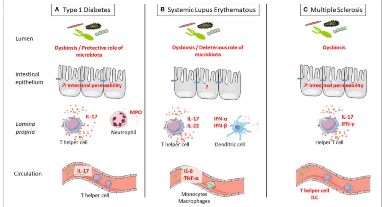
FIGURE 2 | Defect of intestinal barrier and systemic immune response is observed in three examples of autoimmune disorders (ADs): type 1 diabetes (T1D), systemic lupus erythematosus (SLE), and multiple sclerosis (MS). (A) T1D is associated with microbiota dysbiosis, intestinal hyperpermeability, increased IL-17 secretion in the intestine and at systemic level, and increased myeloperoxidase (MPO) in the intestine. Interestingly, colonization by a complex microbiota is protective from type 1 diabetes. (B) SLE is associated with microbiota dysbiosis and increased secretion at the intestinal level of IL-17 and IL-22 by T cells and IFN-α and IFN-β by dendritic cells. At the systemic level, there is a higher secretion of IL-6 and TNF-α by monocytes and macrophages in SLE. Interestingly, colonization by a complex microbiota is deleterious for SLE onset. (C) MS is associated with microbiota dysbiosis, intestinal hyperpermeability, and increased secretion at the intestinal level of IL-17 and IFN-γ by T cells. At the systemic level, the number of innate lymphoid cell (ILC) population was observed in MS. Interestingly, colonization by a complex microbiota is deleterious for MS onset.
important motifs are conserved and overall structures of TCR–
peptide–MHC interaction are similar, suggesting that
cross-reactivity may happen frequently (131). Myelin basic protein, the
immunodominant autoantigen of MS, cross react with Epstein–
Barr virus (EBV), influenza A virus, herpes simplex virus, human
papilloma virus (132), or human herpesvirus-6 (133). Regarding
EBV, MS patients seem to have increased antibody titers against
certain antigens of the virus than have control subjects even
before the onset of MS (134). Despite cross-reactivity, infectious
agents can impair self-antigen tolerance by indirect activation
(135). It has been showed that an integrase expressed by
intestinal Bacteroides encodes a low-avidity mimotope of the
pancreatic β-cell autoantigens and as such might participate to
T1D onset. Colonization of GF mice with Bacteroides promotes
the recruitment of diabetogenic CD8
+T cells to the gut (136).
Intestinal Hyperpermeability in
Autoimmune Disorders
In ADs, intestinal hyperpermeability has been described,
resulting in an increased entry of luminal antigens derived from
food and/or intestinal microbiota or pathogens. The associated
inflammation has been suggested to participate in AD onset
and/or exacerbation.
Even though it is still unclear whether intestinal
hyperpermeability is a trigger or a consequence of T1D
progression (93,
137,
138), epidemiological and preclinical
studies demonstrated that intestinal hyperpermeability occurs
before disease onset (139,
140). Reversion of intestinal
hyperpermeability by treatment with a zonulin 1 (intestinal
homolog of a Vibrio cholerae enterotoxin, which reversibly
increases intestinal permeability) inhibitor ameliorates T1D
manifestation in rat model (141). Microbial translocation in
pancreatic lymph nodes activates NOD2, and IL-17 production
in pancreatic lymph nodes and pancreas which contributes to
T1D development (142).
Intestinal hyperpermeability precedes EAE onset and
increases while disease progresses (143). In this model, increased
intestinal permeability is associated with the increase of crypt
depth and mucosa thickness in jejunum and ileum, as well as
with an overexpression of zonulin 1 (143) as observed for T1D
(141,
144).
Intestinal barrier defect and subsequent exposure to microbial
products play an important role in the pathology of SLE
(145,
146). sCD14, lysozyme, and CXCL16 are markers of
antimicrobial response found increased in SLE subject attesting
to a defect of the intestinal barrier (147).
Intestinal Inflammation
T1D is associated with increased intestinal myeloperoxidase
activity and goblet cell (producing mucus) density, supporting
the idea that early intestinal inflammation might lead to intestinal
hyperpermeability (148,
149). Many studies suggest that the
increased number of Th17 cells is involved in the pathogenesis
of autoimmune diabetes. Higher numbers of IL-17 secreting
cells are detected in recent-onset T1D-promoting inflammatory
response to β-cells (150,
151). Th17 is increased in the peripheral
blood of children with T1D (151,
152). In vitro IL-17 potentiates
inflammatory and proapoptotic responses on human islets
cells (151). Anti-IL-17 treatment reduces islet T cell infiltrates
and GAD65 autoantibodies in NOD mice (153). Neutrophil
extracellular traps (NETs) might contribute to the generation of
ADs by exposing autoantigen (154). The role of NET has been
studied particularly in T1D. Indeed, degradation of NETs in the
gut prevents immune infiltration of pancreatic islet preserving
β-cell mass and systemic inflammation (155).
In a mouse model of SLE developing severe nephritis,
α4β7 expressing T cells is increased in PPs and
pro-inflammatory cytokines (IL-17, IL-22, IFNα, and β) are much
more expressed in distal ileum (156). Furthermore, intestinal
monocytes/macrophages of SLE patients have an altered
expression of type 1 interferon-stimulated genes, HLA-DR,
and Fcγ receptors (157,
158). Monocytes isolated from plasma
of SLE patients release higher pro-inflammatory cytokines
in response to LPS than do healthy patients (159). More
generally, higher production of pro-inflammatory cytokines by
monocytes/macrophages has been described in SLE patients [for
review, see (160)].
In MS, elevated Th1 and Th17 pro-inflammatory responses
are observed in lamina propria, PPs, and mesenteric lymph nodes
(143). GF EAE animals produce lower levels of IFNγ and IL-17 in
the intestines associated with a higher number of Treg cells (104).
Monocolonization of GF animals with segmented filamentous
bacteria, IL-17 inducer in gut (161,
162), induces EAE and
shows that microbiota can affect neurologic inflammation by
recirculation of Th17 to the brain, causing inflammation (106).
Autoreactive T cells from gut could migrate in different organs
depending on pathologies, to brain in the case of MS, to liver in
the case of autoimmune cholestatic liver disease (163,
164), or
to the kidney in the case of SLE (165). Interestingly, not only T
cells seem to be involved in MS but also circulating ILC. Indeed,
a higher number of ILC have been observed in MS patients (166).
CONCLUSION
As a conclusion, compiling evidences highlight the importance
of both intestinal barrier defect and stress in ADs. Stress is
well known to have long-lasting deleterious consequences on
the intestinal barrier. A transversal research on ADs, stress,
and intestinal barrier function would be of great interest and
would bring new understanding in the pathophysiology of
ADs. Identifying stress-induced intestinal barrier dysfunction as
an actor of ADs could bring new possibilities for therapeutic
targets and especially preventing strategies toward the spreading
epidemic of ADs. Therapeutic strategies suggest that probiotics
and FMT treatment might improve AD symptom, but preventive
strategies in an at-risk population still need to be explored. In
this review, we did not mention autoimmune thyroid diseases
(AITDs) that are the most frequent ADs (167). It is difficult to
study AITDs by themselves, as they are often observed together
with other ADs, which are named polyautoimmunity (168).
Then, even though there are sufficient data supporting the role
of stress in AITD onset (20), evidences for a defect of intestinal
barrier functions in AITD are sparse, and only two studies are
available regarding microbiota dysbiosis (169,
170). For those
reasons, we did not use AITD to illustrate the hypothesis of
this review, supporting a role of stress-induced intestinal barrier
disruption in the onset and/or the course of ADs. However, we
wanted to mention the case of AITD as data on intestinal barrier
function would be of great interest in the future.
AUTHOR CONTRIBUTIONS
HI-D and SM reviewed the literature, wrote and corrected the
manuscript, and drew the figures. All authors contributed to the
article and approved the submitted version.
REFERENCES
1. Selye H. Syndrome produced by diverse nocuous agents. Nature. (1936) 138:32. doi: 10.1038/138032a0
2. Kessler RC, Berglund P, Demler O, Jin R, Merikangas KR, Walters EE. Lifetime prevalence and age-of-onset distributions of DSM-IV disorders in the National Comorbidity Survey Replication. Arch Gen Psychiatry. (2005) 62:593. doi: 10.1001/archpsyc.62.6.593
3. Smith SM, Vale WW. The role of the hypothalamic-pituitary-adrenal axis in neuroendocrine responses to stress. Dialogues Clin Neurosci. (2006) 8:383–95. Avialable online at : https://www.ncbi.nlm.nih.gov/pmc/articles/ PMC3181830/
4. Charmandari E, Tsigos C, Chrousos G. Endocrinology of the stress response. Annu Rev Physiol. (2005) 67:259–84. doi: 10.1146/annurev.physiol.67.040403.120816
5. Cain DW, Cidlowski JA. Immune regulation by glucocorticoids. Nat Rev Immunol. (2017) 17:233–47. doi: 10.1038/nri.2017.1
6. Miyata M, Lee J-Y, Susuki-Miyata S, Wang WY, Xu H, Kai H, et al. Glucocorticoids suppress inflammation via the upregulation of negative regulator IRAK-M. Nat Commun. (2015) 6:6062. doi: 10.1038/ncomms7062 7. Beaulieu E, Morand EF. Role of GILZ in immune regulation, glucocorticoid actions and rheumatoid arthritis. Nat Rev Rheumatol. (2011) 7:340–8. doi: 10.1038/nrrheum.2011.59
8. Atsuta J, Plitt J, Bochner BS, Schleimer RP. Inhibition of VCAM-1 expression in human bronchial epithelial cells by glucocorticoids. Am J Respir Cell Mol Biol. (1999) 20:643–50. doi: 10.1165/ajrcmb.20.4.3265
9. Mukaida N, Morita M, Ishikawa Y, Rice N, Okamoto SI, Kasahara T, et al. Novel mechanism of glucocorticoid-mediated gene repression. Nuclear factor-κB is target for glucocorticoid-mediated interleukin 8 gene repression. J Biol Chem. (1994) 269:13289–95.
10. Martinez FO, Sica A, Mantovani A, Locati M. Macrophage activation and polarization. Front Biosci. (2008) 13:453–61. doi: 10.2741/2692
11. Gillis S, Crabtree GR, Smith KA. Glucocorticoid-induced inhibition of T cell growth factor production. II. The effect on the in vitro generation of cytolytic T cells. J Immunol. (1979) 123:1632–8.
12. Gillis S, Crabtree GR, Smith KA. Glucocorticoid-induced inhibition of T cell growth factor production. I. The effect on mitogen-induced lymphocyte proliferation. J Immunol. (1979) 123:1624–31.
13. Elenkov IJ. Glucocorticoids and the Th1/Th2 Balance. Ann N Y Acad Sci. (2004) 1024:138–46. doi: 10.1196/annals.1321.010
14. Liberman AC, Refojo D, Druker J, Toscano M, Rein T, Holsboer F, et al. The activated glucocorticoid receptor inhibits the transcription factor T-bet by direct protein-protein interaction. FASEB J. (2007) 21:1177–88. doi: 10.1096/fj.06-7452com
15. Frank MG, Miguel ZD, Watkins LR, Maier SF. Prior exposure to glucocorticoids sensitizes the neuroinflammatory and peripheral inflammatory responses to E. coli lipopolysaccharide. Brain Behav Immun. (2010) 24:19–30. doi: 10.1016/j.bbi.2009.07.008
16. Lim H-Y, Müller N, Herold MJ, van den Brandt J, Reichardt HM. Glucocorticoids exert opposing effects on macrophage function dependent on their concentration. Immunology. (2007) 122:47–53. doi: 10.1111/j.1365-2567.2007.02611.x
17. Perroud N, Paoloni-Giacobino A, Prada P, Olié E, Salzmann A, Nicastro R, et al. Increased methylation of glucocorticoid receptor gene (NR3C1) in adults with a history of childhood maltreatment: a link with the severity and type of trauma. Transl Psychiatry. (2011) 1:e59. doi: 10.1038/tp.2011.60 18. Melas PA, Wei Y, Wong CCY, Sjöholm LK, Åberg E, Mill J, Schalling M, et al.
Genetic and epigenetic associations of MAOA and NR3C1 with depression and childhood adversities. Int J Neuropsychopharmacol. (2013) 16:1513–28. doi: 10.1017/S1461145713000102
19. McGowan PO, Sasaki A, D’Alessio AC, Dymov S, Labonté B, Szyf M, et al. Epigenetic regulation of the glucocorticoid receptor in human brain associates with childhood abuse. Nat Neurosci. (2009) 12:342–8. doi: 10.1038/nn.2270
20. Sharif K, Watad A, Coplan L, Lichtbroun B, Krosser A, Lichtbroun M, et al. The role of stress in the mosaic of autoimmunity: an overlooked association. Autoimmun Rev. (2018) 17:967–83. doi: 10.1016/j.autrev.2018.04.005 21. Stojanovich L, Marisavljevich D. Stress as a trigger of autoimmune disease.
Autoimmun Rev. (2008) 7:209–13. doi: 10.1016/j.autrev.2007.11.007 22. Shepshelovich D, Shoenfeld Y. Prediction and prevention of autoimmune
diseases: additional aspects of the mosaic of autoimmunity. Lupus. (2006) 15:183–90. doi: 10.1191/0961203306lu2274rr
23. Huerta PT, Kowal C, DeGiorgio LA, Volpe BT, Diamond B. Immunity and behavior: antibodies alter emotion. Proc Natl Acad Sci USA. (2006) 103:678–83. doi: 10.1073/pnas.0510055103
24. Wang Z, Xie Z, Lu Q, Chang C, Zhou Z. Beyond genetics: what causes type 1 diabetes. Clin Rev Allergy Immunol. (2017) 52:273–86. doi: 10.1007/s12016-016-8592-1
25. Teruel M, Alarcón-Riquelme ME. The genetic basis of systemic lupus erythematosus: what are the risk factors and what have we learned. J Autoimmun. (2016) 74:161–75. doi: 10.1016/j.jaut.2016.08.001
26. Giannelou M, Mavragani CP. Cardiovascular disease in systemic lupus erythematosus: a comprehensive update. J Autoimmun. (2017) 82:1–12. doi: 10.1016/j.jaut.2017.05.008
27. Goverman J. Autoimmune T cell responses in the central nervous system. Nat Rev Immunol. (2009) 9:393–407. doi: 10.1038/nri2550
28. Selmi C, Barin JG, Rose NR. Current trends in autoimmunity and the nervous system. J Autoimmun. (2016) 75:20–9. doi: 10.1016/j.jaut.2016.08.005
29. Willis T. Pharmaceutice Rationalis: or, an Exercitation of the Operations of Medicines in Humane Bodies. Shewing the Signs, Causes, and Cures of Most Distempers Incident Thereunto: In Two Parts As Also a Treatise of the Scurvy and the Several Sorts Thereof, With Their Symptoms, Causes and Cure. . . . London: Printed for T. Dring, C. Harper, and J. Leigh, and are to be sold by R. Clavell (1679). p. 1621–75. Available online at: https://quod.lib.umich. edu/e/eebo2/A66509.0001.001?view=toc.
30. Hägglöf B, Blom L, Dahlquist G, Lönnberg G, Sahlin B. The Swedish childhood diabetes study: indications of severe psychological stress as a
risk factor for type 1 (insulin-dependent) diabetes mellitus in childhood. Diabetologia. (1991) 34:579–83. doi: 10.1007/BF00400277
31. Thernlund GM, Dahlquist G, Hansson K, Ivarsson SA, Ludvigsson J, Sjöblad S, et al. Psychological stress and the onset of IDDM in children. Diabetes Care. (1995) 18:1323–9. doi: 10.2337/diacare.18.10.1323
32. Portha B, Chavey A, Movassat J. Early-life origins of type 2 diabetes: fetal programming of the beta-cell mass. Exp Diabetes Res. (2011) 2011:1–16. doi: 10.1155/2011/105076
33. Roberts AL, Malspeis S, Kubzansky LD, Feldman CH, Chang S-C, Koenen KC, et al. Association of trauma and posttraumatic stress disorder with incident systemic lupus erythematosus in a longitudinal cohort of women. Arthritis Rheumatol. (2017) 69:2162–69. doi: 10.1002/art. 40222
34. Mills SD, Azizoddin D, Racaza GZ, Wallace DJ, Weisman MH, Nicassio PM. The psychometric properties of the perceived stress scale-10 among patients with systemic lupus erythematosus. Lupus. (2017) 26:1218–23. doi: 10.1177/0961203317701844
35. Grant I, Brown GW, Harris T, McDonald WI, Patterson T, Trimble MR. Severely threatening events and marked life difficulties preceding onset or exacerbation of multiple sclerosis. J Neurol Neurosurg Psychiatry. (1989) 52:8–13. doi: 10.1136/jnnp.52.1.8
36. Warren S, Greenhill S, Warren KG. Emotional stress and the development of multiple sclerosis: case-control evidence of a relationship. J Chronic Dis. (1982) 35:821–31. doi: 10.1016/0021-9681(82)90047-9
37. Mitsonis CI, Zervas IM, Mitropoulos PA, Dimopoulos NP, Soldatos CR, Potagas CM, et al. The impact of stressful life events on risk of relapse in women with multiple sclerosis: a prospective study. Eur Psychiatry. (2008) 23:497–504. doi: 10.1016/j.eurpsy.2008.06.003
38. Bibolini MJ, Chanaday NL, Báez NS, Degano AL, Monferran CG, Roth GA. Inhibitory role of diazepam on autoimmune inflammation in rats with experimental autoimmune encephalomyelitis. Neuroscience. (2011) 199:421– 8. doi: 10.1016/j.neuroscience.2011.08.076
39. Winkley K, Ismail K, Landau S, Eisler I. Psychological interventions to improve glycaemic control in patients with type 1 diabetes: systematic review and meta-analysis of randomised controlled trials. BMJ. (2006) 333:65. doi: 10.1136/bmj.38874.652569.55
40. Kurashima Y, Kiyono H. Mucosal ecological network of epithelium and immune cells for gut homeostasis and tissue healing. Annu Rev Immunol. (2016) 35:119–47. doi: 10.1146/annurev-immunol-051116-052424
41. Jandhyala SM, Talukdar R, Subramanyam C, Vuyyuru H, Sasikala M, Reddy DN. Role of the normal gut microbiota. World J Gastroenterol. (2015) 21:8787. doi: 10.3748/wjg.v21.i29.8787
42. Gehart H, Clevers H. Tales from the crypt: new insights into intestinal stem cells. Nat Rev Gastroenterol Hepatol. (2019) 16:19–34. doi: 10.1038/s41575-018-0081-y
43. Gordon JI, Hermiston ML. Differentiation and self-renewal in the mouse gastrointestinal epithelium. Curr Opin Cell Biol. (1994) 6:795–803. doi: 10.1016/0955-0674(94)90047-7
44. Ménard S, Cerf-Bensussan N, Heyman M. Multiple facets of intestinal permeability and epithelial handling of dietary antigens. Mucosal Immunol. (2010) 3:247–59. doi: 10.1038/mi.2010.5
45. Mora JR, von Andrian UH. Differentiation and homing of IgA-secreting cells. Mucosal Immunol. (2008) 1:96–109. doi: 10.1038/mi.2007.14 46. Ma H, Tao W, Zhu S. T lymphocytes in the intestinal mucosa:
defense and tolerance. Cell Mol Immunol. (2019) 16:216–24. doi: 10.1038/s41423-019-0208-2
47. Rankin L, Joanna Groom, Mielke LA, Seillet C, Belz GT. Diversity, function, and transcriptional regulation of gut innate lymphocytes. Front Immunol. (2013) 4:22. doi: 10.3389/fimmu.2013.00022
48. Annibali V, Policano C, Buscarinu MC, Lionetto L, Mechelli R, Capi M, et al. Intestinal permeability in multiple sclerosis. J Neuroimmunol. (2014) 275:54. doi: 10.1016/j.jneuroim.2014.08.143
49. Secher T, Kassem S, Benamar M, Bernard I, Boury M, Barreau F, et al. Oral administration of the probiotic strain Escherichia coli nissle 1917 reduces susceptibility to neuroinflammation and repairs experimental autoimmune encephalomyelitis-induced intestinal barrier dysfunction. Front Immunol. (2017) 8:1–10. doi: 10.3389/fimmu.2017.01096
50. Li X, Atkinson MA. The role for gut permeability in the pathogenesis of type 1 diabetes - a solid or leaky concept? Pediatr Diabetes. (2015) 16:485–92. doi: 10.1111/pedi.12305
51. Gitter AH, Bendfeldt K, Schulzke J-D, Fromm M. Leaks in the epithelial barrier caused by spontaneous and TNF-α-induced single-cell apoptosis. FASEB J. (2000) 14:1749–53. doi: 10.1096/fj.99-0898com
52. Winter SE, Winter MG, Xavier MN, Thiennimitr P, Poon V, Keestra AM, et al. Host-derived nitrate boosts growth of E. coli in the Inflamed Gut. Science (80-). (2013) 339:708–11. doi: 10.1126/science.1232467
53. Lowman BC, Drossman DA, Cramer EM, McKee DC, DC McKee. Recollection of childhood events in adults with irritable bowel syndrome. J Clin Gastroenterol. (1987) 9:324–30. doi: 10.1097/00004836-198706000-00017
54. Videlock EJ, Adeyemo M, Licudine A, Hirano M, Ohning G, Mayer M, et al. Childhood trauma is associated with hypothalamic-pituitary-adrenal axis responsiveness in irritable bowel syndrome. Gastroenterology. (2009) 137:1954–62. doi: 10.1053/j.gastro.2009.08.058
55. Hislop IG. Childhood deprivation: an antecedent of the irritable bowel syndrome. Med J Aust. (1979) 1:372–4. doi: 10.5694/j.1326-5377.1979.tb126963.x
56. Sgambato D, Miranda A, Ranaldo R, Federico A, Romano M. The role of stress in inflammatory bowel diseases. Curr Pharm Des. (2017) 23:3997– 4002. doi: 10.2174/1381612823666170228123357
57. Mayer EA, Naliboff BD, Chang L, Coutinho SV. V. Stress and irritable bowel syndrome. Am J Physiol Liver Physiol. (2001) 280:G519–24. doi: 10.1152/ajpgi.2001.280.4.G519
58. Wood JD. Visceral pain: spinal afferents, enteric mast cells, enteric nervous system and stress. Curr Pharm Des. (2011) 17:1573–5. doi: 10.2174/138161211796196918
59. Bonaz BL, Bernstein CN. Brain-gut interactions in inflammatory bowel disease. Gastroenterology. (2013) 144:36–49. doi: 10.1053/j.gastro.2012.10.003
60. Drossman DA. Functional gastrointestinal disorders: history, pathophysiology, clinical features, and Rome IV. Gastroenterology. (2016) 150:1262–79.e2. doi: 10.1053/j.gastro.2016.02.032
61. Riba A, Olier M, Lacroix-Lamandé S, Lencina C, Bacquié V, Harkat C, et al. Paneth cell defects induce microbiota dysbiosis in mice and promote visceral hypersensitivity. Gastroenterology. (2017) 153:1594–606.e2. doi: 10.1053/j.gastro.2017.08.044
62. Ilchmann-Diounou H, Olier M, Lencina C, Riba A, Barretto S, Nankap M, et al. Early life stress induces type 2 diabetes-like features in ageing mice. Brain Behav Immun. (2019) 80:452:63. doi: 10.1016/j.bbi.2019. 04.025
63. Moussaoui N, Jacobs JP, Larauche M, Biraud M, Million M, Mayer E, et al. Chronic early-life stress in rat pups alters basal corticosterone, intestinal permeability, and fecal microbiota at weaning: influence of sex. J Neurogastroenterol Motil. (2017) 23:135–43. doi: 10.5056/ jnm16105
64. Yoshikawa K, Kurihara C, Furuhashi H, Takajo T, Maruta K, Yasutake Y, et al. Psychological stress exacerbates NSAID-induced small bowel injury by inducing changes in intestinal microbiota and permeability via glucocorticoid receptor signaling. J Gastroenterol. (2017) 52:61–71. doi: 10.1007/s00535-016-1205-1
65. Sudo N, Chida Y, Aiba Y, Sonoda J, Oyama N, Yu X-N, et al. Postnatal microbial colonization programs the hypothalamic-pituitary-adrenal system for stress response in mice. J Physiol. (2004) 558:263–75. doi: 10.1113/jphysiol.2004.063388
66. Bailey MT, Dowd SE, Galley JD, Hufnagle AR, Allen RG, Lyte M. Exposure to a social stressor alters the structure of the intestinal microbiota: implications for stressor-induced immunomodulation. Brain Behav Immun. (2011) 25:397–407. doi: 10.1016/j.bbi.2010.10.023
67. Hemmings SMJ, Malan-Müller S, van den Heuvel LL, Demmitt BA, Stanislawski MA, Smith DG, et al. The microbiome in posttraumatic stress disorder and trauma-exposed controls. Psychosom Med. (2017) 79:936–46. doi: 10.1097/PSY.0000000000000512
68. Collins SM. A role for the gut microbiota in IBS. Nat Rev Gastroenterol Hepatol. (2014) 11:497–505. doi: 10.1038/nrgastro.2014.40
69. Zheng G, Wu S-P, Hu Y, Smith DE, Wiley JW, Hong S. Corticosterone mediates stress-related increased intestinal permeability in a region-specific manner. Neurogastroenterol Motil. (2013) 25:e127–39. doi: 10.1111/nmo.12066
70. Cameron HL. Stress impairs murine intestinal barrier function: improvement by glucagon-like peptide-2. J Pharmacol Exp Ther. (2005) 314:214–20. doi: 10.1124/jpet.105.085373
71. Barreau F, Ferrier L, Fioramonti J, Bueno L. Neonatal maternal deprivation triggers long term alterations in colonic epithelial barrier and mucosal immunity in rats. Gut. (2004) 53:501–6. doi: 10.1136/gut.2003.024174 72. Øines E, Murison R, Mrdalj J, Grønli J, Milde AM. Neonatal maternal
separation in male rats increases intestinal permeability and affects behavior after chronic social stress. Physiol Behav. (2012) 105:1058–66. doi: 10.1016/j.physbeh.2011.11.024
73. Riba A, Olier M, Lacroix-Lamandé S, Lencina C, Bacquié V, Harkat C, et al. Early life stress in mice is a suitable model for Irritable Bowel Syndrome but does not predispose to colitis nor increase susceptibility to enteric infections. Brain Behav Immun. (2018) 73:403–15. doi: 10.1016/j.bbi.2018.05.024 74. Moussaoui N, Braniste V, Ait-Belgnaoui A, Gabanou M, Sekkal S, Olier M,
et al. Changes in intestinal glucocorticoid sensitivity in early life shape the risk of epithelial barrier defect in maternal-deprived rats. PLoS ONE. (2014) 9:1–9. doi: 10.1371/journal.pone.0088382
75. Lauffer A, Vanuytsel T, Vanormelingen C, Vanheel H, Salim Rasoel S, Tóth J, et al. Subacute stress and chronic stress interact to decrease intestinal barrier function in rats. Stress. (2016) 19:225–34. doi: 10.3109/10253890.2016.1154527
76. Lafuse WP, Gearinger R, Fisher S, Nealer C, Mackos AR, Bailey MT. Exposure to a social stressor induces translocation of commensal lactobacilli to the spleen and priming of the innate immune system. J Immunol. (2017) 198:2383–93. doi: 10.4049/jimmunol.1601269
77. Vanuytsel T, van Wanrooy S, Vanheel H, Vanormelingen C, Verschueren S, Houben E, et al. Psychological stress and corticotropin-releasing hormone increase intestinal permeability in humans by a mast cell-dependent mechanism. Gut. (2014) 63:1293–9. doi: 10.1136/gutjnl-2013-305690 78. Bischoff SC, Barbara G, Buurman W, Ockhuizen T, Schulzke J-D, Serino
M, et al. Intestinal permeability – a new target for disease prevention and therapy. BMC Gastroenterol. (2014) 14:189. doi: 10.1186/s12876-014-0189-7
79. Eutamene H, Theodorou V, Fioramonti J, Bueno L. Acute stress modulates the histamine content of mast cells in the gastrointestinal tract through interleukin-1 and corticotropin-releasing factor release in rats. J Physiol. (2003) 553:959–66. doi: 10.1113/jphysiol.2003.052274
80. Miranda S, Roux ME. Acoustic stress induces long term severe intestinal inflammation in the mouse. Toxicol Lett. (2017) 280:1–9. doi: 10.1016/j.toxlet.2017.07.898
81. Barbara G, Cremon C, Carini G, Bellacosa L, Zecchi L, Giorgio R De, et al. The immune system in Irritable Bowel Syndrome. J Neurogastroenterol Motil. (2011) 17:349–59. doi: 10.5056/jnm.2011.17.4.349
82. de Oliveira JF, Wiener CD, Jansen K, Portela LV, Lara DR, Souza LD de M, et al. Serum levels of interleukins IL-6 and IL-10 in individuals with posttraumatic stress disorder in a population-based sample. Psychiatry Res. (2018) 260:111–5. doi: 10.1016/j.psychres.2017.11.061
83. Marsland AL, Walsh C, Lockwood K, John-Henderson NA. The effects of acute psychological stress on circulating and stimulated inflammatory markers: a systematic review and meta-analysis. Brain Behav Immun. (2017) 64:208–19. doi: 10.1016/j.bbi.2017.01.011
84. Baldwin JR, Arseneault L, Caspi A, Fisher HL, Moffitt TE, Odgers CL, et al. Childhood victimization and inflammation in young adulthood: a genetically sensitive cohort study. Brain Behav Immun. (2017) 67:211–7. doi: 10.1016/j.bbi.2017.08.025
85. Slopen N, Loucks EB, Appleton AA, Kawachi I, Kubzansky LD, Non AL, et al. Early origins of inflammation: an examination of prenatal and childhood social adversity in a prospective cohort study. Psychoneuroendocrinology. (2015) 51:403–13. doi: 10.1016/j.psyneuen.2014.10.016
86. Rizzetto L, Fava F, Tuohy KM, Selmi C. Connecting the immune system, systemic chronic inflammation and the gut microbiome: the role of sex. J Autoimmun. (2018) 92:12–34. doi: 10.1016/j.jaut.2018.05.008
87. Yurkovetskiy LA, Pickard JM, Chervonsky A V. Microbiota and autoimmunity: exploring new avenues. Cell Host Microbe. (2015) 17:548–52. doi: 10.1016/j.chom.2015.04.010
88. Rosser EC, Mauri C. A clinical update on the significance of the gut microbiota in systemic autoimmunity. J Autoimmun. (2016) 74:85–93. doi: 10.1016/j.jaut.2016.06.009
89. Carding S, Verbeke K, Vipond DT, Corfe BM, Owen LJ. Dysbiosis of the gut microbiota in disease. Microb Ecol Heal Dis. (2015) 26:26191. doi: 10.3402/mehd.v26.26191
90. Gianchecchi E, Fierabracci A. On the pathogenesis of insulin-dependent diabetes mellitus: the role of microbiota. Immunol Res. (2017) 65:242–56. doi: 10.1007/s12026-016-8832-8
91. Ochoa-Repáraz J, Magori K, Kasper LH. The chicken or the egg dilemma: intestinal dysbiosis in multiple sclerosis. Ann Transl Med. (2017) 5:145. doi: 10.21037/atm.2017.01.18
92. de Goffau MC, Fuentes S, van den Bogert B, Honkanen H, de Vos WM, Welling GW, et al. Aberrant gut microbiota composition at the onset of type 1 diabetes in young children. Diabetologia. (2014) 57:1569–77. doi: 10.1007/s00125-014-3274-0
93. Harbison JE, Roth-Schulze AJ, Giles LC, Tran CD, Ngui KM, Penno MA, et al. Gut microbiome dysbiosis and increased intestinal permeability in children with islet autoimmunity and type 1 diabetes : a prospective cohort study. Pediatr Diabetes. (2019) 20:574–83. doi: 10.1111/pedi.12865 94. Endesfelder D, Engel M, Davis-Richardson AG, Ardissone AN, Achenbach
P, Hummel S, et al. Towards a functional hypothesis relating anti-islet cell autoimmunity to the dietary impact on microbial communities and butyrate production. Microbiome. (2016) 4:17. doi: 10.1186/s40168-016-0163-4 95. Kostic AD, Gevers D, Siljander H, Vatanen T, Hyötyläinen T, Hämäläinen
A-M, et al. The dynamics of the human infant gut microbiome in development and in progression toward type 1 diabetes. Cell Host Microbe. (2015) 17:260– 73. doi: 10.1016/j.chom.2015.01.001
96. Brown K, Godovannyi A, Ma C, Zhang Y, Ahmadi-Vand Z, Dai C, et al. Prolonged antibiotic treatment induces a diabetogenic intestinal microbiome that accelerates diabetes in NOD mice. ISME J. (2016) 10:321– 32. doi: 10.1038/ismej.2015.114
97. Peng J, Narasimhan S, Marchesi JR, Benson A, Wong FS, Wen L. Long term effect of gut microbiota transfer on diabetes development. J Autoimmun. (2014) 53:85–94. doi: 10.1016/j.jaut.2014.03.005
98. Brugman S, Klatter FA, Visser JTJ, Wildeboer-Veloo ACM, Harmsen HJM, Rozing J, et al. Antibiotic treatment partially protects against type 1 diabetes in the Bio-Breeding diabetes-prone rat. Is the gut flora involved in the development of type 1 diabetes? Diabetologia. (2006) 49:2105–8. doi: 10.1007/s00125-006-0334-0
99. Schwartz RF, Neu J, Schatz D, Atkinson MA, Wasserfall C. Comment on: Brugman S et al. (2006) Antibiotic treatment partially protects against type 1 diabetes in the Bio-Breeding diabetes-prone rat. Is the gut flora involved in the development of type 1 diabetes? Diabetologia 49:2105–2108. Diabetologia. (2006) 50:220–1. doi: 10.1007/s00125-006-0526-7
100. Miyake S, Kim S, Suda W, Oshima K, Nakamura M, Matsuoka T, et al. Dysbiosis in the gut microbiota of patients with multiple sclerosis, with a striking depletion of species belonging to clostridia XIVa and IV clusters. PLoS ONE. (2015) 10:e0137429. doi: 10.1371/journal.pone.0137429 101. Tremlett H, Fadrosh DW, Faruqi AA, Hart J, Roalstad S, Graves J, et al.
Associations between the gut microbiota and host immune markers in pediatric multiple sclerosis and controls. BMC Neurol. (2016) 16:182. doi: 10.1186/s12883-016-0703-3
102. Chen J, Chia N, Kalari KR, Yao JZ, Novotna M, Soldan MMP, et al. Multiple sclerosis patients have a distinct gut microbiota compared to healthy controls. Sci Rep. (2016) 6:28484. doi: 10.1038/srep28484
103. Jangi S, Gandhi R, Cox LM, Li N, von Glehn F, Yan R, et al. Alterations of the human gut microbiome in multiple sclerosis. Nat Commun. (2016) 7:12015. doi: 10.1038/ncomms12015
104. Berer K, Mues M, Koutrolos M, Rasbi Z Al, Boziki M, Johner C, et al. Commensal microbiota and myelin autoantigen cooperate to trigger autoimmune demyelination. Nature. (2011) 479:538–41. doi: 10.1038/nature10554
105. Yokote H, Miyake S, Croxford JL, Oki S, Mizusawa H, Yamamura T. NKT cell-dependent amelioration of a mouse model of multiple
sclerosis by altering gut flora. Am J Pathol. (2008) 173:1714–23. doi: 10.2353/ajpath.2008.080622
106. Lee YK, Menezes JS, Umesaki Y, Mazmanian SK. Proinflammatory T-cell responses to gut microbiota promote experimental autoimmune encephalomyelitis. Proc Natl Acad Sci USA. (2011) 108:4615–22. doi: 10.1073/pnas.1000082107
107. Gandy KAO, Zhang J, Nagarkatti P, Nagarkatti M. The role of gut microbiota in shaping the relapse-remitting and chronic-progressive forms of multiple sclerosis in mouse models. Sci Rep. (2019) 9:6923. doi: 10.1038/s41598-019-43356-7
108. He Z, Shao T, Li H, Xie Z, Wen C. Alterations of the gut microbiome in Chinese patients with systemic lupus erythematosus. Gut Pathog. (2016) 8:64. doi: 10.1186/s13099-016-0146-9
109. Hevia A, Milani C, López P, Cuervo A, Arboleya S, Duranti S, et al. Intestinal dysbiosis associated with systemic lupus erythematosus. MBio. (2014) 5:e01548–14. doi: 10.1128/mBio.01548-14
110. Luo XM, Edwards MR, Mu Q, Yu Y, Vieson MD, Reilly CM, et al. Gut microbiota in human systemic lupus erythematosus and a mouse model of lupus. Appl Environ Microbiol. (2018) 84:2288. doi: 10.1128/AEM.02288-17 111. Azzouz D, Omarbekova A, Heguy A, Schwudke D, Gisch N, Rovin BH,
et al. Lupus nephritis is linked to disease-activity associated expansions and immunity to a gut commensal. Ann Rheum Dis. (2019) 78:947–56. doi: 10.1136/annrheumdis-2018-214856
112. Johnson BM, Gaudreau M-C, Al-Gadban MM, Gudi R, Vasu C. Impact of dietary deviation on disease progression and gut microbiome composition in lupus-prone SNF 1 mice. Clin Exp Immunol. (2015) 181:323–37.
doi: 10.1111/cei.12609
113. Abraham BP, Quigley EMM. Probiotics in inflammatory Bowel Disease. Gastroenterol Clin North Am. (2017) 46:769–82. doi: 10.1016/j.gtc.2017.08.003
114. Toumi R, Abdelouhab K, Rafa H, Soufli I, Raissi-Kerboua D, Djeraba Z, et al. Beneficial role of the probiotic mixture Ultrabiotique on maintaining the integrity of intestinal mucosal barrier in DSS-induced experimental colitis. Immunopharmacol Immunotoxicol. (2013) 35:403–9. doi: 10.3109/08923973.2013.790413
115. Riedel C-U, Foata F, Philippe D, Adolfsson O, Eikmanns B-J, Blum S. Anti-inflammatory effects of bifidobacteria by inhibition of LPS-induced NF-kappaB activation. World J Gastroenterol. (2006) 12:3729–35. doi: 10.3748/wjg.v12.i23.3729
116. O’Hara AM, Bhattacharyya A, Mifflin RC, Smith MF, Ryan KA, Scott KG-E, et al. Interleukin-8 induction by Helicobacter pylori in gastric epithelial cells is dependent on apurinic/apyrimidinic endonuclease-1/redox factor-1. J Immunol. (2006) 177:7990–9. doi: 10.4049/jimmunol.177.11.7990 117. Khokhlova EV, Smeianov VV, Efimov BA, Kafarskaia LI, Pavlova SI,
Shkoporov AN. Anti-inflammatory properties of intestinal Bifidobacterium strains isolated from healthy infants. Microbiol Immunol. (2012) 56:27–39. doi: 10.1111/j.1348-0421.2011.00398.x
118. Roselli M, Finamore A, Britti MS, Mengheri E. Probiotic bacteria Bifidobacterium animalis MB5 and Lactobacillus rhamnosus GG protect intestinal Caco-2 cells from the inflammation-associated response induced by enterotoxigenic Escherichia coli K88. Br J Nutr. (2006) 95:1177–84. doi: 10.1079/BJN20051681
119. Toumi R, Soufli I, Rafa H, Belkhelfa M, Biad A, Touil-Boukoffa C. Probiotic bacteria lactobacillus and bifidobacterium attenuate inflammation in dextran sulfate sodium-induced experimental colitis in mice. Int J Immunopathol Pharmacol. (2014) 27:615–27. doi: 10.1177/039463201402700418
120. Lavasani S, Dzhambazov B, Nouri M, Fåk F, Buske S, Molin G, et al. A novel probiotic mixture exerts a therapeutic effect on experimental autoimmune encephalomyelitis mediated by IL-10 producing regulatory T cells. PLoS ONE. (2010) 5:e9009. doi: 10.1371/journal.pone.0009009
121. Ezendam J, de Klerk A, Gremmer ER, van Loveren H. Effects of Bifidobacterium animalis administered during lactation on allergic and autoimmune responses in rodents. Clin Exp Immunol. (2008) 154:424–31. doi: 10.1111/j.1365-2249.2008.03788.x
122. Takata K, Kinoshita M, Okuno T, Moriya M, Kohda T, Honorat JA, et al. The lactic acid bacterium Pediococcus acidilactici suppresses autoimmune encephalomyelitis by inducing IL-10-producing regulatory T cells. PLoS ONE. (2011) 6:e27644. doi: 10.1371/journal.pone.0027644
123. Rezende RM, Oliveira RP, Medeiros SR, Gomes-Santos AC, Alves AC, Loli FG, et al. Hsp65-producing Lactococcus lactis prevents experimental autoimmune encephalomyelitis in mice by inducing CD4+LAP+ regulatory T cells. J Autoimmun. (2013) 40:45–57. doi: 10.1016/j.jaut.2012. 07.012
124. Wang Y, Telesford KM, Ochoa-Repáraz J, Haque-Begum S, Christy M, Kasper EJ, et al. An intestinal commensal symbiosis factor controls neuroinflammation via TLR2-mediated CD39 signalling. Nat Commun. (2014) 5:4432. doi: 10.1038/ncomms5432
125. Kouchaki E, Tamtaji OR, Salami M, Bahmani F, Daneshvar Kakhaki R, Akbari E, et al. Clinical and metabolic response to probiotic supplementation in patients with multiple sclerosis: a randomized, double-blind, placebo-controlled trial. Clin Nutr. (2017) 36:1245–9. doi: 10.1016/j.clnu.2016. 08.015
126. Valladares R, Sankar D, Li N, Williams E, Lai K-K, Abdelgeliel AS, et al. Lactobacillus johnsonii N6.2 mitigates the development of Type 1 diabetes in BB-DP rats. PLoS ONE. (2010) 5:e10507. doi: 10.1371/journal.pone.00 10507
127. Dolpady J, Sorini C, Di Pietro C, Cosorich I, Ferrarese R, Saita D, et al. Oral probiotic VSL#3 prevents autoimmune diabetes by modulating microbiota and promoting indoleamine 2,3-dioxygenase-enriched tolerogenic intestinal environment. J Diabetes Res. (2016) 2016:7569431. doi: 10.1155/2016/7569431
128. Uusitalo U, Liu X, Yang J, Aronsson CA, Hummel S, Butterworth M, et al. Association of early exposure of probiotics and islet autoimmunity in the TEDDY Study. JAMA Pediatr. (2016) 170:20. doi: 10.1001/jamapediatrics.2015.2757
129. Mu Q, Zhang H, Liao X, Lin K, Liu H, Edwards MR, et al. Control of lupus nephritis by changes of gut microbiota. Microbiome. (2017) 5:73. doi: 10.1186/s40168-017-0300-8
130. Borody TJ, Khoruts A. Fecal microbiota transplantation and emerging applications. Nat Rev Gastroenterol Hepatol. (2012) 9:88–96. doi: 10.1038/nrgastro.2011.244
131. Birnbaum ME, Mendoza JL, Sethi DK, Dong S, Glanville J, Dobbins J, et al. Deconstructing the peptide-MHC specificity of T cell recognition. Cell. (2014) 157:1073–87. doi: 10.1016/j.cell.2014.03.047
132. Wucherpfennig KW, Strominger JL. Molecular mimicry in T cell-mediated autoimmunity: viral peptides activate human T cell clones specific for myelin basic protein. Cell. (1995) 80:695–705. doi: 10.1016/0092-8674(95)90348-8 133. Tejada-Simon M V, Zang YCQ, Hong J, Rivera VM, Zhang JZ.
Cross-reactivity with myelin basic protein and human herpesvirus-6 in multiple sclerosis. Ann Neurol. (2003) 53:189–97. doi: 10.1002/ana.10425
134. Ascherio A, Munger KL, Lennette ET, Spiegelman D, Hernán MA, Olek MJ, et al. Epstein-Barr virus antibodies and risk of multiple sclerosis: a prospective study. JAMA. (2001) 286:3083–8. doi: 10.1001/jama.286.24.3083 135. Haring JS, Pewe LL, Perlman S. Bystander CD8 T cell-mediated demyelination after viral infection of the central nervous system. J Immunol. (2002) 169:1550–5. doi: 10.4049/jimmunol.169.3.1550
136. Hebbandi Nanjundappa R, Ronchi F, Wang J, Clemente-Casares X, Yamanouchi J, Sokke Umeshappa C, et al. A gut microbial mimic that hijacks diabetogenic autoreactivity to suppress colitis. Cell. (2017) 171:655–67.e17. doi: 10.1016/j.cell.2017.09.022
137. Odenwald MA, Turner JR. Intestinal permeability defects: is it time to treat? Clin Gastroenterol Hepatol. (2013) 11:1075–83. doi: 10.1016/j.cgh.2013.07.001
138. Buckner JH, Greenbaum CJ. Stacking the deck: studies of patients with multiple autoimmune diseases propelled our understanding of type 1 diabetes as an autoimmune disease. J Immunol. (2017) 199:3011–13. doi: 10.4049/jimmunol.1701299
139. Paun A, Yau C, Danska JS. The influence of the microbiome on type 1 diabetes. J Immunol. (2017) 198:590–5. doi: 10.4049/jimmunol.1601519 140. Kuhn C, Besançon A, Lemoine S, You S, Marquet C, Candon S, et al.
Regulatory mechanisms of immune tolerance in type 1 diabetes and their failures. J Autoimmun. (2016) 71:69–77. doi: 10.1016/j.jaut.2016. 05.002
141. Watts T, Berti I, Sapone A, Gerarduzzi T, Not T, Zielke R, et al. Role of the intestinal tight junction modulator zonulin in the pathogenesis of
type I diabetes in BB diabetic-prone rats. Proc Natl Acad Sci USA. (2005) 102:2916–21. doi: 10.1073/pnas.0500178102
142. de Goffau MC, Luopajarvi K, Knip M, Ilonen J, Ruohtula T, Harkonen T, et al. Fecal microbiota composition differs between children with -cell autoimmunity and those without. Diabetes. (2013) 62:1238–44. doi: 10.2337/db12-0526
143. Nouri M, Bredberg A, Weström B, Lavasani S. Intestinal barrier dysfunction develops at the onset of experimental autoimmune encephalomyelitis, and can be induced by adoptive transfer of auto-reactive T cells. PLoS ONE. (2014) 9:e106335. doi: 10.1371/journal.pone.0106335
144. Visser J, Rozing J, Sapone A, Lammers K, Fasano A. Tight junctions, intestinal permeability, and autoimmunity. Ann N Y Acad Sci. (2009) 1165:195–205. doi: 10.1111/j.1749-6632.2009.04037.x
145. Manfredo Vieira S, Hiltensperger M, Kumar V, Zegarra-Ruiz D, Dehner C, Khan N, et al. Translocation of a gut pathobiont drives autoimmunity in mice and humans. Science (80-). (2018) 359:1156–61. doi: 10.1126/science. aar7201
146. Mu Q, Tavella VJ, Kirby JL, Cecere TE, Chung M, Lee J, et al. Antibiotics ameliorate lupus-like symptoms in mice. Sci Rep. (2017) 7:13675. doi: 10.1038/s41598-017-14223-0
147. Ayyappan P, Harms RZ, Buckner JH, Sarvetnick NE. Coordinated induction of antimicrobial response factors in systemic lupus erythematosus. Front Immunol. (2019) 10:658. doi: 10.3389/fimmu.2019.00658
148. Neu J, Reverte CM, Mackey AD, Liboni K, Tuhacek-Tenace LM, Hatch M, et al. Changes in intestinal morphology and permeability in the biobreeding rat before the onset of type 1 diabetes. J Pediatr Gastroenterol Nutr. (2005) 40:589–95. doi: 10.1097/01.MPG.0000159636.19346.C1
149. Secondulfo M, Iafusco D, Carratù R, DeMagistris L, Sapone A, Generoso M, et al. Ultrastructural mucosal alterations and increased intestinal permeability in non-celiac, type I diabetic patients. Dig Liver Dis. (2004) 36:35–45. doi: 10.1016/j.dld.2003.09.016
150. Marwaha AK, Crome SQ, Panagiotopoulos C, Berg KB, Qin H, Ouyang Q, et al. Cutting edge: increased IL-17-secreting T cells in children with new-onset type 1 diabetes. J Immunol. (2010) 185:3814–8. doi: 10.4049/jimmunol.1001860
151. Honkanen J, Nieminen JK, Gao R, Luopajarvi K, Salo HM, Ilonen J, et al. IL-17 immunity in human type 1 diabetes. J Immunol. (2010) 185:1959–67. doi: 10.4049/jimmunol.1000788
152. Reinert-Hartwall L, Honkanen J, Salo HM, Nieminen JK, Luopajärvi K, Härkönen T, et al. Th1/Th17 plasticity is a marker of advanced β cell autoimmunity and impaired glucose tolerance in humans. J Immunol. (2015) 194:68–75. doi: 10.4049/jimmunol.1401653
153. Emamaullee JA, Davis J, Merani S, Toso C, Elliott JF, Thiesen A, et al. Inhibition of Th17 cells regulates autoimmune diabetes in NOD mice. Diabetes. (2009) 58:1302–11. doi: 10.2337/db08-1113
154. Giaglis S, Hahn S, Hasler P. “The NET Outcome”: are neutrophil extracellular traps of any relevance to the pathophysiology of autoimmune disorders in childhood? Front Pediatr. (2016) 4:97. doi: 10.3389/fped.2016. 00097
155. Liang Y, Wang X, He D, You Q, Zhang T, Dong W, et al. Ameliorating gut microenvironment through staphylococcal nuclease-mediated intestinal NETs degradation for prevention of type 1 diabetes in NOD mice. Life Sci. (2019) 221:301–10. doi: 10.1016/j.lfs.2019.02.034
156. Gaudreau M-C, Johnson BM, Gudi R, Al-Gadban MM, Vasu C. Gender bias in lupus: does immune response initiated in the gut mucosa have a role? Clin Exp Immunol. (2015) 180:393–407. doi: 10.1111/cei.12587
157. Hepburn AL, Mason JC, Davies KA. Expression of Fc and complement receptors on peripheral blood monocytes in systemic lupus erythematosus and rheumatoid arthritis. Rheumatology. (2004) 43:547–54. doi: 10.1093/rheumatology/keh112
158. Lee PY, Li Y, Kumagai Y, Xu Y, Weinstein JS, Kellner ES, et al. Type I interferon modulates monocyte recruitment and maturation in chronic inflammation. Am J Pathol. (2009) 175:2023–33. doi: 10.2353/ajpath.2009.090328
159. Jiang W, Zhang L, Lang R, Li Z, Gilkeson G. Sex differences in monocyte activation in systemic lupus erythematosus (SLE). PLoS ONE. (2014) 9:e114589. doi: 10.1371/journal.pone.0114589